ORGANIZING THE MEDICAL IMAGING DEPARTMENT THE MEDICAL IMAGING DEPARTMENT 1
|
|
|
- Fay Wright
- 5 years ago
- Views:
Transcription

1 ORGANIZING THE MEDICAL IMAGING DEPARTMENT THE MEDICAL IMAGING DEPARTMENT 1
2 Modern Medical Imaging methods Modern Medical Imaging includes a lot of methods: Conventional and Digital Radiology. Nuclear Medicine Imaging Methods (SPECT, PET) Sonography. Magnetic Resonance Imaging. There are also other interesting methods, including Endoscopic Imaging, Brain Mapping, Biomagnetic Diagnostics, Impedance Imaging, Thermography etc. that are either closer related to other Departments (Operating Theater, Outpatient Department), or they are not used intensively. THE MEDICAL IMAGING DEPARTMENT 2
 in")

3 The birth of X-rays (Roentgen 1895) in the Crookes tube THE MEDICAL IMAGING DEPARTMENT 3
4 X-ray Bremsstrahlung Spectra THE MEDICAL IMAGING DEPARTMENT 4
5 X-ray Anode material characteristic Spectral lines THE MEDICAL IMAGING DEPARTMENT 5
")

6 Operation principle of the contemporary (Coolidge) X-ray Tubes (V = kv) THE MEDICAL IMAGING DEPARTMENT 6

7 X-ray Tubes in the beginning of the 20 th Century THE MEDICAL IMAGING DEPARTMENT 7


8 Plomb-shielded & Oil-cooled modern X-ray Tubes THE MEDICAL IMAGING DEPARTMENT 8


9 Absorption and Scattering of X-rays THE MEDICAL IMAGING DEPARTMENT 9


10 Intensifying Screens and film Processing Unit THE MEDICAL IMAGING DEPARTMENT 10


11 Various Power Supplies THE MEDICAL IMAGING DEPARTMENT 11

12 Two examination rooms with common Power Supply THE MEDICAL IMAGING DEPARTMENT 12
 THE MEDICAL IMAGING DEPARTMENT")
13 External Power Supply Quality Control (V p, I a ) THE MEDICAL IMAGING DEPARTMENT 13
14 Classification of Radiography Equipment General radiography equipment (bucky systems) for skeletal, chest and head examination. Mammography systems. Dental X-ray equipment (simple and panoramic imaging). Mobile Radiographic Equipment. THE MEDICAL IMAGING DEPARTMENT 14

15 Bucky Systems THE MEDICAL IMAGING DEPARTMENT 15

16 Ceiling suspended X-ray tube THE MEDICAL IMAGING DEPARTMENT 16


17 Typical Bucky System and a usual Cranial Radiography THE MEDICAL IMAGING DEPARTMENT 17


18 Typical Mammography System and corresponding images THE MEDICAL IMAGING DEPARTMENT 18

 THE MEDICAL")
19 Dental Orthopantograph System (Panoramic Image) THE MEDICAL IMAGING DEPARTMENT 19


20 Dose distribution around a Dental System THE MEDICAL IMAGING DEPARTMENT 20


21 Mobile X-ray System and corresponding image THE MEDICAL IMAGING DEPARTMENT 21
22 Systems equipped with Image Intensifier Remote controlled (mainly over-table X-ray tube) fluoroscopy systems. Mobile C-arm Operating Theater X-ray Equipment. Conventional (mainly under-table X-ray tube) mono or biplane Angiography systems. THE MEDICAL IMAGING DEPARTMENT 22


23 Thorax Fluoroscopic image on Fluorescent Screen and on Image Intensifier THE MEDICAL IMAGING DEPARTMENT 23
 THE MEDICAL IMAGING")
24 Operation principle of an Image Intensifier (ZnS:CdS:Ag or CsI) THE MEDICAL IMAGING DEPARTMENT 24


25 Video Camera of an Image Intensifier THE MEDICAL IMAGING DEPARTMENT 25


26 Various size Image Intensifiers and a corresponding Image THE MEDICAL IMAGING DEPARTMENT 26

27 Typical Examination Room THE MEDICAL IMAGING DEPARTMENT 27


28 Over-table and Under-table systems THE MEDICAL IMAGING DEPARTMENT 28

29 Scattered Dose Distribution around an Over-table System THE MEDICAL IMAGING DEPARTMENT 29

30 Scattered Dose Distribution around an Under-table System THE MEDICAL IMAGING DEPARTMENT 30

31 Protection zones according to DIN 6811 THE MEDICAL IMAGING DEPARTMENT 31

32 Ionization chamber THE MEDICAL IMAGING DEPARTMENT 32

33 Geiger-Mueller detector THE MEDICAL IMAGING DEPARTMENT 33

34 Other Detectors THE MEDICAL IMAGING DEPARTMENT 34


35 Radiation Protection Counters THE MEDICAL IMAGING DEPARTMENT 35

36 Dosimetry Phantoms THE MEDICAL IMAGING DEPARTMENT 36


37 Radiological System Maintenance THE MEDICAL IMAGING DEPARTMENT 37


38 One and two level Angiography Systems THE MEDICAL IMAGING DEPARTMENT 38


39 Angiography Systems equipped with synchronized contrast media distributor THE MEDICAL IMAGING DEPARTMENT 39
40 Digital X-ray equipment Computer assisted tomography (CT-scanner). Digital Subtraction Angiography. Digital Fluoroscopy Systems (including remote controlled systems, C-arms etc). THE MEDICAL IMAGING DEPARTMENT 40

41 Digital Angiography Systems THE MEDICAL IMAGING DEPARTMENT 41


42 Two differently processed Digital Subtraction Angiography Images THE MEDICAL IMAGING DEPARTMENT 42


43 Digital Radiology Systems and Digital C-arm System for the Operation Room THE MEDICAL IMAGING DEPARTMENT 43

44 Conventional Tomography: Principle THE MEDICAL IMAGING DEPARTMENT 44


45 Conventional Tomography: Implementation THE MEDICAL IMAGING DEPARTMENT 45

46 2 - D Object Reconstruction based on its multiple projections (Algorithm: J. Radon, Austria, 1917) THE MEDICAL IMAGING DEPARTMENT 46
 and the Evolution of CT THE MEDICAL IMAGING")
47 The Invention (Sir Godfrey Hounsfield, EMI, London, and Alan Cormack, Tufts University, Medford, MA) and the Evolution of CT THE MEDICAL IMAGING DEPARTMENT 47

48 Modern CT THE MEDICAL IMAGING DEPARTMENT 48


49 CT Gantry and Patient Table Movements THE MEDICAL IMAGING DEPARTMENT 49
50 CT Detectors Scintillators (NaI, CaF2, CdWO4, CsI) connected to Photomultiplier or Photodiodes. High Pressure Ionization Chambers (Xe or Xe-Kr). THE MEDICAL IMAGING DEPARTMENT 50

51 Typical CT Examination Room THE MEDICAL IMAGING DEPARTMENT 51

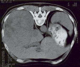
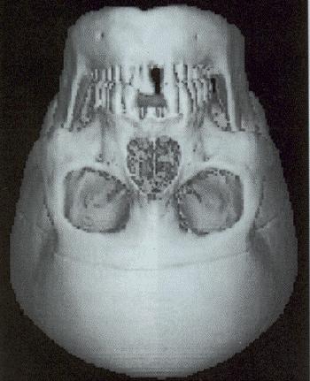
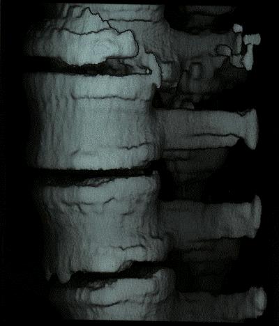
52 CT Slices and 3-D Image Reconstruction THE MEDICAL IMAGING DEPARTMENT 52

53 CT Service and Maintenance Contracts THE MEDICAL IMAGING DEPARTMENT 53

54 Bone Densitometry Systems THE MEDICAL IMAGING DEPARTMENT 54
")

55 80 kv filtered X-ray spectrum and NaI(Tl) detector THE MEDICAL IMAGING DEPARTMENT 55


56 40 kv and 70 kv X-ray spectra and 80 kv 400 mg/cm 2 Ce filtered X-ray spectrum THE MEDICAL IMAGING DEPARTMENT 56

57 Typical Layout of a Bone Densitometry Examination Room THE MEDICAL IMAGING DEPARTMENT 57

58 Typical Bone Densitometry Examination Room THE MEDICAL IMAGING DEPARTMENT 58


59 Typical Bone Densitometry Examination Print-outs THE MEDICAL IMAGING DEPARTMENT 59
60 Management of X-ray Diagnostic Equipment Workflow planning to optimize existing architectural design. Selection of appropriate system, fitting to the Hospital needs. Selection and sharing of X-ray equipment power supply (generator). Film processing method and associated quality control. Conditions of X-ray equipment maintenance contract. Conditions of X-ray tube guarantee. Archiving and Digital Image handling. Radiation Protection. Quality Assurance in everyday Operation. THE MEDICAL IMAGING DEPARTMENT 60


61 Marie and Pierre Curie / Ernest Rutherford THE MEDICAL IMAGING DEPARTMENT 61

62 Nuclear Medicine ƒ - Decay Nuclear Medicine is the specialty that uses radioactive tracers to medical situations, mainly for imaging purposes, but also for therapeutic ones. A complete nuclear medical service includes several sections and rooms. THE MEDICAL IMAGING DEPARTMENT 62
63 Nuclear Medical Service Sections and Rooms. Radio-pharmaceutical preparation: hot-lab, application room and in some major University Hospitals also a Cyclotron Facility for the production of positron emitting isotopes for Positron Emmission Tomography (PET). Examination area: -Camera examination rooms, also for planar, SPECT, etc., image-processing room, physical examination room, dark room. Physics area: Radiation shielded store for isotopes, laboratory, electronic shop. Patient area: Waiting rooms for ambulant and for non-ambulant patients, lavatories. Therapy: Preparation and treatment rooms, treatment planning room, radiation protected isotope store, nuclear medicine ward. THE MEDICAL IMAGING DEPARTMENT 63

64 Planar Bone scanning gamma- Camera I THE MEDICAL IMAGING DEPARTMENT 64

65 Modern Bone scanning gamma - Camera II THE MEDICAL IMAGING DEPARTMENT 65

66 -Camera Tl 204 examination combined to stress test THE MEDICAL IMAGING DEPARTMENT 66

67 Mobile gamma - Camera THE MEDICAL IMAGING DEPARTMENT 67


68 Typical gamma - Camera Images THE MEDICAL IMAGING DEPARTMENT 68

69 Gamma - Camera round NaI(Tl) crystal and the associated Photomultipliers THE MEDICAL IMAGING DEPARTMENT 69

70 Photomultiplier Amplifier THE MEDICAL IMAGING DEPARTMENT 70

71 Gamma - Camera Gantry THE MEDICAL IMAGING DEPARTMENT 71

72 Tomographic gamma - Camera with round crystal THE MEDICAL IMAGING DEPARTMENT 72

73 Tomographic gamma - Camera with rectangular crystal THE MEDICAL IMAGING DEPARTMENT 73


74 Tomographic gamma - Camera with double round and rectangular crystals THE MEDICAL IMAGING DEPARTMENT 74
75 Positron Emission Tomography (PET) Positron Emission Tomography (PET) has proven to be a unique tool in analyzing biomedical metabolic function or dysfunction, qualitatively and quantitatively. It finds application in cardiology for tissue viability and perfusion studies, in neurology for studying epilepsy, dementia, and other investigative brain studies, and in oncology for early tumor diagnosis, tumor grading, and extent of metastasis. THE MEDICAL IMAGING DEPARTMENT 75
76 PET Implementation Positron emission tomography begins with isotopes such as Rubidium 82, Oxygen 15, and Fluorine 18. The typical isotopes used in PET scanning have a half-life that ranges from 75 seconds to 110 minutes. Cyclotron is a cost effective, easy to operate, shelfshielded radioisotope delivery system. A commercially available modern machine, produces substantial quantities of the major positron emitting isotopes and compounds in a variety of chemical forms under full automation. THE MEDICAL IMAGING DEPARTMENT 76

77 A typical commercially available Cyclotron THE MEDICAL IMAGING DEPARTMENT 77


78 PET System Console THE MEDICAL IMAGING DEPARTMENT 78

79 PET scan of a heart following acute myocardial infarction and thrombolytic therapy The Rubidium areas show that the tissue, although mechanically abnormal, is metabolically alive. THE MEDICAL IMAGING DEPARTMENT 79

80 PET 3-D Brain Image Reconstruction THE MEDICAL IMAGING DEPARTMENT 80

81 Magnetic Resonance Imaging (MRI) THE MEDICAL IMAGING DEPARTMENT 81
82 Magnetic Resonance Imaging In common with all other imaging processes in medical diagnostics, magnetic resonance tomography makes use of the interaction of anatomical structures within the human body with a radiation field. With the aid of a radio-frequency field in the Mhz range and a locally variable magnetic field, the sharp resonance absorption of magnetic nuclei in biological tissue is used in order to obtain the spatial distribution of the nuclear magnetization. THE MEDICAL IMAGING DEPARTMENT 82
83 MRI Principle In particular, Hydrogen atoms, which occur naturally in large numbers, allow medically meaningful images to be formed, with a spatial resolution comparable to that of X- ray computed tomography, and which show previously unseen contrasts between tissues. In addition, it is even possible to detect magnetic nuclei, such as 13 C, 19 F, 23 Na, 31 P, in spite of their low concentration in biological tissue; nevertheless, the value of the information thus gained does not approach that obtained through proton resonance. THE MEDICAL IMAGING DEPARTMENT 83
84 Magnetic Moment alignment of a probe with Nuclear Spins In the case of thermal equilibrium, the magnetic moment of a probe with nuclear spins, aligns itself parallel to the external magnetic field. If this equilibrium is disturbed, for example by suddenly changing the direction of the external magnetic field, then a torque acts on the magnetic moment of the sample, causing a change in time, of the angular momentum of the probe. THE MEDICAL IMAGING DEPARTMENT 84
85 The magnetic moment of a probe with nuclear spins aligns itself parallel to the external magnetic field THE MEDICAL IMAGING DEPARTMENT 85
86 Precession of the nuclear magnetization This results in, the precession of the nuclear magnetization, around the direction of the magnetic field, with the Larmor frequency. This precession (Nuclear Magnetic Resonance) can be detected easily, by measuring the induced alternating voltage, in a coil surrounding the sample. After a finite period of time the thermal equilibrium which had previously been disturbed is reestablished. THE MEDICAL IMAGING DEPARTMENT 86
87 Changing the direction of the magnetic field a torque acts on the magnetic moment of the sample THE MEDICAL IMAGING DEPARTMENT 87

88 The precession (NMR) can be measured as induced alternating voltage in a coil surrounding the sample THE MEDICAL IMAGING DEPARTMENT 88
89 Relaxation times Two time constants are of interest: The longitudinal relaxation time T 1 is the time constant with which is restored the magnetization along the direction of the basic field, and it is associated with the emission of energy to the crystal lattice in which the atomic nuclei are embedded (spin-lattice relaxation). The transverse relaxation time T 2 is the time constant with which the transverse component decays, due to the mutual interactions of the nuclear spins (spin-spin relaxation). THE MEDICAL IMAGING DEPARTMENT 89
90 The physical significance of the Relaxation times The relaxation times express the mobility of the molecules in which the nuclei under consideration are located. Each nucleus is surrounded by other magnetic moments which are in constant thermal Brownian molecular motion, and produce a continuously changing magnetic perturbation field. Spectral components which correspond to the precession frequency of the nuclear magnetization, induce the longitudinal relaxation. Transverse relaxation is determined by the frequency of collisions between the molecules as a whole. THE MEDICAL IMAGING DEPARTMENT 90

91 The signal depends on the electron density of the chemical groups to which belong the protons THE MEDICAL IMAGING DEPARTMENT 91
92 MRI signal of characteristic groups THE MEDICAL IMAGING DEPARTMENT 92

93 Typical MRI System THE MEDICAL IMAGING DEPARTMENT 93

94 Typical MRI Suite in the beginning of the 80s THE MEDICAL IMAGING DEPARTMENT 94

95 A modern MRI examination unit THE MEDICAL IMAGING DEPARTMENT 95

96 Patient positioning THE MEDICAL IMAGING DEPARTMENT 96

97 A typical pass-through MRI system THE MEDICAL IMAGING DEPARTMENT 97

98 Cranial examination coil THE MEDICAL IMAGING DEPARTMENT 98

99 Head fixing accessories and a Cranial examination coil THE MEDICAL IMAGING DEPARTMENT 99

100 An open MRI system THE MEDICAL IMAGING DEPARTMENT 100

101 MRI Console THE MEDICAL IMAGING DEPARTMENT 101

102 Some typical MRI images (a) THE MEDICAL IMAGING DEPARTMENT 102
")

103 Some typical MRI images (b) THE MEDICAL IMAGING DEPARTMENT 103

104 Super-conductive MRI Magnet THE MEDICAL IMAGING DEPARTMENT 104
105 MRI advantages MRI has various attractive attributes: Non-ionizing radiation. Excellent soft tissue contrast. Visualization of any desired plane, without special patient positioning. No bone artifacts. Non invasive character of the examination. Potential for flow measurements. Potential for dynamic studies. THE MEDICAL IMAGING DEPARTMENT 105
106 Sonography The development of Ultra-sound equipment started in the fifties with equipment for examining the skull and the heart. Stimulated by sonar and radar technology, these developments culminated in two-dimensional ultrasound examination of: The abdominal organs. The thyroid glands. Obstetric examinations. Sectional displays of the heart. THE MEDICAL IMAGING DEPARTMENT 106
107 Combination units Today, combination units using several scanning methods and satisfying all diagnostic requirements are widespread. Almost all ultrasound methods used today in medical diagnostics are based on the echo pulse technique. Amplitude modulation (A mode) Time motion mode (M mode) Brightness modulation (B mode) have been used, that evaluate the amplitude of the echo signals received by the transducer. THE MEDICAL IMAGING DEPARTMENT 107
108 Scanning procedures There are two major scanning procedures: Mechanical sector scanning with good price-performance ratio, based on the rotor principle or the wobbler principle. Electronic scanning, i.e. the required scanning width (image width) is covered by a large number of identical individual transducers, formed as a linear or curved array. Scanning may be performed in rectilinear coordinates (linear array) or in polar coordinates (sector, mechanical systems, curved or phased array systems). THE MEDICAL IMAGING DEPARTMENT 108
109 Doppler frequency shift A source for extra information is the frequency content of the ultrasound oscillations and especially the Doppler frequency shift of the moving signal source (e.g. blood). A continuous wave (CW) or a pulse wave (PW) Doppler method can be applied. Duplex and triplex scanners combine independent transducer arrangements allowing for Imaging and Doppler examination. THE MEDICAL IMAGING DEPARTMENT 109

110 A typical ultrasound system THE MEDICAL IMAGING DEPARTMENT 110

111 Various Ultra-sound transducers THE MEDICAL IMAGING DEPARTMENT 111
 THE MEDICAL IMAGING")
112 Josephson Effect and the Super-conducting QUantum Interference Devices (SQUID) THE MEDICAL IMAGING DEPARTMENT 112

113 The Current of the Interference Circuit versus Magnetic Field Intensity V = 0, û = c. ( / 0) THE MEDICAL IMAGING DEPARTMENT 113

114 Block Diagram of SQUID applied in Medicine THE MEDICAL IMAGING DEPARTMENT 114
 THE MEDICAL IMAGING")
115 SQUID based Magneto-cardiogram (MCG) THE MEDICAL IMAGING DEPARTMENT 115
BME101 Introduction to Biomedical Engineering Medical Imaging Özlem BİRGÜL Ankara University Department of Biomedical Engineering
 BME101 Introduction to Biomedical Engineering Medical Imaging Özlem BİRGÜL Ankara University Department of Biomedical Engineering Outline What is Medical Imaging? History of Medical Imaging X-Ray Imaging
Part 3 Oral Exam Content Guide
 Initial Certification in Medical Physics Part 3 Oral Exam Content Guide The oral examination is designed to test your knowledge and fitness to practice applied medical physics in the specified specialty(ies).
RADIATION ONCOLOGY RESIDENCY PROGRAM Competency Evaluation of Resident
 Resident s Name: RADIATION ONCOLOGY RESIDENCY PROGRAM Competency Evaluation of Resident Rotation: PHYS 705: Clinical Rotation 3 Inclusive dates of rotation: Aug. 25, 2015 Feb. 25, 2016 Director or Associate
Computer Assisted Surgery Basics of medical imaging
 Computer Assisted Surgery Basics of medical imaging Prof. Leo Joskowicz School of Engineering and Computer Science The Hebrew University of Jerusalem, ISRAEL Medical Image Processing Basics of medical
Pulsed NMR of Paramagnetic Terbium. Cheyenne Michael Yari
 Pulsed NMR of Paramagnetic Terbium Cheyenne Michael Yari June 26, 2012 1 - Introduction The magnetic properties of atomic nuclei have proven to provide very useful information which can directly be used
PRINCIPLES OF CT AND MR IMAGING Marc-André d Anjou, DMV, DACVR Faculty of Veterinary Medicine, University of Montreal Saint-Hyacinthe, Quebec, Canada
 PRINCIPLES OF CT AND MR IMAGING Marc-André d Anjou, DMV, DACVR Faculty of Veterinary Medicine, University of Montreal Saint-Hyacinthe, Quebec, Canada CT and MR imaging offer superior diagnostic possibilities
Positron Emission Tomography Present status and future prospects
 Positron Emission Tomography Present status and future prospects S. Tavernier VRIJE UNIVERSITEIT BRUSSEL July 2011 NDIP Lyon 1 What is PET Positron Emission Tomography is a non invasive method for imaging
Medical instrumentationi 11/19/2010
 Medical instrumentationi BIOEN 302 11/19/2010 Medical instrumentation Definition: instrument for sensing, diagnostics, therapeutics or surgery of human being. 2 Medical instrumentation Definition: instrument
Radiography Curriculum Analysis
 Program Number Program Name Date / /20 Radiography Curriculum Analysis DIRECTIONS: Determine the course(s) in which each of the following content area is covered and enter the course number(s) and/or title(s).
Date: May 26, 2015 Page 1
 Part I. Answer these questions by marking the best answer among the choices given: [2 points each] 1. Ethics and morals differ in that a. Only one of them should be followed b. Ethics are for professionals
Molecular Imaging: Definition, Overview and Goals
 This tutorial will define what is currently considered molecular imaging. It will provide history and an overview, discuss the goals and the advantages of molecular imaging. It will clarify what is and
FLORIDA HOSPITAL DIAGNOSTIC RADIOLOGY RESIDENCY PROGRAM PHYSICS GOALS AND OBJECTIVES
 FLORIDA HOSPITAL DIAGNOSTIC RADIOLOGY RESIDENCY PROGRAM PHYSICS GOALS AND OBJECTIVES Goals and objectives are based on recommendations and requirements from the AAPM, RSNA, NRC, FL DOH, and ACGME Module
Biomedical Imaging Modalities
 1 Biomedical Imaging Modalities The introduction of advanced imaging techniques has improved significantly the quality of medical care available to patients. Noninvasive imaging modalities allow a physician
Computed Tomography: Optimization of acquisition protocols & Justification of clinical referrals. Koos Geleijns, medical physicist
 Computed Tomography: Optimization of acquisition protocols & Justification of clinical referrals Koos Geleijns, medical physicist CT delivers excellent 3D image quality CT delivers excellent 3D image quality
Portable and Mobile X-ray Units
 RADIOLOGY Portable and Mobile X-ray Units Tecmed Africa offers a full range of mobile Radiology systems from a very select group of high quality suppliers. Specialised mobile systems with either manual
NHS Imaging and Radiodiagnostic activity
 NHS Imaging and Radiodiagnostic activity NHS Imaging and Radiodiagnostic activity 2013/14 Release Version number: 1 First published: 6 th August 2014 Prepared by: NHS England Analytical Services (Operations)
Not for publication in the USA Erlangen, November 26, 2017
 Press Not for publication in the USA Erlangen, November 26, 2017 RSNA 2017 in Chicago: South Building, Hall A, Booth 1937 strengthens its CT portfolio by improving patient experience and expanding precision
PHYSICS OF DIAGNOSTIC RADIOLOGY: MDPH
 PHYSICS OF DIAGNOSTIC RADIOLOGY: MDPH 614 McGill University: Medical Physics Unit GENERAL INFORMATION Physics of Diagnostic Radiology, MDPH 614 (3 credits) Mon & Wed, 11:00-12:30 / Glen Site, DS1-7001
Russia Diagnostic Imaging Market Outlook to 2020
 Russia Diagnostic Imaging Market Outlook to 2020 Reference Code: GDMEMC0022DB Publication Date: March 2014 Page 1 1 Table of Contents 1 Table of Contents... 2 1.1 List of Tables... 5 1.2 List of Figures...
Medical Imaging Technologies in Canada, 2006 Methodological Notes
 Medical Imaging Technologies in Canada, 2006 Methodological Notes Introduction The Canadian Institute for Health Information (CIHI) aims to provide accurate and timely data and information to help shape
Medical Imaging quo vadis?
 Medical Imaging quo vadis? Pascal Fallavollita, PhD Senior Research Scientist Image-Guided Interventions Technology and Applications Tutorial Historical: medical imaging happened in last 100 yrs 2 Historical:
Radiography. 1. Electromagnetic Spectrum. 2. X-ray History. 3. X-ray Physics. 4. Clinical Application
 Radiographs X-RAYS Vincent Carrasco, MD, MSIS NLM Postdoctoral Fellow & Doctoral Candidate Carolina Health Informatics Program The University of North Carolina Chapel Hill Radiography 1. Electromagnetic
ADDIS ABABA UNIVERSITY CENTER OF BIOMEDICAL ENGINEERING
 ADDIS ABABA UNIVERSITY CENTER OF BIOMEDICAL ENGINEERING November 2013 History of Biomedical Engineering Definition of Biomedical Engineering Achievements of Biomedical Engineering Streams in Biomedical
On the Importance of Computation in Clinical Radiology
 On the Importance of Computation in Clinical Radiology Manuel Arreola, Ph.D., DABR Director of Radiological Physics Radiology Department University of Florida/Shands Healthcare My Disclaimer I ve received
Press Presse Press Presse
 Press Presse Press Presse Healthcare Sector Imaging & IT Division Erlangen,, March 4, 2010 European Congress of Radiology 2010: Siemens introduces innovations for imaging and diagnostics One of the most
Medical Imaging. Axial plane: bisects the left from the right. Coronal plane: bisects the front from the back. Medical imaging coordinates
 INF-GEO 4310 Imaging Medical Imaging 18.11.2013 Fritz Albregtsen Department of Informatics University sty of Oso Oslo Overview Medical imaging coordinate system 1. X-ray medical imaging a. Projected X-ray
Radioisotope and Radiation Applications (FS2013)
") École Polytechnique Fédérale de Lausanne Radioisotope and Radiation Applications (FS2013) Applications for Medical Diagnosis (Week 3a) Pavel Frajtag 01.10. 2013 1 Applications for Medical Diagnosis: Outline
École Polytechnique Fédérale de Lausanne Radioisotope and Radiation Applications (FS2013) Applications for Medical Diagnosis (Week 3a) Pavel Frajtag 01.10. 2013 1 Applications for Medical Diagnosis: Outline
Photon-based Medical Imagery
 Photon-based Medical Imagery Photon-based Medical Imagery Edited by Hervé Fanet First published 2011 in Great Britain and the United States by ISTE Ltd and John Wiley & Sons, Inc. Adapted and updated from
Simplicity is efficiency. Analyzing Moisture Content by Using Magnetic Resonance Technology
 Simplicity is efficiency. Analyzing Moisture Content by Using Magnetic Resonance Technology Magnetic Resonance Moisture Why biofuel moisture measurement is important? Moisture of fuel increases total mass
R/F. Experiences Using SONIALVISION safire and the Utility of Tomosynthesis. 1. Introduction. 2. Basics of Tomosynthesis.
 R/F Experiences Using SONIALVISION safire and the Utility of Tomosynthesis Radiology Division, Dokkyo Medical University Koshigaya Hospital Masahiro Nakajima Mr. Masahiro Nakajima 1. Introduction The hospital
Toward Human-oriented Life
 Toward Human-oriented Life 180 Toward Human-oriented Life Minoru Sakairi, Dr. Sci. Keiji Kobashi, Dr. Eng. Yasutaka Hasegawa Kazutoshi Kan OVERVIEW: So that people can live healthy and happy lives, humanoriented
Emerging Applications and Trends Across Medical Imaging
 MEDICAL DEVICES PHARMACEUTICALS CHEMICALS FOOD & BEVERAGE ELECTRONICS Emerging Applications and Trends Across Medical Imaging VPG Publications, Consulting, Clients www.vpgcorp.com VPG Market Research Reports
Occams Business Research & Consultancy
 Occams Business Research & Consultancy http://www.marketresearch.com/occams Business Research Consultancy v4040/ Publisher Sample Phone: 800.298.5699 (US) or +1.240.747.3093 or +1.240.747.3093 (Int'l)
VALLIAMMAI ENGINEERING COLLEGE DEPARTMENT OF MECHANICAL ENGINEERING QUESTION BANK
 VALLIAMMAI ENGINEERING COLLEGE SRM Nagar, Kattankulathur 603 203 DEPARTMENT OF MECHANICAL ENGINEERING QUESTION BANK VIII SEMESTER ME 6019-NON DESTRUCTIVE TESTING AND MATERIALS Regulation 2013 Academic
We are IntechOpen, the world s leading publisher of Open Access books Built by scientists, for scientists. International authors and editors
 We are IntechOpen, the world s leading publisher of Open Access books Built by scientists, for scientists 4,000 116,000 120M Open access books available International authors and editors Downloads Our
Magnetic Resonance Spectroscopy from fundamental developments to improved noninvasive diagnosis and characterisation of children s brain tumours.
 Magnetic Resonance Spectroscopy from fundamental developments to improved noninvasive diagnosis and characterisation of children s brain tumours. Martin Wilson IOP Medical Physics Group Scientific and
SPECIFICATION. SPECT/CT Gamma Camera System
 1. Scope SPECIFICATION SPECT/CT Gamma Camera System This specification describes the requirements for a dual-head SPECT/CT Gamma camera and work station solution (hereinafter referred as the System ),
X-ray production and applications. by: Dr. Ahmed M. Maghraby
 X-ray production and applications by: Dr. Ahmed M. Maghraby I - Discovery During the early 1890 s many physicists had been studying electrical conduction in gases at low pressures. Wilhelm Conrad Roentgen
Diagnostic Medical Image Processing
 Diagnostic Medical Image Processing Introduction WS 2010/11 Joachim Hornegger, Dietrich Paulus, Markus Kowarschik Lehrstuhl für Mustererkennung (Informatik 5) Friedrich-Alexander-Universität Erlangen-Nürnberg
MRI. Magnetic Resonance Imaging
 MRI Magnetic Resonance Imaging Key Points MRI: The magnet is always on Know the essential components of an MRI system Know general idea of how MRI captures an image; three steps: Magnetic Alignment, Proton
An ultrasonic volumetric scanner for image-guided surgery
 International Congress Series 1230 (2001) 190 196 An ultrasonic volumetric scanner for image-guided surgery Jeremy Johnson a,b, *,Ömer Oralkan b, Kambiz Kaviani b, Utkan Demirci b, Mustafa Karaman b, Pierre
2.3.Positron Emission Tomography
 2.3.Positron Emission Tomography K. Wienhard, MPI fuer Neurologische Forschung, Gleuelerstr. 50, 50931 Köln, Germany Positron Emission Tomography ( PET ) is the most sensitive method to image trace amounts
Basics of Technology. Lecturer Mgr. Vladan Bernard, Ph.D. Tuition 2nd semester. Range of education. Type of education
 Subject Basics of Technology Lecturer Mgr. Vladan Bernard, Ph.D. Tuition 2nd semester Type of subject Compulsory Range of education 2 hours Self-study 28 hours Type of education Lecture Completion Oral
Low Dose, Lightning Speed, Latest Applications
 RXL EDITION Low Dose, Lightning Speed, Latest Applications The New Standard for Radiology Toshiba Medical Systems is committed to the development of new technologies to minimize radiation dose while maintaining
New PET/CT from Siemens helps more patients benefit from premium technologies
 Press Healthcare Erlangen, October 9, 2015 EANM 2015, Congress Center Hamburg (CCH) New PET/CT from Siemens helps more patients benefit from premium technologies Versatile new PET/CT system addresses a
Status Update on Proton CT
 Status Update on Proton CT Reinhard W. Schulte, Professor of Radiation Medicine Loma Linda University & Medical Center California, USA Outline What do we gain by developing proton CT (pct)? pct principles
The Unique, New MRI Philips Ingenia 3 Tesla is now in Ayios Therissos! The first-ever digital broadband MR system has been installed in Ayios
 The Unique, New MRI Philips Ingenia 3 Tesla is now in Ayios Therissos! The first-ever digital broadband MR system has been installed in Ayios Therissos-Nicosia that delivers crystal clear images, remarkable
ACPSEM. Diagnostic Imaging Medical Physics. Clinical Training Guide
 ACPSEM Diagnostic Imaging Medical Physics Clinical Training Guide Version 5.0 Approved by the PSB on 9/5/2018 THE ACPSEM The ACPSEM mission is to advance services and professional standards in medical
Magnetic Resonance Imaging of concrete. Coring. Location of flaws. Assessment of Concrete Structures. Systems to monitor concrete modulus
 Magnetic Resonance Imaging of concrete Dr Chris Burgoyne Department of Engineering University of Cambridge Assessment of Concrete Structures How can we tell what is going on inside concrete? We would like
HEALTHCARE IMAGING Products by SIEMENS in Moldova
 HEALTHCARE IMAGING Products by SIEMENS in Moldova http://www.healthcare.siemens.com First big government project (in early 90 s) More than 14 X-ray machines Many UltraSound equipment Angiography, C-Arms,
What s the Issue? Radiation Dose in the OR. Exposure to OR Personnel. Effects of Proper Collimation. Image-guided procedures are increasingly complex
 Radiation Dose in the OR Robert G. Gould, Sc.D. Professor and Vice Chair Department of Radiology and Biomedical Imaging University of California San Francisco What s the Issue? Image-guided procedures
MEDICAL PHYSICS (MED PHYS)
") Medical Physics (MED PHYS) 1 MEDICAL PHYSICS (MED PHYS) MED PHYS/PHYSICS 265 INTRODUCTION TO MEDICAL PHYSICS Primarily for premeds and other students in the medical and biological sciences. Applications
Medical Physics (MED PHYS) 1 MEDICAL PHYSICS (MED PHYS) MED PHYS/PHYSICS 265 INTRODUCTION TO MEDICAL PHYSICS Primarily for premeds and other students in the medical and biological sciences. Applications
Innovating Solutions for Staff and Patient Protection. Radiation Shielding
 Innovating Solutions for Staff and Patient Protection Radiation Shielding Introduction Amray is a leading European manufacturer of Radiation Shielding products to the medical and pharmaceutical sectors.
TENDER # SUPPLY OF MOBILE C-ARM RADIOGRAPHY/FLUOROCOPY SYSTEM FOR WESTERN HEALTH
 TENDER # 0571-1707 SUPPLY OF MOBILE C-ARM RADIOGRAPHY/FLUOROCOPY SYSTEM FOR WESTERN HEALTH Amendment tice issued April 13,2017 1. The closing date time is amended to May 5 th,2017 2:00PM 2. The specifications
Translational & Molecular Imaging Institute
 Translational & Molecular Imaging Institute tmii.mssm.edu Summer 2015 CARDIOVASCULAR IMAGING The Imaging Research Center is the backbone of the Translational & Molecular Imaging Institute at Mount Sinai
The time has come. Philips GEMINI TF PET/CT with TruFlight technology
 The time has come Philips GEMINI TF PET/CT with TruFlight technology TruFlight has arrived Time-of-flight technology has always held the promise of better PET imaging. But it took Philips to harness its
Power of BRANSIST safire in Neuroendovascular Therapy
 Vascular Power of BRANSIST safire in Neuroendovascular Therapy Department of Radiology, Kinki University Hospital Suguru Ueda Mr. Suguru Ueda 1. Introduction Kinki University Hospital is located in the
ASTM Volume 03.03, October 2017 Nondestructive Testing (E94 E2373)
") Table of Contents 1 E94-04(2010) Standard Guide for Radiographic Examination 2 E114-15 Standard Practice for Ultrasonic Pulse-Echo Straight-Beam Contact Testing 3 E125-63(2013) Standard Reference Photographs
Table of Contents 1 E94-04(2010) Standard Guide for Radiographic Examination 2 E114-15 Standard Practice for Ultrasonic Pulse-Echo Straight-Beam Contact Testing 3 E125-63(2013) Standard Reference Photographs
Current Market Dynamics and Future Vision of the Care Cycle
 Current Market Dynamics and Future Vision of the Care Cycle Tim Irish Analysts Meeting June 15 th, 2005 Overview Market and Customer trends The Care Cycle and Molecular Medicine 2 Healthcare is the world
Magnetic Resonance Imaging. D. J. McMahon rev cewood
 Magnetic Resonance Imaging D. J. McMahon 150504 rev cewood 2018-02-15 Key Points MRI: The magnet is always on Know the essential components of an MRI system Know general idea of how MRI captures an image;
Patient doses from CT examinations in region of Prishtina, Kosovo
 Patient doses from CT examinations in region of Prishtina, Kosovo Sehad KADIRI, Gëzim HODOLLI, Kostandin DOLLANI Institute of Occupational Medicine, Radiation Protection Service, Obiliq, Kosovo Institute
Improving protocols and procedures for strengthened radiation protection in interventional procedures
 Improving protocols and procedures for strengthened radiation protection in interventional procedures R. Loose German Commission on Radiological Protection (SSK) German Roentgen Society (DRG) Institute
Medical Applications of Nuclear Technology
 Chapter 14 Medical Applications of Nuclear Technology The exploitation of nuclear technology in medical applications began almost from the moment of Roentgen's discovery of x rays in 1895 and Becquerel's
Handzettel 1. CARE Right Computed Tomography, committed to the right dose Ivo Driesser Austin, July 22 nd 2014
 CARE Right Computed Tomography, committed to the right dose Ivo Driesser Austin, July 22 nd 2014 Answers for life. Answers for life. Is 1 msv the right dose for every patient? Female, 54 Abdominal CT Male,
ACVR Residency Training Program Application
 ACVR Residency Training Program Application Submission Date Institution Name: Succinctly state the objectives of the training program. 03-14-2018 20:11:22 Tufts Cummings School of Veterinary Medicine General
Department of Radio Diagnosis
 Department of Radio Diagnosis S.No. Name of Equipment Specification Qty required Suggested Manufacturers Name Approx Unit cost (Rs.) Usage 1 Digital substraction angiography (D.S.A.) A single plane, ceiling
PoS(PhotoDet 2012)055
055") Multi-pixel Geiger mode imager for medical application Khalil JRADI 1 Laboratoire d Electronique Informatique et Image (LE2I) Avenue Alain Savary, UMR CNRS 6306, Université de Bourgogne, Dijon, France
Multi-pixel Geiger mode imager for medical application Khalil JRADI 1 Laboratoire d Electronique Informatique et Image (LE2I) Avenue Alain Savary, UMR CNRS 6306, Université de Bourgogne, Dijon, France
GE Healthcare. Introducing Discovery MI DISCOVERY MI
 GE Healthcare Introducing Discovery MI DISCOVERY MI Introducing Discovery MI Introducing Discovery MI MEANINGFUL INSIGHTS. FROM YOUR PATIENT TO EVERY PATIENT. Meet Discovery TM MI. A PET/CT system conceptualized
MEDICAL EQUIPMENT (1) TOPIC 1: RECORDING AND PROCESSING OF BIOSIGNALS
 TOPIC 1: RECORDING AND PROCESSING OF BIOSIGNALS") MEDICAL EQUIPMENT (1) TOPIC 1: RECORDING AND PROCESSING OF BIOSIGNALS Term 1 2013/14 Prof. Yasser Mostafa Kadah www.k-space.org Measurement Basics Measuring is the experimental determination of a measured
MEDICAL EQUIPMENT (1) TOPIC 1: RECORDING AND PROCESSING OF BIOSIGNALS Term 1 2013/14 Prof. Yasser Mostafa Kadah www.k-space.org Measurement Basics Measuring is the experimental determination of a measured
BIOMEDICAL SIGNAL AND IMAGE PROCESSING
 BIOMEDICAL SIGNAL AND IMAGE PROCESSING EE 5390-001 SYLLABUS Instructor: Wei Qian, Ph.D. Professor of Electrical and Computer Engineering Medical Signal and Image Computerized Processing Scheme for Medical
Medical Electronic Devices
 Medical Electronic Devices Basic Electronics (Electronic devices, Amplifiers, A/D, D/A) Electrodes EEG Deep brain stimulator ECG Cardiac Pacemakers External Defibrillators EMG Neuromuscular Electrical
Applications And Comparison of Medical Imaging Modalities
 International Journal of Engineering Science Invention (IJESI) ISSN (Online): 2319 6734, ISSN (Print): 2319 6726 Volume 7 Issue 1 January 2018 PP. 94-100 Applications And Comparison of Medical Imaging
Table of Contents. Adaptive Diagnostics...7 Integrated Dose Reduction Streamlined Workflow Clinical Images... 26
 2 Table of Contents Adaptive Diagnostics...7 Integrated Dose Reduction... 17 Streamlined Workflow... 21 Clinical Images... 26 3 AQUILION TM PRIME PROVIDES CLINICAL FLEXIBILITY, ENHANCED WORKFLOW FEATURES
Press. Innovative systems for radiology from Siemens. Healthcare Erlangen, March 3, ECR 2016 in Vienna: Hall X5, Booth 12
 Press Healthcare Erlangen, March 3, 2016 ECR 2016 in Vienna: Hall X5, Booth 12 Innovative systems for radiology from Siemens Faster MRI applications for neurology provide better diagnostic results First
Simple, intuitive and accessible MRI solution for preclinical research. M-Series Compact MRI Systems
 Simple, intuitive and accessible MRI solution for preclinical research M-Series Compact MRI Systems Application Oriented Imaging Anatomy and Morphology In vivo soft tissue imaging for morphological characterization.
1. Executive Summary
 1. Executive Summary 1.1 General The fluoroscope is defined as an instrument used chiefly in industry and in the practice of medicine for observing the internal structure of objects (such as the living
Simple, intuitive and accessible MRI solution for preclinical research. M-Series Compact MRI Systems
 Simple, intuitive and accessible MRI solution for preclinical research M-Series Compact MRI Systems Application Oriented Imaging Molecular Imaging Using Contrast Agents Detection and quantification of
THE NEW 640 SLICE CT SCANNER
 THE NEW 640 SLICE CT SCANNER www.ahdubai.com The American Hospital Dubai has recently acquired a state-ofthe-art computerized tomography (CT) scanner that houses a variety of intelligent and industry-leading
SPECIFICATION DIGITAL MAMMOGRAPHY UNIT AND ASSOCIATED SERVICES
 1 Digital Mammography Unit 1. Scope SPECIFICATION DIGITAL MAMMOGRAPHY UNIT AND ASSOCIATED SERVICES This specification describes the requirements for a digital mammography equipment and associated services
Radiation Shielding Glass RD 30 RD 50
 Radiation Shielding Glass RD 30 RD 50 2 SCHOTT is a leading international technology group in the areas of specialty glass and glass-ceramics. With more than 130 years of outstanding development, materials
Radiopharmaceuticals: Production and Availability
 Radiopharmaceuticals: Production and Availability A. Introduction 1. The use of specific radiotracers called radiopharmaceuticals for imaging organ function and disease states is a unique capability of
SPECIFICATION CT SIMULATOR FOR BLACK LION HOSPITAL, ADDIS ABEBA, ETHIOPIA
 SPECIFICATION CT SIMULATOR FOR BLACK LION HOSPITAL, ADDIS ABEBA, ETHIOPIA 1. Scope This specification describes the requirements for the supply, delivery, installation, and acceptance testing of a CT Simulator
Medical imaging - Wikipedia, the free encyclopedia
 Page 1 Medical imaging From Wikipedia, the free encyclopedia Medical imaging is the technique and process used to create images of the human body (or parts and function thereof) for clinical purposes (medical
MEASURING RADIATION DOSE IN COMPUTED TOMOGRAPHY USING ELLIPTIC PHANTOM AND FREE-IN-AIR, AND EVALUATING ITERATIVE METAL ARTIFACT REDUCTION ALGORITHM
 MEASURING RADIATION DOSE IN COMPUTED TOMOGRAPHY USING ELLIPTIC PHANTOM AND FREE-IN-AIR, AND EVALUATING ITERATIVE METAL ARTIFACT REDUCTION ALGORITHM ASHRAF MORGAN Bachelor of Science in Physics Cleveland
Advantages and challenges of whole-body MR-PET. Gaspar Delso Axel Martinez-Möller Stephan Nekolla Sibylle Ziegler
 Advantages and challenges of whole-body MR-PET Gaspar Delso Axel Martinez-Möller Stephan Nekolla Sibylle Ziegler Why MR+PET? MR High spatial resolution (~1mm) Good imaging of anatomy. Excellent soft-tissue
Performance in CT SOMATOM Sensation
 Performance in CT SOMATOM Sensation With the SOMATOM Sensation and z-sharp Technology, the future of volume imaging for cardiac and noncardiac applications has arrived. This scanner is the standard to
Modern. Easy. Reliable.
 www.siemens.com/healthcare Modern. Easy. Reliable. SOMATOM Spirit 2 Spirit SOMATOM Spirit 3 CT Vision Siemens CT Vision Today s reality The justification for the existence of the entire medical industry
RADIOISOTOPES in MEDICINE:
 RADIOISOTOPES in MEDICINE: Requirements - Production - Application and future prospectives 2 Imaging with Radiotracers Gerd-Jürgen BEYER Prof.Dr.rer.nat.habil.(i.R.) Geneva, Switzerland THIRD INTERNATIONAL
F. Jensen 1 ) 100 years of X-rays
 100 years of X-rays") F. Jensen 1 ) 100 years of X-rays The phenomenon that Wilhelm Conrad Roentgen, soon after his discovery, referred to as A New Kind of Rays has, as we now know, existed since the beginning of the universe.
F. Jensen 1 ) 100 years of X-rays The phenomenon that Wilhelm Conrad Roentgen, soon after his discovery, referred to as A New Kind of Rays has, as we now know, existed since the beginning of the universe.
Computer-Aided Surgical Navigation Coding Guide Neurosurgery. May 1, 2009
 Computer-Aided Surgical Navigation Coding Guide Neurosurgery May 1, 2009 Please direct any questions to: Kim Brew Manager, Reimbursement and Therapy Access Medtronic Surgical Technologies (904) 279-7569
Geneva January 10 11, th Swiss Experimental. Surgery Symposium. MGY / 4thSESS /
 4th Swiss Experimental Surgery Symposium Geneva January 10 11, 2008 Source: Reflex issue 2, 2007 The 3 Rs Reduction Replacement Refinement NC3R http://www.nc3rs.org.uk/category.asp?catid =9 No specific
Introduction to Medical Imaging. Lecture 6: Radiography ??? hc λ. X-Ray Discovery. Discovered by Wilhelm Röntgen in 1895.
 X-Ray Discovery Introduction to Medical Imaging Lecture 6: Radiography Discovered by Wilhelm Röntgen in 1895 accidentally, when performing experiments with cathode tubes and fluorescent screens the light
Comprehensive Dose Management for Improved Patient Care.
 Comprehensive Dose Management for Improved Patient Care. Set the standard for dose management Toshiba is committed to continuously advancing dose reduction technologies and incorporating these into its
TRIO. SPECT/CT, SPECT/CT/PET and standalone SPECT 3-head SPECT-based imager family for clinical imaging & clinical/preclinical research use
 TRIO SPECT/CT, SPECT/CT/PET and standalone SPECT 3-head SPECT-based imager family for clinical imaging & clinical/preclinical research use The first and only clinical Triple-Detector SPECT/CT system with
U.S. Census Bureau Electromedical Equipment and Analytical Instruments MA334A(10) Issued July 2011
 Issued July 2011") U.S. Census Bureau Electromedical Equipment and Analytical Instruments - 2010 MA334A(10) Issued July 2011 Address inquiries concerning these data to Investment Goods Industries Branch, U.S. Department
U.S. Census Bureau Electromedical Equipment and Analytical Instruments - 2010 MA334A(10) Issued July 2011 Address inquiries concerning these data to Investment Goods Industries Branch, U.S. Department
Study Guide Imaging Physics and Biophysics for the Master-Study Programmes
 Study Guide Imaging Physics and Biophysics for the Master-Study Programmes Imaging Physics is one of the main areas of research of the Faculty for Physics and Astronomy at the Julius-Maximilians-University
The Role of Tomosynthesis Imaging at Our Institute and X-ray Dose Optimization
 R/F The Role of Tomosynthesis Imaging at Our Institute and X-ray Dose Optimization Tetsuta Izumi R.T. Osaka International Cancer Institute, Diagnostic and Interventional Radiology Section 1 Osaka International
INVALUABLE VERSATILITY. GO DIGITAL. YOUR WAY. DRX-Evolution
 DRX-Evolution X FAC T O R GO DIGITAL. YOUR WAY. The power of the X-Factor has delivered the CARESTREAM DRX-Evolution an exceptionally versatile, flexible DR solution with modular components to fit your
Introduction. The use of the imaging department. X-rays
 DIE1 3/10/04 09:22 AM Page 1 Introduction The use of the imaging department Good communication between clinicians and radiologists is vital because the radiology department needs to understand the clinical
Artifacts Caused by Eddy Current in Diffusion MRI
 Artifacts Caused by Eddy Current in Diffusion MRI Xi Tan ABSTRACT Magnetic resonance diffusion imaging is potentially an important tool for the noninvasive characterization of normal and pathological tissue.