A Role for Focal Adhesion Kinase in Phenylephrine-Induced Hypertrophy of Rat Ventricular Cardiomyocytes.
|
|
|
- Nelson Simon
- 6 years ago
- Views:
Transcription
1 JBC Papers in Press. Published on April 3, 2000 as Manuscript M A Role for Focal Adhesion Kinase in Phenylephrine-Induced Hypertrophy of Rat Ventricular Cardiomyocytes. Joan M. Taylor, Joshua D. Rovin and J. Thomas Parsons * Department of Microbiology, Health Sciences Center, University of Virginia Charlottesville, Virginia, * To whom correspondence should be addressed: J. Thomas Parsons Department of Microbiology Box 441, Health Sciences Center University of Virginia Charlottesville, VA tel. # : (804) fax #: (804) jtp@virginia.edu Copyright 2000 by The American Society for Biochemistry and Molecular Biology, Inc.
2 The abbreviations used are: MAP, mitogen-activated protein; EGF, epidermal growth factor; PDGF, platelet-derived growth factor; GRB2, growth receptor bound protein-2; PAGE, polyacrylamide gel electrophoresis; EGTA, ethylenebis(oxyethylenenitrilo)-tetraacetic acid; PBS, phosphate-buffered saline; TBS, tris-buffered saline; RIPA, radioimmunoprecipitation assay buffer; Ab, antibody; Mab, monoclonal antibody; HRP, horseradish peroxidase; PE, phenylephrine; FAK, focal adhesion kinase; ECM, extracellular matrix; ANF, atrial naturetic factor; FRNK, FAK related non-kinase; PI3 Kinase, phosphoinositide 3-kinase; CAS, p130 Crk associated substrate; ERK, extracellular signal regulated kinase; BrdU, 5-Bromo-2 - Deoxyuridine, DMEM, Dulbecco s modified eagles medium; GFP, green fluorescent protein; JNK, Jun NH2-terminal kinase; MEK, mitogen activated kinase kinase; FN, fibronectin; LAM, laminin; COL, collagen; MBP, myelin basic protein. Running title : FAK regulates myocyte hypertrophy Keywords : integrins, cardiomyocyte, heart, hypertrophy, FAK, FRNK, adenovirus 2
3 ABSTRACT A variety of agonists including phenylephrine (PE) induce hypertrophy in neonatal ventricular cardiomyocytes. Here we report that signals provided by extracellular matrix proteins (ECM) augment the PE-induced hypertrophic response of cardiomyocytes and provide evidence that ECM-dependent signaling is mediated in part by the protein tyrosine kinase, focal adhesion kinase (FAK). Addition of PE to cultured neonatal cardiomyocytes stimulated sarcomeric organization, increase in cell size, and induction of atrial naturetic factor (ANF) in cardiomyocytes plated on the ECM protein laminin or fibronectin. In contrast, cardiomyocytes plated on the non-adhesive substrate gelatin exhibited a reduced capacity to undergo these PEstimulated hypertrophic changes. In cardiomyocytes cultured on ECM, PE stimulated a rapid increase in tyrosine phosphorylation of focal adhesion proteins including FAK, paxillin and p130 Crk associated substrate (CAS) and subsequent formation of peripheral focal complexes. Inhibition of the PE-induced hypertrophic response by genestein and herbimycin-a indicated a requirement for protein tyrosine kinases in PE signaling. To determine whether activation of FAK is required for PE-induced hypertrophy a dominant-interfering mutant form of FAK; termed FRNK (FAK-related non-kinase) was ectopically expressed in cardiomyocytes using a replication-defective adenovirus expression system. FRNK expression attenuated PEstimulated hypertrophy as assessed by cell size, sarcomeric organization and induction of ANF. These data indicate that the signal transduction pathways leading to cardiomyocyte hypertrophy are strongly influenced by and/or dependent upon an integrin-mediated signaling process requiring FAK. 3
4 INTRODUCTION Cardiomyocyte hypertrophy, characterized by increased volume and myofibrillar protein content, is important for the normal developmental growth of the heart. However, in the fully developed adult heart, pressure or volume overload, myocardial infarction or hormonal imbalance can lead to chronic pathological hypertrophy characterized by induction of immediate early genes, re-activation of an embryonic gene program, and reorganization of myocyte cytoskeletal architecture (1). The ability of integrins, extracellular matrix receptors, to regulate cytoskeletal architecture has been well characterized and integrin signaling has clearly been implicated in hormone and growth factor-induced alterations in gene transcription in variety of cell types (2). Integrins are a family of heterodimeric transmembrane receptors (composed of α and β subunits) containing extracellular ligand binding domains that show binding specificity for extracellular matrix (ECM) components and a short cytoplasmic domain that serves to couple integrins with the actin cytoskeleton (3). Several studies have shown that cardiac hypertrophy is accompanied by alterations in ECM components and cardiac integrin expression (4-6). Specifically, enhanced expression of collagen III, fibronectin, osteopontin, and β 1, α 3 and α 5 integrin subunits correlates with advancement of hypertrophy (4-6). A direct role for integrins in the development of hypertrophy has been demonstrated by transgenic studies in which cardiac specific expression of activated α5 integrin leads to profound ventricular hypertrophy (7). Taken together, these observations support a model whereby alterations in signaling pathways responsive to ECM may 4
5 influence the progression of cardiac hypertrophy. Integrin signaling in many cell types involves activation of focal adhesion kinase (FAK), a cytoplasmic non-receptor protein tyrosine kinase. Clustering of integrins leads to the recruitment of FAK to the newly formed focal adhesion and results in autophosphorylation and concomitant activation of FAK. Tyrosine phosphorylation of FAK at Tyr397 (proximal to the kinase domain) creates a binding site for the SH2 domain of another non-receptor tyrosine kinase, Src, resulting in formation of a bipartite complex of two tyrosine kinases (8). The formation of the FAK/Src complex results in the activation of Src and the subsequent activation of downstream signals leading to the regulation of cellular processes such as growth, migration and apoptosis (2,3,9). The C-terminal domain of FAK contains binding sites for a number of signaling molecules including phosphoinositide 3-kinase (PI3 kinase), p130 Crk associated substrate (CAS), GRB2, paxillin and the GTPase regulator associated with FAK (Graf; ref 10). The recruitment and activation of these molecules are thought to be important for the subsequent activation of other downstream signaling pathways. For example, association of FAK with CAS is required for integrin-mediated activation of ERK2 (11). In some cells, the C-terminal domain of FAK is expressed as a separate protein termed FRNK (FAK-related non kinase), whose overexpression attenuates integrin-mediated tyrosine phosphorylation of FAK and paxillin (12,13) and results in the inhibition of the rate of cell spreading and cell migration (12,13). These data indicate that FRNK can act as a biological inhibitor of FAK signaling and highlight the importance of FAK in ECM-mediated signaling events. In addition to providing specific signaling from ECM, recent evidence suggests that 5
6 integrin engagement is required to mediate efficient growth factor and hormonal-mediated signaling. When fibroblasts are held in suspension, EGF-, PDGF- and lysophosphatidic acidstimulated ERK2 activity is markedly reduced compared to cells plated on fibronectin (14,15). Importantly, the inhibition observed in suspended cells can be rescued by over-expression of activated FAK (16). These data indicate that growth factors (or hormones) synergize with cell adhesion signals to stimulate cell growth and survival. We report herein that PE-induced hypertrophy in isolated neonatal cardiomyocytes requires signaling through FAK. Plating cells on the integrin-dependent ECM proteins laminin or fibronectin supports the PE-induced hypertrophic response of the cultured cardiomyocytes, whereas plating cells on the integrin-independent substrate gelatin fails to sustain cellular hypertrophy. Addition of PE stimulates tyrosine phosphorylation of focal adhesion proteins including FAK, paxillin and CAS and alters the subcellular localization of paxillin in cardiomyocytes plated on ECM. In addition, we show that ectopic expression of FRNK, a dominant-interfering mutant for FAK, attenuates PE-induced hypertrophy. These data support a role for integrin-mediated, FAK-dependent signaling for induction of cardiac hypertrophy. 6
7 EXPERIMENTAL PROCEDURES Antibodies Antibody (9E10) to the myc-epitope tag was purchased from Santa Cruz Biotechnology; the anti-anf antibody (IHC9103) from Peninsula Laboratories; the 4G10 phosphotyrosine specific antibody from Upstate Biology; the anti-paxillin antibody from Transduction Laboratories and the anti-gfp antibody from Clontech. An anti-fak tyrosine 397-specific antibody was purchased from Biosource International and an anti-human FAK antibody was purchased from UBI. The ERK1/2 antibodies used include a phosphorylation-specific antibody purchased from Calbiochem and polyclonal (TR10) and monoclonal (1B3B9) antibodies generously provided by Dr. Michael Weber (University of Virginia). CAS was detected using a mixture of two polyclonal antibodies generated against the C-terminal domain of CAS (CAS-F and CAS-P; provided by Dr. Amy Bouton, University of Virginia, ref 17). Expression Constructs and Adenovirus Production The cdna constructs encoding the amino-terminal Myc- and GFP-tagged variants of FRNK were generated by cloning chicken FRNK into the BamHI/NotI or BglII/EcoRI sites of the mammalian expression vectors pcmv-neo-bamhi or pegfp-c1 respectively. For adenovirus production, GFP and GFP-tagged FRNK were generated by PCR from pegfp-c1 and pegfp- 7
8 C1-FRNK respectively using primers that added 5 and 3 BamHI restriction sites. The resultant PCR products were digested with BamHI and ligated with BamHI digested pad-lox (an adenoviral shuttle vector generously provided by Stephen Hardy, Somatix Therapy Corporation, Almeda CA). Correct orientation of all reading frames were confirmed by sequencing and western blot analysis of expressed proteins. The GFPAd-lox and GFP-FRNKAd-lox constructs were subsequently co-transfected with replication-defective ψ5 virus into HEK293 cells that produce stable overexpression of the Cre-recombinase. After recombination, plaque purified virus (1 x 10¹⁰pfu/ml) was generated and purified by cesium-chloride gradients as described elsewhere (18). Cell Culture, Infection and DNA Transfection Ventricular cardiomyocytes were isolated from neonatal rats (1-3 days old) by trypsin and collagenase digestion and purified as previously described (19). The cells were resuspended in DMEM:Media 199 (4:1) containing 10% fetal calf serum and 1% penicillin-streptomycin and plated on tissue culture plastic for two consecutive 1 h periods followed to remove noncardiomyocyte cells. For the biochemical experiments, cardiomyocytes were treated with 100 µm BrdU as described previously to obtain cultures with >95% myocytes (20). The cardiomyocytes were then plated on tissue culture dishes or chamber slides pre-coated with ECM or gelatin as required. Cardiomyocytes were plated at low density (2 x 10 4 cells/ml) for immunofluorescent studies and at high density (1 x 10 6 cells/ml) for biochemical studies. After h the cells were rinsed in serum free DMEM:Media 199 (4:1) and plated in serum free DMEM:Media 199 (4:1) containing 1% penicillin-streptomycin. After a 24 h incubation period, cells were treated with vehicle or PE for the indicated time periods. 8
9 For adenoviral infection, cells were infected in serum free media with either GFP or GFP-FRNK (4 MOI) for 12 h prior to treatment with PE. In some experiments cardiomyocytes were transfected with a myc-frnk expression vector using the FuGene transfection reagent (Boehringer Mannheim). Typically 3 µg of DNA/20 mm chamber slide was combined with 6 µl FuGene and incubated with the cells in serum free DMEM:Media 199 (4:1) for 24 h prior to treatment with vehicle or PE. Immunocytochemistry For immunofluorescent staining, cells were washed three times with phosphate-buffered saline (PBS, calcium and magnesium free) and fixed using 4% paraformaldehyde in PBS for 20 min at RT. Cells were washed three times in PBS and permeabilized with 0.4% Triton X-100 in PBS for 3 min at RT. Slides were then washed three times in PBS to remove the detergent and incubated with the either the primary anti-myc monoclonal antibody, 9E10 (1µg/ml) or the polyclonal anti-anf Ab (1:500, Peninsula Laboratories) for 1 h. Cells were washed three times in PBS and incubated with Cascade-blue conjugated Goat anti-mouse Ab (2µg/ml) and/or Texas- Red conjugated Donkey anti-rabbit Ab (2 µg/ml) for 1h. In some experiments, Texas Red- or FITC-conjugated phalloidin (1:1000; Molecular Probes) was used to visualize filamentous actin. Immunoprecipitation and Western blots Cardiomyocytes were lysed by homogenization in a modified RIPA buffer (50 mm Hepes, 0.15 M NaCl, 2 mm EDTA, 0.1% NP-40 and 0.05% sodium deoxycholate, ph 7.2) containing 1 mm Na 3 VO 4, 40 mm NaF, 10 mm Na 2 pyrophosphate. Paxillin or CAS or ERK1/2 were 9
10 immunoprecipitated by incubation of 1 to 2 mg cell extract with 5 µg of the appropriate antibody for 2 h at 4 C, followed by a 1 h incubation with Protein A-sepharose conjugated beads (Pharmacia). For paxillin immunoprecipitation the beads were pre-coupled to rabbit anti-mouse antibody (10 µg/ml, Jackson Laboratories). The immune complexes were collected by centrifugation, the beads were washed three times with RIPA buffer and once with TBS buffer (0.2 M NaCl and 50 mm NaCl, ph 7.4), boiled in sample buffer and the proteins resolved by 10% SDS-PAGE. Proteins were transferred to nitrocellulose, and Western blots were performed using the appropriate primary antibody at a 1/1000 dilution followed by incubation with either HRPconjugated rabbit anti-mouse Ab or HRP-conjugated Protein A-sepharose (Amersham) at a 1/1000 dilution. Blots were visualized after incubation with chemiluminescence reagents (ECL, Amersham). Immune Complex Kinase Assay ERK was immunoprecipitated by incubation of cardiomyocyte extracts (100 µg) from GFP or GFP-FRNK infected cells with a polyclonal antibody specific for ERK1 and ERK2 (TR10) as described above. The immune complexes were resuspended in MAP kinase reaction buffer (25mM Hepes, ph 7.5, 10 mm magnesium acetate, and 50 µμ unlabeled ATP) containing myelin basic protein (2 µg) and [γ- 32 P]ATP (1 µci, 6000 Ci/mmol) and incubated at 30 C for 20 min. The reaction mixtures were boiled in SDS sample buffer, resolved by 15% SDS-PAGE and visualized by autoradiography. ANF Secretion Cardiomyocytes were isolated as previously described and plated at a density of 2.5x10 5 cells/well in a 24-well plate. Cells were treated with either GFP virus or GFP-FRNK virus (4 MOI) or 10
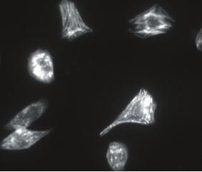
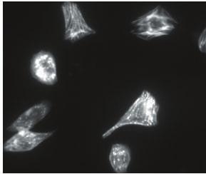
11 vehicle control in serum free media (300 µl/well). 12 h after infection, cells were treated with either vehicle alone or 100 µm PE for 24 h. The conditioned media was collected and centrifuged to pellet any possible debris. The concentration of ANF in the conditioned media was then determined using a peptide enzyme immunoassay (Peninsula Laboratories, San Carlos, CA). RESULTS Many features of pathological hypertrophy can be reproduced in isolated neonatal cardiomyocytes by treatment with a variety of agents including phenylephrine (PE), angiotensin II, and endothelin-1 (21). These cells respond by an increase in cell size, sarcomeric organization, increased expression of immediate early genes (c-fos, c-jun, Egr-1, jun-β and Nur-7) and re-expression of embryonic genes (atrial naturetic factor; ANF; skeletal α actin, smooth muscle α-actin and β-myosin heavy chain; ref. 22). Using this experimental paradigm, we examined to what extent integrin-mediated signaling is involved in the hypertrophic response in vitro. Initially, we examined the ability of PE to stimulate hypertrophy in neonatal cardiomyocytes plated on integrin-dependent (e.g. laminin, fibronectin and collagen type I) or integrin-independent (e.g. gelatin) substrates. PE treatment of cardiomyocytes plated on laminin 11
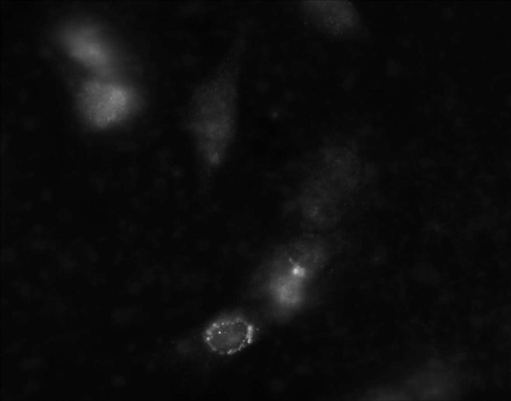
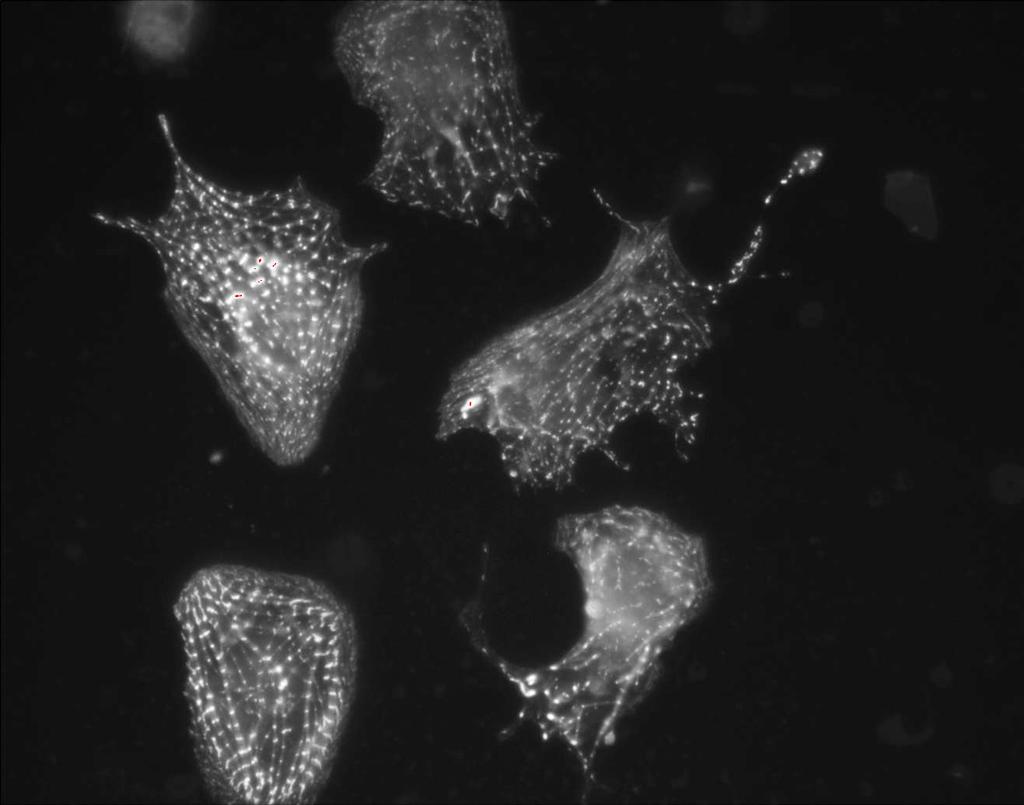
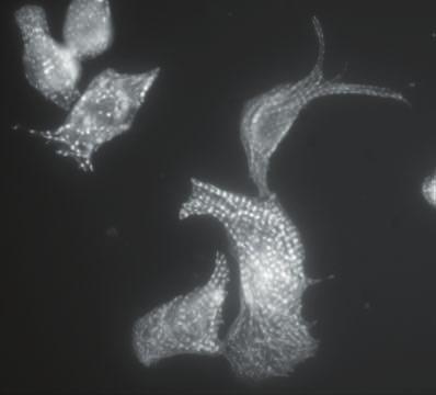
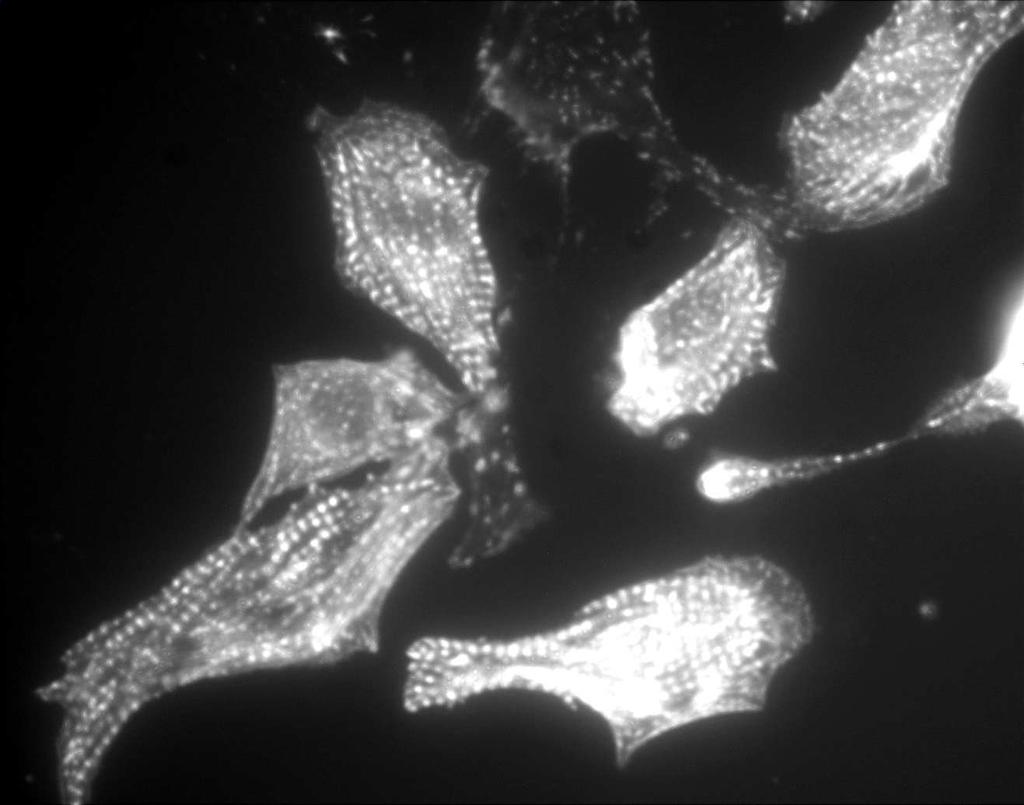
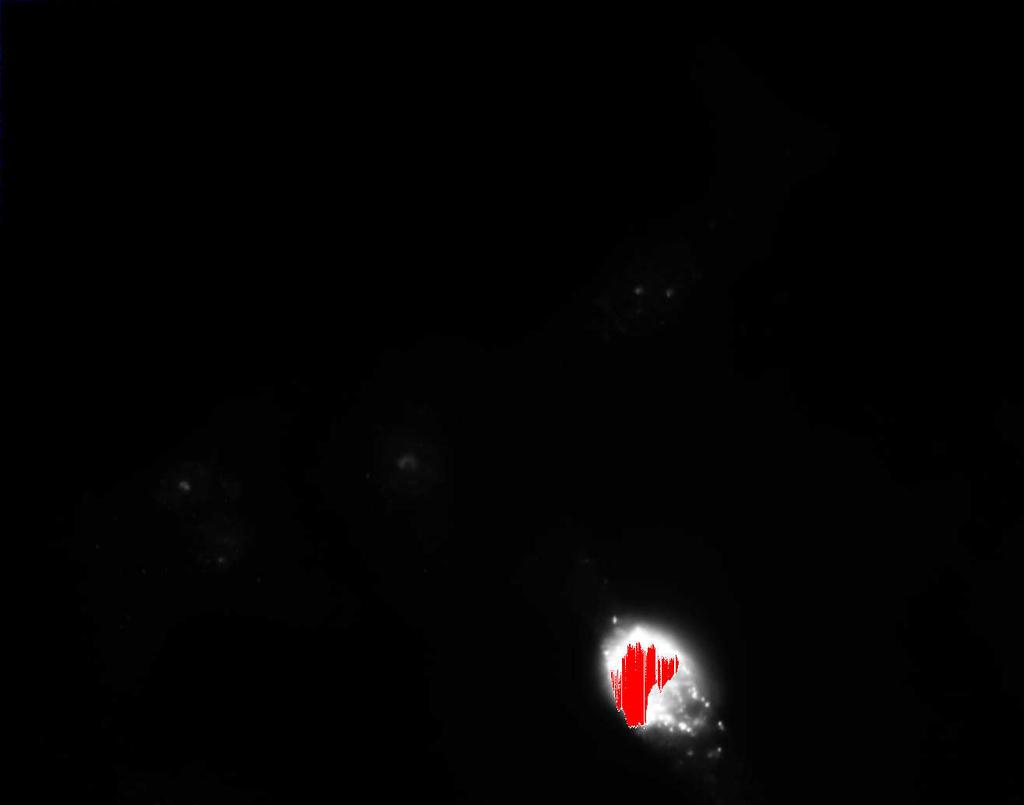
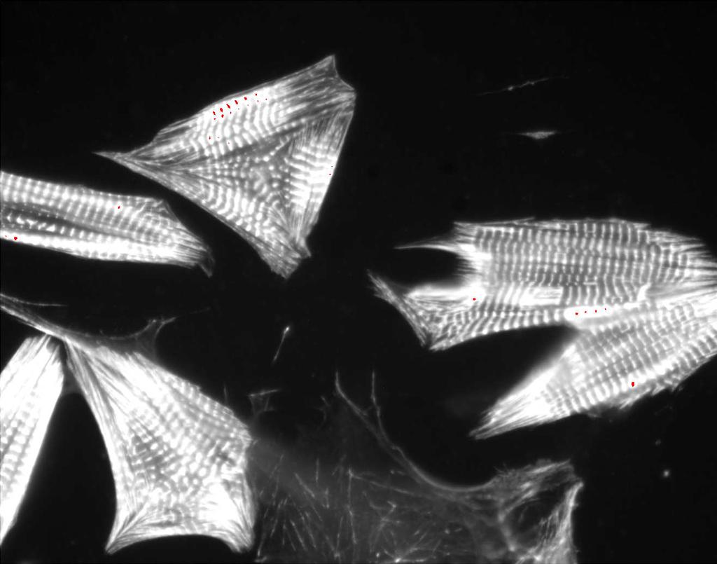
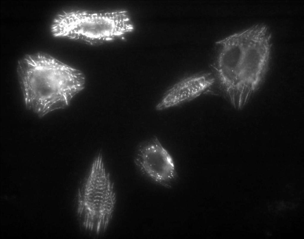
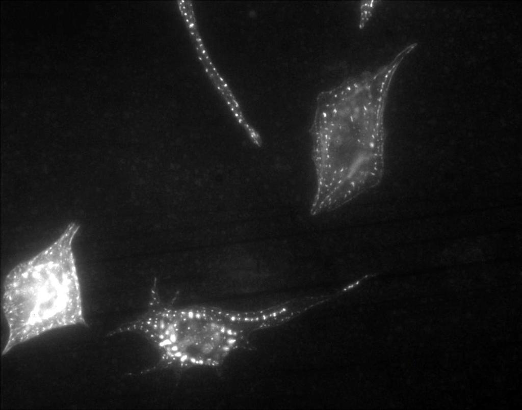
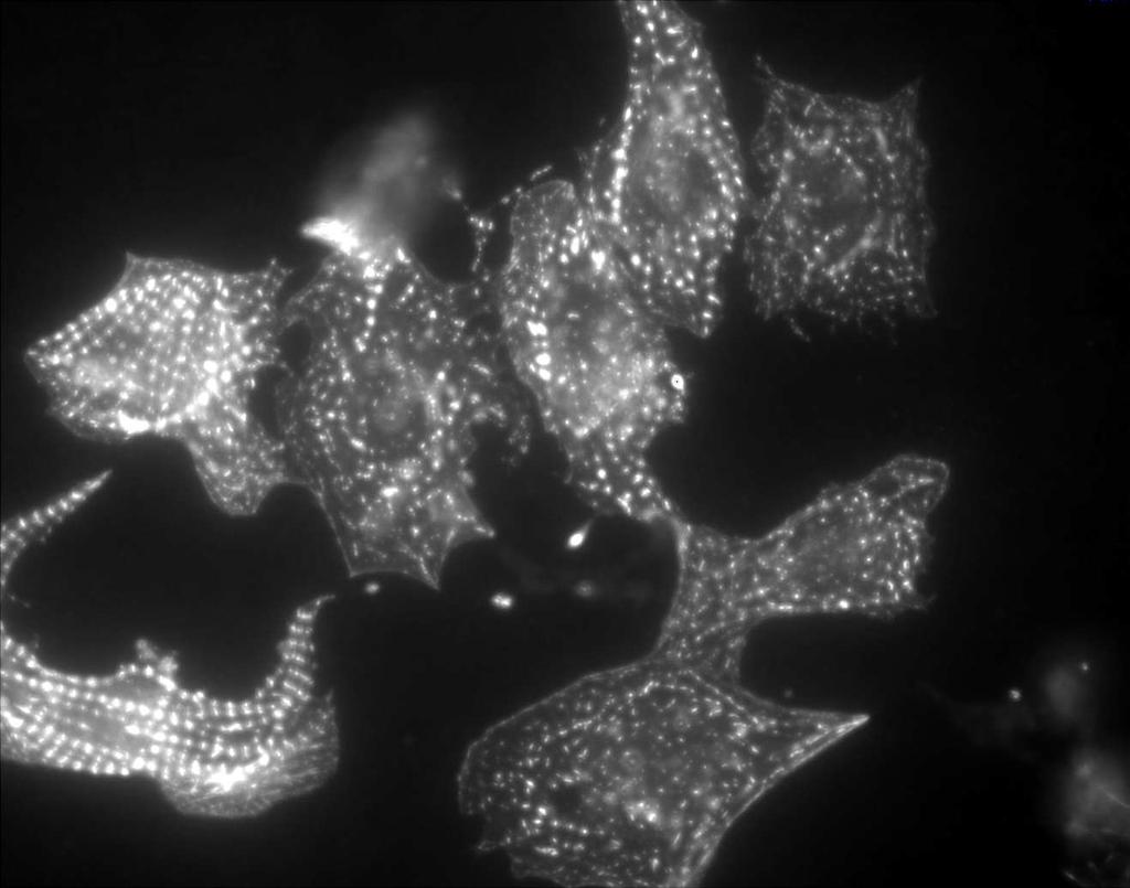
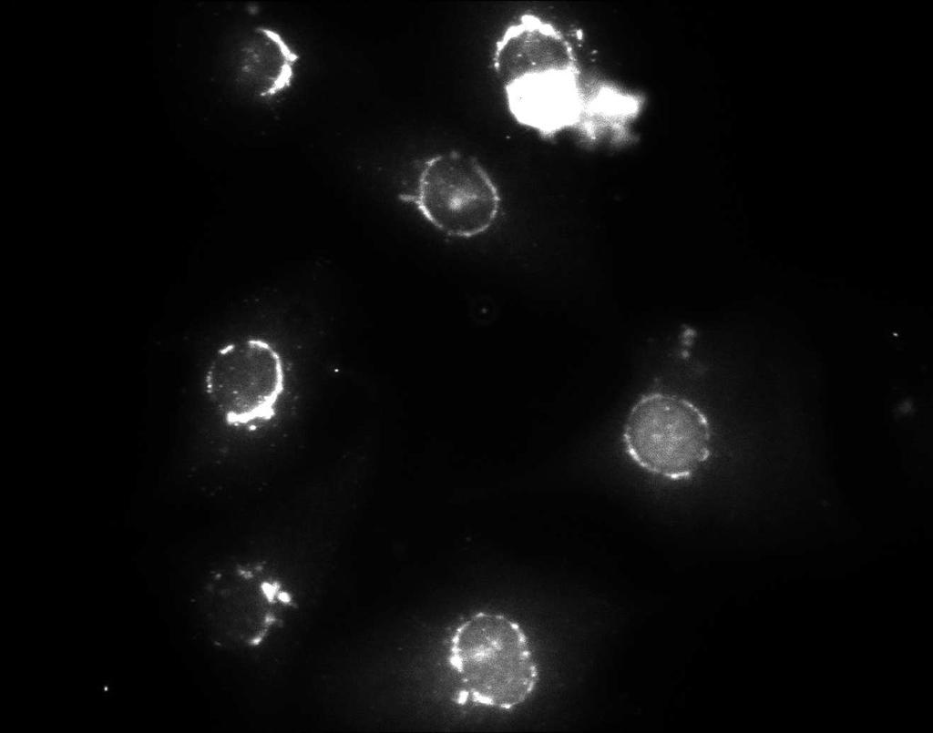
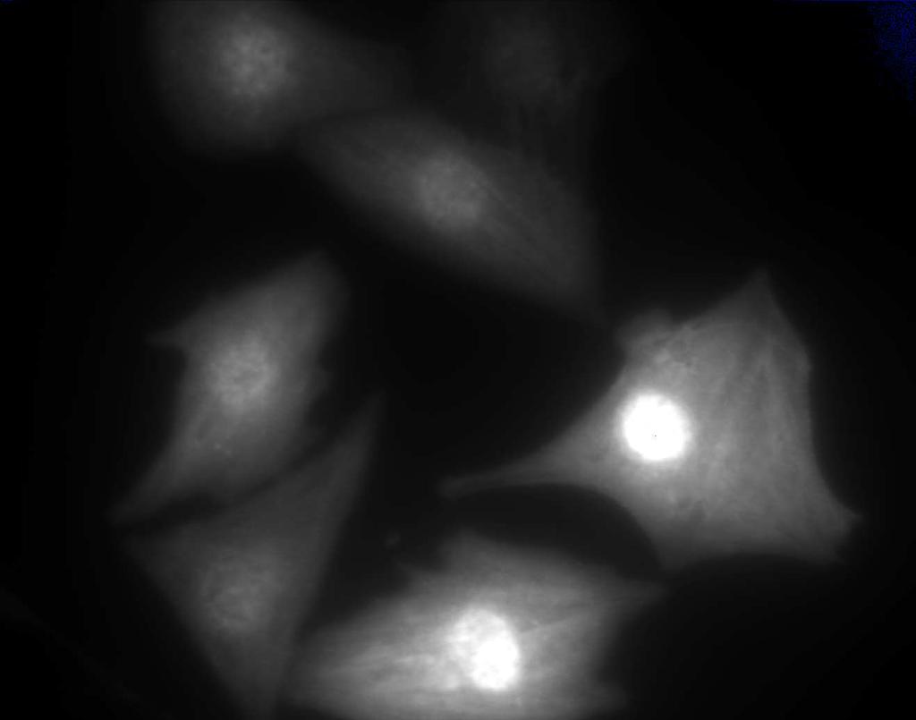
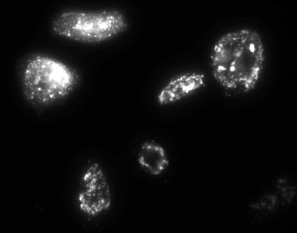
12 (Fig.1A, middle panels) stimulated sarcomeric organization, increase in cell size and ANF production compared to control, vehicle treated cells (Fig 1A, top panels). In contrast, cardiomyocytes plated on gelatin exhibited a reduced capacity to undergo these PE-stimulated hypertrophic changes (Fig 1A, bottom panels). To quantitate the extent of hypertrophy on different matricies, a total of cells in each treatment group (representing cells from three separate experiments) were scored for sarcomeric organization and ANF induction using fluorescent microscopy (Fig. 1B). Sarcomeric reorganization was observed in approximately 90% of PE-treated cells plated on laminin versus 35% of the cells plated on gelatin. Cells not treated with PE showed little actin reorganization whether plated on laminin or gelatin ( Fig. 1B, left panel). ANF production was observed in approximately 70% of PE-treated cells plated on laminin compared to 15% of the cells plated on gelatin. Fibronectin supported PE-stimulated sarcomeric reorganization and ANF production to a similar extent as laminin, whereas type I collagen was far less efficient in supporting these hypertrophic responses (Fig 1B). Thus these initial experiments provide support for the role of ECM-directed integrin signaling in PE-induced hypertrophy. Since maximal PE-induced cardiomyocyte hypertrophy required ECM, we examined whether components of the integrin-signaling cascade were altered after PE stimulation. First we examined whether PE treatment altered the subcellular localization of focal adhesion proteins during the hypertrophic response. Fig. 2 shows immuostaining of actin stress fibers and the focal adhesion protein paxillin during the progression of PE-induced hypertrophy in cells plated on laminin. Typically, cellular changes indicative of hypertrophy including increases in cell size and sarcomeric organization are complete within hours after PE treatment in cultured 12
13 neonatal cells. As shown in Fig. 2A, myofibrillar reorganization was enhanced relative to controls as early as 1h after PE treatment and the organization was virtually complete by 4 h. The recruitment of the focal adhesion protein paxillin to complexes at the periphery of cells appeared to be concomitant with the enhanced sarcomeric reorganization (Fig. 2B). Paxillin staining was generally dispersed in untreated cells but after 1 h of PE treatment, staining appeared more focal in nature, localized to the periphery of cells. At 4 h of PE treatment paxillin appeared in distinct complexes at the termini of actin fibers. A similar staining pattern was observed with antibodies directed against another prominent focal adhesion protein, vinculin (data not shown). The PE-stimulated paxillin complexes observed in cardiomyocytes are reminiscent of (albeit smaller than) classical focal adhesions observed following plating of fibroblasts or epithelial cells on ECM proteins and suggest that PE treatment is giving rise to a rearrangement of the ECM-adhesion structures. Tyrosine phosphorylation is a critical covalent modification driving protein-protein interactions required for cytoskeletal reorganization and focal adhesion assembly (10). As shown in Fig. 2B, PE treatment of cultured cardiomyocytes induced the formation of phosphotyrosine-rich focal adhesion-like complexes at the periphery of cells similar to that observed for paxillin immunostaining. To determine the kinetics and extent of tyrosine phosphorylation of focal adhesion associated proteins, FAK, and CAS were immunoprecipitated from PE-treated cells and analyzed by Western blotting with anti-phosphotyrosine (ptyr) antibodies. As shown in Fig. 3, cells plated on fibronectin, but not gelatin exhibited a PEstimulated, time-dependent increase in tyrosine phosphorylation of FAK (Fig. 3, panel A). Typically, an increase in FAK ptyr is observed within 5-10 min after treatment with PE and is 13
14 sustained for at least 30 min. A similar PE-induced tyrosine phosphorylation of the FAK binding partners, CAS and paxillin were also observed when cells were plated on fibronectin (Fig 3 panels B and C) but not gelatin (data not shown). Importantly, as shown in Figure 3D, PE-induced activation of the downstream signaling molecule ERK2 is also dependent on ECM interactions. Although variability is observed in the onset of ERK phosphorylation (ranging from 5-10 min) a consistent correlation is observed between the time-course for PE-induced FAK and ERK phosphorylation within each experiment. These observations mirror data from numerous studies showing enhanced tyrosine phosphorylation of focal adhesion proteins after integrin ligation induced by plating cells on ECM proteins and are consistent with integrin signaling being a component of PE-induced hypertrophy. Previous studies have shown that tyrosine phosphorylation is central to integrinstimulated focal adhesion formation and downstream signaling. Therefore, we examined the requirement for tyrosine phosphorylation in the induction of PE-induced hypertrophy. Figure 4 shows that the protein tyrosine kinase inhibitors genistein (100µg/ml) or herbimycin A (875 nm) markedly inhibited PE-induced myofibrillar organization and expression of endogenous ANF in cells plated on ECM. However, these inhibitors did not alter viability or morphology of vehicletreated cardiomyocytes (data not shown). These studies corroborate a previous report indicating that induction of an ANF-reporter construct was attenuated by genestein (23). The requirement for tyrosine phosphorylation and the ability of PE to stimulate FAK tyrosine phosphorylation in cardiomyocytes plated on ECM suggested the possibility that FAK is a down-stream mediator of PE stimulated integrin-signaling in the hypertrophy response. To determine whether the integrin signaling required for PE-stimulated hypertrophy is 14
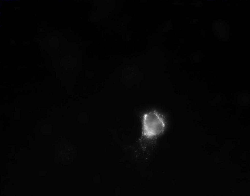
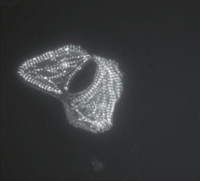
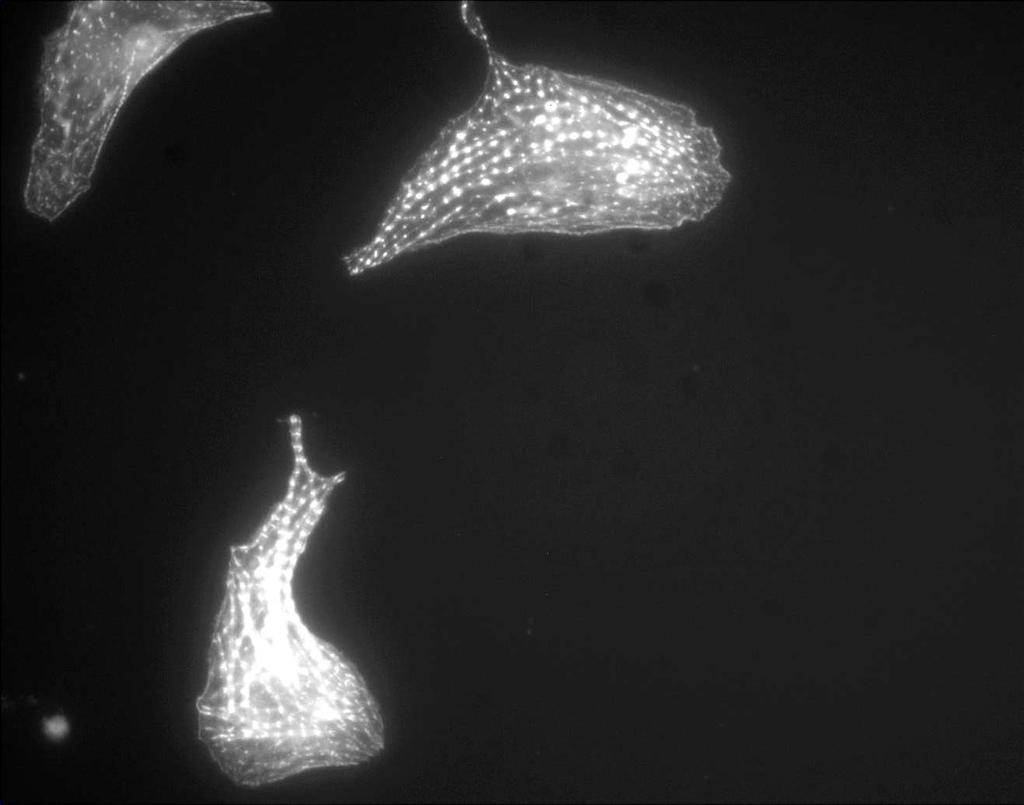
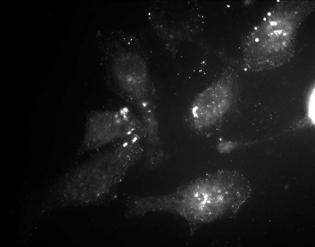
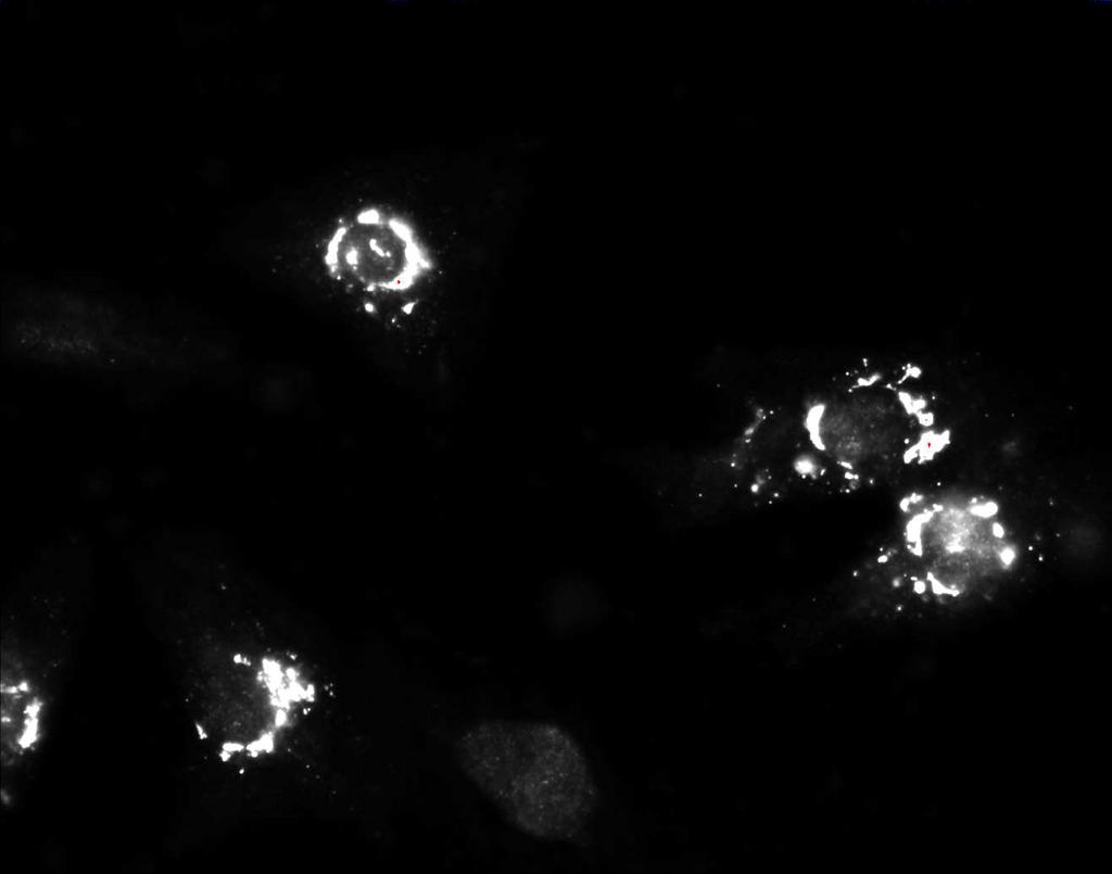
15 dependent on FAK activity, we sought to express a dominant-negative construct corresponding to the C-terminal domain of FAK (termed FRNK) in cardiomyocytes and examine the ability of these cells to undergo PE-mediated hypertrophy. It has been previously shown that FRNK functions to attenuate integrin-mediated tyrosine phosphorylation of FAK and paxillin as well as block integrin-mediated cell spreading and cell migration (12,13). Since this dominant-negative strategy required a high percentage of FRNK-expressing cells in order to observe populational changes in PE-induced signaling, we generated replication-defective adenovirus encoding GFP-tagged FRNK (GFRNK) or GFP alone as a control. GFP and GFRNK were efficiently expressed in % of the cardiomyocyte population as determined by immunofluorescence (data not shown). As shown in Fig 5A, both GFP and GFRNK proteins are expressed in cardiomyocytes within 12 h after adenoviral infection. Importantly, infection of cardiomyocytes with GFP-FRNK but not GFP alone attenuates PE-stimulated FAK activation as assessed by immunoblotting with antibodies specific for FAK ptyr at position 397 (Fig 5B). These data indicate that infection of cardiomyocytes with GFRNK is an effective means of inhibiting FAK signaling in these cells. To determine whether FAK activity was required for PE-stimulated hypertrophy, cardiomyocytes plated on fibronectin were infected with GFP or GFRNK virus prior to PE stimulation. Figure 6 shows that PE stimulates actin rearrangement and ANF-production in GFP-infected cardiomyocytes, indicating that adenovirus alone does not interfere with PEstimulated hypertrophy and is not cytotoxic to these cells. However, infection of cardiomyocytes with GFRNK resulted in a near complete inhibition of PE-stimulated hypertrophy as assessed by cell size, sarcomeric reorganization and endogenous ANF staining (Fig 6). Lack of PE- 15
16 stimulated ANF production in GFRNK infected cells compared to those infected with GFP or uninfected control cells was confirmed by quantitating the amount of ANF secretion in these cells using a peptide enzyme immunoassay. PE stimulated a 3.2±0.9 fold increase in ANF secretion over vehicle treated uninfected control cells (n=5; p<0.05) and a 2.6±0.4 fold increase in GFP-infected cells (n=5, p<0.05). However, no increase (0.94±0.1) in ANF secretion was observed following PE treatment of GFRNK-infected cells (n=5; p>0.05). Basal ANF secretion was not significantly altered by infection with either GFP or GFRNK viruses (n=5, p>0.05). A similar inhibition of PE-stimulated hypertrophy was observed when cells were transfected (rather than infected) with a myc-tagged FRNK cdna construct and scored visually for organized actin structures or ANF production (data not shown). Taken together these data indicate that activation of FAK is critical for maximal PE-stimulated hypertrophy in isolated cardiomyocytes. It is possible that the requirement for functional integrin signaling through FAK in PEstimulated hypertrophy may be due to the ability of FAK to cooperatively activate an essential PE-stimulated pathway. One candidate PE-stimulated pathway involves signaling from Ras to ERK. Activation of ERK2 has been implicated in the regulation of hypertrophic gene expression, and as shown above, PE-stimulated ERK2 activity is dependent on ECM. As shown in Figure 7, PE stimulated a time dependent increase in ERK1/2 activity in GFP-infected cardiomyocytes. However, in cardiomyocytes infected with GFRNK, no increase in ERK1/2 activity was observed following PE treatment. These data indicate that PE-induced signaling to ERK is uncoupled by expression of FRNK and suggest that ERK activation may be a critical point of convergence between FAK and PE signaling. 16
17 DISCUSSION We have shown that PE-induced hypertrophy in isolated neonatal cardiomyocytes requires ECM signaling through FAK. Specifically, plating cells on laminin or fibronectin supports maximal hypertrophic growth, whereas plating on gelatin or type I collagen does not. In addition, in cardiomyocytes plated on laminin or fibronectin, PE enhances the tyrosine phosphorylation of the focal adhesion proteins FAK and CAS and the subsequent formation of focal adhesion complexes. Furthermore, inhibition of signaling through FAK (by ectopic 17
18 expression of a dominant-interfering mutant) attenuates PE-stimulated hypertrophy in isolated cardiomyocytes. The observation that collagen type I is less efficient in supporting PE-induced hypertrophy is consistent with data which demonstrate that neonatal cardiomyocytes have a reduced affinity for type I collagen compared to other ECM such as laminin, fibronectin and type III or IV collagen (6). Our data presented above are also consistent with a recent report showing that PE-mediated ANF production was observed in cardiomyocytes plated on laminin but not in cells plated on the non-adhesive substrate, bovine serum albumin (24). Others, however have reported agonist-induced hypertrophic responses in cardiomyocytes plated on gelatin (25,26). It is possible that the increased cardiomyocyte plating density and the increased duration of the agonist treatment in these latter experiments led to modifications in the ECM environment (e.g., the secretion of ECM proteins) by the myocytes during culture, allowing for the support of hypertrophic growth. The studies described above point to a critical role for integrin signaling and specifically the activation of FAK in cardiomyocyte hypertrophy. Our work confirms and extends that of others indicating that overexpression of the wild type β 1 integrin potentiated PE-induced expression of an ANF reporter gene, whereas ectopic expression of a dominant-interfering integrin mutant attenuated the PE-induced response (24). Interestingly, recent reports have demonstrated that ectoptic expression of wild type or activated Src (F527) stimulate a hypertrophic pattern in neonatal cardiomyocytes (23,27). Src is recruited to activated FAK following integrin ligation and results in the formation of a FAK/Src signaling complex. The apparent requirement for FAK and Src in the hypertrophic response may reflect the need to 18
19 activate signaling pathways downstream of FAK in concert with pathways directly induced by hypertrophic stimuli. A large number of molecules stimulate growth (and hypertrophy) of cardiomyocytes in vitro including a variety of hormones, cytokines and growth factors. Included in the list of mitogens are PE, angiotensin II-, and endothelin I, prostaglandin growth factor 2α and transforming growth factor β among others (5). In regard to signaling, several of these agonists including PE-, angiotensin II- and endothelin I- activate receptors coupled to the heterotrimeric G protein, Gq, and the small molecular weight G protein, Ras. Importantly, the involvement of Gαq and Ras in the hypertrophic response has been shown not only in vitro but also in vivo in several elegant transgenic mouse models (28-30). The signaling molecules downstream of the Gq-coupled receptor responsible for activation of hypertrophy are reasonably delineated and appear to involve signaling through the small molecular weight G protein Ras to the ERK, Jun NH2-terminal kinase (JNK) and p38β MAP kinase pathways (19, 31-34). Our data indicates that activation of ERK1/2 may be a critical point of convergence between integrin- and PE- signaling. We have shown that maximal activation of ERK2 by PE in cardiomyocytes requires FAK-dependent ECM signaling. A similar cooperativity between integrin signaling and growth factor/hormone signaling to ERK2 has been previously observed in other cell types. Indeed, when cultured fibroblasts are held in suspension, PDGF-, EGF- and lysophosphatidic acid-stimulated ERK2 activity is markedly reduced compared to cells plated on fibronectin (14,15). At present, the precise mechanism for the anchorage-dependent activation of ERK2 is not clear. Evidence suggests that activation of the membrane bound receptors and subsequent activation of the small G protein Ras are independent of anchorage 19
20 (14,15). However maximal Ras-mediated activation of the serine/threonine kinase Raf and the downstream kinase MEK requires adhesion signaling (14,15). It is possible that the attenuation of MEK activity observed in suspended cells is due to the absence of integrin-mediated signaling through the Rho family of small GTP binding proteins. Recently it has been shown that members of the Rho family synergize with Raf to activate MEK (35). Importantly, others have shown that the inhibition of ERK2 activation observed in suspended cells can be rescued by over-expression of FAK and our data shows that FRNK attenuates agonist induced ERK1/2 activity in adherent cells (16). Taken together, these data clearly indicate that growth factors (or hormones) synergize with cell adhesion signals to stimulate cell signaling and highlight a possible role for FAK in MAP kinase activation and growth regulation. The precise point of convergence between FAK and PE-signaling to ERK1/2 and other putative downstream signaling molecules that may be important in the development of hypertrophy is currently unclear. A number of signaling molecules including the known FAK binding partners CAS, PI3 kinase, GRB2, paxillin and the Rho-GAP, Graf have been suggested as mediators of FAK signaling (10). We have shown in cardiomyocytes, that PE stimulates a rapid activation of FAK as assessed by enhanced auto-phosphorylation of tyrosine 397 and the subsequent accumulation of phosphotyrosine-rich focal adhesion complexes at the periphery of cells. In addition, PE stimulates tyrosine phosphorylation of two FAK-associated adapter proteins CAS and paxillin in an ECM-dependent fashion. Putative signaling pathways downstream of either CAS or paxillin could involve activation of the ERK, JNK, and p38 MAP kinase pathways, each of which have been implicated in hypertrophic signal transduction (32-34). In certain cell types, association of FAK with CAS is required for integrin-dependent 20
21 ERK2 activation (11). Future experiments will address whether this association is required for PE-stimulated hypertrophy. Interestingly, deletion of CAS by homologous recombination results in embryonic lethality an event likely due in part to abnormal development of the heart (36). Histological examination of hearts from CAS-deficient embryos revealed a thin myocardial wall accompanied by disorganized myofibrils and disrupted Z-disks in the ventricular cardiomyocytes, indicating that CAS may be involved in cardiomyocyte growth and possibly hypertrophy (36). An alternative explanation for the reduced extent of hypertrophy in the absence of FAK signaling is that the cardiomyocytes are undergoing apoptosis. There is a growing body of evidence indicating that the switch from hypertrophy to cardiac failure may involve an apoptotic pathway (34,37,38). For instance, aortic banding of mice with heart-restricted deletion of the gp130 gene results in dilated cardiomyopathy accompanied by cellular apoptosis, an event presumably due to lack of activation of a critical survival signaling pathway during the hypertrophic process (37). Therefore, it is possible that FAK (like gp130) may activate an essential survival pathway that is critical to observe PE-stimulated hypertrophy. Indeed, in other cell types, activation of FAK has been shown to be important for cell survival, and attenuating FAK signaling can lead to apoptosis (39). However, to date we have found no evidence of FRNK-induced apoptosis in cardiomyocytes in our experiments as assessed by DNA content using flow cytometry (unpublished observations). We have shown that PE signaling in cardiomyocytes activates FAK and that activation of FAK is required for PE-stimulated hypertrophy of cultured cells. Since angiotensin II, PGF-α and hypo-osmotic stress also enhance tyrosine phosphorylation of FAK in myocytes, it is 21
22 possible that activation of FAK may be a common requirement for a variety of hypertrophyinducing agents (40-42). The idea that FAK activation may play a role in the development of hypertrophy in vivo is supported by a recent report which demonstrates enhanced cytoskeletal association of β 3 integrin, FAK and Src in pressure overloaded hypertrophying hearts (43). Taken together, these data support a role for integrin-mediated signaling through FAK in the development of cardiac hypertrophy. Future studies will determine the utility of targeting FAK signaling as a potential therapeutic intervention for the treatment of cardiac hypertrophy. ACKNOWLEDGEMENTS This work was supported in part by Grants CA29243 and CA40042 from the DHHS. 22
23 JMT was supported by National Research Award HI-F32-GM18297 and by an American Heart Mid-Atlantic Affiliate Beginning Grant-In-Aid ( U). JDR was supported by a Resident Research Scholarship from the American College of Surgeons. We thank Larry Karns, Allen Everett, and Lou Hammerschold for helpful suggestions. We also thank Wen Xiong and Karen Martin for providing the myc-frnk and GFP-FRNK constructs respectively, Andrew Catling and Michael Weber for providing the ERK reagents, and Stephanie Morton for technical assistance. FIGURE LEGENDS 23
24 Figure 1. PE-stimulated hypertrophy requires ECM. A. Cardiomyocytes were plated on laminin (LAM; 10µg/ml) or gelatin (GEL; 0.1%) in media containing serum for 18 h prior to incubation in the absence of serum for an additional 24 h as described in Experimental Procedures. Following this incubation period, cells were treated with vehicle (top panel) or PE (100 µm; middle and bottom panels). After 30 h, cells were fixed, permeabilized and stained with FITC-phalloidin (left) to visualize sarcomeric reorganization and anti-anf Ab to visualize the induction of ANF (right). B. Cardiomyocytes, plated on 10 µg/ml laminin (LAM), fibronectin (FN), collagen type I (COL), or 0.1% gelatin (GEL), were treated with vehicle or PE as described above. Cells were visually scored for organized sarcomeric structures (left panel) or positive ANF staining (right panel). Data are presented as mean +/- SEM for three separate experiments. For each treatment a total of cells were scored. Figure 2. PE alters subcellular localization of paxillin and phosphotyrosine-containing proteins in isolated cardiomyocytes. Cardiomyocytes were isolated and plated on laminin (10 µg/ml) as described in Experimental Procedures. Cells were treated with PE for 1 or 4 h in serum free media. Cells were fixed, permeabilized and stained with FITC- phalloidin (top panels of A and B), anti-paxillin Ab (bottom panel of A) or anti-phosphotyrosine (PTyr) Ab (bottom panel of B) as described Experimental Procedures. Arrows denote focal adhesion-like complexes at the periphery of cells. Data are representative of three separate experiments. Figure 3. PE stimulates FAK, paxillin, CAS and ERK2 tyrosine phosphorylation in an ECMdependent fashion. Cardiomyocytes were plated on fibronectin (10 µg/ml) in media containing 24
25 10% FCS for 18 h prior to incubation in the absence of serum for an additional 24 h. Following this period, cells were treated with PE (100 µm) for the times indicated. Cells were lysed in RIPA buffer and proteins were separated by SDS-PAGE followed by immunoblotting with a FAK Y397 ptyr Ab (A, top panel). Blots were then stripped and re-probed with an Ab that recognizes total FAK protein (A, bottom panel). CAS (panel B) and paxillin (panel C) were immunoprecipitated from the extracts as described in Experimental Procedures followed by immunoblotting with anti-ptyr Ab (top panels) and the appropriate Ab for loading controls (bottom panels). In panel D, lysates were immunoblotted with a phosphorylation-specific Ab for ERK2 (top panel) and re-probed an Ab that recognizes total ERK1/2 (bottom panel). Data shown are representative of three separate experiments with similar results. Figure 4. Tyrosine kinase inhibitors block PE-stimulated hypertrophy in isolated cardiomyocytes. Cardiomyocytes were isolated and plated on LAM (10 µg/ml) in media containing 10% FCS for 18 h prior to incubation in the absence of serum for an additional 24 h. During this time, cells were pre-treated with vehicle (top panel), Genistein (100 µg/ml for 15 min; middle panels) or Herbimycin A (875 nm for 18 h; bottom panels). Following this incubation period, cells were treated with PE (100 µμ). After 30 h, cells were fixed, permeabilized, and stained with fluorescent phalloidin to visualize actin (left) or and anti-anf Ab (right). Figure 5. Adenoviral-mediated expression of GFP-FRNK attenuates FAK activity in isolated cardiomyocytes. Cardiomyocytes were isolated and plated on FN (10 µg/ml) as described above. 25
26 Cells were infected with GFP or GFP-FRNK (GFRNK) adenovirus (4 MOI) for 12 h. A. Immunoblotting was performed on lysates (50 µg) from GFP infected cells or GFRNK expressing cells using antibodies specific for GFP (left panel) or the C-terminus of FAK (right panel). In panel B, immunoblotting was performed on lysates (100 µg) from uninfected control cells (C), GFP infected cells (GFP) and GFRNK infected cells using a FAK Y397 ptyr Ab (top panel) and an Ab which recognizes total FAK protein (bottom panel). Data are representative of three separate experiments. Figure 6. Adenoviral-mediated expression of GFP-FRNK attenuates PE-stimulated hypertrophy. Cardiomyocytes were isolated, plated on FN (10 µg/ml) and infected with GFP or GFRNK viruses (4 MOI) as described in Experimental Procedures. 12 h post-infection, cells were treated with vehicle (-PE) or 100 µm PE (+PE) for 30 h. Cells were fixed, permeabilized and stained with Texas Red-conjugated phalloidin (panel A) or anti-anf Ab (panel B). GFP and GFRNK expressing cells were visualized by immunofluorescence (left panels). The * in panel A indicates un-infected control cells and the arrow a GFRNK expressing cell. The data presented are representative of four separate experiments. Figure 7. Adenoviral-mediated expression of GFP-FRNK attenuates ERK1/2 kinase activity in isolated cardiomyocytes. Cardiomyocytes were isolated, plated on FN (10 µg/ml) and infected with GFP or GFRNK viruses (4 MOI) as described in Experimental Procedures. 12 h postinfection, cells were treated with 100 µm PE for the times indicated. Cells were lysed in RIPA buffer and ERK1/2 was immunoprecipitated from the extracts. ERK immunoprecipitates were 26
27 incubated with 2 µg of myelin basic protein (MBP) and [γ 32 P]ATP as described in Experimental Procedures. Samples were analyzed by SDS-PAGE, and the gel was transferred to nitrocellulose and exposed to Kodak XAR film for 1 h (top). The membrane was then probed with anti-erk1/2 Ab for a loading control (bottom). Data shown are representative of five separate experiments with similar results. 27
28 REFERENCES 1. Komuro, I. and Yazaki, Y. (1993) Annu Rev Physiol 55, Chrzanowska-Wodnicka, M. and Burridge, K. (1996) J. Cell Biol. 133(6), Hynes, R. O. (1992) Cell 69, Graf, K., Do, Y. S., Ashizawa, N., Meehan, W. P., Giachelli, C. M., Marboe, C. C., Fleck, E., and Hsueh, W. A. (1997) Circulation 96, Mamuya, W., Chobanian, A., and Brecher, P. (1992) Circ Res 71, Terracio, L., Rubin, K., Gullberg, D., Balog, E., Carver, W., Jyring, R., and Borg, T. K. (1991) Circ Res 68, McDonald, J. A. and Valencik, M. (1998) Keystone Symposium X7, A220(Abstract) 8. Schaller, M. D., Hildebrand, J. D., Shannon, J. D., Fox, J. W., Vines, R. R., and Parsons, J. T. (1994) Mol Cell Biol 14, Jockusch, B. M., Bubeck, P., Giehl, K., Kroemker, M., Moschner, J., Rothkegel, M., Rudiger, M., Schluter, K., Stanke, G., and Winkler, J. (1995) Ann Rev Cell Dev Biol 11, Parsons J.T. (1996) Curr. Opin. Cell Biol. 8, Schlaepfer, D. D. and Hunter, T. (1997) J. Biol. Chem. 272, Richardson, A., Malik, R. K., Hildebrand, J. D., and Parsons, J. T. (1997) Mol Cell Biol 17, Richardson, A. and Parsons, T. (1996) Nature 380, Renshaw, M. W., Ren, X. D., and Schwartz, M. A. (1997) EMBO J 16,
29 15. Lin TH, Chen Q, Howe A, and Juliano RL. (1997) J. Biol Chem 272, Renshaw MW, Price LS and Schwartz MA. (1999) J. Cell Biol. In Press. 17. Bouton, A. H. and Burnham, M. R. (1997) Hybridoma 16, Hardy S, Kitamura M, Harris-Stansil T, Dai Y, and Phipps ML (1997) J. Virology 71, Thorburn J, McMahon M, and Thorburn A. (1994) J. Biol. Chem 269, Simpson PC, McGrath A, and Savion S. (1982) Circ. Res. 51, Sadoshima, J. and Izumo, S. (1997) Annu Rev Physiol 59, Chien, K. R., Zhu, H., Knowlton, K. U., Miller-Hance, W., van-bilsen, M., O Brien, T. X., and Evans, S. M. (1993) Annu Rev Physiol 55, Fuller, S. J., Gillespie-Brown, J., and Sugden, P. H. (1998) J. Biol. Chem. 273, Ross, R. S., Pham, C., Shai, S. Y., Goldhaber, J. I., Fenczik, C., Glembotski, C. C., Ginsberg, M. H., and Loftus, J. C. (1998) Circ Res 82, Hoshijima, M., Sah, V. P., Wang, Y., Chien, K. R., and Brown, J. H. (1998) J Biol Chem 273, Sah, V. P., Hoshijima, M., Chien, K. R., and Brown, J. H. (1996) J. Biol. Chem. 271, Kovacic, B., Ilic, D., Damsky, C. H., and Gardner, D. G. (1998) J. Biol. Chem. 273, Akhter SA, Luttrell LM, Rockman HA, Iaccarino G, Lefkowitz RJ, and Koch WJ. (1998) Science 280,
30 29. Thorburn A, Thorburn J, Chen S-Y, Powers S, Shubeita HE, Feramisco JR, and Chien KR. (1993) J. Biol. Chem. 268, Hunter JJ, Tanaka N, Rockman HA, Ross J, and Chien KR. (1995) J. Biol. Chem. 270, Zechner D, Thuerauf DJ, Hanford DS, McDonough PM, and Glembotski CC. (1997) J. Cell Biol. 139, Ramirez, M. T., Sah, V. P., Zhao, X. L., Hunter, J. J., Chien, K. R., and Brown, J. H. (1997) J Biol Chem 272, Wang, Y., Su, B., Sah, V. P., Brown, J. H., Han, J., and Chien, K. R. (1998) J Biol Chem 273, Wang, Y., Huang, S., Sah, V. P., Ross, J.,Jr., Brown, J. H., Han, J., and Chien, K. R. (1998) J Biol Chem 273, Frost JA, Steen H, Shapiro P, Lewis T, Ahn N, Shaw PE, and Cobb MH (1997) EMBO 16, Honda, H., Oda, H., Nakamoto, T., Honda, Z., Sakai, R., Suzuki, T., Saito, T., Nakamura, K., Nakao, K., Ishikawa, T., Katsuki, M., Yazaki, Y., and Hirai, H. (1998) Nat Genet 19, Hirota H, Chen J, Betz UAK, Rajewsky K, Gu Y, Ross J JR, Muller W and Chen K. (1999) Cell 97, Adams, JW, Sakata Y, Davis MG, Sah VP, Wang Y, Liggett SB, Chien KR, Brown JH, Dorn GW II. (1998) PNAS 95, Ilic D, Almeida EA, Schlaepfer DD, Aizawa S, Damsky CH. (1998) J. Cell Biol. 143,
31 40. Adams, J. W., Sah, V. P., Henderson, S. A., and Brown, J. H. (1998) Circ Res 83, Sadoshima, J., Qiu, Z., Morgan, J. P., and Izumo, S. (1996) EMBO J 15, Polte, T. R., Naftilan, A. J., and Hanks, S. K. (1994) J. Cell. Biochem. 55, Kuppuswamy, D., Kerr, C., Narishige, T., Kasi, V. S., Menick, D. R., and Cooper, G. (1997) J Biol Chem 272,


32 A. E GEL + PE LAM + PE LAM ACTIN ANF Taylor et. al. FIG 1A
33 B. ACTIN ORGANIZATION ANF PRODUCTION C PE C PE % TOTAL CELLS % TOTAL CELLS LAM FN COL GEL 0 LAM FN COL GEL Taylor et. al. FIG 1B


34 A. ACTIN PAXILLIN Time (hr) Taylor et. al. FIG 2A


35 B. ACTIN PTYR Time (hr) Taylor et. al. FIG 2B


 PE (100 µm) PE (100 µm) D.")
36 Taylor et. al. FIG 3 A. IB:FAKpY GEL FN FAK IB:FAK FAK PE (100 µm) Time (min) B. FN C. FN IP:CAS IB:PTyr CAS IP:Pax IB:PTyr Pax IP:CAS IB:CAS CAS IP:Pax IB:Pax Pax 0 15 Time (min) 0 15 Time (min) PE (100 µm) PE (100 µm) D. GEL FN IB:ERKpY ERK2 IB:ERK ERK PE (100 µm) Time (min)


37 PE + HERB PE + GEN PE ACTIN ANF Taylor et. al. FIG 4
38 Taylor et. al. FIG 5 A. IB:GFP IB:FAK FAK GFRNK GFP GFRNK GFP GFRNK GFP B. IB:FAKpY FAK IB:FAK FAK C GFP GFRNK
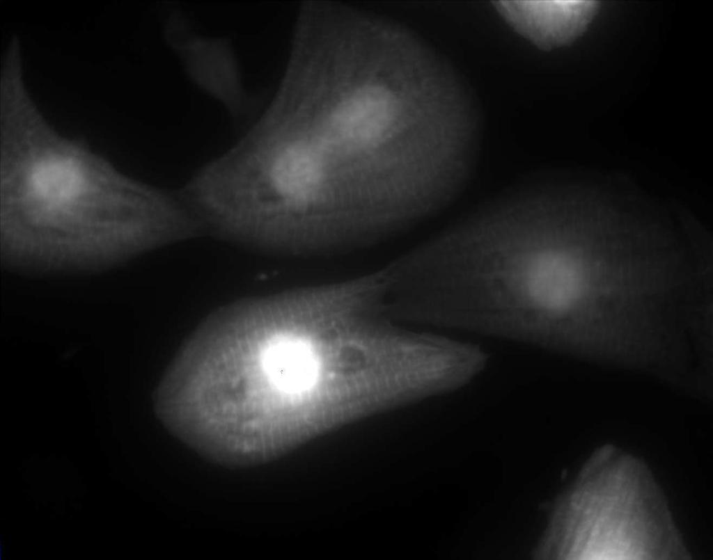
39 A. Fluorescence Actin GFRNK + PE GFP + PE GFRNK - PE GFP - PE * * * * Taylor et. al. FIG 6A

40 B. Fluorescence ANF GFRNK + PE GFP + PE Taylor et. al. FIG 6B
41 Taylor et. al. FIG 7 GFP GFRNK IP:ERK 32 P-MBP MBP IP:ERK IB:ERK ERK Time (min)
Post-translational modification
 Protein expression Western blotting, is a widely used and accepted technique to detect levels of protein expression in a cell or tissue extract. This technique measures protein levels in a biological sample
Cdc42 Activation Assay Kit
 A helping hand for your research Product Manual Configuration-specific Monoclonal Antibody Based Cdc42 Activation Assay Kit Catalog Number: 80701 20 assays 1 Table of Content Product Description 3 Assay
Anti-HB-EGF (Human) mab
 mab") Page 1 For Research Use Only. Not for use in diagnostic procedures. CODE No. D308-3 Anti-HB-EGF (Human) mab CLONALITY CLONE ISOTYPE QUANTITY SOURCE IMMUNOGEN FORMURATION STORAGE Monoclonal 3H4 Mouse IgG1
Page 1 For Research Use Only. Not for use in diagnostic procedures. CODE No. D308-3 Anti-HB-EGF (Human) mab CLONALITY CLONE ISOTYPE QUANTITY SOURCE IMMUNOGEN FORMURATION STORAGE Monoclonal 3H4 Mouse IgG1
Gα 13 Activation Assay Kit
 A helping hand for your research Product Manual Configuration-specific Monoclonal Antibody Based Gα 13 Activation Assay Kit Catalog Number: 80401 20 assays NewEast Biosciences 1 Table of Content Product
Gα i Activation Assay Kit
 A helping hand for your research Product Manual Configuration-specific Monoclonal Antibody Based Gα i Activation Assay Kit Catalog Number 80301 20 assays NewEast Biosciences, Inc 1 Table of Content Product
Supplemental Online Material. The mouse embryonic fibroblast cell line #10 derived from β-arrestin1 -/- -β-arrestin2 -/-
 #1074683s 1 Supplemental Online Material Materials and Methods Cell lines and tissue culture The mouse embryonic fibroblast cell line #10 derived from β-arrestin1 -/- -β-arrestin2 -/- knock-out animals
At E17.5, the embryos were rinsed in phosphate-buffered saline (PBS) and immersed in
 and immersed in") Supplementary Materials and Methods Barrier function assays At E17.5, the embryos were rinsed in phosphate-buffered saline (PBS) and immersed in acidic X-gal mix (100 mm phosphate buffer at ph4.3, 3 mm
Supplementary Materials and Methods Barrier function assays At E17.5, the embryos were rinsed in phosphate-buffered saline (PBS) and immersed in acidic X-gal mix (100 mm phosphate buffer at ph4.3, 3 mm
Supplementary Table 1. The Q-PCR primer sequence is summarized in the following table.
 Supplementary Table 1. The Q-PCR primer sequence is summarized in the following table. Name Sequence (5-3 ) Application Flag-u ggactacaaggacgacgatgac Shared upstream primer for all the amplifications of
Supplementary Information (Ha, et. al) Supplementary Figures Supplementary Fig. S1
 Supplementary Figures Supplementary Fig. S1") Supplementary Information (Ha, et. al) Supplementary Figures Supplementary Fig. S1 a His-ORMDL3 ~ 17 His-ORMDL3 GST-ORMDL3 - + - + IPTG GST-ORMDL3 ~ b Integrated Density (ORMDL3/ -actin) 0.4 0.3 0.2 0.1
Supplementary Information (Ha, et. al) Supplementary Figures Supplementary Fig. S1 a His-ORMDL3 ~ 17 His-ORMDL3 GST-ORMDL3 - + - + IPTG GST-ORMDL3 ~ b Integrated Density (ORMDL3/ -actin) 0.4 0.3 0.2 0.1
RheB Activation Assay Kit
 A helping hand for your research Product Manual Configuration-specific Monoclonal Antibody Based RheB Activation Assay Kit Catalog Number: 81201 20 assays NewEast Biosciences 1 FAX: 610-945-2008 Table
T H E J O U R N A L O F C E L L B I O L O G Y
 T H E J O U R N A L O F C E L L B I O L O G Y Supplemental material Bays et al., http://www.jcb.org/cgi/content/full/jcb.201309092/dc1 Figure S1. Specificity of the phospho-y822 antibody. (A) Total cell
*Corresponding author. Tel: ;
 1 SUPPLEMENTARY DATA 2 3 4 5 6 7 8 9 10 11 Integrin 2 1 in nonactivated conformation can induce focal adhesion kinase signaling Maria Salmela 1, Johanna Jokinen 1,2, Silja Tiitta 1, Pekka Rappu 1, Holland
Rab5 Activation Assay Kit
 A helping hand for your research Product Manual Configuration-specific Monoclonal Antibody Based Rab5 Activation Assay Kit Catalog Number: 83701 20 assays 24 Whitewoods Lane 1 Table of Content Product
Sarker et al. Supplementary Material. Subcellular Fractionation
 Supplementary Material Subcellular Fractionation Transfected 293T cells were harvested with phosphate buffered saline (PBS) and centrifuged at 2000 rpm (500g) for 3 min. The pellet was washed, re-centrifuged
Arf6 Activation Assay Kit
 A helping hand for your research Product Manual Configuration-specific Monoclonal Antibody Based Arf6 Activation Assay Kit Catalog Number: 82401 20 assays NewEast Biosciences 1 Table of Content Product
Supplementary Figure 1. Soft fibrin gels promote growth and organized mesodermal differentiation. Representative images of single OGTR1 ESCs cultured
 Supplementary Figure 1. Soft fibrin gels promote growth and organized mesodermal differentiation. Representative images of single OGTR1 ESCs cultured in 90-Pa 3D fibrin gels for 5 days in the presence
LINGO-1, A TRANSMEMBRANE SIGNALING PROTEIN, INHIBITS OLIGODENDROCYTE DIFFERENTIATION AND MYELINATION THROUGH INTERCELLULAR SELF- INTERACTIONS.
 Supplemental Data: LINGO-1, A TRANSMEMBRANE SIGNALING PROTEIN, INHIBITS OLIGODENDROCYTE DIFFERENTIATION AND MYELINATION THROUGH INTERCELLULAR SELF- INTERACTIONS. Scott Jepson, Bryan Vought, Christian H.
CD93 and dystroglycan cooperation in human endothelial cell adhesion and migration
 /, Supplementary Advance Publications Materials 2016 CD93 and dystroglycan cooperation in human endothelial cell adhesion and migration Supplementary Materials Supplementary Figure S1: In ECs CD93 silencing
Modeling Cardiac Hypertrophy: Endothelin-1 Induction with qrt-pcr Analysis
 icell Cardiomyocytes Application Protocol Modeling Cardiac Hypertrophy: Endothelin-1 Induction with qrt-pcr Analysis Introduction Cardiac hypertrophy is characterized by several different cellular changes,
System. Dynamic Monitoring of Receptor Tyrosine Kinase Activation in Living Cells. Application Note No. 4 / March
 System Application Note No. 4 / March 2008 Dynamic Monitoring of Receptor Tyrosine Kinase Activation in Living Cells www.roche-applied-science.com Dynamic Monitoring of Receptor Tyrosine Kinase Activation
SUPPLEMENTAL MATERIAL. Supplemental Methods:
 SUPPLEMENTAL MATERIAL Supplemental Methods: Immunoprecipitation- As we described but with some modifications [22]. As part of another ongoing project, lysate from human umbilical vein endothelial cells
Supplemental Information
 Supplemental Information Intrinsic protein-protein interaction mediated and chaperonin assisted sequential assembly of a stable Bardet Biedl syndome protein complex, the BBSome * Qihong Zhang 1#, Dahai
RhoC Activation Assay Kit
 Product Manual RhoC Activation Assay Kit Catalog Number STA-403-C 20 assays FOR RESEARCH USE ONLY Not for use in diagnostic procedures Introduction Small GTP-binding proteins (or GTPases) are a family
MEK1/2 (MAPK Kinase) Activity Assay Kit
 Activity Assay Kit") MEK1/2 (MAPK Kinase) Activity Assay Kit For 96 tests Cat. No. SGT440 FOR RESEARCH USE ONLY Not for use in diagnostic procedures USA & Canada Phone: +1(800) 437-7500 Fax: +1 (951) 676-9209 Europe +44 (0)
MEK1/2 (MAPK Kinase) Activity Assay Kit For 96 tests Cat. No. SGT440 FOR RESEARCH USE ONLY Not for use in diagnostic procedures USA & Canada Phone: +1(800) 437-7500 Fax: +1 (951) 676-9209 Europe +44 (0)
Time allowed: 2 hours Answer ALL questions in Section A, ALL PARTS of the question in Section B and ONE question from Section C.
 UNIVERSITY OF EAST ANGLIA School of Biological Sciences Main Series UG Examination 2017-18 CELL BIOLOGY BIO-5005B Time allowed: 2 hours Answer ALL questions in Section A, ALL PARTS of the question in Section
Rabbit (monoclonal) Anti-FAK [py 397 ] Phosphospecific Antibody, Unconjugated
![Rabbit (monoclonal) Anti-FAK [py 397 ] Phosphospecific Antibody, Unconjugated](/thumbs/78/77450403.jpg "Rabbit (monoclonal) Anti-FAK [py 397 ] Phosphospecific Antibody, Unconjugated") Rabbit (monoclonal) Anti-FAK [py 397 ] Phosphospecific Antibody, Unconjugated PRODUCT ANALYSIS SHEET Catalog Number: Lot Number: Volume: 44-625G (10 mini-blot size) See product label 100 μl Clone Number:
Rabbit (monoclonal) Anti-FAK [py 397 ] Phosphospecific Antibody, Unconjugated PRODUCT ANALYSIS SHEET Catalog Number: Lot Number: Volume: 44-625G (10 mini-blot size) See product label 100 μl Clone Number:
IgG TrueBlot Protocol for Mouse, Rabbit or Goatderived Antibodies - For Research Use Only
 IgG TrueBlot Protocol for Mouse, Rabbit or Goatderived Antibodies - For Research Use Only Introduction The IgG TrueBlot for mouse, rabbit, or goat-derived antibodies represents unique series of respective
Beta3 integrin promotes long-lasting activation and polarization of Vascular Endothelial Growth Factor Receptor 2 by immobilized ligand
 SUPPLEMENTAL FIGURES Beta3 integrin promotes long-lasting activation and polarization of Vascular Endothelial Growth Factor Receptor 2 by immobilized ligand C. Ravelli et al. FIGURE S. I Figure S. I: Gremlin
RNA was isolated using NucleoSpin RNA II (Macherey-Nagel, Bethlehem, PA) according to the
 according to the") Supplementary Methods RT-PCR and real-time PCR analysis RNA was isolated using NucleoSpin RNA II (Macherey-Nagel, Bethlehem, PA) according to the manufacturer s protocol and quantified by measuring the
Supplementary Methods RT-PCR and real-time PCR analysis RNA was isolated using NucleoSpin RNA II (Macherey-Nagel, Bethlehem, PA) according to the manufacturer s protocol and quantified by measuring the
T H E J O U R N A L O F C E L L B I O L O G Y
 Supplemental material Thompson et al., http://www.jcb.org/cgi/content/full/jcb.200909067/dc1 T H E J O U R N A L O F C E L L B I O L O G Y Figure S1. Modification-specific antibodies do not detect unmodified
Apoptosis assay: Apoptotic cells were identified by Annexin V-Alexa Fluor 488 and Propidium
 Apoptosis assay: Apoptotic cells were identified by Annexin V-Alexa Fluor 488 and Propidium Iodide (Invitrogen, Carlsbad, CA) staining. Briefly, 2x10 5 cells were washed once in cold PBS and resuspended
Recruitment of Grb2 to surface IgG and IgE provides antigen receptor-intrinsic costimulation to class-switched B cells
 SUPPLEMENTARY FIGURES Recruitment of Grb2 to surface IgG and IgE provides antigen receptor-intrinsic costimulation to class-switched B cells Niklas Engels, Lars Morten König, Christina Heemann, Johannes
ab Ran Activation Assay Kit
 ab173247 Ran Activation Assay Kit Instructions for Use For the simple and fast measurement of Ran activation. This product is for research use only and is not intended for diagnostic use. Version 1 Last
MAP Kinase (ERK1/2) Activity Assay Kit
 Activity Assay Kit") MAP Kinase (ERK/2) Activity Assay Kit For 96 tests Cat. No. SGT45 FOR RESEARCH USE ONLY Not for use in diagnostic procedures USA & Canada Phone: +(800) 437-7500 Fax: + (909) 676-9209 Europe +44 (0) 23
MAP Kinase (ERK/2) Activity Assay Kit For 96 tests Cat. No. SGT45 FOR RESEARCH USE ONLY Not for use in diagnostic procedures USA & Canada Phone: +(800) 437-7500 Fax: + (909) 676-9209 Europe +44 (0) 23
Supporting Information
 Supporting Information Su et al. 10.1073/pnas.1211604110 SI Materials and Methods Cell Culture and Plasmids. Tera-1 and Tera-2 cells (ATCC: HTB- 105/106) were maintained in McCoy s 5A medium with 15% FBS
SUPPLEMENTARY INFORMATION
 The Supplementary Information (SI) Methods Cell culture and transfections H1299, U2OS, 293, HeLa cells were maintained in DMEM medium supplemented with 10% fetal bovine serum. H1299 and 293 cells were
A guide to selecting control, diluent and blocking reagents
 Specializing in Secondary Antibodies and Conjugates A guide to selecting control, diluent and blocking reagents Optimize your experimental protocols with Jackson ImmunoResearch Secondary antibodies and
A guide to selecting control, diluent and blocking reagents
 Specializing in Secondary Antibodies and Conjugates A guide to selecting control, diluent and blocking reagents Optimize your experimental protocols with Jackson ImmunoResearch Secondary antibodies and
Adenovirus Titration Kit
 Adenovirus Titration Kit Catalog # LF-RK0001(1 kit) Immunostaining method for Quantitative Detection of Adenovirus For research use only Not for diagnostic or therapeutic procedures AbFrontier Science
Supplementary Appendix
 Supplementary Appendix This appendix has been provided by the authors to give readers additional information about their work. Supplement to: Svegliati Baroni S, Santillo M, Bevilacqua F, et al. Stimulatory
Supplementary data. sienigma. F-Enigma F-EnigmaSM. a-p53
 Supplementary data Supplemental Figure 1 A sienigma #2 sienigma sicontrol a-enigma - + ++ - - - - - - + ++ - - - - - - ++ B sienigma F-Enigma F-EnigmaSM a-flag HLK3 cells - - - + ++ + ++ - + - + + - -
ab G alpha i Activation Assay Kit
 ab173234 G alpha i Activation Assay Kit Instructions for Use For the simple and fast measurement of G alpha i activation. This product is for research use only and is not intended for diagnostic use. Version
Supplementary Information: Materials and Methods. Immunoblot and immunoprecipitation. Cells were washed in phosphate buffered
 Supplementary Information: Materials and Methods Immunoblot and immunoprecipitation. Cells were washed in phosphate buffered saline (PBS) and lysed in TNN lysis buffer (50mM Tris at ph 8.0, 120mM NaCl
CytoSelect 48-Well Cell Adhesion Assay (ECM Array, Fluorometric Format)
") Product Manual CytoSelect 48-Well Cell Adhesion Assay (ECM Array, Fluorometric Format) Catalog Number CBA-071 CBA-071-5 48 assays 5 x 48 assays FOR RESEARCH USE ONLY Not for use in diagnostic procedures
Product Manual CytoSelect 48-Well Cell Adhesion Assay (ECM Array, Fluorometric Format) Catalog Number CBA-071 CBA-071-5 48 assays 5 x 48 assays FOR RESEARCH USE ONLY Not for use in diagnostic procedures
Electrophoretic Mobility Shift Assay (EMSA). Nuclear extracts were. oligonucleotide spanning the NF-kB site (5 -GATCC-
. Nuclear extracts were. oligonucleotide spanning the NF-kB site (5 -GATCC-") SUPPLEMENTARY MATERIALS AND METHODS Electrophoretic Mobility Shift Assay (EMSA). Nuclear extracts were prepared as previously described. (1) A [ 32 P] datp-labeled doublestranded oligonucleotide spanning
SUPPLEMENTARY MATERIALS AND METHODS Electrophoretic Mobility Shift Assay (EMSA). Nuclear extracts were prepared as previously described. (1) A [ 32 P] datp-labeled doublestranded oligonucleotide spanning
Segments of the obstructed intestinal loops were fixed in 4% paraformaldehyde
 Supplementary text Supplementary materials and methods Histopathological examination Segments of the obstructed intestinal loops were fixed in 4% paraformaldehyde (PFA) and embedded in paraffin wax with
Supplemental Materials and Methods
 Supplemental Materials and Methods In situ hybridization In situ hybridization analysis of HFE2 and genin mrna in rat liver tissues was performed as previously described (1). Briefly, the digoxigenin-labeled
T H E J O U R N A L O F C E L L B I O L O G Y
 T H E J O U R N A L O F C E L L B I O L O G Y Supplemental material Nakajima and Tanoue, http://www.jcb.org/cgi/content/full/jcb.201104118/dc1 Figure S1. DLD-1 cells exhibit the characteristic morphology
We performed RT-PCR, cloning, sequencing and qrt-pcr in murine melanoma. cell lines and melanocytic tumors from RET-mice in accordance with the method
 Supplementary Material and Methods Quantitative RT-PCR (qrt-pcr) We performed RT-PCR, cloning, sequencing and qrt-pcr in murine melanoma cell lines and melanocytic tumors from RET-mice in accordance with
Lab Module 7: Cell Adhesion
 Lab Module 7: Cell Adhesion Tissues are made of cells and materials secreted by cells that occupy the spaces between the individual cells. This material outside of cells is called the Extracellular Matrix
Supplementary Material
 Supplementary Material Supplementary Methods Cell synchronization. For synchronized cell growth, thymidine was added to 30% confluent U2OS cells to a final concentration of 2.5mM. Cells were incubated
Description of supplementary material file
 Description of supplementary material file In the supplementary results we show that the VHL-fibronectin interaction is indirect, mediated by fibronectin binding to COL4A2. This provides additional information
Supplementary Fig.1 Luton
 Supplementary Fig.1 Luton a 175 Brain Thymus Spleen Small Intestine Kidney Testis HeLa b 250 Lung Kidney MDCK c EFA6B si Control si Mismatch #637 #1564 #1770 83 62 47.5 175 IB: anti-efa6b #B1 130 66 Lysates
Figure S1. Verification of ihog Mutation by Protein Immunoblotting Figure S2. Verification of ihog and boi
 Figure S1. Verification of ihog Mutation by Protein Immunoblotting Extracts from S2R+ cells, embryos, and adults were analyzed by immunoprecipitation and immunoblotting with anti-ihog antibody. The Ihog
Supplementary Materials and Methods
 Supplementary Materials and Methods sirna sequences used in this study The sequences of Stealth Select RNAi for ALK and FLOT-1 were as follows: ALK sense no.1 (ALK): 5 -AAUACUGACAGCCACAGGCAAUGUC-3 ; ALK
Figure S1. Figure S2. Figure S3 HB Anti-FSP27 (COOH-terminal peptide) Ab. Anti-GST-FSP27(45-127) Ab.
 Ab. Anti-GST-FSP27(45-127) Ab.") / 36B4 mrna ratio Figure S1 * 2. 1.6 1.2.8 *.4 control TNFα BRL49653 Figure S2 Su bw AT p iw Anti- (COOH-terminal peptide) Ab Blot : Anti-GST-(45-127) Ab β-actin Figure S3 HB2 HW AT BA T Figure S4 A TAG
/ 36B4 mrna ratio Figure S1 * 2. 1.6 1.2.8 *.4 control TNFα BRL49653 Figure S2 Su bw AT p iw Anti- (COOH-terminal peptide) Ab Blot : Anti-GST-(45-127) Ab β-actin Figure S3 HB2 HW AT BA T Figure S4 A TAG
Anti-CLOCK (Mouse) mab
 mab") Page 1 For Research Use Only. Not for use in diagnostic procedures. Anti-CLOCK (Mouse) mab CODE No. D349-3 CLONALITY CLONE ISOTYPE QUANTITY SOURCE IMMUNOGEN FORMURATION STORAGE Monoclonal CLSP4 Mouse IgG1
Page 1 For Research Use Only. Not for use in diagnostic procedures. Anti-CLOCK (Mouse) mab CODE No. D349-3 CLONALITY CLONE ISOTYPE QUANTITY SOURCE IMMUNOGEN FORMURATION STORAGE Monoclonal CLSP4 Mouse IgG1
Supplementary information
 Supplementary information Table of Content: Supplementary Results... 2 Supplementary Figure S1: Experimental validation of AP-MS results by coimmunprecipitation Western blot analysis.... 3 Supplementary
Checkpoint Kinase Activity Immunoblot Kit
 Product Manual Checkpoint Kinase Activity Immunoblot Kit Catalog Number STA- 413 20 assays FOR RESEARCH USE ONLY Not for use in diagnostic procedures Introduction Cdc25C is a protein phosphatase responsible
Apoptosis And Anti-tumor Effect Induced By Mtor Inhibitor And Autophagy Inhibitor In Human Osteosarcoma Cells
 Apoptosis And Anti-tumor Effect Induced By Mtor Inhibitor And Autophagy Inhibitor In Human Osteosarcoma Cells Ryosuke Horie. Kagawa University of medecine, Kita-gun, Japan. Disclosures: R. Horie: None.
Tumor tissues or cells were homogenized and proteins were extracted using
 SUPPLEMENTAL MATERIALS AND METHODS Western Blotting Tumor tissues or cells were homogenized and proteins were extracted using T-PER tissue protein extraction buffer. Protein concentrations were determined
Supplementary Fig. 1. Multiple five micron sections of liver tissues of rats treated
 Supplementary Figure Legends Supplementary Fig. 1. Multiple five micron sections of liver tissues of rats treated with either vehicle (left; n=3) or CCl 4 (right; n=3) were co-immunostained for NRP-1 (green)
7.06 Problem Set #3, 2006
 7.06 Problem Set #3, 2006 1. You are studying the EGF/Ras/MAPK pathway in cultured cells. When the pathway is activated, cells are signaled to proliferate. You generate various mutants described below.
For Research Use Only. Not for use in diagnostic procedures. Anti-NRF2 mab
 Page 1 For Research Use Only. Not for use in diagnostic procedures. Anti-NRF2 mab CODE No. M200-3 CLONALITY CLONE ISOTYPE QUANTITY SOURCE IMMUNOGEN FORMURATION STORAGE Monoclonal 1F2 Mouse IgG1 100 L,
Analysing protein protein interactions using a GST-fusion protein to pull down the interacting target from the cell lysate Hong Wang and Xin Zeng
 Analysing protein protein interactions using a GST-fusion protein to pull down the interacting target from the cell lysate Hong Wang and Xin Zeng Department of Molecular Genetics, Biochemistry and Microbiology,
CytoSelect 48-Well Cell Adhesion Assay (Collagen IV-Coated, Fluorometric Format)
") Product Manual CytoSelect 48-Well Cell Adhesion Assay (Collagen IV-Coated, Fluorometric Format) Catalog Number CBA-061 48 assays FOR RESEARCH USE ONLY Not for use in diagnostic procedures Introduction
Product Manual CytoSelect 48-Well Cell Adhesion Assay (Collagen IV-Coated, Fluorometric Format) Catalog Number CBA-061 48 assays FOR RESEARCH USE ONLY Not for use in diagnostic procedures Introduction
Protocol(Research use only)
") Immunohistochemistry (without pretreatment) p2 Immunohistochemistry (Microwave pretreatment) p3 Immunohistochemistry (Autoclave pretreatment) p4 Immunohistochemistry (Trypsin pretreatment) p5 Immunohistochemistry
Immunohistochemistry (without pretreatment) p2 Immunohistochemistry (Microwave pretreatment) p3 Immunohistochemistry (Autoclave pretreatment) p4 Immunohistochemistry (Trypsin pretreatment) p5 Immunohistochemistry
invasion and motility
 Supplementary Data Maria José Oliveira, Jozef Van Damme, Tineke Lauwaet, Veerle De Corte, Georges De Bruyne, Gerda Verschraegen, Mario Vaneechoutte, Marc Goethals, Mohammad Reza Ahmadian, Oliver Müller,
Materials Dulbecco s Modified Eagle Medium (DMEM) and fetal calf serum (FCS) were
 and fetal calf serum (FCS) were") SUPPLEMENT Materials and Methods Materials Dulbecco s Modified Eagle Medium (DMEM) and fetal calf serum (FCS) were obtained from GIBCO-BRL (Paisley, UK). Cell culture plastic wares were from Greiner (Frickenhausen,
SUPPLEMENT Materials and Methods Materials Dulbecco s Modified Eagle Medium (DMEM) and fetal calf serum (FCS) were obtained from GIBCO-BRL (Paisley, UK). Cell culture plastic wares were from Greiner (Frickenhausen,
Supplemental material
 Supplemental material THE JOURNAL OF CELL BIOLOGY Taylor et al., http://www.jcb.org/cgi/content/full/jcb.201403021/dc1 Figure S1. Representative images of Cav 1a -YFP mutants with and without LMB treatment.
Immunoprecipitation Protocol
 Immunoprecipitation Protocol Immunoprecipitation is a general method to obtain the enrichment of a specific protein from tissue lysate and cell lysate. It can be used to purify a specific protein, to identify
Supporting Online Material Y. Tang et al., published 1/24/03
 Y. Tang SOM, p. 1 Supporting Online Material Y. Tang et al., published 1/24/03 MATERIALS AND METHODS Construction of the Targeting Vector and Generation of Mice Carrying Mutations Targeting vector. Recombinant
SANTA CRUZ BIOTECHNOLOGY, INC.
 TECHNICAL SERVICE GUIDE: Western Blotting 2. What size bands were expected and what size bands were detected? 3. Was the blot blank or was a dark background or non-specific bands seen? 4. Did this same
Protein A Agarose Immunoprecipitation Kit
 Protein A Agarose Immunoprecipitation Kit Catalog Number KA0568 20 Reactions Version: 01 Intended for research use only www.abnova.com Table of Contents Introduction... 3 Background... 3 General Information...
Human/Mouse/Rat Phospho-CREB (S133) Immunoassay. An ELISA-based assay using fluorogenic substrates to measure phosphorylated CREB in whole cells.
 Immunoassay. An ELISA-based assay using fluorogenic substrates to measure phosphorylated CREB in whole cells.") Cell-Based ELISA Human/Mouse/Rat Phospho-CREB (S133) Immunoassay Catalog Number KCB2510 An ELISA-based assay using fluorogenic substrates to measure phosphorylated CREB in whole cells. This package insert
Cell-Based ELISA Human/Mouse/Rat Phospho-CREB (S133) Immunoassay Catalog Number KCB2510 An ELISA-based assay using fluorogenic substrates to measure phosphorylated CREB in whole cells. This package insert
1. Cross-linking and cell harvesting
 ChIP is a powerful tool that allows the specific matching of proteins or histone modifications to regions of the genome. Chromatin is isolated and antibodies to the antigen of interest are used to determine
B. ADM: C. D. Apoptosis: 1.68% 2.99% 1.31% Figure.S1,Li et al. number. invaded cells. HuH7 BxPC-3 DLD-1.
 A. - Figure.S1,Li et al. B. : - + - + - + E-cadherin CK19 α-sma vimentin β -actin C. D. Apoptosis: 1.68% 2.99% 1.31% - : - + - + - + Apoptosis: 48.33% 45.32% 44.59% E. invaded cells number 400 300 200
TECHNICAL BULLETIN. MEK Activity Assay Kit. Product Code CS0490 Storage Temperature 20 C
 MEK Activity Assay Kit Product Code CS0490 Storage Temperature 20 C TECHNICAL BULLETIN Product Description The MAP kinase kinases (MAPKK, mitogen-activated protein kinase kinase, also termed MEK) are a
Recombinant adenoviruses. A TRB3-expressing adenovirus was generated through
 Materials and Methods Recombinant adenoviruses. A -expressing adenovirus was generated through homologous recombination between a linearized transfer vector pad-track and the adenoviral backbone vector
SUPPLEMENTARY INFORMATION
 DOI: 10.1038/ncb2271 Supplementary Figure a! WM266.4 mock WM266.4 #7 sirna WM266.4 #10 sirna SKMEL28 mock SKMEL28 #7 sirna SKMEL28 #10 sirna WM1361 mock WM1361 #7 sirna WM1361 #10 sirna 9 WM266. WM136
Short hairpin RNA (shrna) against MMP14. Lentiviral plasmids containing shrna
 against MMP14. Lentiviral plasmids containing shrna") Supplemental Materials and Methods Short hairpin RNA (shrna) against MMP14. Lentiviral plasmids containing shrna (Mission shrna, Sigma) against mouse MMP14 were transfected into HEK293 cells using FuGene6
Supplemental Materials and Methods Short hairpin RNA (shrna) against MMP14. Lentiviral plasmids containing shrna (Mission shrna, Sigma) against mouse MMP14 were transfected into HEK293 cells using FuGene6
MitoBiogenesis In-Cell ELISA Kit (Colorimetric)
") PROTOCOL MitoBiogenesis In-Cell ELISA Kit (Colorimetric) DESCRIPTION 1850 Millrace Drive, Suite 3A Eugene, Oregon 97403 MS643 Rev.2 For identifying inhibitors and activators of mitochondrial biogenesis
PROTOCOL MitoBiogenesis In-Cell ELISA Kit (Colorimetric) DESCRIPTION 1850 Millrace Drive, Suite 3A Eugene, Oregon 97403 MS643 Rev.2 For identifying inhibitors and activators of mitochondrial biogenesis
Supplemental Data Supplementary Figure Legends and Scheme Figure S1.
 Supplemental Data Supplementary Figure Legends and Scheme Figure S1. UTK1 inhibits the second EGF-induced wave of lamellipodia formation in TT cells. A and B, EGF-induced lamellipodia formation in TT cells,
CytoSelect 48-Well Cell Adhesion Assay (Collagen IV-Coated, Fluorometric Format)
") Product Manual CytoSelect 48-Well Cell Adhesion Assay (Collagen IV-Coated, Fluorometric Format) Catalog Number CBA-061 48 assays FOR RESEARCH USE ONLY Not for use in diagnostic procedures Introduction
Product Manual CytoSelect 48-Well Cell Adhesion Assay (Collagen IV-Coated, Fluorometric Format) Catalog Number CBA-061 48 assays FOR RESEARCH USE ONLY Not for use in diagnostic procedures Introduction
SUPPLEMENTAL MATERIALS SIRTUIN 1 PROMOTES HYPEROXIA-INDUCED LUNG EPITHELIAL DEATH INDEPENDENT OF NRF2 ACTIVATION
 SUPPLEMENTAL MATERIALS SIRTUIN PROMOTES HYPEROXIA-INDUCED LUNG EPITHELIAL DEATH INDEPENDENT OF NRF ACTIVATION Haranatha R. Potteti*, Subbiah Rajasekaran*, Senthilkumar B. Rajamohan*, Chandramohan R. Tamatam,
SUPPLEMENTARY INFORMATION FIGURE 1 - 1
 SUPPLEMENTARY INFORMATION FIGURE 1-1 SUPPLEMENTARY INFORMATION FIGURE 2-2 SUPPLEMENTARY INFORMATION METHODS GST-Pull-Down. Cultures of E. Coli (BL21) were transformed with pgex (Clontech) and pgex recombinant
Supplementary Information: Materials and Methods. GST and GST-p53 were purified according to standard protocol after
 Supplementary Information: Materials and Methods Recombinant protein expression and in vitro kinase assay. GST and GST-p53 were purified according to standard protocol after induction with.5mm IPTG for
Supplemental Materials and Methods
 Supplemental Materials and Methods Co-immunoprecipitation (Co-IP) assay Cells were lysed with NETN buffer (20 mm Tris-HCl, ph 8.0, 0 mm NaCl, 1 mm EDT, 0.5% Nonidet P-40) containing 50 mm β-glycerophosphate,
Supplementary methods
 Supplementary methods Cell culture, infection, transfection, and RNA interference HEK293 cells and its derivatives were grown in DMEM supplemented with 10% FBS. Various constructs were introduced into
Supplementary Figure S1. N-terminal fragments of LRRK1 bind to Grb2.
 Myc- HA-Grb2 Mr(K) 105 IP HA 75 25 105 1-1163 1-595 - + - + - + 1164-1989 Blot Myc HA total lysate 75 25 Myc HA Supplementary Figure S1. N-terminal fragments of bind to Grb2. COS7 cells were cotransfected
SUPPLEMENTARY INFORMATION
 DOI: 10.1038/ncb2743 Figure S1 stabilizes cellular protein level, post-transcriptionally. (a, b) and DDR1 were RNAi-depleted from HEK.293.-CBG cells. Western blots with indicated antibodies (a). RT-PCRs
This Document Contains:
 This Document Contains: 1. In-Cell Western Protocol II. Cell Seeding and Stimulation Supplemental Protocol III. Complete Assay Example: Detailing the Seeding, Stimulation and Detection of the A431 Cellular
Figure S1. Sequence alignments of ATRIP and ATR TopBP1 interacting regions.
 A H. sapiens 204 TKLQTS--ERANKLAAPSVSH VSPRKNPSVVIKPEACS-PQFGKTSFPTKESFSANMS LP 259 B. taurus 201 TKLQSS--ERANKLAVPTVSH VSPRKSPSVVIKPEACS-PQFGKPSFPTKESFSANKS LP 257 M. musculus 204 TKSQSN--GRTNKPAAPSVSH
Supplementary Information for Structural and Mechanistic Insights into Cooperative Assembly of Dimeric Notch Transcription Complexes
 Supplementary Information for Structural and Mechanistic Insights into Cooperative Assembly of Dimeric Notch Transcription Complexes Kelly L. Arnett 1,2, Matthew Hass 3, Debbie G. McArthur 1,2, Ma. Xenia
Application Note Enhanced Transfection Efficiency on Protein Coated Microplates
 Application Note Enhanced Transfection Efficiency on Protein Coated Microplates www.gbo.com/bioscience 1 Introduction Gaussia Luciferase is a reporter luciferase from the marine copepod Gaussia princeps.
2 mg/ml solution in PBS, ph 7.2, 5 mm azide. 2 mg/ml solution in PBS, ph 7.2, 5 mm azide
 Anti-GFP Antibodies Table 1. Contents and storage information. Material Amount Concentration Storage Stability Anti-GFP rabbit polyclonal serum (A6455) 100 μl, with 0.01% thimerosal Not applicable When
CytoSelect 48-Well Cell Adhesion Assay (Laminin-Coated, Colorimetric Format)
") Product Manual CytoSelect 48-Well Cell Adhesion Assay (Laminin-Coated, Colorimetric Format) Catalog Number CBA-056 48 assays FOR RESEARCH USE ONLY Not for use in diagnostic procedures Introduction Cell
Product Manual CytoSelect 48-Well Cell Adhesion Assay (Laminin-Coated, Colorimetric Format) Catalog Number CBA-056 48 assays FOR RESEARCH USE ONLY Not for use in diagnostic procedures Introduction Cell
Description. Lipodin-Pro TM - Protein Transfection Reagent. 1. Kit Benefits
 Description Lipodin-Pro TM - Protein Transfection Reagent The delivery of proteins inside living cells represents an alternative to nucleic acids transfection and a powerful strategy for functional studies
Application Note Enhanced Transfection Efficiency on Protein Coated Microplates
 Application Note Enhanced Transfection Efficiency on Protein Coated Microplates www.gbo.com/bioscience 1 Introduction Gaussia Luciferase is a reporter luciferase from the marine copepod Gaussia princeps.
Stargazin regulates AMPA receptor trafficking through adaptor protein. complexes during long term depression
 Supplementary Information Stargazin regulates AMPA receptor trafficking through adaptor protein complexes during long term depression Shinji Matsuda, Wataru Kakegawa, Timotheus Budisantoso, Toshihiro Nomura,