Introduction. Figure 1. Oris Cell Migration Assay Principle
|
|
|
- Esmond Martin
- 6 years ago
- Views:
Transcription

1 Optimizing Performance of the Membrane-free, Oris Cell Migration Assay for High Throughput Screening using the BioTek Synergy HT Multi-Mode Microplate Reader Keren I. Hulkower, Renee L. Herber, and Scott Gehler, Platypus Technologies, Madison, WI Paul Held, Ph.D and Xavier Amouretti, BioTek Instruments, Inc., Winooski, VT Introduction Dysregulated cellular migration has been implicated in the failure of diabetic wound healing and in metastasis of cancer cells. Identification of potential therapeutic compounds that regulate cell motility would benefit from improved methods for high throughput screening (HTS). The Oris Cell Migration Assay is a microplate-based tool suitable for screening. The assay comprises a 96-well plate with silicone stoppers in each well that facilitate seeding cells annularly while excluding them from a 2 mm diameter centrally located detection zone. Following cell seeding and cell attachment, the stoppers are removed and cells migrate into the detection zone. Cells are then stained and an opaque mask, providing apertures that align with the detection zones, is attached to the bottom of the plate. The fluorescent signal is measured by using BioTek Instruments Synergy HT Multi-Mode Microplate Reader. Capture of fluorescence is limited to cells that have migrated into the detection zone based upon restrictive apertures of the opaque mask. Cell migration is a fundamental activity intrinsic to development and maintenance of homeostasis in processes such as wound healing, neovascularization and the workings of the immune system. The failure of cells to migrate, or the movement of invasive cells into inappropriate locations, is central to many disease processes such as dysregulated wound healing and cancer. Additionally, understanding cell migration is critical to emerging technologies such as tissue engineering and the successful bio-integration of prosthetic devices. There is a need for new technologies that will enable migration and invasion assays that are more reliable, less labor intensive, consume fewer cells and reagents, can be performed in less time and are amenable to high throughput formats.we demonstrate here the differential effects of (i) cell seeding on fibronectin, collagen I and tissue culture treated polystyrene substrates; (ii) staining with cytoplasmic, nuclear and cytoskeletal fluorescent dyes; and (iii) using a variety of mask aperture sizes on the robustness of the Oris Cell Migration Assay with HT-1080, MDA-MB-231 and NMuMG cells. Z-factors of > 0.5 were achieved for some combinations of these test parameters demonstrating the value of the Oris Cell Migration Assay for HTS applications. Figure 1. Oris Cell Migration Assay Principle The Oris Cell Migration Assay (Figure 1) is a multistep process that starts with the application of a mask to the bottom of the wells of a 96-well microplate. In addition, a polymeric insert is fitted to the inside of the wells of the microplate. This insert prevents cells from seeding the inner analytic zone of the well when cells are added to the wells of the microplate. After cell attachment has occurred, the inserts are removed, allowing cells to freely migrate into the central analytic zone of the wells. Cells that have not migrated into the analytic zone are blocked from view by the mask initially applied to the plate bottom. Migrated cells are analyzed by microscopy or by detection with a Synergy HT Multi-Mode Microplate Reader using fluorescence mode. BioTek Instruments, Inc., P.O. Box 998, Highland Park, Winooski, Vermont USA COPYRIGHT 2009 TEL: FAX: Outside the USA: customercare@biotek.com
 and allowed to adhere. Inserts were removed, and the plate was incubated to permit cell migration.")
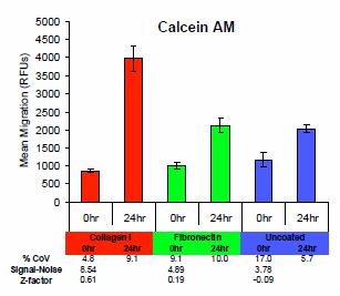
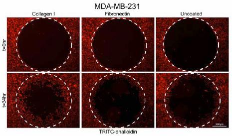
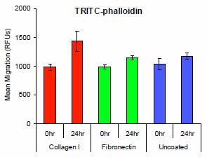
2 Methods Kinetics of HT-1080 Migration HT-1080 cells were pre-treated with mytomycin C (MMC) for 2 hours to inhibit proliferation and then stained with 2.5 mm Cell Tracker Green (Invitrogen). The cells were seeded at 50,000 cells/well in media containing 10% Fetal Bovine Serum (FBS) and allowed to adhere. Inserts were removed, and the plate was incubated to permit cell migration. The numbers of migrated cells were interpolated from standard curves relating Cell Tracker Green fluorescence to cell number and measured on Synergy HT. Each time point represents the mean of nine replicate measurements. Results Kinetics of HT-1080 Migration Initial experiments demonstrated the kinetics of cell migration into the center analytical zone. MDA-MB-231 cell migration optimization using Oris TriCoated Plates The Oris Cell Migration Assay TriCoated (Platypus Technologies, LLC) was used to assess cell migration of MDA-MB-231 breast epithelial cells. Cells were seeded at 20,000 cells/well and allowed to attach overnight onto plates coated with either type I collagen (Collagen I), fibronectin, or tissue culture treated (uncoated) wells. Once the cells formed a confluent monolayer, the silicone stoppers were removed and migration proceeded for 24 hours. Following migration, cells were labeled with Calcein AM (Invitrogen), and migration in the detection zone was then quantified by using a BioTek Synergy HT Multi-Mode Microplate Reader with the Oris Detection Mask attached to the bottom of the plate. Next, cells were fixed and stained for filamentous actin with TRITC-phalloidin (Sigma), and a nuclear DAPI stain (Thermo Fisher). Data represent mean ± STD from a minimum of 8 wells for each condition. Images were acquired, in the absence of the mask, by use of a Zeiss Axiovert inverted microscope. Figure 2. Fluorescent photomicrographs taken at t=0 and t=21 hrs in the absence of the mask. The analytic zone into which cells have migrated is encircled by the white dotted line. As demonstrated in Figure 2, FBS-stimulated, nonproliferating HT1080 cells almost completely fill the analytical zone within 21 hours of removal of the mask. Furthermore, when the migration is examined over time, migration was detectable in less than 4 hours and linear over a 28.5-hour period (Figure 3). NMuMG and HT1080 cell migration optimization using Oris TriCoated Plates NMuMG breast epithelial cells were seeded at 25,000 cells/well and allowed to attach for 6 hours on TriCoated plates. Following 16 hour migration, cells were labeled with Calcein AM and migration in the detection zone was then quantified as previously described. HT1080 human fibrosarcoma cells were seeded at 35,000 cells/well and allowed to attach for 4 hours on TriCoated plates. Following 20 hour migration, cells were labeled with Calcein AM and migration in the detection zone was then quantified as previously described. Oris Mask Aperture Size Effect MDA-MB-231 breast epithelial cells were seeded at 25,000 cells/well and allowed to attach for 7 hours onto collagen I-coated wells. Once the cells formed a confluent monolayer, the silicone stoppers were removed and migration proceeded for 20 hours. Following migration, cells were labeled with Calcein AM (Invitrogen), and migration in the detection zone was then quantified as previously described. For all experiments, S/N ratios and z factors were calculated according to the following equations: Figure 3. Kinetics of HT-1080 cell migration MDA-MB-231 cell migration optimization using Oris TriCoated Plates MDA-MB-231 cell migration is demonstrated in Figure 4 both visually and as a fluorometric readout from Synergy HT. It is apparent from the inverted microscope images for all three cellular stains that the most cell migration is occurring with the collagen I coating of the TriCoated plates. For the Synergy HT readings, there is a definite preference for using the Calcein AM cellular stain. All three stains provide similar background readings (cell migration at t = 0), but the Calcein AM stain provides greater intensities for cells which migrate into the detection volume defined by the mask aperture. It is evident however, that collagen I is the best surface for enhanced cell migration in agreement with the images. CVs in replicate measurements were less than 10% and S/N and z factors were consistent with screening applications. 2

3 Figure 4. A B 3
4 C D 4
 and")
, TRITC-phalloidin (C, D), and a")
5 E F Figure 4. MDA-MB-231 breast epithelial cell migration as measured by inverted microscope (A, C, E) and Synergy HT Multi-Mode Microplate Reader (B, D, F). Three different cellular stains were used: Calcein AM (A, B), TRITC-phalloidin (C, D), and a nuclear DAPI stain (E, F). In A-F, the three different surfaces of the TriCoated plates were tested (Collagen I, Fibronectin and uncoated). 5


6 NMuMG and HT1080 cell migration optimization using Oris TriCoated Plates A Different cell types tend to have different cell migration behavior, which is why the TriCoated plates were developed to allow for the optimization of surface for specific cell type. In this study, NMuMG and HT1080 cell migration behavior was monitored using the Synergy HT and TriCoated plates. It is evident from Figure 5 that different surfaces are optimal for the two cell lines: NMuMG cells provide better performance with collagen I; while fibronectin appears to be a better surface for HT1080 cells. It is recommended that for new assays using untested cell lines one should start with TriCoated plates to optimize surface. B Figure 6. Cell migration as measured by microscopy (A) and Synergy HT (B) for different mask aperture sizes. Figure 5. NMuMG and HT1080 cell migration as measured on Synergy HT. Oris Mask Aperture Size Effect Figure 6 demonstrates the effect of different mask aperture sizes on cell migration signals obtained by both microscopy and Synergy HT. It is evident that although the background is higher for the larger aperture mask, it does provide the best performance. This mask aperture size would also allow for the quantification of early cell migration events also. Discussion The Oris Cell Migration Assay based on a 96-well microplate design allows for sample throughput and assay performance suitable for HTS when used in conjunction with microplate readers, such as the Synergy HT Multi-Mode Reader. Cell migration is cell line specific and thus it is highly recommended to test cell migration on the different surfaces provided in the TriCoated microplates. Calcein AM is the dye recommended for use with Synergy HT. Rev. 4/8/09 6
Investigation of Cell Migration using a High Density Cell Exclusion Assay and Automated Microplate Imager
 A p p l i c a t i o n N o t e Investigation of Cell Migration using a High Density Cell Exclusion Assay and Automated Microplate Imager Peter J. Brescia and Peter Banks, Applications Department, BioTek
Brightfield and Fluorescence Imaging using 3D PrimeSurface Ultra-Low Attachment Microplates
 A p p l i c a t i o n N o t e Brightfield and Fluorescence Imaging using 3D PrimeSurface Ultra-Low Attachment Microplates Brad Larson, BioTek Instruments, Inc., Winooski, VT USA Anju Dang, S-BIO, Hudson,
Automated, Kinetic Imaging of Cell Migration and Invasion Assays using Corning FluoroBlok Permeable Supports
 A p p l i c a t i o n N o t e Automated, Kinetic Imaging of Cell Migration and Invasion Assays using Corning FluoroBlok Permeable Supports Brad Larson, Principal Scientist, Applications Department, BioTek
Oris TM Cell Migration Assay - Fibronectin Coated
 Bringing Science to the Surface TM Oris TM Cell Migration Assay - Fibronectin Coated Product No.: CMAFN1.101 & CMAFN5.101 96-well, 2-D Assay for Investigating Cell Migration of Adherent Cell Lines on Fibronectin
Oris TM Pro Cell Migration Assay Collagen I Coated
 Oris TM Pro Cell Migration Assay Product No.: PROCMACC1 & PROCMACC5 96-well, 2-D Assay for Investigating Cell Migration of Adherent Cell Lines on Collagen I PROTOCOL & INSTRUCTIONS Table of Contents I.
Oris TM Cell Migration Assay Collagen I Coated
 Bringing Science to the Surface TM Oris TM Cell Migration Assay Collagen I Coated Product No.: CMACC1.101 & CMACC5.101 96-well, 2-D Assay for Investigating Cell Migration of Adherent Cell Lines on Collagen
Measuring Wound Healing and Cell Migration using Celigo Imaging Cytometer
 Measuring Wound Healing and Cell Migration using Celigo Imaging Cytometer Nexcelom Bioscience LLC. 360 Merrimack Street, Building 9 Lawrence, MA 01843 T: 978.327.5340 F: 978.327.5341 E: info@nexcelom.com
Oris TM Cell Invasion & Detection Assay Product No.: CIA101DE & CIA200DE
 Bringing Science to the Surface TM Oris TM Cell Invasion & Detection Assay Product No.: CIA101DE & CIA200DE 96-well, 3-D Assay for Investigating Cell Invasion of Adherent Cell Lines PROTOCOL & INSTRUCTIONS
Oris TM Cell Migration Assay Collagen I Coated
 Bringing Science to the Surface TM Oris TM Cell Migration Assay Collagen I Coated Product No.: CMACC1.101 & CMACC5.101 96-well, 2-D Assay for Investigating Cell Migration of Adherent Cell Lines on Collagen
Oris TM Pro Cell Migration Assay Tissue Culture Treated
 Oris TM Pro Cell Migration Assay Tissue Culture Treated Product No.: PROCMA1 & PROCMA5 96-well, 2-D Assay for Investigating Cell Migration of Adherent Cell Lines PROTOCOL & INSTRUCTIONS Table of Contents
Oris TM Collagen I Cell Invasion Assay Product No.: CIA101CC & CIA200CC
 Bringing Science to the Surface TM Oris TM Collagen I Cell Invasion Assay Product No.: CIA101CC & CIA200CC 96-well, 3-D Assay for Investigating Cell Invasion of Adherent Cell Lines on Collagen I PROTOCOL
Oris TM Pro 384 Cell Migration Assay Tissue Culture Treated
 Oris TM Pro 384 Cell Migration Assay Tissue Culture Treated Product No.: PRO384CMA5 384-well, 2-D Assay for Investigating Cell Migration of Adherent Cell Lines PROTOCOL & INSTRUCTIONS Table of Contents
CytoSelect 24-Well Cell Haptotaxis Assay (8 µm, Collagen I-Coated, Fluorometric Format)
") Product Manual CytoSelect 24-Well Cell Haptotaxis Assay (8 µm, Collagen I-Coated, Fluorometric Format) Catalog Number CBA-101-COL 12 assays FOR RESEARCH USE ONLY Not for use in diagnostic procedures Introduction
Product Manual CytoSelect 24-Well Cell Haptotaxis Assay (8 µm, Collagen I-Coated, Fluorometric Format) Catalog Number CBA-101-COL 12 assays FOR RESEARCH USE ONLY Not for use in diagnostic procedures Introduction
Comparison of Oridonin Cytotoxicity in U-2 OS and HepG2 Cells
 A p p l i c a t i o n N o t e Comparison of Oridonin Cytotoxicity in U-2 OS and HepG2 Cells Using the BioSpa 8 to Manage Repeated Reagent Additions to Live Cells Paul Held, PhD, Laboratory Manager, Applications
Oris TM Pro 384 Cell Migration Assay Collagen I Coated
 Oris TM Pro 384 Cell Migration Assay Collagen I Coated Product No.: PRO384CMACC1 & PRO384CMACC5 384-well, 2-D Assay for Investigating Cell Migration of Adherent Cell Lines on Collagen I PROTOCOL & INSTRUCTIONS
Oris TM Cell Migration Assembly Kit FLEX
 Bringing Science to the Surface TM Oris TM Cell Migration Assembly Kit FLEX Product No.: CMAUFL4 96-well Assay for Investigating Cell Migration and Cell Invasion of Adherent Cell Lines PROTOCOL & INSTRUCTIONS
Performance of a Label-Free Image-Based 2D Scratch Wound Healing Assay to monitor Cell Migration and its Inhibition
 A p p l i c a t i o n N o t e Performance of a Label-Free Image-Based 2D Scratch Wound Healing Assay to monitor Cell Migration and its Inhibition Leonie Rieger and Brad Larson, Applications Department,
Platypus Technologies, LLC 5520 Nobel Drive, Suite 100 Madison, WI Toll Free: (866) Phone: (608) Fax: (608)
 Phone: (608) Fax: (608)") Universal Cell Migration Assembly Kit Product No.: CMAU101 & CMAU505 96-well Assay for Investigating Cell Migration, Cell Invasion and 2-D Closure of Adherent Cell Lines Protocol & Instructions Patent
Universal Cell Migration Assembly Kit Product No.: CMAU101 & CMAU505 96-well Assay for Investigating Cell Migration, Cell Invasion and 2-D Closure of Adherent Cell Lines Protocol & Instructions Patent
Quantification of Cell Migration and Invasion Using the IncuCyte Chemotaxis Assay
 APPLICATION NOTE IncuCyte ZOOM Live-Cell Imaging System Quantification of Cell Migration and Invasion Using the IncuCyte Chemotaxis Assay Lindy O Clair, Meagan Roddy, Maria Tikhonenko, Clare Syzbut, Nicola
Radius 24-Well Cell Migration Assay (Fibronectin Coated)
") Product Manual Radius 24-Well Cell Migration Assay (Fibronectin Coated) Catalog Number CBA-125-FN 24 assays FOR RESEARCH USE ONLY Not for use in diagnostic procedures Introduction Cell migration is a highly
Product Manual Radius 24-Well Cell Migration Assay (Fibronectin Coated) Catalog Number CBA-125-FN 24 assays FOR RESEARCH USE ONLY Not for use in diagnostic procedures Introduction Cell migration is a highly
Oris TM 3D Embedded Invasion Assay Product No.: EIA1 & EIA3
 Oris TM 3D Embedded Invasion Assay Product No.: EIA1 & EIA3 96-well, 3D Assay for Investigating Embedded Cell Movement through Collagen I PROTOCOL & INSTRUCTIONS I. MATERIALS PROVIDED... 2 II. MATERIALS
Validation of High-Throughput Wound Healing Assay using 3D Cell Patterning and Automated, Kinetic Imaging
 A p p l i c a t i o n N o t e Validation of High-Throughput Wound Healing Assay using 3D Cell Patterning and Automated, Kinetic Imaging Brad Larson and Peter Banks, BioTek Instruments, Inc., Winooski,
Quantification of Cell Migration and Invasion Using the IncuCyte Chemotaxis Assay
 Quantification of Cell Migration and Invasion Using the IncuCyte Chemotaxis Assay Lindy O Clair, Meagan Roddy, Maria Tikhonenko, Clare Syzbut, Nicola Bevan, Kirk Schroeder and Daniel M. Appledorn Essen
Radius 24-Well Cell Migration Assay (Laminin Coated)
") Product Manual Radius 24-Well Cell Migration Assay (Laminin Coated) Catalog Number CBA-125-LN 24 assays FOR RESEARCH USE ONLY Not for use in diagnostic procedures Introduction Cell migration is a highly
Product Manual Radius 24-Well Cell Migration Assay (Laminin Coated) Catalog Number CBA-125-LN 24 assays FOR RESEARCH USE ONLY Not for use in diagnostic procedures Introduction Cell migration is a highly
Use of Phase Contrast Imaging to Track Morphological Cellular Changes due to Apoptotic Activity
 A p p l i c a t i o n N o t e Use of Phase Contrast Imaging to Track Morphological Cellular Changes due to Apoptotic Activity Brad Larson and Peter Banks, Applications Department, BioTek Instruments, Inc.,
CytoSelect 48- Well Cell Adhesion Assay (Laminin- Coated, Colorimetric Format)
") Product Manual CytoSelect 48- Well Cell Adhesion Assay (Laminin- Coated, Colorimetric Format) Catalog Number CBA- 056 48 assays FOR RESEARCH USE ONLY Not for use in diagnostic procedures Introduction Cell
Product Manual CytoSelect 48- Well Cell Adhesion Assay (Laminin- Coated, Colorimetric Format) Catalog Number CBA- 056 48 assays FOR RESEARCH USE ONLY Not for use in diagnostic procedures Introduction Cell
Automated Imaging and Dual-Mask Analysis of γh2ax Foci to Determine DNA Damage on an Individual Cell Basis
 A p p l i c a t i o n N o t e Automated Imaging and Dual-Mask Analysis of γh2ax Foci to Determine DNA Damage on an Individual Cell Basis Brad Larson, BioTek Instruments, Inc., Winooski, VT USA Asha Sinha
Limit of Detection. Theoretical Lowest Concentration
 Nucleic Acid Quantitation Detection Limits Today s biomedical testing has resulted in sample sizes becoming smaller and smaller, driving the need to measure samples with ever-lower detection limits. The
OrisTM Cell Migration Assay
 OrisTM Cell Migration Assay Product No: CMA0 & CMA50 96-well, -D Assay for Investigating Cell Migration of Adherent Cell Lines PROTOCOL & INSTRUCTIONS Table of Contents I II III IV V VI VII VIII INTRODUCTION
CytoSelect 48-Well Cell Adhesion Assay (Laminin-Coated, Colorimetric Format)
") Product Manual CytoSelect 48-Well Cell Adhesion Assay (Laminin-Coated, Colorimetric Format) Catalog Number CBA-056 48 assays FOR RESEARCH USE ONLY Not for use in diagnostic procedures Introduction Cell
Product Manual CytoSelect 48-Well Cell Adhesion Assay (Laminin-Coated, Colorimetric Format) Catalog Number CBA-056 48 assays FOR RESEARCH USE ONLY Not for use in diagnostic procedures Introduction Cell
Radius 384-Well Cell Migration Assay
 Product Manual Radius 384-Well Cell Migration Assay Catalog Number CBA-127 CBA-127-5 384 assays 5 x 384 assays FOR RESEARCH USE ONLY Not for use in diagnostic procedures Introduction Cell migration is
Optimizing Dual-Glo Luciferase Assays with the Synergy HT Multi-Detection Microplate Reader
 Optimizing Dual-Glo Luciferase Assays with the Synergy HT Multi-Detection Microplate Reader Introduction Today s biological science and drug discovery research often involves the measurement of large numbers
An Automated, Cell-based Platform for the Rapid Detection of Novel Androgen Receptor Modulators
 A p p l i c a t i o n N o t e An Automated, Cell-based Platform for the Rapid Detection of Novel Androgen Receptor Modulators Brad Larson and Peter Banks, BioTek Instruments, Inc., Winooski, VT Bruce Sherf,
CytoSelect 24-Well Cell Invasion Assay (Laminin, Colorimetric Format)
") Product Manual CytoSelect 24-Well Cell Invasion Assay (Laminin, Colorimetric Format) Catalog Number CBA-110-LN 12 assays FOR RESEARCH USE ONLY Not for use in diagnostic procedures Introduction The ability
Product Manual CytoSelect 24-Well Cell Invasion Assay (Laminin, Colorimetric Format) Catalog Number CBA-110-LN 12 assays FOR RESEARCH USE ONLY Not for use in diagnostic procedures Introduction The ability
CytoSelect 48-Well Cell Adhesion Assay (Collagen IV-Coated, Fluorometric Format)
") Product Manual CytoSelect 48-Well Cell Adhesion Assay (Collagen IV-Coated, Fluorometric Format) Catalog Number CBA-061 48 assays FOR RESEARCH USE ONLY Not for use in diagnostic procedures Introduction
Product Manual CytoSelect 48-Well Cell Adhesion Assay (Collagen IV-Coated, Fluorometric Format) Catalog Number CBA-061 48 assays FOR RESEARCH USE ONLY Not for use in diagnostic procedures Introduction
CytoSelect 48-Well Cell Adhesion Assay (Collagen IV-Coated, Colorimetric Format)
") Product Manual CytoSelect 48-Well Cell Adhesion Assay (Collagen IV-Coated, Colorimetric Format) Catalog Number CBA-060 48 assays FOR RESEARCH USE ONLY Not for use in diagnostic procedures Introduction
Product Manual CytoSelect 48-Well Cell Adhesion Assay (Collagen IV-Coated, Colorimetric Format) Catalog Number CBA-060 48 assays FOR RESEARCH USE ONLY Not for use in diagnostic procedures Introduction
CytoSelect 24-Well Wound Healing Assay
 Product Manual CytoSelect 24-Well Wound Healing Assay Catalog Number CBA-120 CBA-120-5 24 assays 5 x 24 assays FOR RESEARCH USE ONLY Not for use in diagnostic procedures Introduction Wounded tissue initiates
CytoSelect 48-Well Cell Adhesion Assay (Collagen IV-Coated, Fluorometric Format)
") Product Manual CytoSelect 48-Well Cell Adhesion Assay (Collagen IV-Coated, Fluorometric Format) Catalog Number CBA-061 48 assays FOR RESEARCH USE ONLY Not for use in diagnostic procedures Introduction
Product Manual CytoSelect 48-Well Cell Adhesion Assay (Collagen IV-Coated, Fluorometric Format) Catalog Number CBA-061 48 assays FOR RESEARCH USE ONLY Not for use in diagnostic procedures Introduction
Membrane Potential Assays Using the ValiScreen Human Kv1.3 Voltage-Gated K + Channel Cell Line on the EnVision Multilabel Plate Reader
 TECHNICAL BRIEF ValiScreen Stable Recombinant Ion Channel Cell Line and the EnVision Multilabel Plate Reader Membrane Potential Assays Using the ValiScreen Human Kv1.3 Voltage-Gated K + Channel Cell Line
A Cost-effective Workflow for High-Throughput Screening of G- Protein Coupled Receptors (GPCRs)
") A p p l i c a t i o n N o t e A Cost-effective Workflow for High-Throughput Screening of G- Protein Coupled Receptors (GPCRs) Monitoring Receptor Mediated Calcium Flux Paul Held and Peter Banks, BioTek
A p p l i c a t i o n N o t e A Cost-effective Workflow for High-Throughput Screening of G- Protein Coupled Receptors (GPCRs) Monitoring Receptor Mediated Calcium Flux Paul Held and Peter Banks, BioTek
Imaging of BacMam Transfected U-2 OS Cells
 A p p l i c a t i o n N o t e Imaging of BacMam Transfected U-2 OS Cells Optimization of Transfection Conditions Using the Cytation 3 Multi- Mode Reader and Gen5 Data Analysis Software Paul Held Ph. D.
Platypus Technologies, LLC 5520 Nobel Drive, Suite 100 Madison, WI Toll Free: (866) Phone: (608) Fax: (608)
 Phone: (608) Fax: (608)") Cell Invasion & Detection Assay Kit Product No.: CIA101DE & CIA200DE 96-well, 3-D Assay for Investigating Cell Invasion of Adherent Cell Lines Protocol & Instructions Patent Pending Platypus Technologies,
Cell Invasion & Detection Assay Kit Product No.: CIA101DE & CIA200DE 96-well, 3-D Assay for Investigating Cell Invasion of Adherent Cell Lines Protocol & Instructions Patent Pending Platypus Technologies,
In Vitro Angiogenesis Assay Kit
 In Vitro Angiogenesis Assay Kit Catalog Number KA1323 100 assays Version: 02 Intended for research use only www.abnova.com Table of Contents Introduction... 3 Background... 3 Principle of the Assay...
CytoSelect 48-Well Cell Adhesion Assay (ECM Array, Fluorometric Format)
") Product Manual CytoSelect 48-Well Cell Adhesion Assay (ECM Array, Fluorometric Format) Catalog Number CBA-071 CBA-071-5 48 assays 5 x 48 assays FOR RESEARCH USE ONLY Not for use in diagnostic procedures
Product Manual CytoSelect 48-Well Cell Adhesion Assay (ECM Array, Fluorometric Format) Catalog Number CBA-071 CBA-071-5 48 assays 5 x 48 assays FOR RESEARCH USE ONLY Not for use in diagnostic procedures
CytoSelect 24-Well Cell Migration Assay (5 µm, Fluorometric Format)
") Revised Protocol Product Manual CytoSelect 24-Well Cell Migration Assay (5 µm, Fluorometric Format) Catalog Number CBA-102 12 assays FOR RESEARCH USE ONLY Not for use in diagnostic procedures Introduction
Revised Protocol Product Manual CytoSelect 24-Well Cell Migration Assay (5 µm, Fluorometric Format) Catalog Number CBA-102 12 assays FOR RESEARCH USE ONLY Not for use in diagnostic procedures Introduction
Assessment of HDAC1 Inhibition Using an Automated, Bioluminescent Histone Deacetylase I/II Assay
 A p p l i c a t i o n N o t e Assessment of HDAC1 Inhibition Using an Automated, Bioluminescent Histone Deacetylase I/II Assay Brad Larson, BioTek Instruments, Inc., Winooski, VT Tracy Worzella, Promega
Focus Application. Cell Migration. Featured Study: Inhibition of Cell Migration by Gene Silencing. xcelligence System Real-Time Cell Analyzer
 xcelligence System Real-Time Cell Analyzer Focus Application Cell Migration Featured Study: Inhibition of Cell Migration by Gene Silencing Markus Greiner and Richard Zimmermann Department of Medical Biochemistry
ab Cell Viability Assay Kit Fluorometric Dual Green/Red
 ab112121 Cell Viability Assay Kit Fluorometric Dual Green/Red Instructions for Use For detecting cell viability in suspension and adherent cells by using dual proprietary green and red fluorescence probes.
Peter Brescia, Applications Scientist and Peter Banks, Scientific Director, BioTek Instruments, Inc.
 Guidelines for the Provision of Residual Volumes of Assay Buffer in Microplate Wash Assays Setting aspiration manifold z-heights in ELx45 / EL46 software to provide predetermined residual volumes in the
Quantitation of ssdna using OliGreen Fluorescent Stain
 Quantitation of ssdna using OliGreen Fluorescent Stain Several different techniques require the use of short synthetic oligonucleotide molecules, often referred to as primers. In each case, the use of
Versatile, Cost-Effective Automation of Avian Influenza and Mycoplasma Gallispeticum-Synovaie ELISAs
 Versatile, Cost-Effective Automation of Avian Influenza and Mycoplasma Gallispeticum-Synovaie ELISAs Wendy Goodrich, Applications Scientist, BioTek Instruments, Inc. Matt Durgin, PAS Technical Service
Determination of Endogenous Cellular Kinase Activity Through the Use of an Automated, High Throughput AlphaScreen - Based Assay
 A p p l i c a t i o n N o t e Determination of Endogenous Cellular Kinase Activity Through the Use of an Automated, High Throughput AlphaScreen - Based Assay Brad Larson and Peter Banks, Applications Department,
Cell Migration, Chemotaxis and Invasion Assay Using Staining
 Cell Migration, Chemotaxis and Invasion Assay Using Staining Protocol Hillary Sherman and Mark Rothenberg Corning Life Sciences 836 North St. Bldg. 300, Suite 3401 Tewksbury, MA 01876 Introduction Cell
Promotion of HDF Cell Attachment and Proliferation
 Promotion of HDF Cell Attachment and Proliferation Objectives To qualitatively assess the effect of fibronectin (Fn) on HDF cell attachment Fn Attachment Assay To observe HDF cell proliferation and position
Label-free, real-time live-cell assays for spheroids: IncuCyte bright-field analysis
 Introduction APPLICATION NOTE IncuCyte Live-Cell Analysis System Label-free, real-time live-cell assays for spheroids: IncuCyte bright-field analysis Susana L. Alcantara, Miniver Oliver, Kalpana Patel,
Cultrex BME Cell Invasion Assay
 Cultrex BME Cell Invasion Assay Catalog Number 3455-096-K 96-well assay for investigating chemotaxis, cell migration, and/or cell invasion for adhesive cell types. This package insert must be read in its
Monitoring Cell Cycle Progression in Cancer Cells
 A p p l i c a t i o n N o t e Monitoring Cell Cycle Progression in Cancer Cells Using Nuclear Staining to Assess Cellular DNA Content Paul Held, Ph.D., Laboratory Manager, Applications Department, BioTek
Automated, Multi-Parameter, Kinetic Methods to Quantify Cell Death
 A p p l i c a t i o n N o t e Automated, Multi-Parameter, Kinetic Methods to Quantify Cell Death Brad Larson, Principal Scientist, BioTek Instruments, Inc., Winooski, VT USA Abstract Automated, multi-parameter
UV Fluorescence Polarization as a Means to Investigate Protein Conformational and Mass Change
 A p p l i c a t i o n N o t e UV Fluorescence Polarization as a Means to Investigate Protein Conformational and Mass Change Using Intrinsic Tryptophan Fluorescence in Conjunction with UV-capable Polarizers
Cholesterol Uptake Cell-Based Assay Kit
 Cholesterol Uptake Cell-Based Assay Kit Item No. 600440 www.caymanchem.com Customer Service 800.364.9897 Technical Support 888.526.5351 1180 E. Ellsworth Rd Ann Arbor, MI USA TABLE OF CONTENTS GENERAL
Corning PureCoat Cultureware. Where life thrives on the surface
 Corning PureCoat Cultureware Where life thrives on the surface Corning PureCoat Surfaces Optimized for Basic Research and Drug Discovery Applications Advantages of Corning PureCoat Amine and Carboxyl Surfaces
Apoptosis And Anti-tumor Effect Induced By Mtor Inhibitor And Autophagy Inhibitor In Human Osteosarcoma Cells
 Apoptosis And Anti-tumor Effect Induced By Mtor Inhibitor And Autophagy Inhibitor In Human Osteosarcoma Cells Ryosuke Horie. Kagawa University of medecine, Kita-gun, Japan. Disclosures: R. Horie: None.
System. Dynamic Monitoring of Receptor Tyrosine Kinase Activation in Living Cells. Application Note No. 4 / March
 System Application Note No. 4 / March 2008 Dynamic Monitoring of Receptor Tyrosine Kinase Activation in Living Cells www.roche-applied-science.com Dynamic Monitoring of Receptor Tyrosine Kinase Activation
Automated Imaging Assay for Characterizing Ca 2+ Flux with R-GECO Biosensor
 A p p l i c a t i o n N o t e Automated Imaging Assay for Characterizing Ca 2+ Flux with R-GECO Biosensor Joe Clayton, Ph.D., and Peter Banks, Ph.D., BioTek Instruments, Inc., Winooski, VT USA Abstract
Kinetic measurement of cytotoxicity using CellTox Green Cytotoxicity Assay on the IncuCyte TM FLR or ZOOM
 Kinetic measurement of cytotoxicity using CellTox Green Cytotoxicity Assay on the IncuCyte TM FLR or ZOOM The CellPlayer TM cytotoxicity assay described in this protocol utilizes CellTox Green Dye (Promega,
CytoSelect 24-Well Cell Migration Assay (12 µm, Colorimetric Format)
") Product Manual CytoSelect 24-Well Cell Migration Assay (12 µm, Colorimetric Format) Catalog Number CBA-107 12 assays FOR RESEARCH USE ONLY Not for use in diagnostic procedures Introduction Cell migration
Product Manual CytoSelect 24-Well Cell Migration Assay (12 µm, Colorimetric Format) Catalog Number CBA-107 12 assays FOR RESEARCH USE ONLY Not for use in diagnostic procedures Introduction Cell migration
Immunofluorescence Staining Protocol for 3 Well Chamber, removable
 Immunofluorescence Staining Protocol for 3 Well Chamber, removable This Application Note presents a simple protocol for the cultivation, fixation, and staining of cells using the 3 Well Chamber, removable.
CytoSelect 24-Well Anoikis Assay
 Product Manual CytoSelect 24-Well Anoikis Assay Catalog Number CBA-080 24 assays FOR RESEARCH USE ONLY Not for use in diagnostic procedures Introduction Adhesion to the extracellular matrix (ECM) is essential
Synergy NEO HTS Multi-Mode Microplate Reader and Life Technologies Reagents
 A p p l i c a t i o n G u i d e Synergy NEO HTS Multi-Mode Microplate Reader and Life Technologies Reagents High Performance Multi-Detection Capabilities that Span the Small Molecule Drug Discovery Process
Cell Viability Assay Kit Fluorometric Blue
 ab112120 Cell Viability Assay Kit Fluorometric Blue Instructions for Use For detecting cell viability in suspension and adherent cells by using our proprietary Blue fluorescence probe This product is for
Supplemental Methods:
 Supplemental Methods: Cell Culture 84A human mammary epithelial cells (HMEC s) were a kind gift from Martha Stampfer (Lawrence Berkeley Laboratory, Berkeley CA). Cells were maintained in DFCI- medium supplemented
Celigo Assays.
 Celigo Assays April 2017 www.nexcelom.com/celigo Nexcelom's team of Field Applications Scientists, R&D Specialists and Product Managers are in frequent contact with researchers in the field, developing
Development of Novel Advanced Cell Culture Surfaces that Provide Better Cell Growth and Attachment for Cell-Based Assays
 Development of Novel Advanced Cell Culture Surfaces that Provide Better Cell Growth and Attachment for Cell-Based Assays Elizabeth J. Abraham, Ph.D. BD Biosciences Outline Overview of surfaces and culture
Lab Module 7: Cell Adhesion
 Lab Module 7: Cell Adhesion Tissues are made of cells and materials secreted by cells that occupy the spaces between the individual cells. This material outside of cells is called the Extracellular Matrix
CytoSelect 96-Well Cell Migration Assay (8 µm, Fluorometric Format)
") Product Manual CytoSelect 96-Well Cell Migration Assay (8 µm, Fluorometric Format) Catalog Number CBA-106 96 assays FOR RESEARCH USE ONLY Not for use in diagnostic procedures Introduction Cell migration
Product Manual CytoSelect 96-Well Cell Migration Assay (8 µm, Fluorometric Format) Catalog Number CBA-106 96 assays FOR RESEARCH USE ONLY Not for use in diagnostic procedures Introduction Cell migration
Utility of Variable Bandwidth Monochromators for Quantification of Fluorescent Probes in Produced Effluent Water
 A p p l i c a t i o n N o t e Utility of Variable Bandwidth Monochromators for Quantification of Fluorescent Probes in Produced Effluent Water Brad Larson and Peter Banks, BioTek Instruments, Inc., Winooski,
Automated Image Processing to Quantify Cell Migration
 Automated Image Processing to Quantify Cell Migration Minmin Shen 1, Bastian Zimmer 2, Marcel Leist 2, Dorit Merhof 1 1 Interdiscipinary Center for Interative Data Analysis, Modelling and Visual Exploration
Ratio and Spectral Scanning
 A p p l i c a t i o n N o t e Micro-Volume Purity Assessment of Nucleic Acids using Ratio and Spectral Scanning Peter Brescia, BioTek Instruments, Inc., Winooski, VT A common method to determine the purity
ab Autophagy/ Cytotoxicity Dual Staining Kit
 ab133075 Autophagy/ Cytotoxicity Dual Staining Kit Instructions for Use For the study of the regulation of autophagy and cytotoxicity at the cellular level. This product is for research use only and is
Multiplex Fluorescence Assays for Adherence Cells without Trypsinization
 Multiplex Fluorescence Assays for Adherence Cells without Trypsinization The combination of a bright field and three fluorescent channels allows the Celigo to perform many multiplexed assays. A gating
Using the xcelligence RTCA SP Instrument to Perform Cytotoxicity Assays
 xcelligence Real-Time Cell Analysis Cytotoxicity Assay Protocol Using the xcelligence RTCA SP Instrument to Perform Cytotoxicity Assays Overview The xcelligence RTCA SP Instrument The xcelligence Real-Time
Cell Viability Assay Kit Fluorometric Near InfraRed
 ab112123 Cell Viability Assay Kit Fluorometric Near InfraRed Instructions for Use For detecting cell viability in suspension and adherent cells by using Near InfraRed fluorescence probes This product is
Live Cell Imaging of RNA Expression
 A p p l i c a t i o n N o t e Live Cell Imaging of RNA Expression Using the Cytation 3 Cell Imaging Multi-Mode Reader to Image SmartFlare RNA Probes Paul Held Ph. D, Laboratory Manager, Applications Dept.,
APPLICATION SPECIFIC PROTOCOL CELL MIGRATION FOR ADHERENT CELLS
 APPLICATION SPECIFIC PROTOCOL CELL MIGRATION FOR ADHERENT CELLS AIM 3D Cell Culture Chips are very useful for the study of 3D cell invasion and migration. The chips are not only suitable for endpoint measurement;
Using the xcelligence RTCA SP Instrument to Perform GPCR Assays
 xcelligence Real-Time Cell Analysis GPCR Assay Protocol Using the xcelligence RTCA SP Instrument to Perform GPCR Assays Overview The xcelligence RTCA SP Instrument The xcelligence Real-Time Cell Analyzer
Quantification of MMP Activity and Inhibition in a 3D Tumor Invasion Model
 A p p l i c a t i o n N o t e Quantification of MMP Activity and Inhibition in a 3D Tumor Invasion Model Using a Novel Fluorescence Detection Technology and Digital Widefield Microscopy Brad Larson and
Normalization of Agilent Seahorse XF Data by In-situ Cell Counting Using a BioTek Cytation 5
 Normalization of Agilent Seahorse XF Data by In-situ Cell Counting Using a BioTek Cytation Application Note Authors Yoonseok Kam 1, Ned Jastromb 1, Joe Clayton, Paul Held, and Brian P. Dranka 1 1 Agilent
CloneSelect Imager. Objective, quantitative assessment of cell growth. Genetix Now part of Molecular Devices.
 CloneSelect Imager Objective, quantitative assessment of cell growth www.moleculardevices.com / genetix Genetix Now part of Molecular Devices Assess cell confluence objectively and quantitatively Rapid
The Synergy 2, A Novel Approach to Microplate Multi- Detection for HTS and Drug Discovery
 The Synergy 2, A ovel Approach to Microplate Multi- Detection for HTS and Drug Discovery Paul Held and Xavier Amouretti, BioTek Instruments, Inc., Winooski, VT 05404 Abstract The Synergy 2 is a new type
IncuCyte Live-Cell Immunocytochemistry Assay
 IncuCyte Live-Cell Immunocytochemistry Assay For cell surface marker analysis This protocol describes a solution for measuring immunocytochemistry in live-cells expressing a surface antigen of interest.
The Product and Experiment Guide
 The Product and Experiment Guide Providing Solutions for Your Research p. 3 p. 10 p. 4 CELL CULTURE & MICROSCOPY IMMUNO- FLUORESCENCE LIVE CELL IMAGING p. 8 p. 7 p. 11 ANGIOGENESIS ASSAYS CHEMOTAXIS ASSAYS
Validation and Comparison of Single-Step Flash and Dual-Spectral Luciferase Reporter Gene Assays using the Synergy Line of Microplate Readers
 A p p l i c a t i o n N o t e Validation and Comparison of Single-Step Flash and Dual-Spectral Luciferase Reporter Gene Assays using the Synergy Line of Microplate Readers Peter J. Brescia and Peter Banks,
BD PureCoat Amine and Carboxyl Cell Culture Surfaces
 BD PureCoat Amine and Carboxyl Cell Culture Surfaces Guidelines for Use BD Biosciences Discovery Labware Two Oak Park, Bedford, MA 01730, tel: 877.232.8995, fax: 800.325.9637, bdbiosciences.com INTENDED
Long-Term Hepatotoxicity Studies using Cultured Human ipsc- Derived Hepatocytes
 A p p l i c a t i o n N o t e Long-Term Hepatotoxicity Studies using Cultured Human ipsc- Derived Hepatocytes Brad Larson and Peter Banks, BioTek Instruments, Inc., Winooski, VT USA Coby Carlson and Michael
Vol 6, Iss 4, Feb 20, 2016
 In vitro Flow Adhesion Assay for Analyzing Shear-resistant Adhesion of Metastatic Cancer Cells to Endothelial Cells Shin-Ae Kang, Sandra Bajana and Takemi Tanaka * Stephenson Cancer Center, University
Automated Digital Microscopy
 A p p l i c a t i o n G u i d e Peter Banks, Ph.D. and Peter J. Brescia, Applications Department, BioTek Instruments, Inc., Winooski, VT Table of Contents Introduction ----------------------------------------------------------------------------------------------------------------------
ab Glucose Uptake Assay Kit (Cell-based)
") ab204702 Glucose Uptake Assay Kit (Cell-based) Instructions for Use For measuring glucose uptake via flow cytometry and fluorescent microscopy. This product is for research use only and is not intended
ab204702 Glucose Uptake Assay Kit (Cell-based) Instructions for Use For measuring glucose uptake via flow cytometry and fluorescent microscopy. This product is for research use only and is not intended
The Effects of Different Sources of Fetal Bovine Serum on Chondrocyte Growth
 The Effects of Different Sources of Fetal Bovine Serum on Chondrocyte Growth Stacey T. Chu, B.A., Nita Chen, B.A., Ruobin Wu, B.A., Alexis B.C. Dang, M.D.. University of California, San Francisco, San
In Vitro Angiogenesis Assay Kit. Item No
 In Vitro Angiogenesis Assay Kit Item No. 10009964 TABLE OF CONTENTS GENERAL INFORMATION 3 Materials Supplied 3 Precautions 4 If You Have Problems 4 Storage and Stability 4 Materials Needed but Not Supplied
Corning Microplates for Microscopy and High Content Imaging. Improve results with microplates for high resolution cell imaging
 Corning Microplates for Microscopy and High Content Imaging Improve results with microplates for high resolution cell imaging High Performance for Cell-based Assays Within the drug discovery process, high