The MAP Kinase Family
|
|
|
- Kory Morgan
- 6 years ago
- Views:
Transcription
1 The MAP Kinase Family Extracellular stimuli Classical MAP kinases Atypical MAP kinases MAPKKK MLK1/2/3/7; LZK RAF-1/A/B TAK1; TPL2 c-mos MEKK1-4; DLK ASK1/2; MLTK TAO1/2 ASK1 TAK1 MEKK1-4 MEKK2/3 TPL2??? TAK1??? MAPKK MEK1/2 MKK4/7 MKK3/6 MEK5 ERK3 K (?) HIPK2 (?)??? MAPK ERK1/2 JNK1/2/3 p38α/β/γ/δ ERK5 ERK3/4 NLK ERK7 Effectors

2 Pleiotropic Functions of MAP Kinases proliferation, angiogenesis embryo growth, Wnt signaling, lung function HSC stroma?? proliferation, survival, senescence inflammation, development, cell cycle neural apoptosis, obesity, T-cell defects

3 Multiple Roles of ERK1/2 in Cell Proliferation

4 Activation of the Ras-ERK1/2 Pathway in Cancer From Roberts and Der. Oncogene 2007

5 Regulation of ERK1/2 MAP Kinases by Mitogens Mitogens Raf MEK1 MEK2 ERK1 ERK2 > 160 cellular targets growth proliferation survival

6 Activation of MAP Kinases by Phosphorylation
7 Dephosphorylation and Inactivation of MAP Kinases
 GST- ERK2, GST-ERK2 K52A,GST-SAPK (JNK2), GST-SAPK (JNK3), and GST-p38 MAP kinases immobilized on glutathione-sepharose beads (9) were incubated with free His-tagged MKP-3 (10) After extensive")
8 Activation of MKPs by MAP Kinases ERK-spec fic binding and activation of MKP-3. (A) GST- ERK2, GST-ERK2 K52A,GST-SAPK (JNK2), GST-SAPK (JNK3), and GST-p38 MAP kinases immobilized on glutathione-sepharose beads (9) were incubated with free His-tagged MKP-3 (10) After extensive washing, binding was assessed by protein immunoblotting Bound MKP-3 was detected with a polyclonal antibody to MKP-3 Binding of antibody to rabbit immunoglobulin G coupled to peroxidase was detected by chemilumines-cence His-tagged MKP-3 (5 ng) was used as a positive control (B) Hydrolysis of p-npp by MKP-3 in the presence of theindicated concentrations of eluted GST-ERK2, catalytically inactive GST-ERK2 K52A, GST-SAPK (JNK2), GST- SAPK (JNK3), or GST-p38 MAP kinase Interaction of MKPs with MAPKs. (A) The docking interaction between MAPKs and MKPs. The docking surface in the MKB domain of MKPs, which can be divided into three modules, binds to the corresponding sites in MAPKs. (B) Activation of MKPs by MAPKs. The dual-specificity phosphatase (DSP) domain in MKPs is inactive without its substrate. Binding of activated MAPKs to the MKB domain induces conformational changes in the DUSP domain, which causes the increase of its catalytic activity.

9 Regulation of ERK1/2 Pathway by Feedback Loops
, MEK1 (Xenopus), MKP3 (rat) and MNK1 (human).")
10 Docking Sites of MAP Kinase Substrates Docking sites of substrates and regulators of MAP kinases D box (hydrophobic-basic-hydrophobic) DEF (motif F-X-F-P) Figure 2: Docking of ERK2 to MEK1, MKP3 and MNK1. a, The amino-acid sequences of putative docking sites in ERK2 (Xenopus), MEK1 (Xenopus), MKP3 (rat) and MNK1 (human). The asterisks indicate the charged amino acids presumed to be essential for the docking. b, Lysates of NIH3T3 cells co-transfected with the indicated combinations of SR plasmid DNA encoding wild-type (wt) ERK2 or ERK2 D321N and HA-tagged wild-type MEK1 or mutant MEK1 (K3M or K4,5M) were immunoprecipitated (IP) with anti-ha antibody (bottom gel). Co-immunoprecipitated wild-type ERK2 or ERK2 D321N was detected by immunoblotting (IB) with anti- Xenopus ERK2 antibody (top gel). The expression level of ERK2 in each sample was similar (middle gel). Comparable amounts of MEK1 were immunoprecipitated in each lane (bottom gel). c, Binding capacity of ERK2 D321N for wild-type (wt) and mutant (mut) MKP3. Arg 64 and Arg 65 were replaced by methionines in MKP3 mut. MKP3 was tagged with the Myc epitope. d, The binding capacity of ERK2 D321N for wild-type and mutant MNK1. HA-tagged ERK2 and Myc -tagged MNK1 were used. Arg 405, Arg 406 and Arg 407 were replaced by methionines in MNK1 mut. e, The binding capacity of ERK2 D321N, ERK2 D324N and ERK2 D321,324N for MEK1. f, The binding capacity of wild-type ERK2 or ERK2 S323D (Ser 323 replaced by Asp) for MEK1 or MKK6. Methodology for c f as in b, with immunoprecipitation by the appropriate antibodies.


11 Common Docking Domain of MAP Kinases The CD domains in the three-dimensional structures of ERK2 and p38. Three-dimensional structures of rat ERK2 and human p38. White arrowheads indicate acidic amino acids of the CD domain (Asp 316 and Asp 319 in rat ERK2, and Asp 313, 315 and 316 in human p38). The CD domain of the MAPK family. Identification of two conserved hydrophobic residues near the CD domain in ERK2. A, sequence alignment of the CD domain sequence among several MAP kinases ranging from budding yeast to human. The numbers mark the residues in the ERK2 sequence. The two conserved acidic residues (Asp316 and Asp319 in ERK2) are in bold type, and the two conserved hydrophobic residues (Tyr314 and Tyr315 in ERK2) are underlined. B, ERK2 structure with Asp316 and Asp319 shown in red and Tyr314 and Tyr315 shown in yellow.
.")

12 Binding Domain to DEF Motif Distinct from CD Molecular-Surface Representation of Activated ERK2 Docking Domains ERK2 interacts with the D-domain substrate RSK through the CD domain (residues D316 and D319, colored green). The DEF docking domain (residues M197, L198, Y231, L232, L235, and Y261, colored yellow) is necessary for interaction with and transactivation of the Ets family transcription factor Elk-1. Importantly, DEF-domain interactions are also required for the induction and activation of immediate early genes (IEGs) such as Egr1 and c- Fos. The dual phosphorylated activation loop of ERK2, pt183-e-py185, is colored red.


")

13 Docking Domain of MAP Kinase Kinases DVD domain (Domain for Versatile Docking) required for activation of MAPKKs by MAPKKKs

14 Mechanisms Determining Response Specificity to ERK1/2 Signaling Output


15 MAP Kinase Scaffolds and Specificity MAP kinases are activated by common extracellular stimuli A single MAP kinase can be used in multipler modules role of protein scaffolds Roles of protein scaffolds 1. specificity of modules 2. signal amplification 3. Sub-cellular localization


16 MAP Kinase Scaffolds in Mammals Fig. 4. Specific activation of ERK1, but not ERK2, after cotransfection with MP1. Duplicate dishes of NIH 3T3 cells were transiently cotransfected with HA-tagged ERK1 or ERK2 and either MP1 or control vector. After 24 hours, cells were deprived of serum for 4 hours and then either left untreated or stimulated with fetal calf serum (FCS, 10%) for 10 min. HA-tagged ERKs were immunoprecipitated and assayed with MBP as substrate, essentially as described (10).
17 Effect of Scaffold or Kinase Concentration on Cellular Signaling Combinatorial inhibition. Signaling down a scaffolded protein kinase cascade is a question of balance. If there is too little scaffold, signaling will be low (left). At an intermediate concentration of scaffold, signaling will be high (center). Once the concentration of scaffold exceeds that of the kinases it binds, the signaling begins to decrease (right).

18 Spatio-temporal Organization of MAP Kinase Modules the biological response is dictated by the intensity, duration and localization of MAP kinase activity proliferation vs differentiation proliferation vs arrest in G1/senescence


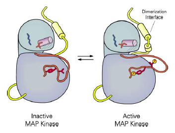
19 Spatial Control of MAP Kinase Signaling ERK2 (quiescent) ERK2 (GF 60 min) Regulatory mechanisms of nuclear translocation of ERK1/2. In unstimulated conditions, ERK1/2 is bound to MEK1/2 and localizes in the cytoplasm. Upon stimulation, ERK1/2 dissociates from MEK1/2 and translocates to the nucleus by the use of three distinct mechanisms. I, ERK1/2 dimerizes and is actively transported to the nucleus; II, ERK1/2 passively diffuses into the nucleus; III, ERK1/2 passes through the nuclear pore by directly interacting with the NPC. Then, ERK1/2 is dephosphorylated and actively exported from the nucleus.


20 PEA-15 Sequesters ERK1/2 in the Cytoplasm


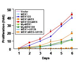
21 Nuclear MEK1 Induces Cell Transformation


22 Sustained Activation of ERK1/2 Repress Antiproliferative Genes
, the transient signal decays before c-fos can be produced and translocated to the nucleus.")

23 Interpretation of the Sustained Activation of ERK1/2 MAP Kinases A working model of how c-fos functions as a sensor for sustained ERK activity. Although both transient and sustained ERK signals stimulate c-fos gene expression (step 1), the transient signal decays before c-fos can be produced and translocated to the nucleus. Sustained ERK activity allows c-fos's Ser 374 and Ser 362 residues to be phosphorylated in ERK- and RSK-dependent manners, respectively (step 2). In addition to stabilizing the protein, these phosphorylations allow ERK to interact with the DEF domain. Recruited ERK then phosphorylates c-fos at Thr 325 and Thr 331, amino-terminal of the DEF domain (step 3). These DEF-domain-directed phosphorylations, as well as other uncharacterized ERK/DEFdomain-associated effects, regulate c-fos function.

24 Amplitude of ERK1/2 Signal Determines Cellular Response


25 Cross-talk between PKA and ERK1/2 Pathways Model for signal integration by HePTP. a, In the absence of external stimuli,the 'classical' pathway of MAP-kinase activation (originating at a receptor, R1) through Ras, Raf and Mek has a low basal activity, which is counteracted by HePTP. b, Stimulation of receptors (R2) that couple to adenylate cyclase (AC) turn on the new pathway that involves phosphorylation of HePTP at Ser 23 by PKA, leading to dissociation of Erk from HePTP. This release from inhibition results in MAP-kinase activation and subsequent c-fos induction.
26 Inactivation of MAP Kinases by Phosphorylation of MAP Kinase Kinases A 3 A Blot: αha MEK1 activity (units) MEK1 activity (units) h 3h 6h exp Time (min) C B h 3h 6h exp Time (min) Blot: P -Ser218/222 Blot: MEK1 Luciferase activity (fold increase)


27 Specific Roles of ERK1 and ERK2 Isoforms? Erk1 -/- Erk2 -/- +/+ +/- -/- Erk2 +/+ Erk2 +/-
 Schematic representation (top) of the proviral vector form used in shrna-mediated RNA interference.")
28 Redondant Roles of Erk1 and Erk2 Isoforms? ERK-specific gene silencing unmasks differential roles for ERK1 and ERK2 in cell signaling and proliferation. (a) Schematic representation (top) of the proviral vector form used in shrna-mediated RNA interference. (b) Wild type (+), ERK1 KD or ERK2 KD MEFs were serum starved for 24 h and then stimulated with 20% serum for 5, 10, 30, 60 and 120 min. Western blots were analyzed with anti-phospho-erk and anti-erk antibodies. (c) Bands from (b) were quantified and fold increases in phospho-erk2 or phospho-erk1 levels over total ERK2 or total ERK1 levels calculated. (d) Growth curve of wild-type, ERK1 and ERK2 KD fibroblasts and their corresponding controls, seeded in triplicate in the presence of 10% serum and 2 µg/ml puromycin and counted after the indicated times. Analysis of MEFs KO for Erk1 and Erk2 Cell proliferation (fold) WT ERK1 -/ Time (days) * Cell proliferation (fold) WT ERK2 -/- * * Time (days) Cell proliferation (fold) ERK1 +/+ ERK2 f/f ERK1 -/- ERK2 Δ/Δ Time (days)
29 Cell Proliferation Rate Correlates with the Total Level of Activated ERK1/2


30 Jnk1 et Jnk2: Effects on c-jun and Proliferation? Results KO MEFs: opposite effects of Jnk1 and Jnk2 Chemical genetic approach: redondant roles of Jnk1 and Jnk2
Figure 1: TDP-43 is subject to lysine acetylation within the RNA-binding domain a) QBI-293 cells were transfected with TDP-43 in the presence or
 QBI-293 cells were transfected with TDP-43 in the presence or") Figure 1: TDP-43 is subject to lysine acetylation within the RNA-binding domain a) QBI-293 cells were transfected with TDP-43 in the presence or absence of the acetyltransferase CBP and acetylated TDP-43
Figure 1: TDP-43 is subject to lysine acetylation within the RNA-binding domain a) QBI-293 cells were transfected with TDP-43 in the presence or absence of the acetyltransferase CBP and acetylated TDP-43
Supplementary Information
 Supplementary Information Supplementary Figure S1 (a) P-cRAF colocalizes with LC3 puncta. Immunofluorescence (IF) depicting colocalization of P-cRAF (green) and LC3 puncta (red) in NIH/3T3 cells treated
SUPPLEMENTARY INFORMATION
 SUPPLEMENTARY INFORMATION Dynamic Phosphorylation of HP1 Regulates Mitotic Progression in Human Cells Supplementary Figures Supplementary Figure 1. NDR1 interacts with HP1. (a) Immunoprecipitation using
DNA Binding Domains: Structural Motifs. Effector Domain. Zinc Fingers. Zinc Fingers, continued. Zif268
 DNA Binding Domains: Structural Motifs Studies of known transcription factors have found several motifs of protein design to allow sequence-specific binding of DNA. We will cover only three of these motifs:
Supplementary Figure 1. GST pull-down analysis of the interaction of GST-cIAP1 (A, B), GSTcIAP1
, GSTcIAP1") Legends Supplementary Figure 1. GST pull-down analysis of the interaction of GST- (A, B), GST mutants (B) or GST- (C) with indicated proteins. A, B, Cell lysate from untransfected HeLa cells were loaded
Legends Supplementary Figure 1. GST pull-down analysis of the interaction of GST- (A, B), GST mutants (B) or GST- (C) with indicated proteins. A, B, Cell lysate from untransfected HeLa cells were loaded
supplementary information
 DOI: 10.1038/ncb2172 Figure S1 p53 regulates cellular NADPH and lipid levels via inhibition of G6PD. (a) U2OS cells stably expressing p53 shrna or a control shrna were transfected with control sirna or
Post-translational modification
 Protein expression Western blotting, is a widely used and accepted technique to detect levels of protein expression in a cell or tissue extract. This technique measures protein levels in a biological sample
Flag-Rac Vector V12 V12 N17 C40. Vector C40 pakt (T308) Akt1. Myc-DN-PAK1 (N-SP)
 Akt1. Myc-DN-PAK1 (N-SP)") a b FlagRac FlagRac V2 V2 N7 C4 V2 V2 N7 C4 p (T38) p (S99, S24) p Flag (Rac) NIH 3T3 COS c +Serum p (T38) MycDN (NSP) Mycp27 3 6 2 3 6 2 3 6 2 min p Myc ( or p27) Figure S (a) Effects of Rac mutants on
a b FlagRac FlagRac V2 V2 N7 C4 V2 V2 N7 C4 p (T38) p (S99, S24) p Flag (Rac) NIH 3T3 COS c +Serum p (T38) MycDN (NSP) Mycp27 3 6 2 3 6 2 3 6 2 min p Myc ( or p27) Figure S (a) Effects of Rac mutants on
T H E J O U R N A L O F C E L L B I O L O G Y
 T H E J O U R N A L O F C E L L B I O L O G Y Supplemental material Nakajima and Tanoue, http://www.jcb.org/cgi/content/full/jcb.201104118/dc1 Figure S1. DLD-1 cells exhibit the characteristic morphology
Supplementary data. sienigma. F-Enigma F-EnigmaSM. a-p53
 Supplementary data Supplemental Figure 1 A sienigma #2 sienigma sicontrol a-enigma - + ++ - - - - - - + ++ - - - - - - ++ B sienigma F-Enigma F-EnigmaSM a-flag HLK3 cells - - - + ++ + ++ - + - + + - -
T H E J O U R N A L O F C E L L B I O L O G Y
 T H E J O U R N A L O F C E L L B I O L O G Y Supplemental material Han et al., http://www.jcb.org/cgi/content/full/jcb.201311007/dc1 Figure S1. SIVA1 interacts with PCNA. (A) HEK293T cells were transiently
Supplementary Figure 1.
 Supplementary Figure 1. Quantification of western blot analysis of fibroblasts (related to Figure 1) (A-F) Quantification of western blot analysis for control and IR-Mut fibroblasts. Data are expressed
ASPP1 Fw GGTTGGGAATCCACGTGTTG ASPP1 Rv GCCATATCTTGGAGCTCTGAGAG
 Supplemental Materials and Methods Plasmids: the following plasmids were used in the supplementary data: pwzl-myc- Lats2 (Aylon et al, 2006), pretrosuper-vector and pretrosuper-shp53 (generous gift of
Supplementary Information
 Supplementary Information Supplementary Figures Supplementary Figure 1. MLK1-4 phosphorylate MEK in the presence of RAF inhibitors. (a) H157 cells were transiently transfected with Flag- or HA-tagged MLK1-4
Development of an AlphaLISA MEK1 Kinase Assay Using Full-Length ERK2 Substrate
 TECHNICAL NOTE Development of an AlphaLISA MEK1 Kinase Assay Using Full-Length ERK2 Substrate AlphaLISA Technology Author: Jeanine Hinterneder, PhD PerkinElmer, Inc. Hopkinton, MA Introduction The mitogen-activated
b alternative classical none
 Supplementary Figure. 1: Related to Figure.1 a d e b alternative classical none NIK P-IkBa Total IkBa Tubulin P52 (Lighter) P52 (Darker) RelB (Lighter) RelB (Darker) HDAC1 Control-Sh RelB-Sh NF-kB2-Sh
Figure S1. Sequence alignments of ATRIP and ATR TopBP1 interacting regions.
 A H. sapiens 204 TKLQTS--ERANKLAAPSVSH VSPRKNPSVVIKPEACS-PQFGKTSFPTKESFSANMS LP 259 B. taurus 201 TKLQSS--ERANKLAVPTVSH VSPRKSPSVVIKPEACS-PQFGKPSFPTKESFSANKS LP 257 M. musculus 204 TKSQSN--GRTNKPAAPSVSH
Supplementary Figure S1. N-terminal fragments of LRRK1 bind to Grb2.
 Myc- HA-Grb2 Mr(K) 105 IP HA 75 25 105 1-1163 1-595 - + - + - + 1164-1989 Blot Myc HA total lysate 75 25 Myc HA Supplementary Figure S1. N-terminal fragments of bind to Grb2. COS7 cells were cotransfected
Nature Structural & Molecular Biology: doi: /nsmb.1583
 Acetylation by GCN5 regulates CDC6 phosphorylation in the S-phase of the cell cycle Roberta Paolinelli 1,2, Ramiro Mendoza-Maldonado 2, Anna Cereseto 1 and Mauro Giacca 2 1 Molecular Biology Laboratory,
SUPPLEMENTARY INFORMATION
 DOI: 10.1038/ncb2271 Supplementary Figure a! WM266.4 mock WM266.4 #7 sirna WM266.4 #10 sirna SKMEL28 mock SKMEL28 #7 sirna SKMEL28 #10 sirna WM1361 mock WM1361 #7 sirna WM1361 #10 sirna 9 WM266. WM136
7.06 Cell Biology EXAM #2 March 20, 2003
 7.06 Cell Biology EXAM #2 March 20, 2003 This is an open book exam, and you are allowed access to books, a calculator, and notes but not computers or any other types of electronic devices. Please write
7.06 Problem Set #3, 2006
 7.06 Problem Set #3, 2006 1. You are studying the EGF/Ras/MAPK pathway in cultured cells. When the pathway is activated, cells are signaled to proliferate. You generate various mutants described below.
TECHNICAL BULLETIN. MEK Activity Assay Kit. Product Code CS0490 Storage Temperature 20 C
 MEK Activity Assay Kit Product Code CS0490 Storage Temperature 20 C TECHNICAL BULLETIN Product Description The MAP kinase kinases (MAPKK, mitogen-activated protein kinase kinase, also termed MEK) are a
TECHNICAL BULLETIN. JNK 1&2 Activity Assay Kit. Product Number CS0380 Storage Temperature 20 C
 JNK 1&2 Activity Assay Kit Product Number CS0380 Storage Temperature 20 C TECHNICAL BULLETIN Product Description The c-jun N-terminal kinases (JNKs), also known as stress activated protein kinases (SAPKs),
Sarker et al. Supplementary Material. Subcellular Fractionation
 Supplementary Material Subcellular Fractionation Transfected 293T cells were harvested with phosphate buffered saline (PBS) and centrifuged at 2000 rpm (500g) for 3 min. The pellet was washed, re-centrifuged
SUPPLEMENTARY INFORMATION
 DOI: 10.1038/ncb3363 Supplementary Figure 1 Several WNTs bind to the extracellular domains of PKD1. (a) HEK293T cells were co-transfected with indicated plasmids. Flag-tagged proteins were immunoprecipiated
Supplementary methods Shoc2 In Vitro Ubiquitination Assay
 Supplementary methods Shoc2 In Vitro Ubiquitination Assay 35 S-labelled Shoc2 was prepared using a TNT quick Coupled transcription/ translation System (Promega) as recommended by manufacturer. For the
Stargazin regulates AMPA receptor trafficking through adaptor protein. complexes during long term depression
 Supplementary Information Stargazin regulates AMPA receptor trafficking through adaptor protein complexes during long term depression Shinji Matsuda, Wataru Kakegawa, Timotheus Budisantoso, Toshihiro Nomura,
Fig. S1. Effect of p120-catenin overexpression on the interaction of SCUBE2 with E-cadherin. The expression plasmid encoding FLAG.
 Fig. S1. Effect of p120-catenin overexpression on the interaction of SCUBE2 with E-cadherin. The expression plasmid encoding FLAG.SCUBE2, E-cadherin.Myc, or HA.p120-catenin was transfected in a combination
of signal transduction pathways?
 CELL BIOLOGY Signal Transduction + How do you study activation of signal transduction pathways? + IDENTIFICATION OF PATHWAY-SPECIFIC TRANSCRIPTION ACTIVATION + EASIER, FASTER THAN BLOTTING OR GEL-SHIFT
Supplementary Materials for
 advances.sciencemag.org/cgi/content/full/4/9/eaat5401/dc1 Supplementary Materials for GLK-IKKβ signaling induces dimerization and translocation of the AhR-RORγt complex in IL-17A induction and autoimmune
Supplementary Fig. 1
 a FL (1-2266) NL (1-1190) CL (1191-2266) HA-ICE1: - HA-ICE1: - - - FLAG-ICE2: + + + + FLAG-ELL: + + + + + + IP: anti-ha FLAG-ICE2 HA-ICE1-FL HA-ICE1-NL HA-ICE1-CL FLAG-ICE2 b IP: anti-ha FL (1-2266) NL
Supplementary Materials for
 www.sciencesignaling.org/cgi/content/full/8/404/ra120/dc1 Supplementary Materials for The subcellular localization and activity of cortactin is regulated by acetylation and interaction with Keap1 Akihiro
SUPPLEMENTARY INFORMATION
 doi:10.1038/nature09732 Supplementary Figure 1: Depletion of Fbw7 results in elevated Mcl-1 abundance. a, Total thymocytes from 8-wk-old Lck-Cre/Fbw7 +/fl (Control) or Lck-Cre/Fbw7 fl/fl (Fbw7 KO) mice
Notes on experimental technique: 1. Enzyme activity was measured by noting the increase in absorbance at 341 nm, as NADPH +
 Case 2 Glucose-6-phosphate dehydrogenase activity and cell growth Focus concept The activity of the pentose phosphate pathway enzyme glucose-6-phosphate dehydrogenase has been found to be important in
Supplemental Material Igreja and Izaurralde 1. CUP promotes deadenylation and inhibits decapping of mrna targets. Catia Igreja and Elisa Izaurralde
 Supplemental Material Igreja and Izaurralde 1 CUP promotes deadenylation and inhibits decapping of mrna targets Catia Igreja and Elisa Izaurralde Supplemental Materials and methods Functional assays and
Supplementary Figure 1, related to Figure 1. GAS5 is highly expressed in the cytoplasm of hescs, and positively correlates with pluripotency.
 Supplementary Figure 1, related to Figure 1. GAS5 is highly expressed in the cytoplasm of hescs, and positively correlates with pluripotency. (a) Transfection of different concentration of GAS5-overexpressing
Supplementary Fig. 1 Identification of Nedd4 as an IRS-2-associated protein in camp-treated FRTL-5 cells.
 Supplementary Fig. 1 Supplementary Fig. 1 Identification of Nedd4 as an IRS-2-associated protein in camp-treated FRTL-5 cells. (a) FRTL-5 cells were treated with 1 mm dibutyryl camp for 24 h, and the lysates
Coleman et al., Supplementary Figure 1
 Coleman et al., Supplementary Figure 1 BrdU Merge G1 Early S Mid S Supplementary Figure 1. Sequential destruction of CRL4 Cdt2 targets during the G1/S transition. HCT116 cells were synchronized by sequential
Supplementary Fig. 1 Kinetics of appearence of the faster migrating form of Bcl-10.
 α-cd3 + α-cd28: Time (min): + + + + + + + + + 0 5 15 30 60 120 180 240 300 360 360 n.s. Supplementary Fig. 1 Kinetics of appearence of the faster migrating form of. Immunoblot of lysates from Jurkat cells
Supplementary Information
 Supplementary Information Sam68 modulates the promoter specificity of NF-κB and mediates expression of CD25 in activated T cells Kai Fu 1, 6, Xin Sun 1, 6, Wenxin Zheng 1, 6, Eric M. Wier 1, Andrea Hodgson
supplementary information
 DOI: 10.1038/ncb2116 Figure S1 CDK phosphorylation of EZH2 in cells. (a) Comparison of candidate CDK phosphorylation sites on EZH2 with known CDK substrates by multiple sequence alignments. (b) CDK1 and
Supplementary Figure 1. TRIM9 does not affect AP-1, NF-AT or ISRE activity. (a,b) At 24h post-transfection with TRIM9 or vector and indicated
 At 24h post-transfection with TRIM9 or vector and indicated") Supplementary Figure 1. TRIM9 does not affect AP-1, NF-AT or ISRE activity. (a,b) At 24h post-transfection with TRIM9 or vector and indicated reporter luciferase constructs, HEK293T cells were stimulated
Supplementary Figure 1. TRIM9 does not affect AP-1, NF-AT or ISRE activity. (a,b) At 24h post-transfection with TRIM9 or vector and indicated reporter luciferase constructs, HEK293T cells were stimulated
Enhancers. Activators and repressors of transcription
 Enhancers Can be >50 kb away from the gene they regulate. Can be upstream from a promoter, downstream from a promoter, within an intron, or even downstream of the final exon of a gene. Are often cell type
Aoki et al.,
 JCB: SUPPLEMENTAL MATERIAL Aoki et al., http://www.jcb.org/cgi/content/full/jcb.200609017/dc1 Supplemental materials and methods Calculation of the raw number of translocated proteins First, the average
THE NEWSLETTER OF ACTIVE MOTIF November 2006 volume 7 number 4
 THE NEWSLETTER OF ACTIVE MOTIF November 2006 volume 7 number 4 New: Magnetic Beads Make Chromatin Immunoprecipitation Faster and Easier Active Motif s new ChIP-IT Express Kits use protein G-coated magnetic
used at a final concentration of 5 ng/ml. Rabbit anti-bim and mouse anti-mkp2 antibodies were
 1 Supplemental Methods Reagents and chemicals: TGFβ was a generous gift from Genzyme Inc. (Cambridge, MA) and was used at a final concentration of 5 ng/ml. Rabbit anti-bim and mouse anti-mkp2 antibodies
CRE/CREB Reporter Assay Kit camp/pka Cell Signaling Pathway Catalog #: 60611
 Data Sheet CRE/CREB Reporter Assay Kit camp/pka Cell Signaling Pathway Catalog #: 60611 Background The main role of the camp response element, or CRE, is mediating the effects of Protein Kinase A (PKA)
Supplementary Table 1. The Q-PCR primer sequence is summarized in the following table.
 Supplementary Table 1. The Q-PCR primer sequence is summarized in the following table. Name Sequence (5-3 ) Application Flag-u ggactacaaggacgacgatgac Shared upstream primer for all the amplifications of
Supplemental Figure 1 Human REEP family of proteins can be divided into two distinct subfamilies. Residues (single letter amino acid code) identical
 identical") Supplemental Figure Human REEP family of proteins can be divided into two distinct subfamilies. Residues (single letter amino acid code) identical in all six REEPs are highlighted in green. Additional
Supplemental Figure Human REEP family of proteins can be divided into two distinct subfamilies. Residues (single letter amino acid code) identical in all six REEPs are highlighted in green. Additional
Chapter 14 Regulation of Transcription
 Chapter 14 Regulation of Transcription Cis-acting sequences Distance-independent cis-acting elements Dissecting regulatory elements Transcription factors Overview transcriptional regulation Transcription
Supplemental Materials and Methods
 Supplemental Materials and Methods Co-immunoprecipitation (Co-IP) assay Cells were lysed with NETN buffer (20 mm Tris-HCl, ph 8.0, 0 mm NaCl, 1 mm EDT, 0.5% Nonidet P-40) containing 50 mm β-glycerophosphate,
The Transfection Collection TCF/LEF Transient Pack Wnt / -catenin Signaling Pathway Catalog #: 79273
 Data Sheet The Transfection Collection TCF/LEF Transient Pack Wnt / -catenin Signaling Pathway Catalog #: 79273 Background The Wnt / -catenin signaling pathway controls a large and diverse set of cell
Data Sheet. SBE Reporter Kit (TGFβ/SMAD signaling pathway) Catalog #: 60654
 Catalog #: 60654") Data Sheet SBE Reporter Kit (TGFβ/SMAD signaling pathway) Catalog #: 60654 Background The transforming growth factor beta (TGFβ) signaling pathway is involved in a diverse range of cell processes such
Data Sheet SBE Reporter Kit (TGFβ/SMAD signaling pathway) Catalog #: 60654 Background The transforming growth factor beta (TGFβ) signaling pathway is involved in a diverse range of cell processes such
SUPPLEMENTARY INFORMATION
 SUPPLEMENTARY INFORMATION Supplementary figures Supplementary Figure 1: Suv39h1, but not Suv39h2, promotes HP1α sumoylation in vivo. In vivo HP1α sumoylation assay. Top: experimental scheme. Middle: we
Table 1. Primers, annealing temperatures, and product sizes for PCR amplification.
 Table 1. Primers, annealing temperatures, and product sizes for PCR amplification. Gene Direction Primer sequence (5 3 ) Annealing Temperature Size (bp) BRCA1 Forward TTGCGGGAGGAAAATGGGTAGTTA 50 o C 292
This is the author's accepted version of the manuscript.
 This is the author's accepted version of the manuscript. The definitive version is published in Nature Communications Online Edition: 2015/4/16 (Japan time), doi:10.1038/ncomms7780. The final version published
Supplementary Materials for
 www.sciencesignaling.org/cgi/content/full/9/429/ra54/dc1 Supplementary Materials for Dephosphorylation of the adaptor LAT and phospholipase C by SHP-1 inhibits natural killer cell cytotoxicity Omri Matalon,
At E17.5, the embryos were rinsed in phosphate-buffered saline (PBS) and immersed in
 and immersed in") Supplementary Materials and Methods Barrier function assays At E17.5, the embryos were rinsed in phosphate-buffered saline (PBS) and immersed in acidic X-gal mix (100 mm phosphate buffer at ph4.3, 3 mm
Supplementary Materials and Methods Barrier function assays At E17.5, the embryos were rinsed in phosphate-buffered saline (PBS) and immersed in acidic X-gal mix (100 mm phosphate buffer at ph4.3, 3 mm
Hossain_Supplemental Figure 1
 Hossain_Supplemental Figure 1 GFP-PACT GFP-PACT Motif I GFP-PACT Motif II A. MG132 (1µM) GFP Tubulin GFP-PACT Pericentrin GFP-PACT GFP-PACT Pericentrin Fig. S1. Expression and localization of Orc1 PACT
HPV E6 oncoprotein targets histone methyltransferases for modulating specific. Chih-Hung Hsu, Kai-Lin Peng, Hua-Ci Jhang, Chia-Hui Lin, Shwu-Yuan Wu,
 1 HPV E oncoprotein targets histone methyltransferases for modulating specific gene transcription 3 5 Chih-Hung Hsu, Kai-Lin Peng, Hua-Ci Jhang, Chia-Hui Lin, Shwu-Yuan Wu, Cheng-Ming Chiang, Sheng-Chung
Supplementary Figure 1. Intracellular distribution of the EPE peptide. HeLa cells were serum-starved (16 h, 0.1%), and treated with EPE peptide,
, and treated with EPE peptide,") Supplementary Figure 1. Intracellular distribution of the EPE peptide. HeLa cells were serum-starved (16 h, 0.1%), and treated with EPE peptide, conjugated with either TAT or Myristic acid and biotin for
Supplementary Figure 1. Intracellular distribution of the EPE peptide. HeLa cells were serum-starved (16 h, 0.1%), and treated with EPE peptide, conjugated with either TAT or Myristic acid and biotin for
Data Sheet. CRE/CREB Reporter Assay Kit (camp/pka Cell Signaling Pathway) Catalog #: 60611
 Catalog #: 60611") Data Sheet CRE/CREB Reporter Assay Kit (camp/pka Cell Signaling Pathway) Catalog #: 60611 Background The main role of the camp response element, or CRE, is mediating the effects of Protein Kinase A (PKA)
Data Sheet CRE/CREB Reporter Assay Kit (camp/pka Cell Signaling Pathway) Catalog #: 60611 Background The main role of the camp response element, or CRE, is mediating the effects of Protein Kinase A (PKA)
Bronchial epithelium and its associated tissues act as a
 The Journal of Immunology A JNK-Independent Signaling Pathway Regulates TNF -Stimulated, c-jun-driven FRA-1 Protooncogene Transcription in Pulmonary Epithelial Cells 1 Pavan Adiseshaiah,* Dhananjaya V.
Figure S1 (related to Fig. 1): The prototypical mitochondrial pathway of apoptosis is involved in cell-death of v-src-transformed cells.
: The prototypical mitochondrial pathway of apoptosis is involved in cell-death of v-src-transformed cells.") Figure S1 (related to Fig. 1): The prototypical mitochondrial pathway of apoptosis is involved in cell-death of v-src-transformed cells. (A) Non-transformed (Control cells) and v-srctransformed 3T3 cells
Figure S1 (related to Fig. 1): The prototypical mitochondrial pathway of apoptosis is involved in cell-death of v-src-transformed cells. (A) Non-transformed (Control cells) and v-srctransformed 3T3 cells
Regulation of transcription by the MLL2 complex and MLL complex-associated AKAP95
 Supplementary Information Regulation of transcription by the complex and MLL complex-associated Hao Jiang, Xiangdong Lu, Miho Shimada, Yali Dou, Zhanyun Tang, and Robert G. Roeder Input HeLa NE IP lot:
Supplementary Table 1. Sequences for BTG2 and BRCA1 sirnas.
 Supplementary Table 1. Sequences for BTG2 and BRCA1 sirnas. Target Gene Non-target / Control BTG2 BRCA1 NFE2L2 Target Sequence ON-TARGET plus Non-targeting sirna # 1 (Cat# D-001810-01-05) sirna1: GAACCGACAUGCUCCCGGA
Supplemental material
 Supplemental material THE JOURNAL OF CELL BIOLOGY Gillespie et al., http://www.jcb.org/cgi/content/full/jcb.200907037/dc1 repressor complex induced by p38- Gillespie et al. Figure S1. Reduced fiber size
Immunoglobulins. (1 of 2)
") Immunoglobulins (1 of 2) Immunoglobulins (Igs) = antibodies Each B cell synthesizes Igs of single specificity for a specific epitope B cell receptors (BCRs) are the Igs on B cell surface Humoral immunity
Immunoglobulins (1 of 2) Immunoglobulins (Igs) = antibodies Each B cell synthesizes Igs of single specificity for a specific epitope B cell receptors (BCRs) are the Igs on B cell surface Humoral immunity
Supplementary Figure 1 PARP1 is involved in regulating the stability of mrnas from pro-inflammatory cytokine/chemokine mediators.
 Supplementary Figure 1 PARP1 is involved in regulating the stability of mrnas from pro-inflammatory cytokine/chemokine mediators. (a) A graphic depiction of the approach to determining the stability of
Supplementary Figure 1. The Hsp70 acetylation level is related to the co-chaperone binding of Hsp70 under various stress conditions.
 Supplementary Figure 1. The Hsp70 acetylation level is related to the co-chaperone binding of Hsp70 under various stress conditions. 1 (a) Etoposide treatment gradually changes acetylation level and co-chaperone
Supplemental Data. Sethi et al. (2014). Plant Cell /tpc
. Plant Cell /tpc") Supplemental Data Supplemental Figure 1. MYC2 Binds to the E-box but not the E1-box of the MPK6 Promoter. (A) E1-box and E-box (wild type) containing MPK6 promoter fragment. The region shown in red denotes
Supplemental Data Supplemental Figure 1. MYC2 Binds to the E-box but not the E1-box of the MPK6 Promoter. (A) E1-box and E-box (wild type) containing MPK6 promoter fragment. The region shown in red denotes
transcription and the promoter occupancy of Smad proteins. (A) HepG2 cells were co-transfected with the wwp-luc reporter, and FLAG-tagged FHL1,
 HepG2 cells were co-transfected with the wwp-luc reporter, and FLAG-tagged FHL1,") Supplementary Data Supplementary Figure Legends Supplementary Figure 1 FHL-mediated TGFβ-responsive reporter transcription and the promoter occupancy of Smad proteins. (A) HepG2 cells were co-transfected
Supplementary Data Supplementary Figure Legends Supplementary Figure 1 FHL-mediated TGFβ-responsive reporter transcription and the promoter occupancy of Smad proteins. (A) HepG2 cells were co-transfected
SUPPLEMENTARY INFORMATION
 doi:.38/nature899 Supplementary Figure Suzuki et al. a c p7 -/- / WT ratio (+)/(-) p7 -/- / WT ratio Log X 3. Fold change by treatment ( (+)/(-)) Log X.5 3-3. -. b Fold change by treatment ( (+)/(-)) 8
Supplementary Materials for
 www.sciencesignaling.org/cgi/content/full/6/304/ra104/dc1 Supplementary Materials for Lysine Methylation Promotes VEGFR-2 Activation and Angiogenesis Edward J. Hartsough, Rosana D. Meyer, Vipul Chitalia,
Transcriptional Regulation (Gene Regulation)
") Experimental Techniques in Biomedical Sciences 의생명과학실험기법 Transcriptional Regulation (Gene Regulation) 4/17/13 Jeong Hoon Kim (jeongkim@skku.edu) Department of Health Sciences and Technology, SKKU Graduate
Experimental Techniques in Biomedical Sciences 의생명과학실험기법 Transcriptional Regulation (Gene Regulation) 4/17/13 Jeong Hoon Kim (jeongkim@skku.edu) Department of Health Sciences and Technology, SKKU Graduate
3 P p25. p43 p41 28 FADD. cflips. PE-Cy5 [Fluorescence intensity]
![3 P p25. p43 p41 28 FADD. cflips. PE-Cy5 [Fluorescence intensity]](/thumbs/87/97394430.jpg "3 P p25. p43 p41 28 FADD. cflips. PE-Cy5 [Fluorescence intensity]") L S p4 3 D3 76 N S L Ve ct or p4 3 D3 76 N S L Ve ct or A aspase 8 FADD TRAF2 D95-R - + Vector D95L TL I S L D376N T RAIL-R1 T RAIL-R2 D95-R E-y5 [Fluorescence intensity] Supplemental Fig. 1 Different
L S p4 3 D3 76 N S L Ve ct or p4 3 D3 76 N S L Ve ct or A aspase 8 FADD TRAF2 D95-R - + Vector D95L TL I S L D376N T RAIL-R1 T RAIL-R2 D95-R E-y5 [Fluorescence intensity] Supplemental Fig. 1 Different
Supplementary Materials
 Supplementary Materials Supplementary Figure 1. PKM2 interacts with MLC2 in cytokinesis. a, U87, U87/EGFRvIII, and HeLa cells in cytokinesis were immunostained with DAPI and an anti-pkm2 antibody. Thirty
Phosphoproteome dynamics reveal novel ERK1/2 MAP kinase substrates with broad spectrum of functions
 Molecular Systems Biology Peer Review Process File Phosphoproteome dynamics reveal novel ERK1/2 MAP kinase substrates with broad spectrum of functions Mathieu Courcelles, Christophe Frémin, Laure Voisin,
Supplemental Figure Legends:
 Supplemental Figure Legends: Fig S1. GFP-ABRO1 localization. U2OS cells were infected with retrovirus expressing GFP- ABRO1. The cells were fixed with 3.6% formaldehyde and stained with antibodies against
Supplementary Information
 Supplementary Information Peroxiredoxin-2 and STAT3 form a redox relay for H 2 O 2 signaling Mirko C. Sobotta 1, Willy Liou 1, Sarah Stöcker 1, Deepti Talwar 1, Michael Oehler 1, Thomas Ruppert 2, Annette
A Phosphatase Holoenzyme Comprised of Shoc2/Sur8 and the Catalytic Subunit of PP1 Functions as an M-Ras Effector to Modulate Raf Activity
 Molecular Cell 22, 217 230, April 21, 2006 ª2006 Elsevier Inc. DOI 10.1016/j.molcel.2006.03.027 A Phosphatase Holoenzyme Comprised of Shoc2/Sur8 and the Catalytic Subunit of PP1 Functions as an M-Ras Effector
Supplementary Fig. 1 Proteomic analysis of ATR-interacting proteins. ATR, ARID1A and
 Supplementary Figure Legend: Supplementary Fig. 1 Proteomic analysis of ATR-interacting proteins. ATR, ARID1A and ATRIP protein peptides identified from our mass spectrum analysis were shown. Supplementary
Regulation of autophagic activity by ζ proteins associated with class III. phosphatidylinositol-3 kinase. Mercedes Pozuelo Rubio
 ONLINE SUPPORTING INFORMATION Regulation of autophagic activity by 14-3-3ζ proteins associated with class III phosphatidylinositol-3 kinase Mercedes Pozuelo Rubio entro Andaluz de Biología Molecular y
Supplemental Information. Pacer Mediates the Function of Class III PI3K. and HOPS Complexes in Autophagosome. Maturation by Engaging Stx17
 Molecular Cell, Volume 65 Supplemental Information Pacer Mediates the Function of Class III PI3K and HOPS Complexes in Autophagosome Maturation by Engaging Stx17 Xiawei Cheng, Xiuling Ma, Xianming Ding,
Supplementary information
 Supplementary information The E3 ligase RNF8 regulates KU80 removal and NHEJ repair Lin Feng 1, Junjie Chen 1 1 Department of Experimental Radiation Oncology, The University of Texas M. D. Anderson Cancer
ncounter Vantage 3D RNA:Protein Solid Tumor Assay
 PRODUCT BULLETIN Product Highlights RNA More data from your precious samples; RNA:Protein profiling requires just 1 μg of protein or FFPE slides (protein-only analysis requires ng or 1 FFPE slide) Simple
Recruitment of Grb2 to surface IgG and IgE provides antigen receptor-intrinsic costimulation to class-switched B cells
 SUPPLEMENTARY FIGURES Recruitment of Grb2 to surface IgG and IgE provides antigen receptor-intrinsic costimulation to class-switched B cells Niklas Engels, Lars Morten König, Christina Heemann, Johannes
Supplemental Online Material. The mouse embryonic fibroblast cell line #10 derived from β-arrestin1 -/- -β-arrestin2 -/-
 #1074683s 1 Supplemental Online Material Materials and Methods Cell lines and tissue culture The mouse embryonic fibroblast cell line #10 derived from β-arrestin1 -/- -β-arrestin2 -/- knock-out animals
SUPPLEMENTARY INFORMATION
 (Supplementary Methods and Materials) GST pull-down assay GST-fusion proteins Fe65 365-533, and Fe65 538-700 were expressed in BL21 bacterial cells and purified with glutathione-agarose beads (Sigma).
Figure S1. Verification of ihog Mutation by Protein Immunoblotting Figure S2. Verification of ihog and boi
 Figure S1. Verification of ihog Mutation by Protein Immunoblotting Extracts from S2R+ cells, embryos, and adults were analyzed by immunoprecipitation and immunoblotting with anti-ihog antibody. The Ihog
Takeshi Sekiguchi 1,2, Eishi Noguchi 2, Toshiro Hayashida 2, Torahiko Nakashima 2, Hideo Toyoshima 1, Takeharu Nishimoto 2 and Tony Hunter 1,*
 D-type cyclin expression is decreased and p21 and p27 CDK inhibitor expression is increased when tsbn462 CCG1/TAF II 250 mutant cells arrest in G1 at the restrictive temperature Takeshi Sekiguchi 1,2,
Data Sheet. TCF/LEF Reporter Kit Wnt / -catenin signaling pathway Catalog #: 60500
 Data Sheet TCF/LEF Reporter Kit Wnt / -catenin signaling pathway Catalog #: 60500 Background The Wnt / -catenin signaling pathway controls a large and diverse set of cell fate decisions in embryonic development,
Supplemental Fig. 1: PEA-15 knockdown efficiency assessed by immunohistochemistry and qpcr
 Supplemental figure legends Supplemental Fig. 1: PEA-15 knockdown efficiency assessed by immunohistochemistry and qpcr A, LβT2 cells were transfected with either scrambled or PEA-15 sirna. Cells were then
SUPPLEMENTARY INFORMATION
 DOI: 10.1038/ncb3562 In the format provided by the authors and unedited. Supplementary Figure 1 Glucose deficiency induced FH-ATF2 interaction. In b-m, immunoblotting or immunoprecipitation analyses were
SUPPLEMENTARY INFORMATION
 DOI: 10.1038/ncb3240 Supplementary Figure 1 GBM cell lines display similar levels of p100 to p52 processing but respond differentially to TWEAK-induced TERT expression according to TERT promoter mutation
Supplementary Figure 1. Drawing of spinal cord open-book preparations and DiI tracing. Nature Neuroscience: doi: /nn.3893
 Supplementary Figure 1 Drawing of spinal cord open-book preparations and DiI tracing. Supplementary Figure 2 In ovo electroporation of dominant-negative PlexinA1 in commissural neurons induces midline
Supplementary Fig. 1. Schematic structure of TRAIP and RAP80. The prey line below TRAIP indicates bait and the two lines above RAP80 highlight the
 Supplementary Fig. 1. Schematic structure of TRAIP and RAP80. The prey line below TRAIP indicates bait and the two lines above RAP80 highlight the prey clones identified in the yeast two hybrid screen.
Solutions to 7.02 Quiz II 10/27/05
 Solutions to 7.02 Quiz II 10/27/05 Class Average = 83 Standard Deviation = 9 Range Grade % 87-100 A 43 74-86 B 39 55-73 C 17 > 54 D 1 Question 1 (56 points) While studying deep sea bacteria, you discover
Cell proliferation was measured with Cell Counting Kit-8 (Dojindo Laboratories, Kumamoto, Japan).
.") 1 2 3 4 5 6 7 8 Supplemental Materials and Methods Cell proliferation assay Cell proliferation was measured with Cell Counting Kit-8 (Dojindo Laboratories, Kumamoto, Japan). GCs were plated at 96-well
1 2 3 4 5 6 7 8 Supplemental Materials and Methods Cell proliferation assay Cell proliferation was measured with Cell Counting Kit-8 (Dojindo Laboratories, Kumamoto, Japan). GCs were plated at 96-well