Overview of Immunohistochemistry. (with a focus on wax-embedded sections)
|
|
|
- Amos Briggs
- 6 years ago
- Views:
Transcription
1 Overview of Immunohistochemistry (with a focus on wax-embedded sections)


2 Overview of Immunohistochemistry (with a focus on wax-embedded sections)

3 Overview of Immunohistochemistry
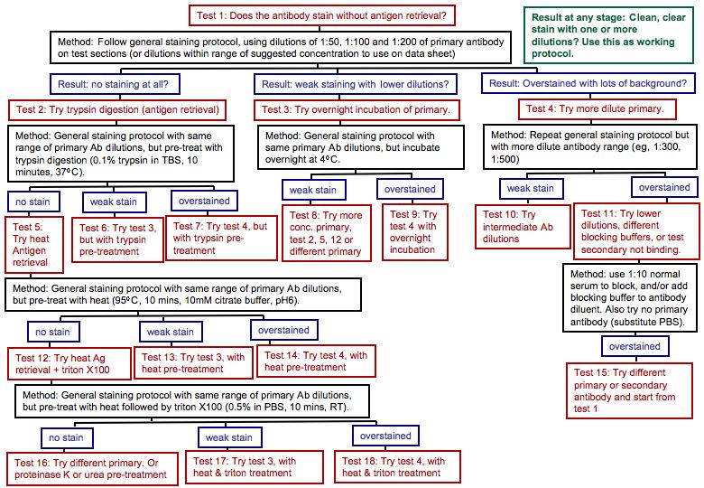
4 IHC is like cooking. There are many recipes out there, but some of them do not work out very well. However, when they do, they are great! You need the right ingredients, a dose of experience, a few tricks from old cooks, and a grain of common sense. There are several traps to avoid. - sensitivity and specificity of the antibodies and technical procedure used are crucial to avoid falsepositive and false-negative results.
5 False-positive and false-negative results can be caused by: Primary antibody failing to detect their target antigen, even if it is present in the tissue Why? conformation changes induced by fixation or embedding low affinity of the antibody for the target failure of antibody to penetrate into tissue Antibodies binding non-specifically to other targets or tissue components (both primary and secondary antibodies) Blindly following an established protocol may prove insufficient. Each different antibody needs optimisation of the general protocol to ensure specific binding.
6 FIXATIVES

7 Influence of tissue preparation - fixatives Fixation changes the chemical properties of tissue constituents and alters 3D protein conformation by cross-linking. It has a major impact on affinity and selectivity of antibodies. Epitope masking can also occur when fixation alters penetration of antibodies into the tissue. European Journal of Neuroscience2008;28:


8 KO mouse - non-specific staining non-specific staining dependence of GABAA receptor 3 subunit-immunoreactivity on fixation European Journal of Neuroscience2008;28:
9 For fixatives consider: Reducing or enhancing fixation: Type of fixative (eg,formalin vs paraformaldehyde) ph Concentration of fixative (eg, 4% vs 10%) duration of fixation use of additives (eg, picric acid) Antigen retrieval
10 ANTIGEN RETRIEVAL

 detergent (eg, triton, Tween20) Choose carefully, depending")
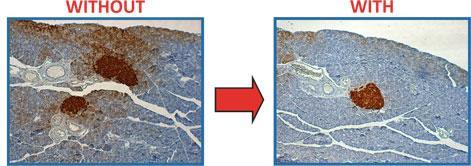
11 Antigen Retrieval Epitope masking can occur when fixation alters penetration of antibodies into the tissue. Antigen retrieval breaks down cross-links to expose the epitope and allow the primary antibody to bind. Several retrieval methods exist, designed to break different types of cross-link. Eg: heating/boiling in acidic buffer enzyme digestion (eg, trypsin, Protease K) detergent (eg, triton, Tween20) Choose carefully, depending on nuclear or cytoplasmic staining repeated freeze/thaw Before retrieval After retrieval Beware false positive after Ag Retrieval
 (D) Formalin-fixed, paraffin-embedded osteosarcoma sample after FOXP3 staining with standard heat induced epitope retrieval at 98 C with optimized epitope retrieval at 127 C.")
12 The type of retrieval matters (A) Formalin-fixed, paraffin-embedded osteosarcoma sample after CD31 staining with standard heat induced epitope retrieval at 98 C (B) with optimized enzymatic epitope retrieval. (C) (D) Formalin-fixed, paraffin-embedded osteosarcoma sample after FOXP3 staining with standard heat induced epitope retrieval at 98 C with optimized epitope retrieval at 127 C. Kunz P, Fellenberg J, Moskovszky L, Sápi Z, et al. (2014) PLoS ONE 9(3): e doi: /journal.pone

13 The type of retrieval matters
14 ANTIBODY SPECIFICITY

15 Antibody Specificity Monoclonal Antibodies: Polyclonal Antibodies:
16 Primary Antibody Specificity: controls Ideally, a tissue section should remain unstained after IHC processing if it is devoid of the target antigen. In practice, this is generally not the case - IgGs bind with low affinity to numerous (mostly unidentified) tissue constituents. Non-specific signals in tissues devoid of their target, such as a section from a knockout mouse, can also display non-specific staining. How to know staining is real? CONTROLS! 1. Knockout mice 2. Two antibodies raised against different epitopes of the antigen of interest (should show identical staining pattern) 3. Inactivate antibody with its antigen prior to use (does not control for several targets sharing a common epitope recognised by the antibody) 4. Positive control tissue - where staining pattern is known. Also: Use blocking solutions Optimise temperature, concentration and duration of incubation, as well as duration of rinsing steps.
 is higher than for primary Abs.")
17 Secondary Antibody Specificity: Blocking background staining Secondary antibodies are raised against IgGs of the species in which the primary antibodies were raised. Used in fairly high concentration - non-specific binding to tissue components (eg, ECM) is higher than for primary Abs. IgG KO mouse Primary Ab = migroglia marker Secondary Ab - without blocking Primary Ab = migroglia marker Secondary Ab - with blocking May also cross-react with IgGs from other species (relevant for dual staining). Use highly adsorbed secondary antibodies. Use blocking solutions. Optimise temperature, concentration and duration of incubation, as well as duration of rinsing steps. Use controls: No primary antibody


18 Secondary Antibody Specificity: Blocking non-specific binding
19 ENDOGENOUS PEROXIDASE

20 False positive: Endogenous peroxidase
21 Remember: Assess for false positives: Does the tissue autofluoresce? Are there endogenous peroxidases? Do the secondary antibodies alone produce staining? Does the primary antibody label the expected structures? Assess for false negatives: Failure to detect an antigen does not mean the antibody doesn t work. Is antigenicity lost during fixing? Does tissue processing have a deleterious effect? (try frozen sections) Does antigen retrieval give positive staining? Did you dilute the antibody too far in advance? (Abs stick to plastic!) Did you add peroxide to DAB substrate? Did you use the correct secondary? Did you make up ABC at least 30 minutes prior to use? Do you need to amplify the signal?
22
THE BASICS OF IMMUNOHISTOCHEMISTRY
 THE BASICS OF IMMUNOHISTOCHEMISTRY Introduction Immunohistochemistry (IHC) identifies specific tissue components by means of a specific antigen/antibody reaction tagged with a visible label. IHC makes
Immunohistochemistry guide
 Immunohistochemistry guide overview immunohistochemistry Overview Immunohistochemistry is a laboratory technique utilized for the visual detection of antigens in tissue. When working with cells this technique
ab Mouse and Rabbit Specific HRP/DAB (ABC) Detection IHC Kit
 Detection IHC Kit") ab64264 - Mouse and Rabbit Specific HRP/DAB (ABC) Detection IHC Kit Instructions for Use For the detection of a specific antibody bound to an antigen in tissue sections. This product is for research use
ab64264 - Mouse and Rabbit Specific HRP/DAB (ABC) Detection IHC Kit Instructions for Use For the detection of a specific antibody bound to an antigen in tissue sections. This product is for research use
CIHRT Exhibit P-1764 Page 1 IMMUNOHISTOCHEMISTRY ACCURATE LOCALIZATION OF TISSUE OR CELLULAR CONSTITUENTS WITH ANTIBODIES
 CIHRT Exhibit P-1764 Page 1 IMMUNOHISTOCHEMISTRY ACCURATE LOCALIZATION OF TISSUE OR CELLULAR CONSTITUENTS WITH ANTIBODIES CIHRT Exhibit P-1764 Page 2 FUNCTIONAL ROLE OF ANTIBODIES Identify the tissue of
ab EXPOSE Rabbit Specific HRP/DAB Detection IHC Kit
 Version 3 Last updated 3 November 2017 ab80437 - EXPOSE Rabbit Specific HRP/DAB Detection IHC Kit For the detection of a specific antibody bound to an antigen in tissue sections. This product is for research
ab EXPOSE Mouse and Rabbit Specific HRP/DAB Detection IHC Kit
 Version 7 Last updated 17 January 2018 ab80436 - EXPOSE Mouse and Rabbit Specific HRP/DAB Detection IHC Kit For the detection of a specific antibody bound to an antigen in tissue sections. This product
ab Mouse and Rabbit AP/Fast-Red (ABC) Detection IHC Kit
 Detection IHC Kit") ab128967 - Mouse and Rabbit AP/Fast-Red (ABC) Detection IHC Kit Instructions for Use For the detection of a specific antibody bound to an antigen in tissue sections. This product is for research use only
ab128967 - Mouse and Rabbit AP/Fast-Red (ABC) Detection IHC Kit Instructions for Use For the detection of a specific antibody bound to an antigen in tissue sections. This product is for research use only
Immunohistochemistry. How does it look like? When do we need IHC? When do we need IHC? In clinic: In research:
 Introduction How does it look like? Immunohistochemistry Smooth muscle actin Parvalbumin Distrophyn Sandrine Bichet Head of Molecular Histology Platform Signal versus background 06.03.2012 IHC basics Introduction
ab Mouse and Rabbit Specific HRP/AEC IHC Detection Kit - Micropolymer
 Version 4 Last updated 21 June 2018 ab236467 Mouse and Rabbit Specific HRP/AEC IHC Detection Kit - Micropolymer For the detection of a specific antibody bound to an antigen in tissue sections. This product
ab TripleStain IHC Kit: M&M&R on human tissue (DAB, Red/AP & DAB/Ni)
") ab183287 TripleStain IHC Kit: M&M&R on human tissue (DAB, Red/AP & DAB/Ni) Instructions for Use For the detection of Rabbit and Mouse Primary antibodies on Human tissue or cell samples. This product is
ab183287 TripleStain IHC Kit: M&M&R on human tissue (DAB, Red/AP & DAB/Ni) Instructions for Use For the detection of Rabbit and Mouse Primary antibodies on Human tissue or cell samples. This product is
ab DoubleStain IHC Kit: R&Rt on Human/Mouse Tissue (Green/HRP & AP/Red)
") ab183285 DoubleStain IHC Kit: R&Rt on Human/Mouse Tissue (Green/HRP & AP/Red) Instructions for Use For the detection of Rat and Rabbit Primary antibodies on Human/Mouse Tissue. This product is for research
ab183285 DoubleStain IHC Kit: R&Rt on Human/Mouse Tissue (Green/HRP & AP/Red) Instructions for Use For the detection of Rat and Rabbit Primary antibodies on Human/Mouse Tissue. This product is for research
ab TripleStain IHC Kit: R&R&M on human tissue (DAB, AP/Red & Green/HRP)
") ab183288 TripleStain IHC Kit: R&R&M on human tissue (DAB, AP/Red & Green/HRP) Instructions for Use For the detection of Rabbit and Mouse Primary antibodies on Human Tissue. This product is for research
ab183288 TripleStain IHC Kit: R&R&M on human tissue (DAB, AP/Red & Green/HRP) Instructions for Use For the detection of Rabbit and Mouse Primary antibodies on Human Tissue. This product is for research
ab Human on human IHC kit (HRP/DAB)
") Version 1 Last updated 13 September 2016 ab214749 Human on human IHC kit (HRP/DAB) For staining human primary antibodies on human tissues without background staining This product is for research use only
Version 1 Last updated 13 September 2016 ab214749 Human on human IHC kit (HRP/DAB) For staining human primary antibodies on human tissues without background staining This product is for research use only
IHC staining protocol. Paraffin, frozen and free-floating sections
 IHC staining protocol Paraffin, frozen and free-floating sections IHC staining protocol Contents Paraffin and frozen sections Immunostaining free-floating sections Signal amplification Paraffin and frozen
Tissue Tackle AEC Mouse Immunohistochemistry System Cat # HCS26
 Tissue Tackle AEC Mouse Immunohistochemistry System Cat # HCS26 Table of Contents Page Intended Use... 1 Background... 2 Principle of the Assay... 2 Materials Provided... 3 Materials Required But Not Provided...
ab64254 Liquid Fast-Red Substrate Kit (75X)
") Version 1 Last updated 6 June 2018 ab64254 Liquid Fast-Red Substrate Kit (75X) For the immunohistochemical staining. This product is for research use only and is not intended for diagnostic use. Table
Version 1 Last updated 6 June 2018 ab64254 Liquid Fast-Red Substrate Kit (75X) For the immunohistochemical staining. This product is for research use only and is not intended for diagnostic use. Table
ab TripleStain IHC Kit: M&M&R on Human tissue (DAB, AP/Red & HRP/Green)
") ab183286 TripleStain IHC Kit: M&M&R on Human tissue (DAB, AP/Red & HRP/Green) Instructions for Use For the detection of Rabbit and Mouse Primary antibodies on Human Tissue. This product is for research
ab183286 TripleStain IHC Kit: M&M&R on Human tissue (DAB, AP/Red & HRP/Green) Instructions for Use For the detection of Rabbit and Mouse Primary antibodies on Human Tissue. This product is for research
Product Datasheet and Instructions for Use
 Product Code: MP-109-CM01 (0.1ml conc) MP-109-CM05 (0.5ml conc) MP-109-CM1 (1ml conc) MP-109-PM6 (6ml RTU) Product Description: Androgen Receptor Concentrated and Prediluted Monoclonal Antibody Control
ab Human on human IHC kit (AP/Permanent
 Version 1 Last updated 13 September 2016 ab214753 Human on human IHC kit (AP/Permanent Red) For staining human primary antibodies on human tissues without background staining This product is for research
HistoMark Double Staining Procedures. Where Better Science Begins.
 HistoMark Double Staining Procedures Where Better Science Begins www.kpl.com HistoMark Double Staining Procedures Researchers often need the ability to visualize multiple proteins in one tissue sample.
Product Datasheet. Melatonin Receptor 1B Antibody NLS932. Unit Size: 0.05 ml
 Product Datasheet Melatonin Receptor 1B Antibody NLS932 Unit Size: 0.05 ml Store at 4C short term. Aliquot and store at -20C long term. Avoid freeze-thaw cycles. Reviews: 1 Publications: 2 Protocols, Publications,
ab DoubleStain IHC Kit: M&R on human tissue (DAB & AP/Red)
") ab210059 DoubleStain IHC Kit: M&R on human tissue (DAB & AP/Red) Instructions for use: For the detection of mouse and rabbit primary antibodies on human tissue. This product is for research use only and
ab210059 DoubleStain IHC Kit: M&R on human tissue (DAB & AP/Red) Instructions for use: For the detection of mouse and rabbit primary antibodies on human tissue. This product is for research use only and
Overview of Immunohistochemistry
 Overview of Immunohistochemistry Immunohistochemistry (IHC) combines anatomical, immunological and biochemical techniques to identify discrete tissue components by the interaction of target antigens with
Contents. 11 The Use of Epitope Tags in Histochemistry References... 98
 Contents 1 Antibodies for Immunohistochemistry... 1 1.1 Structure of Antibodies... 2 1.2 Polyclonal Antibodies... 4 1.3 Mouse Monoclonal Antibodies... 4 1.4 Rabbit Monoclonal Antibodies... 5 1.5 Protein
Product Datasheet and Instructions for Use
 Product Datasheet and Instructions for Use Product Code: MP-138-CM01 (0.1ml conc) MP-138-CM05 (0.5ml conc) MP-138-CM1 (1ml conc) Product Description: Biotinylated Bromodeoxyuridine (BrdU) Concentrated
Anti-Piscirickettsia salmonis monoclonal antibody. Product no: P05
 Anti-Piscirickettsia salmonis monoclonal antibody Product no: P05 Product Description The monoclonal antibody (Mab) against Piscirickettsia salmonis is specific for this bacterium. The specificity of the
Product Datasheet and Instructions for Use
 Product Datasheet and Instructions for Use Product Code: MP-323-CM01 (0.1ml conc) MP-323-CM05 (0.5ml conc) Product Description: CD24 Concentrated Monoclonal Antibody Control Number: 901-323-052510 ISO
How To Optimize Your IMMUNOHISTOCHEMISTRY EXPERIMENT
 How To Optimize Your IMMUNOHISTOCHEMISTRY EXPERIMENT www.ptglab.com 2 How To Optimize Your IHC Experiment ptglab.com 3 CONTENTS 4 5 Introduction To Immunohistochemistry 6 9 General Protocols 10 11 Antigen
Methodology for Immunohistochemistry. Learning Objectives:
 Proteomics Methodology for Immunohistochemistry Methodology for Immunohistochemistry A staining process for identifying the proteins location in cells, tissues by using antigen-antibody property. Immuno
Quinolinic acid polyclonal antibody
 Product Data Sheet IS1010 Quinolinic acid polyclonal antibody Ref: IS1010 Validated for IHC in human brain tissues, the anti-quinolinic acid (QUIN) rabbit polyclonal antibody proved to work at 1/1000 dilution
Frozen tissue section
 IHC Protocol - Frozen Tissue Author : Dan Souw Immunohistochemistry on Frozen tissues IHC Protocol - Frozen Tissue: An introduction This is the second post in a series on immunohistochemistry (IHC). The
Product Datasheet and Instructions for Use
 Product Code: MP-357-CMK01 (conc 0.1ml) MP-357-CMK05 (conc 0.5ml) MP-357-CMK1 (conc 1ml) MP-357-PM6 (RTU 6ml) Product Description: Factor XIIIa Concentrated and Prediluted Monoclonal Antibody Control Number:
BrdU IHC Kit. For the detection and localization of bromodeoxyuridine incorporated into newly synthesized DNA of actively proliferating cells
 K-ASSAY BrdU IHC Kit For the detection and localization of bromodeoxyuridine incorporated into newly synthesized DNA of actively proliferating cells Cat. No. KT-077 For Research Use Only. Not for Use in
Survivin (BIRC5) Immunohistochemistry Kit
 Immunohistochemistry Kit") Survivin (BIRC5) Immunohistochemistry Kit For Immunohistochemical Staining of Survivin (BIRC5) in human FFPE Tissue RUK-KBI01-20 For Research Use Only Riverside Biosciences Inc. 2327 S 5th Ave, North Riverside,
Survivin (BIRC5) Immunohistochemistry Kit For Immunohistochemical Staining of Survivin (BIRC5) in human FFPE Tissue RUK-KBI01-20 For Research Use Only Riverside Biosciences Inc. 2327 S 5th Ave, North Riverside,
Product Datasheet. LMO2 Antibody NB Unit Size: 0.1 ml
 Product Datasheet LMO2 Antibody NB110-83978 Unit Size: 0.1 ml Store at 4C short term. Aliquot and store at -20C long term. Avoid freeze-thaw cycles. Protocols, Publications, Related Products, Reviews,
IMMUNOPRECIPITATION TROUBLESHOOTING TIPS
 IMMUNOPRECIPITATION TROUBLESHOOTING TIPS Creative Diagnostics Abstract Immunoprecipitation (IP) is the technique of precipitating a protein antigen out of solution using an antibody that specifically binds
Product Datasheet and Instructions for Use
 Product Code: MP-066-CM01 (0.1ml conc) MP-066-CM05 (0.5ml conc) MP-066-CM1 (1ml conc) MP-066-PM6 (6ml RTU) Product Description: Neurofilament Concentrated and Prediluted Monoclonal Antibody Control Number:
Anti-Ig HRP Detection Kits
 BD Pharmingen Anti-Ig HRP Detection Kits Instruction Manual Detection Kit Cat. No. Anti-Mouse Ig HRP 551011 Anti-Rat Ig HRP 551013 Anti-Hamster Ig HRP 551012 2014 Becton, Dickinson and Company. All rights
Anti-White Spot Syndrome Virus (WSSV) monoclonal antibody. Product no: P13
 monoclonal antibody. Product no: P13") Anti-White Spot Syndrome Virus (WSSV) monoclonal antibody Product no: P13 Product Description The monoclonal antibody (Mab) against the Vp28 protein of White Spot Syndrome Virus (WSSV) is specific for
Anti-White Spot Syndrome Virus (WSSV) monoclonal antibody Product no: P13 Product Description The monoclonal antibody (Mab) against the Vp28 protein of White Spot Syndrome Virus (WSSV) is specific for
Adenomatous Polyposis Coli (APC) Immunohistochemistry Kit
 Immunohistochemistry Kit") Adenomatous Polyposis Coli (APC) Immunohistochemistry Kit For Immunohistochemical Staining of Adenomatous Polyposis Coli (APC) in human FFPE Tissue RUK-KAP01-20 For Research Use Only Riverside Biosciences
Adenomatous Polyposis Coli (APC) Immunohistochemistry Kit For Immunohistochemical Staining of Adenomatous Polyposis Coli (APC) in human FFPE Tissue RUK-KAP01-20 For Research Use Only Riverside Biosciences
Manufactured by. Zyagen Barnes Canyon Road San Diego, CA 92121, USA
 Alkaline Phosphatase Immunohistochemistry Detection kits For detection of mouse, rabbit, goat, rat, sheep, chicken, guinea pig, and human primary antibodies Size: 500 Tests Catalog #: AK-011, Mouse Kit
Technical Note. Tissue Section Imaging. Published August The most recent version of this Technical Note is posted at licor.com/bio/support.
 Technical Note Tissue Section Imaging Published August 2017. The most recent version of this Technical Note is posted at licor.com/bio/support. Page 2 - Tissue Section Imaging Table of Contents Page I.
Product Datasheet. Somatostatin R1/SSTR1 Antibody NLS994. Unit Size: 0.05 ml
 Product Datasheet Somatostatin R1/SSTR1 Antibody NLS994 Unit Size: 0.05 ml Store at 4C short term. Aliquot and store at -20C long term. Avoid freeze-thaw cycles. Protocols, Publications, Related Products,
Keratin 19 (KRT19) Immunohistochemistry Kit
 Immunohistochemistry Kit") Keratin 19 (KRT19) Immunohistochemistry Kit For Immunohistochemical Staining of Keratin 19 (KRT19) in human FFPE Tissue RUK-KKR01-20 For Research Use Only Riverside Biosciences Inc. 2327 S 5th Ave, North
Keratin 19 (KRT19) Immunohistochemistry Kit For Immunohistochemical Staining of Keratin 19 (KRT19) in human FFPE Tissue RUK-KKR01-20 For Research Use Only Riverside Biosciences Inc. 2327 S 5th Ave, North
Quinolinic acid monoclonal antibody
 Product Data Sheet IS002 Quinolinic acid monoclonal antibody Ref: IS002 Confirmed to be highly specific and affine by competitive ELISA, the monoclonal anti- Quinolinic acid antibody 4E11-G3 was validated
ApoTrack Cytochrome c Apoptosis ICC Antibody Kit
 ab110417 ApoTrack Cytochrome c Apoptosis ICC Antibody Kit Instructions for Use For the Immunocytochemistry analysis of cytochrome c and a mitochondrial marker (Complex Vα) in apoptotic cells and non-apoptotic
SEM Immunocytochemistry for Cells & Materials
 SEM Immunocytochemistry for Cells & Materials R. Geoff Richards AO Research Institute, AO Foundation, Davos, Switzerland. Immunohistochemistry Immunocytochemistry can be performed on a biological specimen,
General Comments. Misinformation in Immunohistochemistry. Common Misinformation s in Immunohistochemistry 4/13/2017
 Common Misinformation s in Immunohistochemistry Tri State Meeting May 3, 2017 Steven Westra Reagent Product Specialist Leica Biosystems Misinformation in Immunohistochemistry Flood of New Markers Diagnostic
Hypoxyprobe Plus Kit
 Updated 2017 1 PRODUCT INSERT Hypoxyprobe, Inc 121 Middlesex Turnpike Burlington, MA 01803 USA www.hypoxyprobe.com Hypoxyprobe Plus Kit (HPI Part # HP2-XXX) Kit contents: Solid pimonidazole HCl (Hypoxyprobe
ab BrdU Immunohistochemistry Kit
 ab125306 - BrdU Immunohistochemistry Kit Instructions for Use For the detection and localization of bromodeoxyuridine incorporated into newly synthesized DNA of actively proliferating cells. This product
1, Run 2. May Colonic mucosa. 2. Tonsil, and 1. Participation: The background. antibody clone/vendor. overall score.
 General module. Cycle 1, Run 2 The slides to be stained for CD3 comprised : 1. Colonic mucosa, 2. Tonsil, and 3. PTCL All tissuess sent were fixed in 10% neutral buffered formalin. www.qcmark.org 1 3 2
ApoTrack Cytochrome c Apoptosis ICC Antibody
 ab110417 ApoTrack Cytochrome c Apoptosis ICC Antibody Instructions for Use For the Immunocytochemistry analysis of cytochrome c and a mitochondrial marker (Complex Vα) in apoptotic cells and nonapoptotic
SANTA CRUZ BIOTECHNOLOGY, INC.
 TECHNICAL SERVICE GUIDE: Western Blotting 2. What size bands were expected and what size bands were detected? 3. Was the blot blank or was a dark background or non-specific bands seen? 4. Did this same
Best IHC Staining Practices
 Best IHC Staining Practices Featuring Cell Marque Tissue & Cellular Diagnostics The life science business of Merck KGaA, Darmstadt, Germany operates as MilliporeSigma in the U.S. and Canada. Immunohistochemistry
1. Paraffin section slides can be stored at room temperature for a long time.
 Immunohistochemistry (IHC) Protocols Immunohistochemistry (IHC) Protocol of Paraffin Section 1. Fix dissected tissues with 10% formalin for no less than 48 hours at room temperature. Inadequately fixation
GenomeMe. GeneAbTM Her2/Neu. Clone: IHC002 Source: Mouse Monoclonal Positive Control: Breast Carcinoma
 GeneAbTM Her2/Neu Clone: IHC002 Source: Mouse Monoclonal Positive Control: Breast Carcinoma 1. Intended Use This antibody is intended for in vitro diagnostic (IVD) use. The Her2/Neu (IHC002) antibody is
IMMUNOPATHOLOGY. This SOP will be applied to npod paraffin samples stained by immunohistochemistry.
 1 PURPOSE IMMUNOPATHOLOGY The purpose of this Standard Operating Procedure (SOP) is to outline procedures for immunopathology preparation and analysis of npod samples. 2 SCOPE This SOP will be applied
Sources of Background in Immunohistochemistry
 Sources of Background in Immunohistochemistry Hydrophobic Interaction Hydrophobic interactions occur between macromolecules in aqueous media when there surface tension is lower than water. This attraction
Can Get Signal immunostain
 Can Get Signal immunostain Immunoreaction Enhancer Solution (Code No. NKB-401, NKB-501, NKB-502, NKB-601, NKB-602) Instruction Manual TOYOBO CO., LTD. Life Science Department OSAKA JAPAN Distributor A3327K
Supporting Protocols
 Supporting Protocols This protocol may be used prior to immunostaining cells, organoids, or patient-derived xenografts cultured in TissueSpec ECM Hydrogels. Introduction Cells and organoids may form complex
ApoTrack Cytochrome c Apoptosis ICC Antibody Kit: 2 color immunocytochemistry of cytochrome c and mitochondria.
 PROTOCOL ApoTrack Cytochrome c Apoptosis ICC Antibody Kit 1850 Millrace Drive, Suite 3A Eugene, Oregon 97403 MSA07 Rev.1 DESCRIPTION ApoTrack Cytochrome c Apoptosis ICC Antibody Kit: 2 color immunocytochemistry
In-Gel Western Detection Using Near-Infrared Fluorescence
 In-Gel Western Detection Using Near-Infrared Fluorescence Developed for: Aerius, and Odyssey Family of Imagers Please refer to your manual to confirm that this protocol is appropriate for the applications
ab BrdU Immunohistochemistry Kit
 ab125306 - BrdU Immunohistochemistry Kit Instructions for Use For the detection and localization of bromodeoxyuridine incorporated into newly synthesized DNA of actively proliferating cells. This product
Basal-Like Cells Constitute the Proliferating Cell Population in Cystic Fibrosis Airways
 Basal-Like Cells Constitute the Proliferating Cell Population in Cystic Fibrosis Airways Judith A. Voynow, Bernard M. Fischer, Bruce C. Roberts, and Alan D. Proia On-line Data Supplement 1 Methods Image
GenomeMe. GeneAb TM BRAF V600E. Clone: IHC600 Source: Mouse Monoclonal Positive Control: Colorectal Adenocarcinoma
 GeneAb TM BRAF V600E Clone: IHC600 Source: Mouse Monoclonal Positive Control: Colorectal Adenocarcinoma 1. Intended Use This antibody is intended for in vitro diagnostic (IVD) use. The BRAF V600E (IHC600)
1. Goat Anti-Caspase-3 (CPP32) Antibody, R&D systems (cat #AF-605-NA), 0.5ug/ml
 Antibody, R&D systems (cat #AF-605-NA), 0.5ug/ml") Western Blot Antibodies: 1. Goat Anti-Caspase-3 (CPP32) Antibody, R&D systems (cat #AF-605-NA), 0.5ug/ml 2. Goat Anti-human LAP (TGF-b1) Antibody, R&D Systems (cat #AF-246-NA), 0.1-0.2 ug/ml 3. Rabbit
Western Blot Antibodies: 1. Goat Anti-Caspase-3 (CPP32) Antibody, R&D systems (cat #AF-605-NA), 0.5ug/ml 2. Goat Anti-human LAP (TGF-b1) Antibody, R&D Systems (cat #AF-246-NA), 0.1-0.2 ug/ml 3. Rabbit
Detection of protein expression
 Detection of protein expression by immunocytochemistry Dennis Brown, Ph. D. Program in Membrane Biology/Renal Unit MGH East Brown@receptor.mgh.harvard.edu http://membranebiology.mgh.harvard.edu Level of
Product Datasheet and Instructions for Use
 Product Code: MP-033-CM01 (0.1ml conc) MP-033-CM05 (0.5ml conc) MP-033-CM1 (1ml conc) MP-033-PM6 (6ml RTU) Product Description: CD68 [KP1] Concentrated and Predilute Monoclonal Antibody Control Number:
Product Datasheet. V2 Vasopressin R/AVPR2 Antibody NLS272. Unit Size: 0.05 ml
 Product Datasheet V2 Vasopressin R/AVPR2 Antibody NLS272 Unit Size: 0.05 ml Store at 4C short term. Aliquot and store at -20C long term. Avoid freeze-thaw cycles. Publications: 1 Protocols, Publications,
Easy-WESTERN-II Super
 Easy-WESTERN-II Super Primary Antibody Detection Reagent for Western Blots User Manual for High Sensitivity and Strong Signal Detection Immediately after receiving the kit, read the section titled COMPONENTS
For Research Use Only. Not for use in diagnostic procedures. Anti-NRF2 mab
 Page 1 For Research Use Only. Not for use in diagnostic procedures. Anti-NRF2 mab CODE No. M200-3 CLONALITY CLONE ISOTYPE QUANTITY SOURCE IMMUNOGEN FORMURATION STORAGE Monoclonal 1F2 Mouse IgG1 100 L,
Easy-WESTERN-II Super
 Easy-WESTERN-II Super Primary Antibody Detection Reagent for Western Blots User Manual for High Sensitivity and Strong Signal Detection Immediately after receiving the kit, read the section titled COMPONENTS
Easy-WESTERN-II Quick
 Easy-WESTERN-II Quick Primary Antibody Detection Reagent for Western Blots User Manual for Quick Antigen Detection or Multi Antigen Detection Immediately after receiving the kit, read the section titled
VisUCyte TM HRP Polymer-DAB Cell & Tissue Staining Kit
 VisUCyte TM HRP Polymer-DAB Cell & Tissue Staining Kit For the detection of goat, mouse, rabbit, rat, or sheep primary IgG Antibodies with a biotin-free detection system. Size: 50 Tests Secondary Antibody-HRP
BOVINE SPONGIFORM ENCEPHALOPATHY ANTIGEN TEST KIT, IMMUNOHISTOCHEMISTRY
 FOR VETERINARY USE ONLY USDA Product Code 5430.40 BOVINE SPONGIFORM ENCEPHALOPATHY ANTIGEN TEST KIT, IMMUNOHISTOCHEMISTRY Assay instructions for catalog number: 298 General Description This Bovine Spongiform
Product Datasheet. PIEZO1 Antibody NBP Unit Size: 0.1 ml
 Product Datasheet PIEZO1 Antibody NBP1-78537 Unit Size: 0.1 ml Store at 4C short term. Aliquot and store at -20C long term. Avoid freeze-thaw cycles. Publications: 3 Protocols, Publications, Related Products,
TUNEL Universal Apoptosis Detection Kit ( Biotin-labeled POD )
") TUNEL Universal Apoptosis Detection Kit ( Biotin-labeled POD ) Cat. No. L00290 Technical Manual No. 0267 Version 03112011 I Description. 1 II Key Features.... 1 III Kit Contents.. 1 IV Storage.. 2 V Procedure...
TUNEL Universal Apoptosis Detection Kit ( Biotin-labeled POD ) Cat. No. L00290 Technical Manual No. 0267 Version 03112011 I Description. 1 II Key Features.... 1 III Kit Contents.. 1 IV Storage.. 2 V Procedure...
Product Datasheet. MBP Antibody NBP Unit Size: 0.1 ml. Store at 4C short term. Aliquot and store at -20C long term. Avoid freeze-thaw cycles.
 Product Datasheet MBP Antibody NBP2-33555 Unit Size: 0.1 ml Store at 4C short term. Aliquot and store at -20C long term. Avoid freeze-thaw cycles. Protocols, Publications, Related Products, Reviews, Research
Which hydrogel preparation for immunostaining protocol should I use?
 Protocol: Preparation of TissueSpec hydrogels for immunostaining This protocol may be used prior to immunostaining cells, organoids, or patient-derived xenografts cultured in TissueSpec matrix hydrogels.
2-step or indirect immunofluorescence 1. Substrate on which cells are plated: plastic vs. glass; coating vs. non
 Variables in standard immunostaining protocol 2-step or indirect immunofluorescence 1. Substrate on which cells are plated: plastic vs. glass; coating vs. non 2. Plating density: sparse vs. confluent 3.
LAMININ. For Immunohistochemical Demonstration of Laminin in Paraffin-embedded and Frozen Human Tissue Sections Stock No. IMMH-7
 LAMININ For Immunohistochemical Demonstration of Laminin in Paraffin-embedded and Frozen Human Tissue Sections Stock No. IMMH-7 TABLE OF CONTENTS BACKGROUND AND PRINCIPLE... 4 REAGENTS AND EQUIPMENT PROVIDED...
IR-Blot Secondary antibodies Rev01
 0 About us Cyanagen is a biotech company located in Bologna, dedicated to research, development and production of reagents for molecular diagnostic since 2003 and one of the leading companies in the field
ebioscience BrdU Kit for IHC/ICC Colorimetric Catalog Number: RUO: For Research Use Only. Not for use in diagnostic procedures.
 Page 1 of 1 ebioscience BrdU Kit for IHC/ICC Colorimetric Catalog Number: 8800-6599 RUO: For Research Use Only. Not for use in diagnostic procedures. Product Information Contents: ebioscience BrdU Kit
Supplementary Methods. Li J.-Y. et al. Lewy bodies in grafted neurons in Parkinson s patients suggest host to. graft disease propagation
 1 Supplementary Methods Li J.-Y. et al. Lewy bodies in grafted neurons in Parkinson s patients suggest host to graft disease propagation Neural transplantation and clinical assessment Detailed information
IR-Blot Secondary antibodies Rev00
 0 About us Cyanagen is a biotech company located in Bologna, dedicated to research, development and production of reagents for molecular diagnostic since 2003 and one of the leading companies in the field
MOUSE RAPID STAINING KIT Stock No. QUIK-1. Directions for Use
 MOUSE RAPID STAINING KIT Stock No. QUIK-1 Directions for Use BACKGROUND AND PRINCIPLE The introduction of immunohistochemical techniques has ushered a new era of staining into the laboratory based upon
Western blot troubleshooting guide
 Specializing in Secondary Antibodies and Conjugates Western blot troubleshooting guide Optimize your Western blotting with Jackson ImmunoResearch Secondary antibodies Troubleshooting for better blots Western
Cartilage Staining Kit (Chondrocyte and Cartilage Tissue Staining Kit)
") Cat. # MK310 For Research Use Cartilage Staining Kit (Chondrocyte and Cartilage Tissue Staining Kit) Product Manual v201009 Table of Contents I. Description... 3 II. Kit Components... 3 III. Materials
Cat. # MK310 For Research Use Cartilage Staining Kit (Chondrocyte and Cartilage Tissue Staining Kit) Product Manual v201009 Table of Contents I. Description... 3 II. Kit Components... 3 III. Materials
Novocastra Liquid Mouse Monoclonal Antibody Insulin
 Novocastra Liquid Mouse Monoclonal Antibody Insulin Product Code: NCL-L-INSULIN Leica Biosystems Newcastle Ltd Balliol Business Park West Benton Lane Newcastle Upon Tyne NE12 8EW United Kingdom ( +44 191
Immunohistochemistry: Basics and Methods
 Immunohistochemistry: Basics and Methods Bearbeitet von Igor B Buchwalow, Werner Böcker 1st Edition. 2010. Buch. x, 153 S. Hardcover ISBN 978 3 642 04608 7 Format (B x L): 15,5 x 23,5 cm Gewicht: 445 g
KCC Path-Core Page 1 of 5
 Instructions for Sample preparation for Paraffin embedding PLEASE NOTE: There is no one-size-fits-all method of tissue preparation for all experimental designs. Before harvesting tissue, you need to assess
Hypoxyprobe -1 Green Kit Kit contents:
 Updated 2015 1 PRODUCT INSERT Hypoxyprobe, Inc 121 Middlesex Turnpike Burlington, MA 01803 USA www.hypoxyprobe.com Hypoxyprobe -1 Green Kit Kit contents: Solid pimonidazole HCl (Hypoxyprobe -1) FITC conjugated
BrdU Immunohistochemistry Kit Instruction Manual
 BrdU Immunohistochemistry Kit Instruction Manual Features Easy to use system Reagents titered for success Proven protocol Ordering Information Catalog Number X1545K Size 50 Slides Format Immunohistochemistry
PerkinElmer Life and Analytical Sciences, Inc.
 PerkinElmer Life and Analytical Sciences, Inc. TSA PLUS FLUORESCENCE SYSTEMS Tyramide Signal Amplification For Fluorescence In situ Hybridization and Immunohistochemistry Fluorescein Tetramethylrhodamine
Hypoxyprobe Plus Kit
 Updated 2017 1 PRODUCT INSERT Hypoxyprobe, Inc 121 Middlesex Turnpike Burlington, MA 01803 USA www.hypoxyprobe.com Hypoxyprobe Plus Kit (HPI Part # HP2-XXX) Kit contents: Solid pimonidazole HCl (Hypoxyprobe
TECHNICAL BULLETIN. Goat ExtrAvidin Peroxidase Staining Kit. Product Number EXTRA-1 Storage Temperature 2-8 C
 Goat ExtrAvidin Peroxidase Staining Kit Product Number EXTRA-1 Storage Temperature 2-8 C TECHNICAL BULLETIN Product Description The unique avidin reagent, ExtrAvidin, combines the high specific activity
PREPARATION OF HISTOLOGICAL SPECIMENS
 PREPARATION OF HISTOLOGICAL SPECIMENS Histo-techniques Preparation of tissue for microscopic examination Series of processes Ultimate aim to make tissue visible as it is Pathology Vs Anatomy Steps vary
SOX2 antibody - Embryonic Stem Cell Marker (ab15830) datasheet
 datasheet") -Embryonic Stem Cell Marker (ab15830) 1/8 ページ d Products: Stem Cells >> Germline Stem Cells >> Embryonic Germ Cells SOX2 antibody - Embryonic Stem Cell Marker (ab15830) datasheet Product Name Product type
-Embryonic Stem Cell Marker (ab15830) 1/8 ページ d Products: Stem Cells >> Germline Stem Cells >> Embryonic Germ Cells SOX2 antibody - Embryonic Stem Cell Marker (ab15830) datasheet Product Name Product type
Product Datasheet. TOMM20 Antibody NBP Unit Size: 0.1 ml
 Product Datasheet TOMM20 Antibody NBP1-81556 Unit Size: 0.1 ml Store at 4C short term. Aliquot and store at -20C long term. Avoid freeze-thaw cycles. Reviews: 2 Publications: 2 Protocols, Publications,
Updated PRODUCT INSERT. HPI Catalog # HP4-XXX. Kit Contents: Solid CCI-103F (Hypoxyprobe -F6) Diluted Rabbit anti-cci-103f antisera (PAbF6)
 Diluted Rabbit anti-cci-103f antisera (PAbF6)") Updated 2017 1 PRODUCT INSERT Hypoxyprobe, Inc. 121 Middlesex Turnpike Burlington, MA 01803 USA www.hypoxyprobe.com Hypoxyprobe F6 Kit HPI Catalog # HP4-XXX Kit Contents: Solid CCI-103F (Hypoxyprobe -F6)
Updated 2017 1 PRODUCT INSERT Hypoxyprobe, Inc. 121 Middlesex Turnpike Burlington, MA 01803 USA www.hypoxyprobe.com Hypoxyprobe F6 Kit HPI Catalog # HP4-XXX Kit Contents: Solid CCI-103F (Hypoxyprobe -F6)