Photoacoustic assessment of oxygen saturation: effect of red blood cell aggregation
|
|
|
- Julianna Small
- 6 years ago
- Views:
Transcription
1 Photoacoustic assessment of oxygen saturation: effect of red blood cell aggregation Eno Hysi a, Ratan K Saha b and Michael C Kolios a a Department of Physics, Ryerson University, Toronto, Canada b Applied Material Science Division, Saha Institute of Nuclear Physics, Kolkata, India; mkolios@ryersonca ABSTRACT The simultaneous photoacoustic assessment of oxygen saturation and red blood cell aggregation is presented Aggregation was induced on porcine red blood cells using Dextran-70 at multiple hematocrit levels Samples were exposed to 750 nm and 1064 nm for each hematocrit and aggregate size in order to compute the oxygen saturation As the size of the aggregate increased, the photoacoustic signal amplitude increased monotonically The same trend was observed for increasing hematocrit at each aggregation level The oxygen saturation of aggregated samples was 30% higher than non-aggregated samples at each hematocrit level This suggests that the presence of red blood cell aggregates impairs the release of oxygen to the surrounding environment Such a result has important implications for detecting red blood cell aggregation non-invasively and making clinical decisions based on the simulatenous assessment of oxygen saturation Keywords: Photoacoustics, red blood cell aggregation, oxygen saturation 1 INTRODUCTION Estimating a patient s need for oxygen is an important assessment of the patient s vital signs as life cannot thrive in the absence of oxygen Monitoring the oxygen saturation (SO 2 ) of blood is a routine method for assessing clinical conditions where oxygenation levels fluctuate The technique is useful in many settings where the patient s oxygenation is unstable (ie in intensive care, operating rooms, and emergency or other hospital wards) for determining the effectiveness or need for supplemental oxygen 1 Typically derived from the hemoglobin concentration, SO 2 is crucially important in mapping brain hemodynamics in various applications such as during responses to sensory simulations 2 In addition, monitoring of SO 2 has been used to evaluate the treatment of tumors with chemotherapy and radiation therapy 3, monitor the healing of wounds 4 and even study gene expression 5 Non-invasive monitoring of SO 2 is commonly achieved through the near-infrared spectroscopy (NIRS) method 6 NIRS relies on 2-wavelength emissions, typically through translucent body parts (ie finger tip or earlobes) At the wavelengths typically used (660 nm and 910 nm), the absorption of oxyhemoglobin (OHb) and deoxyhemoglobin (DHb) differs significantly Therefore, the OHb/DHb ratio can be calculated from the ratio of the absorbance at each respective wavelength However, a major limitation of NIRS is the small penetration depth due to significant light scattering in tissue In addition, NIRS is unable to distinguish between venous and arterial blood due to poor spatial resolution 7 Furthermore, this technique does not provide a complete measure of circulatory sufficiency It can provide false SO 2 readings based on the blood that arrives at the instant the measurement is taken despite the fact tissue can be hypoxic or hemoglobin insufficient (ie anemic) 8 Photoacoustic (PA) imaging can potentially overcome some of these limitations of NIRS in monitoring SO 2 The primary advantage of PA imaging lies in the fact that the scattering of sound waves detected by passive ultrasonic transducers is 2-3 orders of magnitude smaller than the scattering of light waves thus enabling the probing of deeper structures due to lower attenuation 9 By using multiple wavelengths of illumination, PA imaging has been able to not Photons Plus Ultrasound: Imaging and Sensing 2013, edited by Alexander A Oraevsky, Lihong V Wang, Proc of SPIE Vol 8581, 85813T 2013 SPIE CCC code: /13/$18 doi: / Proc of SPIE Vol T-1 Downloaded From: on 03/10/2013 Terms of Use:


 in")

 The collected, packed RBCs were then suspended in PBS in")

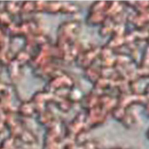
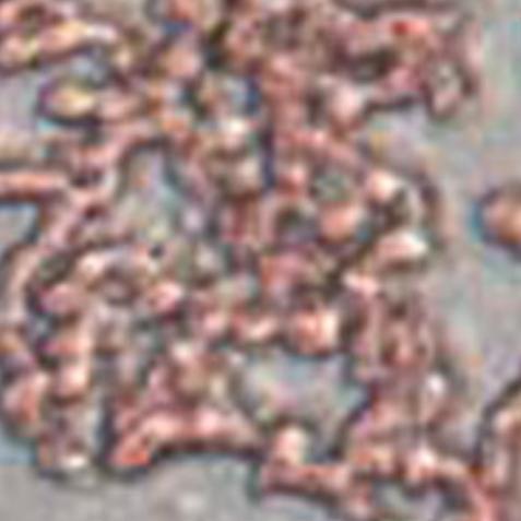
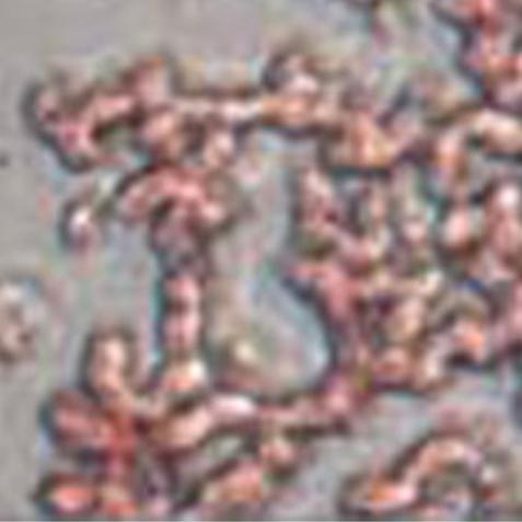
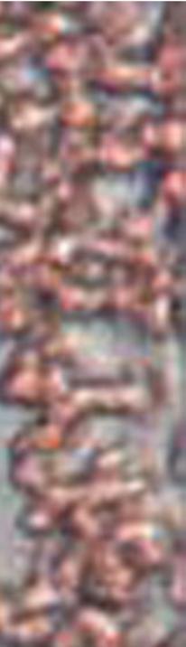
2 only measuree SO 2 for in-vivo samples but also simultaneously form high resolution images using the absorption of the oxygen-carrying cells in circulation, red blood cells (RBCs) Our group has recently shown that the aggregation of RBCs is a phenomenon which affects the PA signals 13,14 The aggregation of RBCs results in the formation of clusters of RBCs of different sizess which can increase the viscosity of blood and impair the circulation It is observed in a variety of disorders such as myocardial infarctions, bacterial infections, type 2 diabetes and sickle cell disease 15 Theoretical simulations showed that as the size of the aggregates increased, the dominant frequency peak of the PA signal power spectrum decreased, approaching the diagnostic frequency range (<30 MHz) This could be attributed to the fact that an aggregatee of RBCs behaves as a single entity rather than individual RBCs 13 Experimentally, we observed that the strength of the PA signal and spectral features of the power spectrum were dependent on the size of the RBC aggregatess and that the signal changes correlated well with independent measurements of aggregation 14 It has been shown that the formation of RBC aggregates contributess to a thickening of the plasma layer around blood aggregates leading to a significant reduction in the diffusion of oxygen in surrounding tissues 16,17 Since PA techniques are capable of measuring the SO 2 of a blood sample in addition to detecting the presence of aggregation, we propose using PA methods for assessing the effect of RBC aggregation on SO 2 This simultaneous detection could potentially provide a non-invasive method for assessing RBC aggregation and thee effect that it has on tissue oxygenation 2 METHODS 21 Sample preparation The guidelines on handling blood weree adapted from the recommendation of the International society for Clinical Hemorheology and the European Society for Clinical Hemorheology y and Microcirculation 18 The source of blood was the femoral vein of Yorkshire pigs (Comparative Research, Toronto,, Canada) Thee blood was drawn into spray-coated EDTA vacutaners (Becton, Dickinson and Company, Franklin Lakes, New Jersey) in order to avoid coagulation In order to separate the RBCs from the plasma, the blood was centrifuged (1500 g for 6 minutes at room temperature) and then washed twice with phosphate-buffered saline (PBS) The collected, packed RBCs were then suspended in PBS in order to achieve three hematocritt levels (10%, 20% and 40%) ) Aggregation was induced using Dextran-70 by changing the concentration of (Sigma-Aldrich, St Louis, Missouri) dissolvedd in PBS Varying degrees of aggregationn were achieved Dextran in PBS ([Dex-PBS]) In this study, the [Dex-PBS] was 1%,, 3% and 8% for each hematocrit level The blood was kept in full contact with air prior to experimentation and all experiments weree completed within 4 hours of blood collection The presence of aggregation was assessed through light microscopy (Olympus Canada Inc, Markham, Canada) Representative images of non-aggregated and aggregated RBCs are shown in figure 1 :: ; :;' T' : : Figure 1: (a) Non-aggregated RBC suspended in PBS and (b) aggregated RBCs suspended in 3% [Dex- PBS] The scale bar denotes 10 µm Proc of SPIE Vol T-2 Downloaded From: on 03/10/2013 Terms of Use:
3 22 PA measurements The PA measurements were conducted using the Imagio Small Animal PA Imaging Device (Seno Medical Instruments Inc, San Antonio, Texas) shown in figure 2 Laser So urce Trans& ucer San iple CCD Camera Wa'ter Figure 2: The Imagio Small Animal PA Imaging Device Thee blood samplee was placed in a cylindrical tube that was placed between the laser (left) and ultrasound transducer (right) The system consisted c of a Q-switched, pulsed Nd:YAG laser deliveredd through an articulated arm coaxial to a transducer 60 mm apartt allowing raster scanning of the sample The laser beam diameter was 9 mm, the pulse width was 6 ns, the pulse repetition rate was 10 Hz and the maximum fluence was 25 mj/cm 2 /pulse Each sample was irradiated to 750 nm and 1064 nmm lasers The PA signals were collected through a 4 element annular array transducer with 5 MHz center frequency and 60% -6 db bandwidth The blood samples were loaded into cylindrical tubes (diameter 05 mm) at the focus of the transducer (295 mm) A vertical raster scan was performed for each sample During each scan, the laser emitted 4 pulses at each location and the 4 PA signals recorded weree averaged to produce 1 PA signal used for analysis A total of 80 laser pulses were fired for each sample thus corresponding to 20 PA signals at different locationss inside each tube (05 mm apart over a length of 10 cm) 23 Signal analysis and estimation of SO 2 The PA signal amplitude (SA) of each recorded signal was computedd as the integration of the Hilbert Transform of the PA signal The average SA and standard deviation was computed by averaging thee SA from all 20 signals recorded for each hematocrit and [Dex-PBS] The normality of the data was confirmed using a Shapiro-Wilk test (normality criterion W > 005) An unpaired t-test was used to compare the SA from non-aggregated and aggregated RBCs Statistical significance was w established for p-values of 005 or less The SO 2 for each exposed sample was computed by taking into account the fact that the SA is directly proportional to the concentrationn of OHb and DHb 12 The SO 2 can be computed by exposing each sample at 2 optical wavelengths ( λ 1 = 750nm and λ 2 = 1064nm ): Proc of SPIE Vol T-3 Downloaded From: on 03/10/2013 Terms of Use:
4 SO 2 [ OHb] SA( λ2) ε( DHb, λ1) SA( λ1) ε( DHb, λ2) = = [ OHb] + [ DHb] SA( λ ) Δε( λ ) SA( λ ) Δε( λ ) (1) Here, Δ ε ( λ) = ε( OHb, λ) ε( DHb, λ) is the difference in extinction coefficient ε for each wavelength of illumination λ 3 RESULTS AND DISCUSSION Figure 3 contains the summary of the PA SA of each RBC sample exposed to both illumination wavelengths The SA is plotted as a function of both hematocrit and aggregation level (determined by the [Dex-PBS]) For both exposure wavelengths, the SA increased monotonically with increasing hematocrit (~13x per doubling hematocrit level) This is in accordance to previous reports which suggest that the increase is due to the increase in the concentration of optical absorbers (the RBCs) 13,14,19 21 The relationship between the PA SA and hematocrit suggests that it might be possible to use PA techniques as a non-invasive means of estimating the hematocrit, an improvement over other non-invasive methods such as ultrasound backscattering where the relationship between the ultrasound backscatter and hematocrit is relatively complex 22 Increasing aggregation levels resulted in changes in the PA SA as shown in figure 3 For a [Dex-PBS] of 3%, the SA was highest for all hematocrit levels The 1% and 8% [Dex-PBS] were nearly identical for all hematocrit levels (p = 02) The SA for the highest aggregation level was ~16x higher than the non-aggregated sample (0% [Dex-PBS]) for both wavelengths of illumination (p = 0001) This can be attributed to the fact that the 3% [Dex-PBS] forms the largest aggregate while the 1% and 8% concentrations yield smaller aggregates as previously reported through independent measures of aggregation 23,24 The SA for the 1064 nm exposure was ~13x smaller than the 750 nm exposure for all samples recorded This could be attributed to the oxygen-depended optical absorption of the RBCs 9 3 (a) SA (au) (b) H=10% H=20% H=40% SA (au) [Dex-PBS] (%) Figure 3: PA SA for (a) 750 nm and (b) 1064 nm exposures of RBC samples at 10%, 20% and 40% hematocrit and 1%, 3% and 8% [Dex-PBS] levels The 0% [Dex-PBS] corresponds to the non-aggregated sample Figure 4 shows the SO 2 computed using equation 1 for the RBC samples examined A linear increase in the SO 2 with increasing hematocrit was observed with an average increase of ~7% per doubling of the hematocrit level The largest Proc of SPIE Vol T-4 Downloaded From: on 03/10/2013 Terms of Use:
5 SO 2 was recorded for the 3% [Dex-PBS] samples which was ~30% higher than the non-aggregated case (p = ) The 1% and 8% samples were nearly identical and ~20% higher than then non-aggregated samples (p = ) These results suggest that the presence of aggregation increases the SO 2 level As this oxygen is bound within the aggregate, the ability of the clustered RBCs to release oxygen to the surrounding environment is diminished This has important implications for adequate oxygen transport to surrounding tissues and further investigations are warranted in order to understand the role of RBC aggregation in vascular pathologies such as atherosclerosis Similar increases in SO 2 with aggregation have been reported using a microscope system coupled with a spectrophotometer 17 The impaired oxygen diffusion due to aggregation is attributed to an increased layer of aggregant/plasma engulfing the RBC aggregates The fact that PA imaging is capable of measuring changes in oxygenation and RBC aggregation simultaneously suggests that it could potentially be used for making assessments of RBC aggregation in diseased tissues while providing information about the level of oxygen and the capacity of the RBCs to deliver oxygen for the tissues of interest H=10% H=20% H=40% SO 2 (%) [Dex-PBS] (%) Figure 4: The SO 2 calculated using equation 1 for all blood samples for 3 hematocrit and 3 aggregation levels The 0% [Dex-PBS] corresponds to the non-aggregated sample 4 CONCLUSIONS This paper presents the potential for using PA techniques for assessing oxygenation of blood samples in the presence of RBC aggregates The results of this study suggest that in the presence of RBC aggregation, the release of oxygen to the surrounding environment is impaired It might be possible to use PA imaging for assessing the presence of RBC aggregation in circulatory disorders while measuring the oxygenation of the same area and thereby monitoring the need for oxygen to the surrounding tissues ACKNOWLEDGEMENTS This work was made possible due to the financial support of the following granting agencies awarded to M C Kolios: Natural Sciences and Engineering Research Council of Canada, Canadian Institutes of Health Research, Canadian Foundation for Innovation, Canada Research Chairs Program and the Terry Fox Foundation E Hysi was supported Proc of SPIE Vol T-5 Downloaded From: on 03/10/2013 Terms of Use:
6 through the Alexander Graham Bell Graduate Scholarship A Worthington is gratefully acknowledged for the technical support provided along with J Barry for providing the porcine blood The authors are also very thankful to M Rui and E Berndl for their assistance with the blood specimens REFERENCES [1] CD Hanning and J M Alexander-Williams, Pulse oximetry: a practical review, Brit Med J 311, (1995) [2] H F Zhang et al, Imaging of hemoglobin saturation variation in single vessels in vivo using photoacoustic microscopy, Appl Opt 90(5), (2007) [3] M Henke et al, Blood hemoglobin level may affect radiosensitivitypreliminary results on acutely reacting normal tissues, Int J Radiat Oncol Biol Phys 48(2), (2000) [4] B Venkatesh et al, Monitoring tissue oxygenation during resuscitation of major burns, J Trauma 50(3), (2001) [5] S S Foo et al, Functional imaging of intratumoral hypoxia, Mol Imaging Biol 6(5), (2004) [6] M E George et al, Noninvasive tissue oxygen saturation measurements identify supply dependency, J Surg Res 160, (2010) [7] S Hu and L V Wang, Photoacoustic imaging and characterization of microvasculature, J Biomed Opt 15(1), (2010) [8] K Maslov et al, Effects of wavelength-dependent fluence attenuation of the noninvasive photoacoustic imaging of hemoglobin oxygen saturation of subcutaneous microvasculature in vivo, Inverse Probl 23, S113-S122 (2007) [9] L V Wang, Prospects of photoacoustic tomography, Med Phys 35(12), (2008) [10] C Li and L V Wang, Photoacoustic tomography and sensing in biomedicine, Phys Med Biol 54(19), R59 R97 (2009) [11] Y Wang et al, In vivo intergrated photoacoustic and confocal microscopy of hemoglobin oxygen saturation and oxygen partial pressure, Opt Let 36(7), (2011) [12] XWang et al, Noninvasive imaging of hemoglobin concentration and oxygenation in rat brain using highresolution photoacoustic tomography, J Biomed Opt 11(2), (2006) [13] R K Saha and M C Kolios, A simulation study on photoacoustic signals from red blood cells, J Acoust Soc Am 129(5), , (2011) [14] E Hysi, R K Saha, and M C Kolios, On the use of photoacoustics to detect red blood cell aggregation, Biomed Opt Express 3(9), (2012) [15] O K Baskurt, B Neu, and H J Meiselman, Red Blood Cell Aggregation, pp 1 304, CRC Press, Boca Raton, FL (2011) [16] N Tateishi et al, Reduced oxygen release from erythrocytes by the acceleration-induced flow shift, observed in an oxygen-permable narrow tube, J Biomech 35(9), (2002) [17] N Tateishi, N Maeda, and T Shiga, A method for measuring the rate of oxygen release from single microvessels, Circ Res 70(4), (1992) Proc of SPIE Vol T-6 Downloaded From: on 03/10/2013 Terms of Use:
7 [18] O K Baskurt et al, New guidelines for hemorheological laboratory techniques, Clin Hemorheol Micro 42(2), (2009) [19] R K Saha and M C Kolios, Effects of erythrocyte oxygenation on optoacoustic signals, J Biomed Opt 16(11), (2011) [20] E Hysi et al, Photoacoustic ultrasound spectroscopy for assessing red blood cell aggregation and oxygenation, J Biomed Opt 17(12), (2012) [21] A B Karpiouk et al, Combined ultrasound and photoacoustic imaging to detect and stage deep vein thrombosis: phantom and ex vivo studies, J Biomed Opt 13(5), (2008) [22] R S C Cobbold, Foundations of Biomedical Ultrasound, pp , Oxford University Press, New York, NY (2007) [23] O K Baskurt, R A Farley, and H J Meisleman, Erythrocyte aggregation tendency and cellular properties in horse, human, and rat: a comparative study, Am J Physiol Heart Circ Physiol 273, H2604 H2612 (1997) [24] O K Baskurt et al, Handbook of Hemorheology and Hemodynamics, pp 1 455, IOS Press, Amsterdam (2007) Proc of SPIE Vol T-7 Downloaded From: on 03/10/2013 Terms of Use:
Photoacoustic Imaging in Biomedicine Critical Review by Saurabh Vyas Group 9: Interventional Photoacoustic Ultrasound CIS II: 600.
 Photoacoustic Imaging in Biomedicine Critical Review by Saurabh Vyas Group 9: Interventional Photoacoustic Ultrasound CIS II: 600.446, Spring 2011 Introduction Photoacoustic imaging (PA Imaging) is the
Photoacoustic imaging of vascular networks in transgenic mice
 Photoacoustic imaging of vascular networks in transgenic mice J.G. Laufer 1, J.O. Cleary 1,2, E.Z. Zhang 1, M.F. Lythgoe 2, P.C. Beard 1 1. Department of Medical Physics and Bioengineering, University
Basic principles of quantification using optical techniques
 Contents Basic principles of quantification using optical techniques Adrian Taruttis Helmholtz Zentrum München Chair for Biological Imaging Technische Universität München Light/ tissue interactions Planar
In vivo label-free photoacoustic flow cytography and on-the-spot laser killing of single circulating. melanoma cells
 In vivo label-free photoacoustic flow cytography and on-the-spot laser killing of single circulating melanoma cells Yun He 1,, Lidai Wang 1,,, Junhui Shi 1,, Junjie Yao 1, Lei Li 1, Ruiying Zhang 1, Chih-Hsien
PREPARED FOR: U.S. Army Medical Research and Materiel Command Fort Detrick, Maryland
 AD Award Number: W81XWH-07-1-0231 TITLE: Spectroscopic Photoacoustic Tomography of Prostate Cancer PRINCIPAL INVESTIGATOR: Xueding Wang CONTRACTING ORGANIZATION: University Of Michigan Ann Arbor, MI 48109-1274
PREPARED FOR: U.S. Army Medical Research and Materiel Command Fort Detrick, Maryland
 AD Award Number: W81XWH-07-1-0231 TITLE: Spectroscopic Photoacoustic Tomography of Prostate Cancer PRINCIPAL INVESTIGATOR: Xueding Wang CONTRACTING ORGANIZATION: University Of Michigan Ann Arbor, MI 48109-1274
Miniature fibre optic probe for minimally invasive photoacoustic sensing
 Miniature fibre optic probe for minimally invasive photoacoustic sensing Sunish J. Mathews*, Edward Z. Zhang, Adrien E. Desjardins and Paul C. Beard Department of Medical Physics and Biomedical Engineering,
IN VIVO BLOOD OXYGENATION LEVEL MEASUREMENTS USING PHOTOACOUSTIC MICROSCOPY. A Thesis MATHANGI SIVARAMAKRISHNAN
 IN VIVO BLOOD OXYGENATION LEVEL MEASUREMENTS USING PHOTOACOUSTIC MICROSCOPY A Thesis by MATHANGI SIVARAMAKRISHNAN Submitted to the Office of Graduate Studies of Texas A&M University in partial fulfillment
Realtime Photoacoustic Microscopy of Murine Cardiovascular Dynamics
 Realtime Photoacoustic Microscopy of Murine Cardiovascular Dynamics R.J. Zemp, 1,3* L. Song, 1* R. Bitton, 2 K.K. Shung, 2 and L.V. Wang 1,** 1 Washington University, Optical Imaging Laboratory, Department
Optoacoustic System for 3D Functional and Molecular Imaging in Nude Mice
 Optoacoustic System for 3D Functional and Molecular Imaging in Nude Mice Matthew P. Fronheiser a, Alan Stein a, Don Herzog a, Scott Thompson a, Anton Liopo b, Mohammad Eghtedari b, Massoud Motamedi b,
1st Faculty of Medicine, Charles University in Prague Center for Advanced Preclinical Imaging (CAPI)
") ADVANTAGES Optical Imaging OI Optical Imaging is based on the detection of weak light by a highly sensitive and high resolution CCD camera DISADVANTAGES High sensitivity Limited penetration depth Easy
ADVANTAGES Optical Imaging OI Optical Imaging is based on the detection of weak light by a highly sensitive and high resolution CCD camera DISADVANTAGES High sensitivity Limited penetration depth Easy
Imaging acute thermal burns by photoacoustic microscopy
 Journal of Biomedical Optics 115, 054033 September/October 2006 Imaging acute thermal burns by photoacoustic microscopy Hao F. Zhang Konstantin Maslov Department of Biomedical Engineering Optical Imaging
Classification of biological cells using a sound wave based flow cytometer
 Classification of biological cells using a sound wave based flow cytometer Eric M. Strohm a,c,d, Vaskar Gnyawali b,c,d, Mia Van De Vondervoort a,c,d, Yasaman Daghighi a,c,d, Scott S. H. Tsai b,c,d, Michael
Biophotonics?? Biophotonics. technology in biomedical engineering. Advantages of the lightwave
 Biophotonics - Imaging: X-ray, OCT, polarimetry, DOT, TIRF, photon migration, endoscopy, confocal microscopy, multiphoton microscopy, multispectral imaging - Biosensing: IR spectroscopy, fluorescence,
REVOLUTIONIZING EARLY CANCER DETECTION
 cover story Written by candace o connor Photos by geoff story IMAGING WITH LIGHT AND SOUND: REVOLUTIONIZING EARLY CANCER DETECTION For years, the field of optical imaging in biological tissue had languished,
Experts in Femtosecond Laser Technology. DermaInspect. Non-invasive multiphoton tomography of human skin
 Experts in Femtosecond Laser Technology DermaInspect Non-invasive multiphoton tomography of human skin In vivo optical biopsies with subcellular spatial resolution based on near infrared femtosecond laser
Enhanced Sensitivity Carbon Nanotubes as Targeted Photoacoustic Molecular Imaging Agents
 Enhanced Sensitivity Carbon Nanotubes as Targeted Photoacoustic Molecular Imaging Agents Adam de la Zerda 1,2,*, Zhuang Liu 3,*, Cristina Zavaleta 1, Sunil Bodapati 1, Robert Teed 1, Srikant Vaithilingam
Biomedical Applications of Molecular Spectroscopy
 Biomedical Applications of Molecular Spectroscopy Mike Kayat B&W Tek, Inc 19 Shea Way Newark, DE 19713 United States of America +1 302 368 7824 mikek@bwtek.com 1 Overview Molecular spectroscopy is a large
SUPPLEMENTAL INFORMATION. Virus-mimicking nano-constructs as a contrast agent for near infrared photoacoustic imaging
 SUPPLEMENTAL INFORMATION Virus-mimicking nano-constructs as a contrast agent for near infrared photoacoustic imaging Sharad Gupta, a Muhammad R. Chatni, b Ayala L. N. Rao, c Valentine I. Vullev, a Lihong
FUNCTIONAL PHOTOACOUSTIC MICROSCOPY. A Dissertation HAO ZHANG
 FUNCTIONAL PHOTOACOUSTIC MICROSCOPY A Dissertation by HAO ZHANG Submitted to the Office of Graduate Studies of Texas A&M University in partial fulfillment of the requirements for the degree of DOCTOR OF
SIMULTANEOUS IMAGING OF A lacz-marked TUMOR AND MICROVASCULATURE MORPHOLOGY IN VIVO BY DUAL-WAVELENGTH PHOTOACOUSTIC MICROSCOPY
 Journal of Innovative Optical Health Sciences Vol. 1, No. 2 (2008) 207 215 c World Scientific Publishing Company SIMULTANEOUS IMAGING OF A lacz-marked TUMOR AND MICROVASCULATURE MORPHOLOGY IN VIVO BY DUAL-WAVELENGTH
Toward High-speed Transcranial Photoacoustic Imaging using Compact Near-infrared Pulsed LED Illumination System
 Toward High-speed Transcranial Photoacoustic Imaging using Compact Near-infrared Pulsed LED Illumination System Jeeun Kang a, Haichong Kai Zhang a, Alexis Cheng a, Arman Rahmim a,c, Dean F. Wong a, Jin
Three-dimensional combined photoacoustic and optical coherence microscopy for in vivo microcirculation studies
 Three-dimensional combined photoacoustic and optical coherence microscopy for in vivo microcirculation studies Li Li 1, Konstantin Maslov 1, Geng Ku 1, Lihong V. Wang 1,* 1 Optical Imaging Laboratory,
TITLE PAGE. Citation Format: Copyright notice: DOI abstract link:
 TITLE PAGE Citation Format: Matthias Rehberger, Martina Giovannella, Marco Pagliazzi, Udo Weigel, Turgut Durduran, Davide Contini, Lorenzo Spinelli, Antonio Pifferi, Alessandro Torricelli, Robert Schmitt,
Absorption of an electromagnetic wave
 In vivo optical imaging?? Absorption of an electromagnetic wave Tissue absorption spectrum Extinction = Absorption + Scattering Absorption of an electromagnetic wave Scattering of an electromagnetic wave
In situ semi-quantitative assessment of single cell viability by resonance
 Electronic Supplementary Material (ESI) for Chemical Communications. This journal is The Royal Society of Chemistry 2018 Electronic Supplementary Information (ESI) In situ semi-quantitative assessment
Measurement of cell aggregation characteristics by analysis of laser-backscattering in a microfluidic rheometry
 Korea-Australia Rheology Journal Vol. 19, No. 2, August 2007 pp. 61-66 Measurement of cell aggregation characteristics by analysis of laser-backscattering in a microfluidic rheometry Sehyun Shin*, J.X.
Time-resolved optical spectroscopy and imaging of breast
 Contributed paper OPTO-ELECTRONICS REVIEW 12(2), 249 253 (2004) Time-resolved optical spectroscopy and imaging of breast P. TARONI *, A. PIFFERI, A. TORRICELLI, and R. CUBEDDU INFM-Dipartimento di Fisica,
7/16/2009, LV WANG. 1 cm
 PHOTOACOUSTIC TOMOGRAPHY: High-resolution Imaging of Optical Contrast In Vivo at New Depths Lihong V. Wang, PhD Gene K. Beare Distinguished Professor Optical Imaging Laboratory Dept of Biomedical Engineering
Laser treatment of gravure-printed ITO films on PET
 Laser treatment of gravure-printed ITO films on PET Howard V Snelling, Anton A Serkov, Jack Eden, Rob J Farley Physics, School of Mathematical and Physical Sciences, University of Hull, HU6 7RX, UK Presentation
DETECTION OF LASER ULTRASONIC SURFACE DISPLACEMENT BY WIDE APERTURE FIBER OPTIC AMPLIFIER M.L. Rizzi and F. Corbani CESI, Milano, Italy
 DETECTION OF LASER ULTRASONIC SURFACE DISPLACEMENT BY WIDE APERTURE FIBER OPTIC AMPLIFIER M.L. Rizzi and F. Corbani CESI, Milano, Italy Abstract: In the frame of the European Project INCA, CESI is in charge
Light dosimetry for Low-Level Laser therapy: Accounting for differences in tissue and depth
 Light dosimetry for Low-Level Laser therapy: Accounting for differences in tissue and depth Robert Weersink a, Roger White b, Lothar Lilge c a Laboratory for Applied Biophotonics, University Health Network,
Translational Photoacoustic Tomography
 Washington University in St. Louis Washington University Open Scholarship Engineering and Applied Science Theses & Dissertations Engineering and Applied Science Spring 5-15-2016 Translational Photoacoustic
Effects of Laser Peening Parameters. on Plastic Deformation in Stainless Steel
 Effects of Laser Peening Parameters on Plastic Deformation in Stainless Steel Miho Tsuyama* 1, Yasuteru Kodama* 2, Yukio Miyamoto* 2, Ippei Kitawaki* 2, Masahiro Tsukamoto* 3 and Hitoshi Nakano* 1 *1 Faculty
Supporting Information
 Supporting Information Wiley-VCH 2013 69451 Weinheim, Germany Quantitative Analysis of the Fate of Gold Nanocages In Vitro and In Vivo after Uptake by U87-MG Tumor Cells** Eun Chul Cho, Yu Zhang, Xin Cai,
 NeonatalHeartRateDetectionUsing AuxiliaryOpticalSystem MridulaGarimella,T.V.HimajaSree,AnushaBandari,IbrahimPatel Abstract: Fetal heart rate (FHR) monitoring is a proven means of assessing fetal health
In-vitro study on the hemorheological characteristics of chicken blood in microcirculation
 Korea-Australia Rheology Journal Vol. 19, No. 2, August 2007 pp. 89-95 In-vitro study on the hemorheological characteristics of chicken blood in microcirculation Ho Seong Ji, Jung Yeop Lee and Sang Joon
University of Michigan
 University of Michigan Department of Mechanical Engineering Low-cost Non-invasive Diagnosis of Malaria Infected Red Blood Cells Han Yu Undergraduate Student Department of Electrical Engineering and Computer
Deliverable 2.1: Definition of paradigms representing exemplary breast lesions cases
 Project title: Smart Optical and Ultrasound Diagnostics of Breast Cancer Grant Agreement: 731877 Call identifier: H2020-ICT-2016-1 Topic: ICT-29-2016 Photonics KET 2016 Deliverable 2.1: Definition of paradigms
Transient and selective suppression of neural activity with infrared light
 Supplementary Material Transient and selective suppression of neural activity with infrared light Authors: Austin R. Duke 1, Michael W. Jenkins 2, Hui Lu 3, Jeffrey M. McManus 3, Hillel J. Chiel 3,2,4,
PATTERNING OF OXIDE THIN FILMS BY UV-LASER ABLATION
 Journal of Optoelectronics and Advanced Materials Vol. 7, No. 3, June 2005, p. 1191-1195 Invited lecture PATTERNING OF OXIDE THIN FILMS BY UV-LASER ABLATION J. Ihlemann * Laser-Laboratorium Göttingen e.v.,
Nayar Prize I Quarterly Progress Report (Quarters 2&3) August, 2016
 August, 2016") Nayar Prize I Quarterly Progress Report (Quarters 2&3) August, 2016 Project: ADEPT Cancer Imager Team: Ken Tichauer, Jovan Brankov, Raju Mehta Students: Lagnojita Sinha, Xiaochun Xu Progress Summary Since
Nayar Prize I Quarterly Progress Report (Quarters 2&3) August, 2016 Project: ADEPT Cancer Imager Team: Ken Tichauer, Jovan Brankov, Raju Mehta Students: Lagnojita Sinha, Xiaochun Xu Progress Summary Since
CENTER FOR BRAIN EXPERIMENT
 CENTER FOR BRAIN EXPERIMENT Section of Brain Structure Associate Professor: ARII, Tatsuo, PhD 1967 Graduated from Tohoku University, Faculty of Science. Completed the doctoral course in Engineering, Nagoya
Principle and technique of NIRS-Imaging for human brain FORCE: fast-oxygen response in capillary event
 International Congress Series 1270 (2004) 85 90 Principle and technique of NIRS-Imaging for human brain FORCE: fast-oxygen response in capillary event Toshinori Kato* www.ics-elsevier.com Ogawa Laboratories
Multiplexed 3D FRET imaging in deep tissue of live embryos Ming Zhao, Xiaoyang Wan, Yu Li, Weibin Zhou and Leilei Peng
 Scientific Reports Multiplexed 3D FRET imaging in deep tissue of live embryos Ming Zhao, Xiaoyang Wan, Yu Li, Weibin Zhou and Leilei Peng 1 Supplementary figures and notes Supplementary Figure S1 Volumetric
Noninvasive label-free imaging of microhemodynamics by optical-resolution photoacoustic microscopy
 Noninvasive label-free imaging of microhemodynamics by optical-resolution photoacoustic microscopy Song Hu, Konstantin Maslov, and Lihong V. Wang* Optical Imaging Laboratory, Department of Biomedical Engineering,
In-situ laser-induced contamination monitoring using long-distance microscopy
 In-situ laser-induced contamination monitoring using long-distance microscopy Paul Wagner a, Helmut Schröder* a, Wolfgang Riede a a German Aerospace Center (DLR), Institute of Technical Physics, Pfaffenwaldring
EFFECT OF LASER RADIATION ON ELECTRICAL CONDUCTIVITY OF HUMAN BLOOD
 International Journal of Science, Environment and Technology, Vol. 3, No 1, 2014, 286 290 ISSN 2278-3687 (O) EFFECT OF LASER RADIATION ON ELECTRICAL CONDUCTIVITY OF HUMAN BLOOD 1 Shikha Rathore and 2 Basharath
Spectroscopy and Imaging IV
 PROGRESS IN BIOMEDICAL OPTICS AND IMAGING Vol. 16 No. 55 Clinical and Biomedical Spectroscopy and Imaging IV J. Quincy Brown Volker Decked Edifors 22-24 June 2015 Munich, Germany Sponsored by SPIE (United
Fs- Using Ultrafast Lasers to Add New Functionality to Glass
 An IMI Video Reproduction of Invited Lectures from the 17th University Glass Conference Fs- Using Ultrafast Lasers to Add New Functionality to Glass Denise M. Krol University of California, Davis 17th
Femtosecond micromachining in polymers
 Femtosecond micromachining in polymers Prof. Dr Cleber R. Mendonca Daniel S. Corrêa Prakriti Tayalia Dr. Tobias Voss Dr. Tommaso Baldacchini Prof. Dr. Eric Mazur fs-micromachining focus laser beam inside
Supplementary Information
 Supplementary Information Trapping and Detection of Nanoparticles and Cells Using a Parallel Photonic Nanojet Array Yuchao Li, Hongbao Xin, Xiaoshuai Liu, Yao Zhang, Hongxiang Lei*, and Baojun Li* State
Qswitched lasers are gaining more interest because of their ability for various applications in remote sensing, environmental monitoring, micro
 90 Qswitched lasers are gaining more interest because of their ability for various applications in remote sensing, environmental monitoring, micro machining, nonlinear frequency generation, laserinduced
ABSTRACT 1. INTRODUCTION
 Biodegradable polymer based theranostic agents for photoacoustic imaging and cancer therapy Yan J. Wang a,b,c, Eric M. Strohm a,b,c, and Michael C. Kolios* a,b,c a Dept. of Physics, Ryerson University,
Evaluation of Ultrasonic Attenuation and Estimation of Ultrasonic Grain Noise in Copper
 Evaluation of Ultrasonic Attenuation and Estimation of Ultrasonic Grain Noise in Copper T. Stepinski and P. Wu Uppsala University, Signals & Systems, SE 751 0, Uppsala, Sweden Abstract. This paper presents
SUPPLEMENTARY INFORMATION
 VOLUME: 1 ARTICLE NUMBER: 0068 In the format provided by the authors and unedited. Precision assessment of label-free psoriasis biomarkers with ultrabroadband optoacoustic mesoscopy Juan Aguirre 1, Mathias
Investigation of the influence of laser irradiation on the relative concentration of oxyhemoglobin in the blood
 Investigation of the influence of laser irradiation on the relative concentration of oxyhemoglobin in the blood MM. Asimov, AT. Gisbrecht*, S.A. Marnilov**, Yu.S. Plaksiy** Institute ofphysics NAS ofbelarus,
Biomedical Optical Imaging Martin Frenz Biomedical Photonics Department Institute of Applied Physics, University of Bern
 1 Biomedical Optical Imaging Martin Frenz Biomedical Photonics Department Institute of Applied Physics, University of Bern Imaging is one of the most powerful tools in biomedical research. The impact and
Skin Imaging NTU
 Skin Imaging Research @ NTU A*STAR-NHG-NTU Joint Skin Research Workshop, Oct 19, 2013 A/Prof. Wee Ser ewser@ntu.edu.sg Programme Lead (MISA), NITHM Director, VALENS Research Centre, EEE Nanyang Technological
Deeply penetrating in vivo photoacoustic imaging using a clinical ultrasound array system
 Deeply penetrating in vivo photoacoustic imaging using a clinical ultrasound array system Chulhong Kim, 1,3 Todd N. Erpelding, 2,3 Ladislav Jankovic, 2 Michael D. Pashley, 2 and Lihong V. Wang 1,* 1 Optical
HHS Public Access Author manuscript Annu Rev Biomed Eng. Author manuscript; available in PMC 2014 August 11.
 Photoacoustic microscopy and computed tomography: from bench to bedside Lihong V. Wang * and Optical Imaging Laboratory, Department of Biomedical Engineering, Washington University in St. Louis Liang Gao
Translational Multimodality Optical Imaging
 Translational Multimodality Optical Imaging Fred S. Azar Xavier Intes Editors 0 ARTECH H O U S E BOSTON LONDON artechhouse.com Contents Foreword Preface xv xvii CHAPTER1 Introduction to Clinical Optical
Laser- and Light-Induced Autofluorescence Spectroscopy of Human Skin in Dependence on Excitation Wavelengths
 Vol. 112 (2007) ACTA PHYSICA POLONICA A No. 5 Proceedings of the International School and Conference on Optics and Optical Materials, ISCOM07, Belgrade, Serbia, September 3 7, 2007 Laser- and Light-Induced
THE USE OF A DIODE LASER IN THE TREATMENT OF HEMORRHOIDAL DISEASE
 International Journal of Civil Engineering and Technology (IJCIET) Volume 9, Issue 11, November 2018, pp. 2108 2112, Article ID: IJCIET_09_11_206 Available online at http://www.iaeme.com/ijciet/issues.asp?jtype=ijciet&vtype=9&itype=11
Submicron-resolution Photoacoustic Microscopy of Endogenous Light-absorbing Biomolecules
 Washington University in St. Louis Washington University Open Scholarship All Theses and Dissertations (ETDs) Spring 3-26-2014 Submicron-resolution Photoacoustic Microscopy of Endogenous Light-absorbing
micromachines ISSN X
 Micromachines 2012, 3, 55-61; doi:10.3390/mi3010055 Article OPEN ACCESS micromachines ISSN 2072-666X www.mdpi.com/journal/micromachines Surface Plasmon Excitation and Localization by Metal-Coated Axicon
Compact Plasmonic Blackbody for Cancer Theranosis in Near-Infrared II Window
 Supporting Information for Compact Plasmonic Blackbody for Cancer Theranosis in Near-Infrared II Window Jiajing Zhou,, Yuyan Jiang,, Shuai Hou, Paul Kumar Upputuri, Di Wu,, Jingchao Li, Peng Wang, Xu Zhen,
Structural changes of austenitic steel obtained by 532 nm and 1064 nm Nd:YAG laser radiation
 JOURNAL OF OPTOELECTRONICS AND ADVANCED MATERIALS Vol. 8, No. 1, February 2006, p, 230-234 Structural changes of austenitic steel obtained by 532 nm and 1064 nm Nd:YAG laser radiation M. I. RUSU *, R.
Enhanced Light Trapping in Periodic Aluminum Nanorod Arrays as Cavity Resonator
 Enhanced Light Trapping in Periodic Aluminum Nanorod Arrays as Cavity Resonator Rosure B. Abdulrahman, Arif S. Alagoz, Tansel Karabacak Department of Applied Science, University of Arkansas at Little Rock,
Contact Details. Dr Alexander Galkin. Office: MBC Room 186. Tel: (028) Frequency and wavelength.
 Frequency and wavelength.") Contact Details The electromagnetic spectrum Biological Spectroscopy Dr Alexander Galkin Email: a.galkin@qub.ac.uk Dr Alexander Galkin MSc Biomolecular Function - BBC8045 Office: MBC Room 186 Tel: (028)
Contact Details The electromagnetic spectrum Biological Spectroscopy Dr Alexander Galkin Email: a.galkin@qub.ac.uk Dr Alexander Galkin MSc Biomolecular Function - BBC8045 Office: MBC Room 186 Tel: (028)
Citation for published version (APA): Bremmer, R. H. (2011). Non-contact spectroscopic age determination of bloodstains
: Bremmer, R. H. (2011). Non-contact spectroscopic age determination of bloodstains") UvA-DARE (Digital Academic Repository) Non-contact spectroscopic age determination of bloodstains Bremmer, R.H. Link to publication Citation for published version (APA): Bremmer, R. H. (2011). Non-contact
UvA-DARE (Digital Academic Repository) Non-contact spectroscopic age determination of bloodstains Bremmer, R.H. Link to publication Citation for published version (APA): Bremmer, R. H. (2011). Non-contact
HHS Public Access Author manuscript Phys Rev Lett. Author manuscript; available in PMC 2014 March 18.
 Photo-imprint Photoacoustic Microscopy for Three-dimensional Label-free Sub-diffraction Imaging Junjie Yao, Lidai Wang, Chiye Li, Chi Zhang, and Lihong V. Wang * Optical Imaging Laboratory, Department
STRUCTURAL DIAGNOSTICS OF GEOMATERIALS BY LASER ULTRASONIC SPECTROSCOPY
 Acústica 2008 20-22 de Outubro, Coimbra, Portugal Universidade de Coimbra STRUCTURAL DIAGNOSTICS OF GEOMATERIALS BY LASER ULTRASONIC SPECTROSCOPY E.B. Cherepetskaya 1, V.N. Inkov 1, A.A. Zakirov 1, A.A.
F* techniques: FRAP, FLIP, FRET, FLIM,
 F* techniques: FRAP, FLIP, FRET, FLIM, FCS Antonia Göhler March 2015 Fluorescence explained in the Bohr model Absorption of light (blue) causes an electron to move to a higher energy orbit. After a particular
SLS-process monitoring and temperature control
 SLS-process monitoring and temperature control Yu. Chivel 1, M. Doubenskaia 2 1 Institute of Molecular & Atomic Physics NAS Belarus 68 Nezavisimosti av., 220072, Minsk, Belarus 2 Ecole Nationale d Ingénieurs
This document is downloaded from DR-NTU, Nanyang Technological University Library, Singapore.
 This document is downloaded from DR-NTU, Nanyang Technological University Library, Singapore. Title Microsphere enabled subdiffraction-limited opticalresolution photoacoustic microscopy: a simulation study
Ultrasound-Aided High-Resolution Biophotonic Tomography
 Ultrasound-Aided High-Resolution Biophotonic Tomography 1 cm Lihong V. Wang, PhD Gene K. Beare Distinguished Professor Optical Imaging Laboratory Department of Biomedical Engineering Washington University
Introduction. (b) (a)
 (a)") Introduction Whispering Gallery modes (WGMs) in dielectric micro-cavities are resonant electromagnetic modes that are of considerable current interest because of their extremely high Q values leading to
Introduction Whispering Gallery modes (WGMs) in dielectric micro-cavities are resonant electromagnetic modes that are of considerable current interest because of their extremely high Q values leading to
Study of SOFC Operational Behavior by Applying Diagnostic Methods
 Study of SOFC Operational Behavior by Applying In-Situ Diagnostic Methods Günter Schiller, Wolfgang Bessler, Caroline Willich, K. Andreas Friedrich Deutsches Zentrum für Luft- und Raumfahrt, Institut für
Examples of the application of light-tissue interaction to biomedical engineering
 Lighting in Engineering, Architecture and the Environment 223 Examples of the application of light-tissue interaction to biomedical engineering A. Cysewska-Sobusiak, A. Hulewicz, Z. Krawiecki & G. Wiczynski
Widefield Microscopy Bleed-Through
 In widefield microscopy the excitation wavelengths which illuminate the sample, and the emission wavelengths which reach the CCD camera are selected throughout a filter cube. A filter cube consists of
HHS Public Access Author manuscript Nat Biomed Eng. Author manuscript; available in PMC 2018 January 12.
 Single-impulse Panoramic Photoacoustic Computed Tomography of Small-animal Whole-body Dynamics at High Spatiotemporal Resolution Lei Li 1,2,, Liren Zhu 2,3,, Cheng Ma 3,4,, Li Lin 2,3,, Junjie Yao 3,5,
Dielectric II-VI and IV-VI Metal Chalcogenide Thin Films in Hollow Glass Waveguides (HGWs) for Infrared Spectroscopy and Laser Delivery
 for Infrared Spectroscopy and Laser Delivery") Dielectric II-VI and IV-VI Metal Chalcogenide Thin Films in Hollow Glass Waveguides (HGWs) for Infrared Spectroscopy and Laser Delivery Carlos M. Bledt * a, Daniel V. Kopp a, and James A. Harrington a
Dielectric II-VI and IV-VI Metal Chalcogenide Thin Films in Hollow Glass Waveguides (HGWs) for Infrared Spectroscopy and Laser Delivery Carlos M. Bledt * a, Daniel V. Kopp a, and James A. Harrington a
Principles of translational medicine: imaging, biomarker imaging, theranostics
 Principles of translational medicine: imaging, biomarker imaging, theranostics Compiled by: Endre Mikus PhD, CEO Budapest, 21/9/2015 Imaging and imaging biomarkers An imaging biomarker is an anatomic,
Genetically targeted all-optical electrophysiology with a transgenic Credependent
 Genetically targeted all-optical electrophysiology with a transgenic Credependent Optopatch mouse Short title: Transgenic Optopatch mouse Shan Lou 1, Yoav Adam 1, Eli N. Weinstein 1,4, Erika Williams 2,
BME101 Introduction to Biomedical Engineering Medical Imaging Özlem BİRGÜL Ankara University Department of Biomedical Engineering
 BME101 Introduction to Biomedical Engineering Medical Imaging Özlem BİRGÜL Ankara University Department of Biomedical Engineering Outline What is Medical Imaging? History of Medical Imaging X-Ray Imaging
Interventional multi-spectral photoacoustic imaging in laparoscopic surgery
 Interventional multi-spectral photoacoustic imaging in laparoscopic surgery Emma R. Hill 1, Wenfeng Xia 1, Daniil I. ikitichev 1, Kurinchi Gurusamy 2, Paul C. Beard 1, David J. Hawkes 1, Brian R. Davidson
Michael T. Anderson, Stephen E. Cumblidge, Steven R. Doctor Pacific Northwest National Laboratory, Richland, WA, USA
 THROUGH WELD INSPECTION OF WROUGHT STAINLESS STEEL PIPING USING PHASED ARRAY ULTRASONIC PROBES Michael T. Anderson, Stephen E. Cumblidge, Steven R. Doctor Pacific Northwest National Laboratory, Richland,
Radio-opaque isotropic fibre optic probes for in vivo photodynamic therapy dosimetry
 Radio-opaque isotropic fibre optic probes for in vivo photodynamic therapy dosimetry E.J. Hudson, M.R. Stringer, H.J. van Staveren*, M.. Smith cademic Unit of Medical Physics, Department of Clinical Medicine,
HELICA BIOSYSTEMS, INC. HIGH SENSITIVITY HUMAN C-REACTIVE PROTEIN FOR RESEARCH USE ONLY (Not for in vitro diagnostic use)
") INTENDED USE HELICA BIOSYSTEMS, INC. HIGH SENSITIVITY HUMAN C-REACTIVE PROTEIN FOR RESEARCH USE ONLY (Not for in vitro diagnostic use) The Helica C-reactive protein assay is intended for the detection
INTENDED USE HELICA BIOSYSTEMS, INC. HIGH SENSITIVITY HUMAN C-REACTIVE PROTEIN FOR RESEARCH USE ONLY (Not for in vitro diagnostic use) The Helica C-reactive protein assay is intended for the detection
Fabrication of Micro and Nano Structures in Glass using Ultrafast Lasers
 Fabrication of Micro and Nano Structures in Glass using Ultrafast Lasers Denise M. Krol University of California, Davis IMI Glass Workshop Washington DC April 15-17, 2007 Femtosecond laser modification
Assessment of Blood Trauma in Circulatory-assist Devices. Sangria Project. Amanda Daly. Advisor: Dr. Marina Kameneva
 Assessment of Blood Trauma in Circulatory-assist Devices Amanda Daly Advisor: Dr. Marina Kameneva Multi-scale Thrombosis Modeling Seminar Series Experimental subgroup meeting June 25, 2010 Motivation for
Optimization of Water based Optical Filter for Concentrated Crystalline Si PV/T System - A Theoretical Approach
 Research Article International Journal of Current Engineering and Technology E-ISSN 2277 46, P-ISSN 2347-56 24 INPRESSCO, All Rights Reserved Available at http://inpressco.com/category/ijcet Optimization
Supporting Information: Gold nanorod plasmonic upconversion microlaser
 Supporting Information: Gold nanorod plasmonic upconversion microlaser 1 Materials Synthesis and Properties Ce Shi, Soheil Soltani, Andrea M. Armani 1.1 Nanorod synthesis First the gold nanorods (NRs)
Electronic Supplementary Information
 Electronic Supplementary Information Enzyme-Responsive Copper Sulphide Nanoparticles for Combined Photoacoustic Imaging, Tumor-Selective Chemotherapy and Photothermal Therapy Zhengbao Zha, b Shuhai Zhang,
The quenching effect in PRESAGE dosimetry of proton beams: Is an empirical correction feasible?
 The quenching effect in PRESAGE dosimetry of proton beams: Is an empirical correction feasible? S J Doran 1,2, T Gorjiara 3, J Adamovics 4,5, Z Kuncic 3, A Kacperek 6 and C Baldock 3 1 CRUK Cancer Imaging
LOW TEMPERATURE GROWTH OF SMOOTH INDIUM TIN OXIDE FILMS BY ULTRAVIOLET ASSISTED PULSED LASER DEPOSITION
 Journal of Optoelectronics and Advanced Materials Vol. 4, No. 1, March 2002, p. 21-25 LOW TEMPERATURE GROWTH OF SMOOTH INDIUM TIN OXIDE FILMS BY ULTRAVIOLET ASSISTED PULSED LASER DEPOSITION V. Craciun,
Formation of Droplets on Thin Film Surface in Pulsed Laser Deposition Using Metal Targets*
 [Quarterly Journal of Japan Welding Society, Vol. 21, No. 3, pp. 338-343 (2003)] Formation of Droplets on Thin Film Surface in Pulsed Laser Deposition Using Metal Targets* by Salim MUSTOFA**, TSUYUGUCHI
Photothermal Optical Coherence Tomography of Nanoparticle Contrast Agents
 Photothermal Optical Coherence Tomography of Nanoparticle Contrast Agents Jason M. Tucker Schwartz Kelsey R. Beavers Craig L. Duvall Melissa C. Skala Vanderbilt University Department of Biomedical Engineering