Practical light microscopy: an introduction
|
|
|
- Tamsyn Garrison
- 6 years ago
- Views:
Transcription
1 Practical light microscopy: an introduction Dr. Mark Leake, Oxford University
2 Aim of today s talk: Explanation of the very (very) basics of how a light microscope works Illustration of the most important modifications that make the microscope more useful--phase contrast, DIC, fluorescence Outline the bacterial chemotaxis practical session
3 What can we do with a light microscope? In a nutshell: Magnify things, resolve details not possible with the naked eye. More specifically: Obtain information about the distribution of specific molecules inside cells, including sub-cellular structures Follow changes in cells or molecules over time.
4 Light has both particle and wave properties Quantum mechanics -- too complicated for today! Light travels in a straight line (rays)--like a bullet Light also has "wave" properties, such as wavelength, and interference amplitude E wavelength λ


5 Lenses utilize refraction of light Interaction of light with matter can alter speed, and path, of light. How? The electrons in the material interact with the vibrations of the electromagnetic field. A prism: different wavelengths are refracted (bent) to different degrees A lens: refraction at curved surfaces can cause the light rays to converge (or diverge)
6 How do lenses magnify? f = "focal length", or "focal distance", of lens. Magnification depends on position of object relative to the lens. This can be seen by "ray tracing" (black lines and arrows). Object at distance greater than 2 x f from the lens f f 2 x f 2 x f Miniature image formed, at distance between f and 2 x f Object exactly at distance 2 x f from the lens f f Same-size image formed, at distance 2 x f from the lens Object at distance between 2 x f and f from the lens f f Magnified object formed, at distance than distance "f" the lens
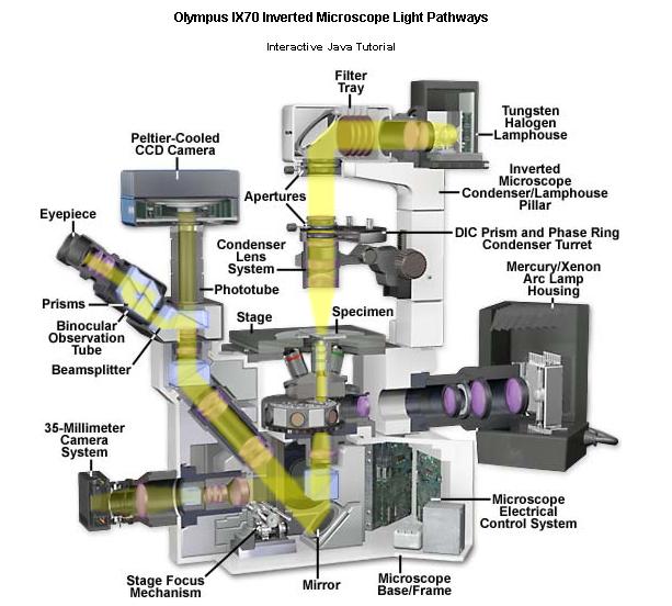
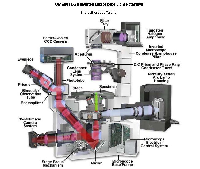
7 The compound microscope Conceptually, the compound microscope is not much different from a magnifying glass, but it has two different stages of magnification (hence "compound")--the objective and the eyepieces. The objective creates an image inside the microscope, and this is further magnified by the ocular (eyepiece). The eye is part of the "imaging system"! In addition, everything is mounted on a stand, which makes it easy to focus, and there is a built-in illumination source. The condenser lens (under the stage) directs the light correctly onto the sample.
 particle Two particles just at resolution limit Two well-resolved")
8 Magnification alone isn t sufficient Resolution: The ability to distinguish two points very close together "Airy rings" Single (tiny) particle Two particles just at resolution limit Two well-resolved particles


9 The Royal Oak
10 What determines the resolution limit? No lens has perfect resolution, even in theory Resolution depends on the angle (θ) of the cone of light that the objective can collect from the specimen. This angle, called Numerical Aperture, in turn depends on the lens diameter and on the distance from the specimen to the lens. q Objective lens Specimen For all practical purposes, improvements of Numerical Aperture in microscope design have reached their limits. The other factor affecting resolution is the wavelength of light itself. amplitude E wavelength λ
11 Useful size range for light microscopy Using standard light microscopy most structures and substructures we observe are within a typical range from about 300 μm down to about 0.3 μm (300 nm), though we can detect single molecules under certain circumstances

12 By 1900 generic microscope designs were very similar to those of today, but.cells are mostly water, and therefore mostly transparent! How can you generate contrast to "see" a transparent object?

 stain Discoveries in")
")
 Eosinophils: granules")
 Neutrophils: not stained with either")
13 Using chemical stains for cells and tissues Mitosis in white blood cells-- Giemsa stain Epithelial tissue--haemotoxylin (basic dye) & Eosin (acidic dye) stain Discoveries in biology emerged from cytochemistry: Different types of granulocytes (white-blood cells) include : Basophils: granules bound basic dyes (e.g. haemotoxylin) Eosinophils: granules bound acid dyes (e.g. eosin) Neutrophils: not stained with either acid- or basic-dyes But these cells are FIXED
14 The big advances over previous 100 years: Imaging live cells (Phase contrast microscopy: 1930s, Differential contrast microscopy: 1950s) Imaging specific molecules inside cells (Immunofluorescence microscopy: 1960s and onwards) Imaging specific molecules inside live cells (Fluorescent labelled proteins:1980s, Green Fluorescent Protein: 1990s)
15 How to see non-fixed living cells? How do stained samples generate contrast? Now need to think about light as electromagnetic radiation, i.e., waves. When stained samples absorb light, they reduce the amplitude of specific wavelengths amplitude E wavelength λ Stained sample
16 Transparent object do interact with light! Light passing through any dense sample is slowed down (this is one aspect of refraction), which changes its phase relative to light not passing through the sample
17 Phase-contrast microscopy: Invented by Zernike, a physicist, in the 1930s (Nobel Prize, 1953) Uses interference of light waves turn "invisible" phase differences into contrast. In essence is sensitive to spatial differences in refractive index Phaseretardation Normal contrast distance Phase contrast
, not absolute phase difference.")
18 Differential interference contrast (DIC) Another phase-dependent method, more recent than phase contrast, and much more complicated technically. Also takes advantage of differences in phase, but measures relative phase difference (i.e. is sensitive to the spatial gradient of refractive index), not absolute phase difference. Differences are greatest at edges, giving 3-D contour effect Normal contrast Phase contrast DIC
19 More specific stains: Beginnings in late 1960s/early 1970s. Starts with chemically-fixed cells. Use antibodies to visualize specific components of cells Incubate with primary" antibody that binds uniquely to a specific protein (e.g. "rabbit anti-actin") Then incubate with labeled secondary" antibody that binds to the primary antibody (e.g. fluorescein-labelled "sheep anti-rabbit") Primary antibody Labeled secondary antibody Seeing this easily required a new type of microscope!
20 Fluorescence Fluorophore absorbs light of a specific wavelength Rapidly emits light of longer wavelength (within nanoseconds) Can have multiple distinct fluorophore in the same experiment. Best results require modifications to the microscope design. Fluorescein Rhodamine Absorb: 490 nm (blue) Emit: 520 nm (green) Absorb: 550 nm (green-yellow) Emit: 580 nm (orange-red)
 mirror Excitation filter Emission")
21 Epifluorescence microscopes Epifluorescence microscopy uses illumination from above ("epi-") and a special cube containing usually two colored filters plus a special beam-splitting ("dichroic") mirror Excitation filter Emission filter
22
23
24 Seeing different molecules types in living cells? Some fluorescent dyes bind to specific compartments or organelles Can also "microinject" labelled protein, or labelled antibody, directly into cells. Difficulties: Requires purified proteins or antibodies. These may perturb protein function. Many types of cells (cells in tissues, microorganisms, many plant cells) cannot be injected.

25 An answer: GFP Green Fluorescent Protein, a naturally fluorescent protein identified in the jellyfish Aequorea victoria.

")

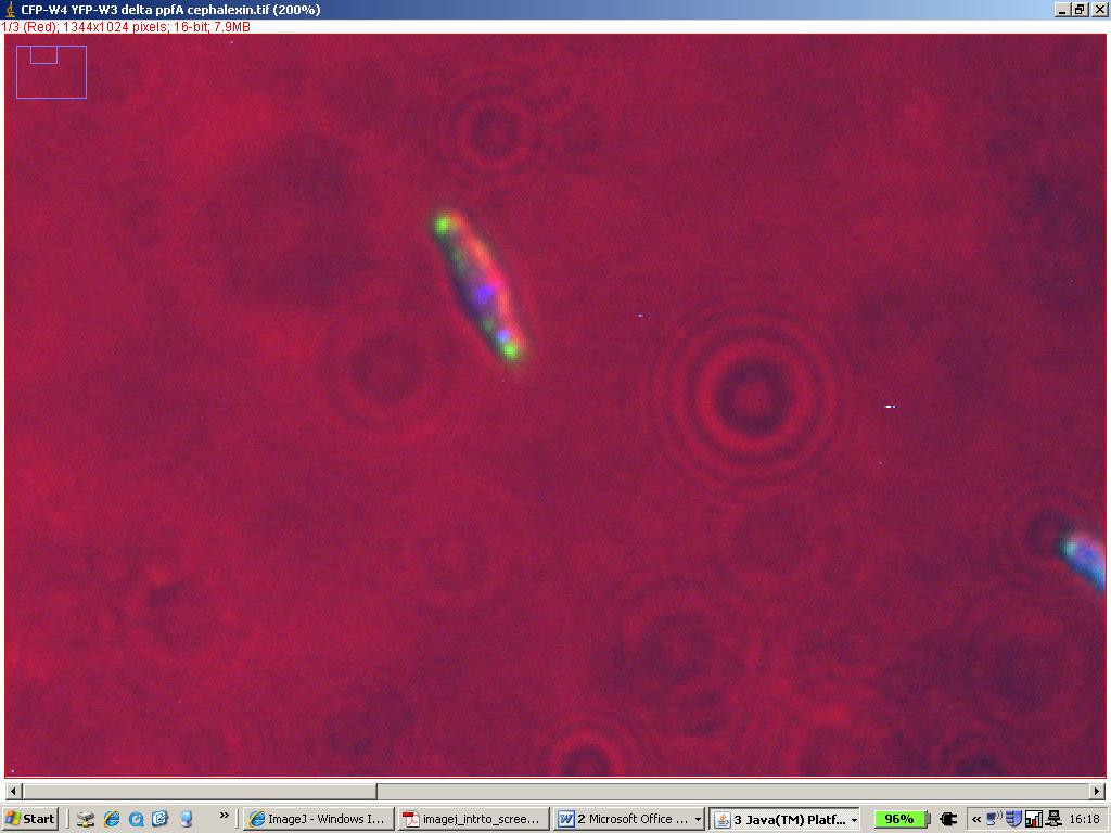
26 Antibody-tethered cell-rotation assay: d) e) (d) Brightfield and (e) TIRF images of GFP-MotB E. coli mutant. Black bar=1μm
 ECFP = enhanced Cyan Fluorescent Protein (GFP derivative)")
 Changing the properties of GFP and RFP by genetic")
27 Extending the palette: EYFP = enhanced Yellow Fluorescent Protein (GFP derivative) ECFP = enhanced Cyan Fluorescent Protein (GFP derivative) DsRed2FP = Red Fluorescent Protein (coral protein, unrelated to GFP, and not monomeric) Changing the properties of GFP and RFP by genetic engineering
28 Advanced methods for fluorescence microscopy: How to improve the quality of fluorescent imaging? Deconvolution microscopy: using computation to put "out-of-focus" light back where it belongs Confocal microscopy: using tricks of geometric optics to remove "out-of-focus" light before it hits the detector TIRF: delimiting the excitation volume to improve image contrast How to measure protein interactions and dynamics? FRET (Foerster Resonance Energy Transfer) FLIM (Fluorescence Lifetime Imaging) FRAP/FLP (Fluorescence Recovery After/Loss In Photobleaching) FCS (Fluorescence Correlation Spectroscopy)

29 off-focus problems Sample object: a "subresolution" fluorescent bead


30 Deconvolution of off-focus light Objective lens X-Z view of sample object X-Z view of raw image stack X-Z view of deconvolved image stack Planes of focus (z stack)

31 Before deconvolution After deconvolution GFP tag in Drosophila embryo
32 Confocal microscope (laser scanning) At the pinhole aperture, in-focus light from the specimen is again "in focus", and all of it goes through the pinhole, but out-of-focus light from the specimen is now "out-of-focus" and spread out, contributing little to the total signal received by the photomultiplier
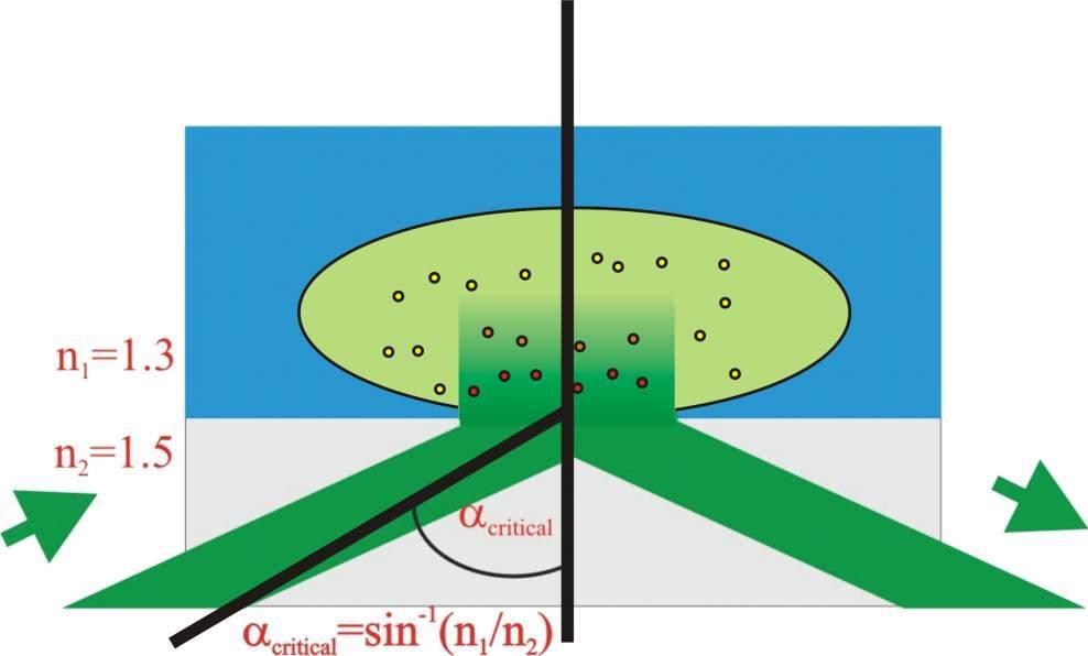

33 Total-internal-reflection-fluorescence (TIRF):
34 Measuring molecular interactions and dynamics Fluorescence correlation spectroscopy (FCS) Fluorescence recovery after/loss in photobleaching (FRAP/FLIP) Foerster resonance energy transfer (FRET) and multi-colour imaging Fluorescence lifetime imaging (FLIM)


35 Fluorescence correlation spectroscopy (FCS) Good for measuring concentrations, diffusion coefficients and turnover Different diffusion coefficients give rise to different fluctuations Experimental set-up Femtoliter volume! Small numbers of fluorescent molecules (maybe 1-500) Reasons for fluctuations in fluorescence P. Schwille and E. Haustein, MPI Goettingen Invitrogen website
36 Fluorescence recovery after photobleaching (FRAP) Fission yeast mitotic spindles Pre-anaphase Anaphase Start with structure of interest uniformly labeled with fluorophore (fluorescent dye or GFP-fusion protein). Then, photobleach a region, and follow recovery in space and time
:")


37 Focused Laser FRAP/FLIP ( Loss In Photobleaching): Either track individual particles directly, or apply Monte-Carlo 2D simulations to estimate diffusion coefficient:
38 Foerster resonance energy transfer (FRET) Uses a pair of distinct but compatible fluorophores (e.g. fluorescein and rhodamine, or CFP and YFP), each attached to a different protein If proteins are close together (6-8 nm), the energy emitted by the shorter-wavelength fluorophores can be "immediately" absorbed by the longer-wavelength fluorophore To be "compatible", the emission spectrum of the shorter-wavelength fluorophore must overlap considerably with the excitation spectrum of the longer-wavelength fluorophore Since FRET occurs only when two proteins are very close together, it can be used to judge whether two proteins are present in the same complex in vivo (far superior to "co-localization at the light level") Fluor Rhod No FRET Fluor Rhod FRET
 can alter the fluorescence lifetime This can be used to assay proteinprotein interactions, among other things Example: the")
 = 4 ns t (1/2) = 2 ns intensity time http://www.cheng.cam.ac.")
39 Fluorescence lifetime imaging (FLIM) Changes in the molecular environment of a fluorochrome (including interactions with FRET partners) can alter the fluorescence lifetime This can be used to assay proteinprotein interactions, among other things Example: the merging of two flowing microchannels of fluorescent dye Conventional fluorescence image Rhodamine in KCl Rhodamine in KI Decay of fluorescence after excitation: Fluorescence lifetime image t (1/2) = 4 ns t (1/2) = 2 ns intensity time
40 The bacterial chemotaxis practical

41

42
43
44
45

46

47
48
49 Useful articles: Bibliography Axelrod D, Burghardt TP & Thompson NL (1984) Total internal reflection fluorescence. Annu. Rev. Biophys. Bioeng. 13, Leake MC. et al (2006) Stoichiometry and turnover in single, functioning membrane protein complexes. Nature 443, Thompson S, Wadhams GH & Armitage JP. (2006) The positioning of cytoplasmic protein clusters in bacteria. PNAS 103, Tsien R. (1998) The green fluorescent protein. Annu. Rev. Bioch. 67, Wadhams GH et al (2003) Targeting of two signal transduction pathways to different region of the bacterial cell. Mol. Microbiol. 50, Wadhams GH & Armitage JP. (2004) making sense of it all: bacterial chemotaxis. Nat. Rev. Mol. Cell Biol. 5, Useful websites:
Fluorescence Light Microscopy for Cell Biology
 Fluorescence Light Microscopy for Cell Biology Why use light microscopy? Traditional questions that light microscopy has addressed: Structure within a cell Locations of specific molecules within a cell
Resolution of Microscopes Visible light is nm Dry lens(0.5na), green(530nm light)=0.65µm=650nm for oil lens (1.4NA) UV light (300nm) = 0.13µm f
, green(530nm light)=0.65µm=650nm for oil lens (1.4NA) UV light (300nm) = 0.13µm f") Microscopes and Microscopy MCB 380 Good information sources: Alberts-Molecular Biology of the Cell http://micro.magnet.fsu.edu/primer/ http://www.microscopyu.com/ Approaches to Problems in Cell Biology
Microscopes and Microscopy MCB 380 Good information sources: Alberts-Molecular Biology of the Cell http://micro.magnet.fsu.edu/primer/ http://www.microscopyu.com/ Approaches to Problems in Cell Biology
Bottom-up systems biology using optical proteomics
 Bottom-up systems biology using optical proteomics Systems biology option, Biochemistry Dr. Mark Leake, Oxford University www.physics.ox.ac.uk/users/leake Some previous single-molecule techniques Leake
Visualizing Cells Molecular Biology of the Cell - Chapter 9
 Visualizing Cells Molecular Biology of the Cell - Chapter 9 Resolution, Detection Magnification Interaction of Light with matter: Absorbtion, Refraction, Reflection, Fluorescence Light Microscopy Absorbtion
Confocal Microscopy & Imaging Technology. Yan Wu
 Confocal Microscopy & Imaging Technology Yan Wu Dec. 05, 2014 Cells under the microscope What we use to see the details of the cell? Light and Electron Microscopy - Bright light / fluorescence microscopy
BIO 315 Lab Exam I. Section #: Name:
 Section #: Name: Also provide this information on the computer grid sheet given to you. (Section # in special code box) BIO 315 Lab Exam I 1. In labeling the parts of a standard compound light microscope
A Brief History of Light Microscopy And How It Transformed Biomedical Research
 A Brief History of Light Microscopy And How It Transformed Biomedical Research Suewei Lin Office: Interdisciplinary Research Building 8A08 Email: sueweilin@gate.sinica.edu.tw TEL: 2789-9315 Microscope
Confocal Microscopes. Evolution of Imaging
 Confocal Microscopes and Evolution of Imaging Judi Reilly Hans Richter Massachusetts Institute of Technology Environment, Health & Safety Office Radiation Protection What is Confocal? Pinhole diaphragm
FLUORESCENCE. Matyas Molnar and Dirk Pacholsky
 FLUORESCENCE Matyas Molnar and Dirk Pacholsky 1 Information This lecture contains images and information from the following internet homepages http://micro.magnet.fsu.edu/primer/index.html http://www.microscopyu.com/
Dino-Lite knowledge & education. Fluorescence Microscopes
 Dino-Lite knowledge & education Fluorescence Microscopes Dino-Lite Fluorescence models Smallest fluorescence microscope in the world Revolution to biomedical and educational applications Flexible Easy
Special Techniques 1. Mark Scott FILM Facility
 Special Techniques 1 Mark Scott FILM Facility SPECIAL TECHNIQUES Multi-photon microscopy Second Harmonic Generation FRAP FRET FLIM In-vivo imaging TWO-PHOTON MICROSCOPY Alternative to confocal and deconvolution
F* techniques: FRAP, FLIP, FRET, FLIM,
 F* techniques: FRAP, FLIP, FRET, FLIM, FCS Antonia Göhler March 2015 Fluorescence explained in the Bohr model Absorption of light (blue) causes an electron to move to a higher energy orbit. After a particular
BIO 315 Lab Exam I. Section #: Name:
 Section #: Name: Also provide this information on the computer grid sheet given to you. (Section # in special code box) BIO 315 Lab Exam I 1. In labeling the parts of a standard compound light microscope
MICROSCOPY. "micro" (small) "scopeo" (to watch)
 scopeo (to watch)") MICROSCOPY "micro" (small) "scopeo" (to watch) THE RELATIVE SIZES OF MOLECULES, CELLS AND ORGANISMS THE RELATIVE SIZES OF MOLECULES, CELLS AND ORGANISMS MICROSCOPY 1590 2012 MICROSCOPY THE LIGHT Light:
MICROSCOPY "micro" (small) "scopeo" (to watch) THE RELATIVE SIZES OF MOLECULES, CELLS AND ORGANISMS THE RELATIVE SIZES OF MOLECULES, CELLS AND ORGANISMS MICROSCOPY 1590 2012 MICROSCOPY THE LIGHT Light:
Fluorescence Microscopy. Terms and concepts to know: 10/11/2011. Visible spectrum (of light) and energy
 and energy") Fluorescence Microscopy Louisiana Tech University Ruston, Louisiana Microscopy Workshop Dr. Mark DeCoster Associate Professor Biomedical Engineering 1 Terms and concepts to know: Signal to Noise Excitation
Fluorescence Microscopy Louisiana Tech University Ruston, Louisiana Microscopy Workshop Dr. Mark DeCoster Associate Professor Biomedical Engineering 1 Terms and concepts to know: Signal to Noise Excitation
Foundations in Microbiology Seventh Edition
 Lecture PowerPoint to accompany Foundations in Microbiology Seventh Edition Talaro Chapter 3 Tools of the Laboratory: The Methods for Studying Microorganisms Copyright The McGraw-Hill Companies, Inc. Permission
The most extensively used technique for tissue analysis is light microscopy.
 Fluorescence Theory Quantum yield Wavelength shift Ligand interactions Membrane interactions Using quenchning effects Fluorescence in-vivo Localization Distance measurements FRET The most extensively used
FLIM Fluorescence Lifetime IMaging
 FLIM Fluorescence Lifetime IMaging Fluorescence lifetime t I(t) = F0 exp( ) τ 1 τ = k f + k nr k nr = k IC + k ISC + k bl Batiaens et al, Trends in Cell Biology, 1999 τ τ = fluorescence lifetime (~ns to
Absorption of an electromagnetic wave
 In vivo optical imaging?? Absorption of an electromagnetic wave Tissue absorption spectrum Extinction = Absorption + Scattering Absorption of an electromagnetic wave Scattering of an electromagnetic wave
Partha Roy
 Fluorescence microscopy http://micro.magnet.fsu.edu/primer/index.html Partha Roy 1 Lecture Outline Definition of fluorescence Common fluorescent reagents Construction ti of a fluorescence microscope Optical
Nodes of regulation in cellular systems
 Nodes of regulation in cellular systems cell membrane signal transduction ligands receptors oligomerization transport signal transduction modified protein Golgi transcription factor transport ER transport
Lab 5: Optical trapping and single molecule fluorescence
 Lab 5: Optical trapping and single molecule fluorescence PI: Matt Lang Lab Instructor: Jorge Ferrer Summary Optical tweezers are an excellent experimental tool to study the biophysics of single molecule
BIOCHEMIST ALL IN ONE ARTICLE
 BIOCHEMIST ALL IN ONE ARTICLE Bringing ease-of-use to microscopy From the Philosopher s Stone to the Researcher s Dream Although naturally occurring luminescence has been observed for many centuries, the
Contact Details. Dr Alexander Galkin. Office: MBC Room 186. Tel: (028) Frequency and wavelength.
 Frequency and wavelength.") Contact Details The electromagnetic spectrum Biological Spectroscopy Dr Alexander Galkin Email: a.galkin@qub.ac.uk Dr Alexander Galkin MSc Biomolecular Function - BBC8045 Office: MBC Room 186 Tel: (028)
Contact Details The electromagnetic spectrum Biological Spectroscopy Dr Alexander Galkin Email: a.galkin@qub.ac.uk Dr Alexander Galkin MSc Biomolecular Function - BBC8045 Office: MBC Room 186 Tel: (028)
Rice/TCU REU on Computational Neuroscience. Fundamentals of Molecular Imaging
 Rice/TCU REU on Computational Neuroscience Fundamentals of Molecular Imaging June 2, 2009 Neal Waxham 713-500-5621 m.n.waxham@uth.tmc.edu Objectives Introduction to resolution in light microscopy Brief
Fluorescence Microscopy
 Fluorescence Microscopy Dr. Arne Seitz Swiss Institute of Technology (EPFL) Faculty of Life Sciences Head of BIOIMAGING AND OPTICS BIOP arne.seitz@epfl.ch Fluorescence Microscopy Why do we need fluorescence
Fluorescence Microscopy
 Fluorescence Microscopy Dr. Arne Seitz Swiss Institute of Technology (EPFL) Faculty of Life Sciences Head of BIOIMAGING AND OPTICS BIOP arne.seitz@epfl.ch Fluorescence Microscopy Why do we need fluorescence
Imaging facilities at WUR
 Imaging facilities at WUR Advanced light microscopy facilities at Wageningen UR Programme Thursday 13 June 2013 Lunch meeting organized by Cat-Agro Food 12.00 Welcome and sandwich lunch 12.10 Introduction
Biophotonics?? Biophotonics. technology in biomedical engineering. Advantages of the lightwave
 Biophotonics - Imaging: X-ray, OCT, polarimetry, DOT, TIRF, photon migration, endoscopy, confocal microscopy, multiphoton microscopy, multispectral imaging - Biosensing: IR spectroscopy, fluorescence,
STORM/PALM. Super Resolution Microscopy 10/31/2011. Looking into microscopic world of life
 Super Resolution Microscopy STORM/PALM Bo Huang Department of Pharmaceutical Chemistry, UCSF CSHL Quantitative Microscopy, 1/31/211 Looking into microscopic world of life 1 µm 1 µm 1 nm 1 nm 1 nm 1 Å Naked
Final Exam, 176 points PMB 185: Techniques in Light Microscopy
 Final Exam, 176 points Name PMB 185: Techniques in Light Microscopy Point value is in parentheses at the end of each question. 1) Order the steps in setting up Köhler illumination. It is not necessary
Chapter 10: Classification of Microorganisms
 Chapter 10: Classification of Microorganisms 1. The Taxonomic Hierarchy 2. Methods of Identification 1. The Taxonomic Hierarchy Phylogenetic Tree of the 3 Domains Taxonomic Hierarchy 8 successive taxa
Microscopy, Staining, and Classification
 CSLO CHECK CSLO1. Describe distinctive characteristics and diverse growth requirements of prokaryotic organisms compared to eukaryotic organisms. PowerPoint Lecture Presentations prepared by Mindy Miller-Kittrell,
FRET measurement between YFP and CFP
 FRET measurement between YFP and CFP EYFP and ECFP function as a donor-acceptor pair for fluorescence resonance energy transfer (FRET), in which excitation of the donor (cyan) molecule leads to emission
Contents. SCHOOL of FLUORESCENCE. For more information, go to lifetechnologies.com/imagingbasics
 MPSF educator packet This packet contains illustrations and figures from the Molecular Probes School of Fluorescence website. They illustrate concepts from the basic physical properties that underlie fluorescence
Multiplexed 3D FRET imaging in deep tissue of live embryos Ming Zhao, Xiaoyang Wan, Yu Li, Weibin Zhou and Leilei Peng
 Scientific Reports Multiplexed 3D FRET imaging in deep tissue of live embryos Ming Zhao, Xiaoyang Wan, Yu Li, Weibin Zhou and Leilei Peng 1 Supplementary figures and notes Supplementary Figure S1 Volumetric
Cell Structure and Function
 Cell Structure and Function Dead White Men Who Discovered (and were made of) Cells: Anton Van Leeuwenhoek Robert Hooke Where the Magic Happened Schleiden Cell Theory All plants are made of cells Schwann
A Thin Layer Imaging with the Total Internal Reflection Fluorescence Microscopy
 Journal of Optoelectronical Nanostructures Islamic Azad University Summer 2017 / Vol. 2, No. 2 A Thin Layer Imaging with the Total Internal Reflection Fluorescence Microscopy Neda Roostaie 1, Elham Sheykhi
Introduction to histology and its methods of study
 Introduction to histology and its methods of study Li shulei lishulei@tom.com Department of Histology & Embryology 1 What is histology Definition Cell: smallest units functions in the human body Tissue
Fluorescence spectroscopy
 Fluorescence spectroscopy The light: electromagnetic wave Zoltán Ujfalusi Biophysics seminar Dept. of Biophysics, University of Pécs 14-16 February 2011 Luminescence: light is not generated by high temperatures!!!
Biophysics of contractile ring assembly
 Biophysics of contractile ring assembly Dimitrios Vavylonis Department of Physics, Lehigh University October 1, 2007 Physical biology of the cell Physical processes in cell organization and function: Transport
Cell analysis and bioimaging technology illustrated
 Cell analysis and bioimaging technology illustrated The Cell Analysis Center Scientific Bulletin Part 1 Sysmex has been studying and exploring principles of automated haematology analysers, making full
Microbiology Chapter 2 Laboratory Equipment and Procedures 2:1 The Light Microscope MICROSCOPE: any tool with a lens to magnify and observe tiny
 Microbiology Chapter 2 Laboratory Equipment and Procedures 2:1 The Light Microscope MICROSCOPE: any tool with a lens to magnify and observe tiny details of specimens Micro tiny, small Scope to see SIMPLE
Workshop advanced light microscopy
 Workshop advanced light microscopy Multi-mode confocal laser scanning microscope Jan Willem Borst Laboratory of Biochemistry Biomolecular Networks www.bic.wur.nl MicroSpectroscopy Centre Wageningen Microspectroscopy
Fluorescence spectroscopy
 Fluorescence spectroscopy The light: electromagnetic wave Tamás Huber Biophysics seminar Dept. of Biophysics, University of Pécs 05-07. February 2013. Luminescence: light emission of an excited system.
Total Internal Reflection Fluorescence Microscopy
 Total Internal Reflection Microscopy Nicole O Neil Indiana University October 24, 2005 Agenda Why use TIRFM? Theory behind TIR Snell s Law Instrumentation Evanescent Wave Excitation of Fluorophores Advantages/Disadvantages
Live cell microscopy
 Live cell microscopy 1. Why do live cell microscopy? 2. Maintaining living cells on a microscope stage. 3. Considerations for imaging living cells. 4. Fluorescence labeling of living cells. 5. Imaging
Intracellular localization and trafficking of proteins or How (and why) to find a needle in a haystack
 to find a needle in a haystack") Intracellular localization and trafficking of proteins or How (and why) to find a needle in a haystack :: The Structure of a Cell :: Relative sizes SUBUNITS 0.2 mm (200 μm) minimum resolvable by unaided
Intracellular localization and trafficking of proteins or How (and why) to find a needle in a haystack :: The Structure of a Cell :: Relative sizes SUBUNITS 0.2 mm (200 μm) minimum resolvable by unaided
Design for Manufacturability (DFM) in the Life Sciences
 in the Life Sciences") T E C H N I C A L N O T E Design for Manufacturability (DFM) in the Life Sciences Fluorescence Spectroscopy Product Platform Realized with TracePro TM Suite of Opto-Mechanical Design Software Tools Authors:
T E C H N I C A L N O T E Design for Manufacturability (DFM) in the Life Sciences Fluorescence Spectroscopy Product Platform Realized with TracePro TM Suite of Opto-Mechanical Design Software Tools Authors:
Fluorescence microscopy
 Fluorescence microscopy 1 Fluorescence microscopies basic fluorescence, fluorophores Deconvolution Confocal Two-photon/multi-photon 4Pi Light sheet Total internal reflection STED FRAP/FLIP/FCS FRET PALM/STORM/iPALM
Widefield Microscopy Bleed-Through
 In widefield microscopy the excitation wavelengths which illuminate the sample, and the emission wavelengths which reach the CCD camera are selected throughout a filter cube. A filter cube consists of
Super Resolution Imaging Solution Provider. Imaging Future
 Super Resolution Imaging Solution Provider Imaging Future Imaging Solution More Than Equipment NanoBioImaging(NBI) is the Industrial Partner of HKUST Super Resolution Imaging Center (SRIC). NBI aims to
Confocal Microscopy Analyzes Cells
 Choosing Filters for Fluorescence A Laurin Publication Photonic Solutions for Biotechnology and Medicine November 2002 Confocal Microscopy Analyzes Cells Reprinted from the November 2002 issue of Biophotonics
Microscopy. CS/CME/BioE/Biophys/BMI 279 Nov. 2, 2017 Ron Dror
 Microscopy CS/CME/BioE/Biophys/BMI 279 Nov. 2, 2017 Ron Dror 1 Outline Microscopy: the basics Fluorescence microscopy Resolution limits The diffraction limit Beating the diffraction limit 2 Microscopy:
Optical microscopy Theoretical background Galina Kubyshkina
 Optical microscopy Theoretical background Galina Kubyshkina Elektromaterial Lendava d.d., Slovenia Crystalline materials presence of a unit (cell), which is periodically repeated in space regular structure
Introduction to Histology
 Introduction to Histology The name "Histology" is derived from the Greek word for a tissue "Histos", and "-logos" = the study of It is tightly bounded to molecular biology, genetics, immunology and other
Biochemistry. Biochemical Techniques. 18 Spectrofluorimetry
 Description of Module Subject Name Paper Name 12 Module Name/Title 1. Objectives 1.1 To understand technique of Spectrofluorimetry. 1.2 To explain instrumentation design 1.3 What are applications of Spectrofluorimetry?
Fluorescence spectroscopy
 Fluorescence spectroscopy The light: electromagnetic wave Tamás Huber Biophysics seminar Dept. of Biophysics, University of Pécs 05-06. February 2014. 1 Luminescence: light emission of an excited system.
Spectral Separation of Multifluorescence Labels with the LSM 510 META
 Microscopy from Carl Zeiss Spectral Separation of Multifluorescence Labels with the LSM 510 META Indians living in the South American rain forest can distinguish between almost 200 hues of green in their
Chapter 4. the biological community to assay for protein-protein interactions. FRET describes the
 31 Chapter 4 Determination of nachr stoichiometry using Normalized Försters Resonance Energy Transfer (NFRET) Försters resonance energy transfer (FRET) has become a technique widely used in the biological
Concept review: Fluorescence
 16 Concept review: Fluorescence Some definitions: Chromophore. The structural feature of a molecule responsible for the absorption of UV or visible light. Fluorophore. A chromophore that remits an absorbed
A quantitative protocol for intensity-based live cell FRET imaging.
 A quantitative protocol for intensity-based live cell FRET imaging. Kaminski CF, Rees EJ, Schierle GS. Methods Mol Biol. 2014; 1076:445-454. Department of Chemical Engineering and Biotechnology, Pembroke
Fluorescence Microscopy: A Biological Perspective
 Fluorescence Microscopy: A Biological Perspective From nanometre to metre: the scale of life Instrumentation and accessible scale limits the questions that can be addressed in biology Why are there limits?
11/19/2013. Janine Zankl FACS Core Facility 13. November Cellular Parameters. Cellular Parameters. Monocytes. Granulocytes.
 DEPARTEMENT BIOZENTRUM Janine Zankl FACS Core Facility 13. November 2013 Cellular Parameters Granulocytes Monocytes Basophils Neutrophils Lymphocytes Eosinophils Cellular Parameters 1 What Is Flow Cytometry?
Chapter 03 - Tools of the Laboratory: Methods for the Culturing of Microscopic Analysis of microorganisms
 Microbiology: A Systems Approach 4th Edition Cowan Test Bank Completed download: https://testbankreal.com/download/microbiology-systems-approach-4thedition-test-bank-cowan/ (Downloadable package TEST BANK
Supplementary Table 1. Components of an FCS setup (1PE and 2PE)
") Supplementary Table 1. Components of an FCS setup (1PE and 2PE) Component and function Laser source Excitation of fluorophores Microscope with xy-translation stage mounted on vibration isolated optical
Supplementary Table 1. Components of an FCS setup (1PE and 2PE) Component and function Laser source Excitation of fluorophores Microscope with xy-translation stage mounted on vibration isolated optical
Chapter 1. A Preview of the Cell. Lectures by Kathleen Fitzpatrick Simon Fraser University Pearson Education, Inc.
 Chapter 1 A Preview of the Cell Lectures by Kathleen Fitzpatrick Simon Fraser University The Cell Theory: A Brief History Robert Hooke (1665) observed compartments in cork, under a microscope, and first
Methods of Culturing Microorganisms. Chapter 3. Five Basic Techniques of Culturing Bacteria. Topics
 Chapter 3 Topics Methods of Culturing Microorganisms Microscope (History, Types, Definitions) Staining (Gram s) Methods of Culturing Microorganisms Five basic techniques of culturing Media Microbial growth
COPYRIGHTED MATERIAL. Tissue Preparation and Microscopy. General Concepts. Chemical Fixation CHAPTER 1
 CHAPTER 1 Tissue Preparation and Microscopy General Concepts I. Biological tissues must undergo a series of treatments to be observed with light and electron microscopes. The process begins by stabilization
Challenges to measuring intracellular Ca 2+ Calmodulin: nature s Ca 2+ sensor
 Calcium Signals in Biological Systems Lecture 3 (2/9/0) Measuring intracellular Ca 2+ signals II: Genetically encoded Ca 2+ sensors Henry M. Colecraft, Ph.D. Challenges to measuring intracellular Ca 2+
cell and tissue imaging by fluorescence microscopy
 cell and tissue imaging by fluorescence microscopy Steven NEDELLEC Plateforme Micropicell SFR Santé François Bonamy Nantes 1 A matter of size Limit of resolution 0.15mm aims: building the image of an object
Principles of flow cytometry: overview of flow cytometry and its uses for cell analysis and sorting. Shoreline Community College BIOL 288
 Principles of flow cytometry: overview of flow cytometry and its uses for cell analysis and sorting Shoreline Community College BIOL 288 Flow Cytometry What is Flow Cytometry? Measurement of cells or particles
Monitoring and Optimizing the Lipopolysaccharides-plasmid DNA interaction by FLIM-FRET
 Transactions on Science and Technology Vol. 4, No. 3-3, 342-347, 2017 Monitoring and Optimizing the Lipopolysaccharides-plasmid DNA interaction by FLIM-FRET Nur Syahadatain Abdul Razak 1#, Clarence M.
D e c N o. 2 8
 D e c. 2 0 0 7 N o. 2 8 CONFOCAL APPLICATION LETTER resolution FRET Acceptor Photobleaching LAS AF Application Wizard FRET with Leica TCS SP5 LAS AF Version 1.7.0 Introduction Fluorescence Resonance Energy
Macromolecular environments influence proteins
 Research & Development Protein Dynamics Macromolecular environments influence proteins 6 www.q-more.com/en/ q&more 01.16 Studying proteins in the presence of high concentrations of macromolecules ( molecular
BASICS OF FLOW CYTOMETRY
 BASICS OF FLOW CYTOMETRY AUTHOR: Ana Isabel Vieira APPROVAL: Henrique Veiga Fernandes Ana Sílvia Gonçalves SOP.UCF.002 03-09-2015 Pag. 1/9 Overview Flow: Fluid Cyto: Cell Metry: Measurement Flow cytometry
1st Faculty of Medicine, Charles University in Prague Center for Advanced Preclinical Imaging (CAPI)
") ADVANTAGES Optical Imaging OI Optical Imaging is based on the detection of weak light by a highly sensitive and high resolution CCD camera DISADVANTAGES High sensitivity Limited penetration depth Easy
ADVANTAGES Optical Imaging OI Optical Imaging is based on the detection of weak light by a highly sensitive and high resolution CCD camera DISADVANTAGES High sensitivity Limited penetration depth Easy
Lab 1A: Microscopy I. Name: Section:
 Lab 1A: Microscopy I A response is required for each item marked: (# ). Your grade for the lab 1 report (1A and 1B combined) will be the fraction of correct responses on a 50 point scale[(# correct/# total)
QImaging Camera Application Notes Multicolor Immunofluorescence Imaging
 QImaging Camera Application Notes Multicolor Immunofluorescence Imaging In order to image localization of intracellular proteins with high specificity, it is frequently necessary to multiplex antibody
Genetically targeted all-optical electrophysiology with a transgenic Credependent
 Genetically targeted all-optical electrophysiology with a transgenic Credependent Optopatch mouse Short title: Transgenic Optopatch mouse Shan Lou 1, Yoav Adam 1, Eli N. Weinstein 1,4, Erika Williams 2,
Transmission Electron Microscopy (TEM) Prof.Dr.Figen KAYA
 Prof.Dr.Figen KAYA") Transmission Electron Microscopy (TEM) Prof.Dr.Figen KAYA Transmission Electron Microscope A transmission electron microscope, similar to a transmission light microscope, has the following components along
Transmission Electron Microscopy (TEM) Prof.Dr.Figen KAYA Transmission Electron Microscope A transmission electron microscope, similar to a transmission light microscope, has the following components along
INTRODUCTION TO FLOW CYTOMETRY
 DEPARTEMENT BIOZENTRUM INTRODUCTION TO FLOW CYTOMETRY F ACS C ore F acility Janine Zankl FACS Core Facility 3. Dezember 2015, 4pm Cellular Parameters Granulocytes Monocytes Basophils Lymphocytes Neutrophils
Two-Photon Microscopy for Deep Tissue Imaging of Living Specimens
 for Deep Tissue Imaging of Living Specimens Tilman Franke* and Sebastian Rhode TILL Photonics GmbH, an FEI company, Lochhamer Schlag 21, D-82166 Gräfelfing, Germany *tilman.franke@fei.com Introduction
Biophotonics I W. Petrich
 Biophotonics I W. Petrich Slides of lecture #6 November 20 th, 2017 http://www.kip.uni-heidelberg.de/biophotonik/teaching Lecture Biophotonics I will be credited with 2 CP subject to successfully passing
Fluorescence Resonance Energy Transfer (FRET) Microscopy
 Microscopy") Applications in Confocal Microscopy Fluorescence Resonance Energy Transfer (FRET) Microscopy Product Info Brochures Confocal Theory Java Tutorials Glossary Applications Image Gallery Resource Links The
Applications in Confocal Microscopy Fluorescence Resonance Energy Transfer (FRET) Microscopy Product Info Brochures Confocal Theory Java Tutorials Glossary Applications Image Gallery Resource Links The
Microscopy from Carl Zeiss
 Microscopy from Carl Zeiss LSM 710 In Tune with Your Application Enjoy new freedom in selecting fluorescent dyes with In Tune, the new laser system for the LSM 710. Whatever the wavelength, you can match
Lab 1: Ensemble Fluorescence Basics
 Lab 1: Ensemble Fluorescence Basics This laboratory module is divided into two sections. The first one is on organic fluorophores, and the second one is on ensemble measurement of FRET (Fluorescence Resonance
Imagerie et spectroscopie de fluorescence par excitation non radiative
 Imagerie et spectroscopie de fluorescence par excitation non radiative comment s affranchir de la limite de diffraction Rodolphe Jaffiol, Cyrille Vézy, Marcelina Cardoso Dos Santos LNIO, UTT, Troyes NanoBioPhotonics
CD93 and dystroglycan cooperation in human endothelial cell adhesion and migration
 /, Supplementary Advance Publications Materials 2016 CD93 and dystroglycan cooperation in human endothelial cell adhesion and migration Supplementary Materials Supplementary Figure S1: In ECs CD93 silencing
The new LSM 700 from Carl Zeiss
 The new LSM 00 from Carl Zeiss Olaf Selchow, Bernhard Goetze To cite this version: Olaf Selchow, Bernhard Goetze. The new LSM 00 from Carl Zeiss. Biotechnology Journal, Wiley- VCH Verlag, 0, (), pp.. .
Introduction to Fluorescence Jablonski Diagram
 ntroduction to Fluorescence Jablonski Diagram Excited Singlet Manifold S1 internal conversion S2 k -isc k isc Excited riplet Manifold 1 S0 k nr k k' f nr fluorescence k p phosphorescence Singlet round
Simultaneous multi-color, multiphoton fluorophore excitation using dual-color fiber lasers
 Multiphoton Microscopy / Fiber Laser Simultaneous multi-color, multiphoton fluorophore excitation using dual-color fiber lasers Matthias Handloser, Tim Paasch-Colberg, Bernhard Wolfring TOPTICA Photonics
The Green Fluorescent Protein. w.chem.uwec.edu/chem412_s99/ppt/green.ppt
 The Green Fluorescent Protein w.chem.uwec.edu/chem412_s99/ppt/green.ppt www.chem.uwec.edu/chem412_s99/ppt/green.ppt Protein (gene) is from a jellyfish: Aequorea victoria www.chem.uwec.edu/chem412_s99/ppt/green.ppt
More on fluorescence
 More on fluorescence Last class Fluorescence Absorption emission Jablonski diagrams This class More on fluorescence Common fluorophores Jablonski diagrams to spectra Properties of fluorophores Excitation
Localization Microscopy
 Localization Microscopy Theory, Sample Prep & Practical Considerations Patrina Pellett & Ann McEvoy Applications Scientist GE Healthcare, Cell Technologies May 27 th, 2015 Localization Microscopy Talk
Microstructural Characterization of Materials
 Microstructural Characterization of Materials 2nd Edition DAVID BRANDON AND WAYNE D. KAPLAN Technion, Israel Institute of Technology, Israel John Wiley & Sons, Ltd Contents Preface to the Second Edition
SURFACE ENHANCED RAMAN SCATTERING NANOPARTICLES AS AN ALTERNATIVE TO FLUORESCENT PROBES AN EVALUATION
 APPLICATION NOTE SURFACE ENHANCED RAMAN SCATTERING NANOPARTICLES AS AN ALTERNATIVE TO FLUORESCENT PROBES AN EVALUATION Summary: Interest in using nanoparticles specifically, Surface Enhanced Raman Scattering
Microscopy, Staining, and Classification. ~10 um. Red Blood Cells = mm 1500 um. Width of penny
 PowerPoint Lecture Presentations prepared by Mindy Miller-Kittrell, North Carolina State University C H A P T E R 4 Microscopy, Staining, and Classification Figure 3.4 Approximate size of various types
Janos Szabad Department of Biology University of Szeged 6720 Szeged, Somogyi str
 Janos Szabad Department of Biology University of Szeged 6720 Szeged, Somogyi str. 4. E-mail: szabad.janos@med.u-szeged.hu - Through the use of antibodies - against the protein - against a fusion partner
Welcome! openmicberkeley.wordpress.com. Open Berkeley
 Welcome! openmicberkeley.wordpress.com Agenda Jen Lee: Introduction to FRET Marla Feller: Using FRET sensors to look at time resolved measurements Becky Lamason: Using FRET to determine if a bacterial