The Animal Cap Assay. Animal Cap Assay 1. Jeremy Green
|
|
|
- Brian Weaver
- 6 years ago
- Views:
Transcription
1 Animal Cap Assay 1 1 The Animal Cap Assay Jeremy Green 1. Introduction Over the last 10 years, the animal cap of the Xenopus laevis embryo has proved to be a versatile test tissue for a variety of molecules involved not only in animal development but also vertebrate cell regulation in general. These molecules include growth factors (1 3), cell surface receptors (4 6), signal transduction molecules (7,8), transcription factors (9), and extracellular matrix molecules (10). The animal cap assay provides a simple, quick, inexpensive, and quantitative bioassay for biological activity of both cloned genes and purified or unpurified proteins. The animal cap is a region of the Xenopus blastula and early gastrula stage embryo (6 12 h after fertilization). It is animal because the upper, pigmented half of the egg and embryo is referred to as the animal hemisphere (as opposed to the lower, vegetal hemisphere). The animal hemisphere is so named both because it contributes most to the final body (the vegetal hemisphere being mostly for yolk storage) and because those cells that it is made of are the most motile, or animated, during development. The animal cap is a cap because it forms the roof of a large cavity the blastocoel throughout blastula and gastrula stages. When excised and depending somewhat on the technique and stage of excision, it has the shape of a rather untidy skullcap. The animal cap, if left in situ, normally contributes to the skin and nervous system of the tadpole. When excised and cultured in normal amphibian media (simple saline solutions), it develops into a ball of skin tissue or atypical epidermis. The basis of the animal cap assay is that the excised animal cap can be diverted from its epidermal fate to other fates by (a) juxtaposition with other tissues, (b) inclusion of soluble growth factors or other reagents in the medium, or (c) by preinjecting the embryo with RNA or DNA encoding developmen- From: Methods in Molecular Biology, Vol. 127: Molecular Methods in Developmental Biology: Xenopus and Zebrafish Edited by: M. Guille Humana Press Inc., Totowa, NJ 1
2 2 Green tally active genes. Importantly, the Xenopus animal cap does not respond promiscuously to nonspecific biological perturbation (see Note 1). Furthermore, it can respond in a number of informatively different ways to molecules that are active; for example, the response might be a change of cell type to neural, mesodermal, or endodermal fate. It might also include a morphological response, such as elongation. Another strength of the assay is that it can be made quantitative. Serial dilution of the test reagent and use of an objective scoring criterion (such as elongation) has proved very effective in quantitating amounts of active ingredient; for example, the mesoderm-inducing growth factor activin causes dramatic elongation of animal caps and is routinely quantitated by making a twofold dilution series and scoring (plus or minus) for any induction detectable as a morphological difference from uninduced control caps (11,12). Although the animal cap assay is a very useful one, some caution and a knowledge of the history of its use is advisable (see Note 2). The history begins with the discovery by Spemann in the 1920s that a transplanted amphibian dorsal lip, or Organizer, can induce a complete extra body axis in its host. The most prominent feature of the induced axis is an extra nervous system. In the 1930s, the hunt for the active ingredient in this inductive process ended in failure because the assay essentially an animal cap assay showed too many false-positive responses. This was because the experiments were done with newt and salamander embryos, not Xenopus embryos. In a number of amphibian species, the animal cap has a strong intrinsic tendency to become neuralized. Importantly, this is not the case for Xenopus. The Xenopus animal cap assay came to prominence when a number of laboratories were trying to identify the active molecule in the mesoderm induction. Nieuwkoop showed that whereas juxtaposition of an animal cap with Spemann s Organizer induces it to become neural tissue, juxtaposition of a cap with the vegetal hemisphere induces it to become mesoderm. Prominently induced among mesodermal tissues is skeletal muscle. In the mid-1980s, mesoderm induction was achieved with soluble growth factors, specifically fibroblast growth factor (FGF) (13) and what later turned out to be activin, a member of the transforming growth factor beta (TGFβ) superfamily of factors (2,14). These two factors induce different spectra of mesodermal cell types and morphological responses. The dose (i.e., concentration and time of incubation) of growth factor is also critical in specifying the kind of response (15). With the identification of mesoderm-inducing factors and the cloning of genes encoding them, it soon became routine to induce caps by injecting in vitro-transcribed RNA into embryos in the first few cell cycles and subsequently excising caps and incubating them without further additions.
3 Animal Cap Assay 3 The animal cap is not a uniform tissue, nor does its specification as epidermis represent an absolute cellular default or ground state. Its outer cells are different from its inner cells and its dorsal half is different from its ventral half by a number of criteria. Outer layer cells are pigmented, linked by tight junctions, and are relatively insensitive to mesoderm induction compared to the inner layer cells. Dorsal half-caps (as identified by labeling the embryo s and cap s dorsal side before explantation) are more readily induced to make dorsal mesoderm and neuroectoderm than the ventral half-caps. The difference is thought to be due to the epidermalizing effects of ventrally expressed bone morphogenetic protein 4 (BMP4) (16 19). Cell dissociation (by incubation of animal caps in a medium lacking calcium) abolishes the dorsoventral differences, presumably by dispersing the secreted BMP. The apparently complex biology of the animal cap response is an indication of how little is known about the ramified regulatory networks that are undoubtedly involved in the regulation of early development. The animal cap assay serves purely as a screen or assay for some biological activity for example, in a screen or purification protocol for new genes and proteins or as the focus in a study of early patterning of the ectoderm, mesoderm, and, even, endoderm. 2. Materials 1. A dissecting microscope (e.g., Nikon SMZ-U or a similar dissecting 10 W-power zoom microscope). 2. Cold light source (e.g., Schott KL1500 or similar fiber-optic gooseneck illuminator). 3. A controlled temperature (refrigerated) incubator (13 25 C). 4. A cooled dissection stage is helpful but not essential to prolong the period during which the embryos may be injected if microinjection is required. 5. In vitro fertilization with testis is normal to produce large numbers of synchronous embryos. 6. Dejellying of embryos is essential and carried out with 2% cysteine (ph with sodium hydroxide). Dejellying after two or three cell divisions is recommended, because it is then easy and desirable to remove sick embryos and unfertilized eggs and to keep the good embryos well dispersed to maximize synchrony. 7. 1X Marc s Modified Ringers (MMR): 100 mm NaCl, 2 mm KCl, 2 mm CaCl 2, 1 mm MgCl 2, 10 mm HEPES ph 7.4 (see Note 3). 8. Plastic Petri dishes lined with fresh 1% agarose (see Note 4). 9. Fine watchmaker s forceps, such as Dumont number 5 Biologie forceps, are essential for removal of the outer vitelline membrane of the embryo and for excision of the cap. (Tungsten or glass needles can also be used, but the dissection is slower and not significantly more precise than using forceps.). The forceps can be used straight out of the box, but a little sharpening on a piece of wet dry abrasive paper or a sharpening stone is helpful in improving or restoring the forceps tips. Note, however, that the sharpening should be minimal (perhaps
4 4 Green two or three gentle strokes of the tips angled at about 30 to the horizontal surface) and done with the forceps tips held together to maintain the meeting points. 10. Pipets: the ends are broken off Pasteur pipets (after scoring with a diamond pencil) to leave a mouth 3 4 mm in diameter. For moving explants, an unmodified Pasteur pipet can be used, although a Gilson Pipetman P10 with a cut off yellow tip is also suitable and somewhat easier to control. For removing explants from the rather deep wells of a multiwell plate, it is a good idea to use a Pasteur pipet that has been bent over a flame. 3. Methods 3.1. Test Material 1. For soluble proteins or protein mixtures, serial twofold dilutions should be prepared in the 1X MMR, 0.1% bovine serum albumin (BSA). If the test substance is prepared in its own medium (e.g., conditioned tissue culture medium, then care must be taken that this medium does not significantly alter the composition of the MMR. Thus, either use dilutions of greater than 1 in 10, dialyze the test substance, or use ultrafiltration and dilution before adding it to MMR. 2. For RNA injections, the RNA is transcribed from a suitable linearized DNA template using an in vitro transcription kit (Message Machine, Ambion, Austin, TX) or components bought separately (see ref. 20, Chapter 9). RNA is phenol extracted and ethanol precipitated and quantified carefully. We usually quantify RNA on an ethidium agarose electrophoresis gel against spectrophotometrically quantified RNA standards. This gives information about integrity as well as quantity. RNAs are injected in amounts varying from 5 pg to 5 ng per embryo to obtain biological effects. It is important to include water-injected and nonsense RNA controls to check for nonspecific effects of the injection. It is very important to note that RNA injected in the one- to two-cell stage embryo and later does not diffuse freely from the site of injection, so that for animal cap assays, the RNA must be injected in the animal hemisphere Embryo Preparation and Explantation The animal cap excision day falls into one of two patterns. Either eggs are fertilized in the evening and kept at C overnight for dissection the following morning, or they are fertilized in the early morning and kept at room temperature or warmer (up to 25 C) for dissection the same day. The evening fertilization is recommended for analysis at gastrula stages, as these are reached in the afternoon or evening of the dissection day. The number of caps to be excised must be estimated together with the stage at which they will be dissected (see Notes 5 and 6). 1. Embryos must be well dejellied to enable removal of the vitelline membrane. About 6 min at room temperature in 2% cysteine ph 8.0 is typically sufficient to do this.
5 Animal Cap Assay 5 2. The removal of the vitelline membrane or envelope is the hardest step in the animal cap assay. The following steps provide a description of one approach, but such a description in words is inevitably a poor substitute for laboratory demonstration by an expert (see Fig. 1). Lots of practice is essential in any case to develop a feel for the procedure. Be warned that the novice will inevitably mash the first few dozens of embryos before a single clean devitellinization is successfully achieved. Fortunately, for an animal cap assay it does not matter if the entire vegetal and marginal regions of the embryo are obliterated as long as the cap itself is intact. Set up the lighting under the dissection microscope to show of the brilliant shine or glint at the embryo surface. This bubblelike shine is due to the vitelline membrane. The membrane itself is quite hard to see, and the glint of reflected light is very helpful in tracking it. 3. Grasp the membrane with the very tips of one pair of forceps in the marginal or vegetal region while bracing the embryo against the side of the other forceps. The vitelline membrane is slippery and the embryo has a tendency to roll with vegetal pole down. Thus, the grabbing/bracing movement has to be coordinated and quite quick. Ideally, the membrane is grabbed cleanly without penetrating the embryo itself, but almost inevitably one of the forceps tips stabs through the membrane and into the yolky vegetal cells. This does not matter as long as a firm grasp of the membrane is achieved. 4. With the other forceps, grasp for the membrane close to where the first pair penetrates and holds the membrane and pull away from the first with a looping movement. This second grasp is best done essentially blind, in that the optimal grabbing point is invisible but always at the surface of the first forceps, just behind the first forceps tips. The looping movement should trace the curvature of the embryo surface at about one embryo diameter s distance from it. The best direction for the looping action will vary from embryo to embryo. This action and distance tears the membrane and maximizes the length of the tear without ripping the embryo itself. Repeating step 3 may be necessary, but with one or two such rips, the vitelline membrane should be loosened and crumpled such that is easy to grab and pull off the embryo with either of the forceps. 5. After vitelline membrane removal, it is a good idea to roll the embryo animal pole up and gently push it back into shape. This helps maintain a good blastocoel, which eases cap explantation. It also prevents contact between the animal cap and the blastocoel floor, which can lead to mesoderm induction. 6. Before excising the cap, it is important to estimate the location of the animal pole and blastocoel. Gently prod the devitellinized embryo to reveal where the blastocoel is, because overlying pigmented tissue is more easily depressible than neighboring marginal regions. Care must be taken to take only animal cap tissue and not marginal zone material because the latter is specified very early in development to become mesoderm. Marginal zone cells are easily recognized because they are larger and more yolky that animal cap cells. If accidentally excised with the animal cap, they should be trimmed off. 7. Make V-shaped cuts around the animal pole using forceps. The cuts are made by pinching the devitellinized embryo about halfway between animal pole and equa-
6 6 Green
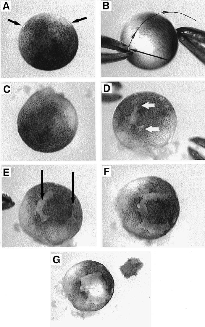
7 Animal Cap Assay 7 tor. A darting movement made during the pinching action gives a cleaner cut and prevents sticking of tissue to the forceps. Make a cut first with one pair of forceps, then at a diametrically opposite position with the other. Rotate the embryo 90 clockwise or anticlockwise and make two more cuts. The cap should lift out from the embryo with the last pinching movement. With practice, the forceps pinching method can be as neat and easy as most of the alternative dissection methods (see Note 7) and is certainly much faster. 8. It is important for induction by soluble factors to transfer animal caps to the inducer-containing medium soon (i.e., within a few minutes) after excision. As soon as caps are excised in calcium-containing medium, they begin to curl up at the edges. Eventually, they roll up into a ball that is impervious to induction by growth factors subsequently added to the medium (11). This rounding up is faster in some embryo batches than others, but typically takes place over min. The rounding up may be delayed in low-calcium medium, but this is not recommended because once a cap starts to round up, it goes to completion quite quickly regardless of the medium. 9. Incubation time depends on what is to be assayed. It is critical that sibling whole embryos are kept at the same temperature to monitor developmental stage. Caps seem to do best when incubated at 18 C, slightly cooler than room temperature. However, this is not a strong effect and the temperature should be adjusted to facilitate harvesting at the appropriate stage. 10. Harvest the explants at the appropriate stage below (see Note 8). Fig. 1. Steps in animal cap excision using the two-forceps method. (A) A stage 8.5 blastula. Note the shining highlights on the vitelline membrane (arrows). (B) The embryo is braced with the right forceps while the vitelline membrane is grabbed by the left forceps. The upper point of the left forceps has penetrated the membrane (tip of straight arrow). The right forceps are brought to grasp at the vitelline membrane just where the left forceps penetrate or meet the embryo surface. Upon grasping, the right forceps are drawn upward and to the right (curved line) in a looping motion. (C) The devitellinized blastula is rolled and shaped so that its animal pole is once again uppermost and it is nearly spherical. Note differences between this and the blastula in panel A, namely no glinting membrane and a flatter, more spread out shape. Debris has leaked from the vegetal pole and is lying around the embryo, but it does not affect the animal cap. (D) After the first pinching cut with the left forceps. White arrows mark where the forceps points first penetrated the animal hemisphere and the limits of the < -shaped cut. (E) After the second cut using the right forceps. The right incision is hard to see in this example, but note that the distance between the cuts encompasses only the middle 50% of the embryo diameter. (F) After rotating the embryo clockwise 90, a third cut (using the left forceps) produces the trapdoor appearance. (G) The pinching action of the fourth cut pulls out the animal cap, on the right. Note the relatively dark color of the inner surface of the animal cap (showing) compared to the very light, yolky blastocoel floor.
8 8 Green Stage Assay Purpose 10.5 RNA Transcription of immediate early genes RNA, immunostaining Analysis of early patterning (e.g., Hox) genes Inspection Elongation (transient for FGF, sustained for activin) 25 onward RNA, immunostaining, histology Terminal differentiation 25 onward Visual inspection Elongation or balloon formation 4. Notes 1. There is a philosophical objection to the animal cap assay, namely that because the animal cap s normal specification is to become epidermal, any change to this is somehow nonphysiological. This argument is, of course, undeniable, but it is not an objection to the animal cap assay as such. Instead, it is an important fact to be borne in mind when choosing among alternative assays and in interpreting data that the animal cap assay generates. Some of the past discoveries about the animal cap (see Subheading 1) have shown that it is not a homogenous naive tissue nor a static one. Some of its salient features are worth reiterating: a. Dorsoventral asymmetry (the dorsal half of an animal cap is much easier to induce to make, for example, dorsal mesoderm than the ventral half) b. Inside outside asymmetry (outer, pigmented cap cells are less responsive to some mesoderm inducers than inner blastocoel roof cells, whereas outer cells may be more responsive to other types of induction such as cement gland) c. Transient sensitivities (responsiveness to mesoderm inducers declines gradually during the beginning of gastrulation; responsiveness to Xwnt8 expression seems to change as early as the midblastula stage) To these should be added some other less obvious properties: d. Changing cell population (the cell movements known as epiboly mean that cells are constantly moving out of the animal cap into the marginal zone and thinning the cap itself) e. Changing extracellular matrix (by very late blastula and early gastrula stages, the cap becomes sticky to dissect, presumably because of deposition of fibronectin and other extracellular matrix components) Fortunately, it is relatively straightforward to control for these factors. Dorsoventral asymmetry can be abolished by ultraviolet-ventralising the embryos (see Chap. 14 of ref. 20). Inside outside differences can be monitored histologically or made physically separate by cell dissociation. Timing factors can, and should, be investigated by taking caps at specific stages. As cap cutting itself can be quite quick, the time resolution of such experiments is good. 2. When should the cap assay be used? Very often, overexpression of a gene in a whole embryo leads to a complex and uninterpretable effect. The animal cap assay can often provide a simpler phenotype. This is particularly true if the ques-
9 Animal Cap Assay 9 tion being asked concerns direct and immediate effects of gene expression or protein application. Furthermore, this kind of direct action assay is much easier to do in Xenopus than in almost any other model embryo species. Another type of use for the animal cap assay is as a pure assay, screen, or reporter without specific reference to normal cap physiology; for example, it can be used in tracing very low quantities of active proteins from non-xenopus species during purification procedures. This type of use has not been greatly exploited because most Xenopus scientists are interested in the biology of the cap and factors themselves. Such a use depends, of course, on the material to be tested having some activity. However, the extreme sensitivity and speed of the assay should recommend it to a wider audience for such materials. Dissociating the cells of excised animal caps has been used extensively to control or eliminate cell cell signaling and increase exposure of cells to soluble factors. For a detailed protocol, see ref. 21). Cells kept dissociated do not survive well and tend to differentiate as neural cells. Relatively transient dissociation maintains the epidermal specification of the cap while allowing other manipulations. Caps can be used in screens for cloning. cdna libraries are made in vectors that enable transcription of mrna in vitro. The libraries are divided into pools (small pools of about 100 clones appear to be optimal). The pools are transcribed and the mrna generated is injected into embryo or oocyte animal hemispheres. From embryos, the caps can be excised and simply assayed. For a paracrine screen (i.e., for secreted factors), a normal animal cap is placed hatlike onto the top of an injected oocyte. Such screens have been used successfully to identify and isolate genes of significant biological interest (22). Caps have been used to investigate the penetration of signals through tissue. One or more caps are juxtaposed with a known source of mesoderm-inducing signal. By lineage labeling either the responding cap or the signal source tissue (which can also be an injected cap) signal penetration or transmission through several cell diameters has been demonstrated (23,24). Caps have also been used to assay signals from chicken embryos. Caps wrapped in the chick s Hensen s node, for example, become neuralized. This assay has the advantage that the conjugated tissues are incubated at a little below room temperature, effectively freezing the chick s development while allowing the Xenopus tissue to develop and respond to chick signals (25). 3. Any full-strength amphibian saline (e.g., MMR, normal amphibian medium [NAM]; see ref. 20) may be used. The high salt levels in these media cause whole embryos to exogastrulate, but in animal cap explants, they encourage healing. Other media can be used to delay rounding up of the explanted cap. This can be helpful experimentally, as rounding up can be rapid and fully rounded cap explants are not responsive to subsequently applied soluble factors. To prolong the process, a onetenth dilution of MMR in calcium-magnesium-free medium (CMFM) is recommended (20). However, it is extremely difficult to stop rounding up entirely and the rate of rounding varies from egg batch to egg batch. (If more controlled cell exposure is important, then a dissociated cell protocol is recommended.) If soluble pro-
10 10 Green tein factors are to be used in the medium, bovine serum albumin (BSA, Sigma) should be added to 0.1% w/v to block nonspecific protein binding. 4. The agarose lining of dissection and incubation plates prevents sticking of explants. Depending on the number of caps to be assayed, it is essential to have sufficient numbers of dissection dishes, as they quickly become full of yolky debris during dissection. At least one 35-mm dish per 20 embryos/caps to be dissected is recommended. Depending on the number of conditions and caps to be assayed, agarose-lined dishes or multiwell plates must also be prepared for the caps after dissection. A critical factor is that explants tend to fuse with one another, which can obscure observation of morphological responses. Cap fusing has two effects. One is that, like rounding up, it excludes penetration or access of soluble factors. The other is that scoring morphological changes is much harder in fused caps than in single caps. Where neither morphology nor factors in the medium are important, cap fusion seems to have little effect on, for example, gene expression. To keep explants separate, they can be assayed as one explant per well in an agarose-lined 96-well tissue culture plate. Alternatively, two or more explants can be placed in separate depressions made in agarose-lined dishes or larger wells. Depressions are made using the sealed, red-hot end of a glass Pasteur pipet or metal fork. Alternatively, they can be cast into the agarose as follows. A mold is drilled in a block of Teflon or similar material consisting of an array of 1.8-mm-diameter 1.0-mm-deep depressions in the floor of a 4-mm-deep recess. The recess is slightly smaller that the Petri dish to be used for the embryos. A nonadhesive silicone compound, such as Dow-Corning Sylgard 184, is cast in the mould to generate a disk or square of rubber about 2 mm thick with 1-mm pimples on the underside. This is floated on the surface of molten 1% agarose and removed after the agarose has set, leaving depressions suitable for embryos and explants. 5. For straightforward morphological assays, such as elongation in response to activin, as few as two caps per condition is sufficient and gives reproducible and quantitative results. For some morphological assays, such as for FGF, several caps are required because the morphological response is weaker and more unreliable. For RNA analysis by reverse transcriptase polymerase chain reaction (RT-PCR), one or two caps per condition is minimally sufficient. However, more caps will improve RNA yield per cap and enable duplicate assays for multiple genes strongly recommended for RT-PCR. For RNA analysis by RNase protection assay (RPA), 10 caps per condition is advised. Although this seems like more work, RPA enables several genes to be quantitatively analyzed in the same tube. This provides better quantitative control than with RT-PCR. For wholemount in situ hybridization, the number of caps needed is largely a matter of taste, provided the gene expression is patently reproducible. Similarly, caps to be harvested for immunostaining or conventional histological staining should be numerous enough to allow for some losses during workup and for persuasive reproducibility to be apparent. Generally speaking, it is better to cut additional caps than to economize. With practice, it should be possible for an average worker to dissect caps per hour.
11 Animal Cap Assay A range of dissection stages is available. It is extremely difficult to dissect an animal cap before Nieuwkoop and Faber (NF) stage 6.5 because, until then, the blastocoel is very small and the animal cap consists of very few, large, fragile cells. Even at stage 7.0, results are less likely to be consistent than at stages The response to soluble mesoderm-inducing factors is constant during the window. After this time, with the onset of gastrulation (stage 10 onward), responsiveness to mesoderm inducers activin and FGF declines. Explantation is further complicated by the involution of mesoderm into the blastocoel underlying the animal cap. Animal cap that is underlain by mesoderm is respecified from epidermal to neural fate so that, although explantation is still possible, the nature of the explant and thus the assay changes. Toward stage 10, the animal cap also becomes sticky, and sticks to the forceps during dissection. Thus, the 3- to 4-h window between stages 7.5 and 9.5 (mid to late blastula) is both the most welldefined and the most convenient dissection period. If assays from caps dissected throughout this period are inconsistent, then more restricted ranges within this range should be compared. 7. There are two variations on the excision method described. One is to use different tools to make the same cuts; for example, instead of forceps, a sharpened tungsten needle can be used to make the cuts. The needle is inserted into the blastocoel and used to cut through it by pressing it up against the underside of either forceps or a second needle held at the cap surface. This method is slower than the forceps-only method and perhaps, because of this, can lead to neater cutting. However, when both methods are mastered, the difference is negligible. The second variation on the above excision method is more radical: The cap is cut from below after first inverting the embryo and then cutting open the blastocoel via vegetal hemisphere. The main merit of this approach is that the precise position of the blastocoel, cap, and marginal zone are apparent before the cap itself is excised. This prevents inclusion of any marginal zone cells in the explant. However, the method is very much slower and messier. Cap size and site of excision can be important for one main reason. Very large or off-center caps inevitably contain some marginal zone cells and can, in some circumstances, be more sensitive to induction than smaller caps. Thus, in general, it is better to err on the small side. However, caps can be too small. Very small caps are physically less robust and can fail to undergo morphological changes such as extension movements. Care is therefore required to make caps by cuts at a latitude of about 45 from the animal pole and thus about 0.5 mm across. Sizing the caps by eye (rather than, say, using a micrometer) is sufficient to get consistent results, although if this turns out to be a problem, one of the alternative excision methods might be appropriate. In any case, it is always a good idea to cut at least two caps for each condition to be assayed. The stage of excision also plays a role. The blastocoel is much larger in late blastula than early blastula and is thus easier to dissect cleanly. 8. For analysis of gene expression, it is important to know what the normal in vivo expression of a gene is before using it as part of an animal cap assay. The dynamic
12 12 Green nature of much gene expression means that the same gene in an animal cap can mean different things at different stages. If possible, more than one gene should be analyzed. Functional tests and differentiation itself must ultimately be more persuasive if the interpretation of gene expression is at all ambiguous. Expression of too few genes in animal caps is, if anything, overused and overinterpreted in the literature. 9. Animal caps can be embedded in wax and sectioned using standard procedures. The sectioning is somewhat difficult due to the small size of the samples. Thus, it is often preferable to do wholemount staining. Staining of these hard-to-handle explants is best done in small baskets. These can be made by heat sealing 70-μm nylon or polyester mesh onto the end of a cut microfuge tube or both ends of a short section of Tygon tubing. Heat sealing is done on a piece of aluminum foil covering a hotplate. Rather large baskets called Netwell inserts (Costar) can also be used, although these require larger volumes of probe and antibody solution. References 1. Kimelman, D. and Kirschner, M. (1987) Synergistic induction of mesoderm by FGF and TGF-beta and the identification of an mrna coding for FGF in the early Xenopus embryo. Cell 51, Smith, J. C., Price, B. M., Van Nimmen, K., and Huylebroeck, D. (1990) Identification of a potent Xenopus mesoderm-inducing factor as a homologue of activin A. Nature 345, Hogan, B. L., Blessing, M., Winnier, G. E., Suzuki, N., and Jones, C. M. (1994) Growth factors in development: the role of TGF-beta related polypeptide signalling molecules in embryogenesis. Development 120, Amaya, E., Musci, T. J., and Kirschner, M. W. (1991) Expression of a dominant negative mutant of the FGF receptor disrupts mesoderm formation in Xenopus embryos. Cell 66, Hemmati-Brivanlou, A. and Melton, D. A. (1992) A truncated activin receptor inhibits mesoderm induction and formation of axial structures in Xenopus embryos. Nature 359, Howard, J. E., Hirst, E. M., and Smith, J. C. (1992) Are beta 1 integrins involved in Xenopus gastrulation? Mech. Devices 38, Graff, J. M., Bansal, A., and Melton, D. A. (1996) Xenopus Mad proteins transduce distinct subsets of signals for the TGF beta superfamily. Cell 85, Massague, J. (1996) TGFbeta signaling: receptors, transducers, and Mad proteins. Cell 85, Cunliffe, V. and Smith, J. C. (1992) Ectopic mesoderm formation in Xenopus embryos caused by widespread expression of a Brachyury homologue. Nature 358, Brickman, MC and Gerhart, JC (1994) Heparitinase inhibition of mesoderm induction and gastrulation in Xenopus laevis embryos. Dev. Biol. 164, Cooke, J., Smith, J. C., Smith, E. J., and Yaqoob, M. (1987) The organization of mesodermal pattern in Xenopus laevis: experiments using a Xenopus mesoderminducing factor. Development 101,
13 Animal Cap Assay Smith, J. C., Yaqoob, M., and Symes, K. (1988) Purification, partial characterization and biological effects of the XTC mesoderm-inducing factor. Development 103, Slack, J. M., Darlington, B. G., Heath, J. K., and Godsave, S. F. (1987) Mesoderm induction in early Xenopus embryos by heparin-binding growth factors. Nature 326, Smith, J. C. (1987) A mesoderm inducing factor is produced by a Xenopus cell line. Development 99, Green, J. B. A., Howes, G., Symes, K., Cooke, J., and Smith, J. C. (1990) The biological effects of XTC-MIF: quantitative comparison with Xenopus bfgf. Development 108, Dale, L., Howes, G., Price, B. M., and Smith, J. C. (1992) Bone morphogenetic protein 4, a ventralizing factor in early Xenopus development. Development 115, Graff, J. M., Thies, R. S., Song, J. J., Celeste, A. J., and Melton, D. A. (1994) Studies with a Xenopus BMP receptor suggest that ventral mesoderm-inducing signals override dorsal signals in vivo. Cell 79, Suzuki, A., Thies, R. S., Yamaji, N., Song, J. J., Wozney, J. M., Murakami, K., and Ueno, N. (1994) A truncated bone morphogenetic protein receptor affects dorsal-ventral patterning in the early Xenopus embryo. Proc. Natl. Acad. Sci. USA 91, 10,255 10, Maeno, M., Ong, R. C., Suzuki, A., Ueno, N., and Kung, H. F. (1994) A truncated bone morphogenetic protein 4 receptor alters the fate of ventral mesoderm to dorsal mesoderm: roles of animal pole tissue in the development of ventral mesoderm. Proc. Natl. Acad. Sci. USA 91, 10,260 10, Peng, H. B. and Kay, B. K. (eds.) (1991) Xenopus laevis: practical uses in cell and molecular biology, in Methods in Cell Biology, Academic, New York. 21. Green, J. B. A., New, H. V., and Smith, J. C. (1992) Responses of embryonic Xenopus cells to activin and FGF are separated by multiple dose thresholds and correspond to distinct axes of the mesoderm. Cell 71, Lustig, K. D., Kroll, K. L., Sun, E. E., and Kirschner, M. W. (1996) Expression cloning of a Xenopus T-related gene (Xombi) involved in mesodermal patterning and blastopore lip formation. Development 122, Gurdon, J. B., Harger, P., Mitchell, A., and Lemaire, P. (1994) Activin signalling and response to a morphogen gradient. Nature 371, Gurdon, J. B., Mitchell, A., and Mahony, D. (1995) Direct and continuous assessment by cells of their position in a morphogen gradient. Nature 376, Kintner, C. R. and Dodd, J. (1991) Hensen s node induces neural tissue in Xenopus ectoderm. Implications for the action of the organizer in neural induction. Development 113,
Mesoderm Formation. Fate map of early gastrula. Only two types of mesoderm are induced. Mesoderm induction by the vegetal hemisphere
 Fate map of early gastrula Mesoderm Formation Animal hemisphere forms ectoderm (lacks ) Sperm Entry Point dbl dbl brachyury Vegetal hemisphere forms endoderm (requires ) dbl goosecoid Marginal zone forms
We are walking and standing with parts of our bodies which could have been used for thinking had they developed in another part of the embryo.
 We are walking and standing with parts of our bodies which could have been used for thinking had they developed in another part of the embryo. Hans Spemann, 1943 Reading from Chapter 3 - types of cell
+ + Development and Evolution Dorsoventral axis. Developmental Readout. Foundations. Stem cells. Organ formation.
 Development and Evolution 7.72 9.11.06 Dorsoventral axis Human issues Organ formation Stem cells Developmental Readout Axes Growth control Axon guidance 3D structure Analysis Model + + organisms Foundations
Xenopus gastrulation. Dorsal-Ventral Patterning - The Spemann Organizer. Two mesoderm inducing signals
 Dorsal- atterning - The Spemann Organizer Two mesoderm inducing signals late-blastula early-gastrula nimal Blood Mesothelium Not Vegetal NC Endoderm Hans Spemann (1869-1941) Hilde Mangold (1898-1924) Dr
Fertilization. Animal hemisphere. Sperm entry point
 Fertilization Animal hemisphere Sperm entry point Establishes the dorsal/ventral axis Ventral side sperm entry Dorsal side gray crescent Organized by sperm centriole Cleavage Unequal radial holoblastic
Neural Induction. Chapter One
 Neural Induction Chapter One Fertilization Development of the Nervous System Cleavage (Blastula, Gastrula) Neuronal Induction- Neuroblast Formation Cell Migration Mesodermal Induction Lateral Inhibition
Readings. Lecture IV. Mechanisms of Neural. Neural Development. September 10, Bio 3411 Lecture IV. Mechanisms of Neural Development
 Readings Lecture IV. Mechanisms of Neural NEUROSCIENCE: References : 5 th ed, pp 477-506 (sorta) 4 th ed, pp 545-575 (sorta) Fainsod, A., Steinbeisser, H., & De Robertis, E. M. (1994). EMBO J, 13(21),
Activity 47.1 What common events occur in the early development of animals? 1. What key events occur at each stage of development?
 Notes to Instructors Chapter 47 Animal Development What is the focus of this activity? Chapter 21 provided a review of how genes act to control development. Chapter 47 reviews some of the major morphological
Neural Induction. Steven McLoon Department of Neuroscience University of Minnesota
 Neural Induction Steven McLoon Department of Neuroscience University of Minnesota 1 Course News Coffee Hour (with Dr. Nakagawa) Friday, Sept 22 8:30-9:30am Surdyks Café in Northrop Auditorium Stop by for
Neural Development. How does a single cell make a brain??? How are different brain regions specified??? Neural Development
 Neural Development How does a single cell make a brain??? How are different brain regions specified??? 1 Neural Development How do cells become neurons? Environmental factors Positional cues Genetic factors
Blastula: An early staged embryo that is made up of sphere shaped cells that surround an inner fluid-filled cavity known as the blastocoel.
 1) Define the following terms: cleavage, blastomere, blastula, blastulation, microlecithal, mesolecithal, megalecithal, centrolecithal, isolecithal, telolecithal, holoblastic cleavage, meroblastic cleavage,
7.22 Example Problems for Exam 1 The exam will be of this format. It will consist of 2-3 sets scenarios.
 Massachusetts Institute of Technology Department of Biology 7.22, Fall 2005 - Developmental Biology Instructors: Professor Hazel Sive, Professor Martha Constantine-Paton 1 of 10 7.22 Fall 2005 sample exam
Early Development and Axis Formation in Amphibians
 Biology 4361 Early Development and Axis Formation in Amphibians October 25, 2006 Overview Cortical rotation Cleavage Gastrulation Determination the Organizer mesoderm induction Setting up the axes: dorsal/ventral
Reading. Lecture III. Nervous System Embryology. Biology. Brain Diseases. September 5, Bio 3411 Lecture III. Nervous System Embryology
 Reading NEUROSCIENCE: 5 th ed, pp. 477-506 NEUROSCIENCE: 4 th ed, pp. 545-575 Bio 3411 Wednesday 2 Summary from Lecture II Biology Understanding the brain is THE major question in biology and science.
Lecture III. Nervous System Embryology
 Bio 3411 Wednesday Reading NEUROSCIENCE: 5 th ed, pp. 477-506 NEUROSCIENCE: 4 th ed, pp. 545-575 2 1 Summary from Lecture II Biology Understanding the brain is THE major question in biology and science.
7.22 Final Exam points
 Massachusetts Institute of Technology Department of Biology 7.22, Fall 2005 - Developmental Biology Instructors: Professor Hazel Sive, Professor Martha Constantine-Paton 1 of 11 7.22 2004 FINAL FOR STUDY
Hemidesmosome. Focal adhesion
 BIOLOGY 52 - - CELL AND DEVELOPMENTAL BIOLOGY - - FALL 2002 Third Examination - - November, 2002 -------------------------------------------------- Put your name at the top of each page. Please read each
Developmental Zoology (ZOO ) Gatrulation
 Gatrulation") Developmental Zoology (ZOO 228.1.0) Gatrulation 1 Developmental Stages Ø Early Development Fertilization Cleavage Gastrulation Neurulation Ø Later Development Organogenesis Larval molts Metamorphosis Aging
Developmental Zoology (ZOO 228.1.0) Gatrulation 1 Developmental Stages Ø Early Development Fertilization Cleavage Gastrulation Neurulation Ø Later Development Organogenesis Larval molts Metamorphosis Aging
Lecture 3 MOLECULAR REGULATION OF DEVELOPMENT
 Lecture 3 E. M. De Robertis, M.D., Ph.D. August 16, 2016 MOLECULAR REGULATION OF DEVELOPMENT GROWTH FACTOR SIGNALING, HOX GENES, AND THE BODY PLAN Two questions: 1) How is dorsal-ventral (D-V) cell differentiation
In Situ Hybridization
 In Situ Hybridization Modified from C. Henry, M. Halpern and Thisse labs April 17, 2013 Table of Contents Reagents... 2 AP Buffer... 2 Developing Solution... 2 Hybridization buffer... 2 PBT... 2 PI Buffer
Note: for laboratory research use only. RNA High-purity Total RNA Rapid Extraction Kit (Spin-column) Signalway Biotechnology
 Signalway Biotechnology") Note: for laboratory research use only RNA High-purity Total RNA Rapid Extraction Kit (Spin-column) Cat. #: RP1202 (50preps) Signalway Biotechnology I. Kit Content, Storage Condition and Stability Content
Note: for laboratory research use only RNA High-purity Total RNA Rapid Extraction Kit (Spin-column) Cat. #: RP1202 (50preps) Signalway Biotechnology I. Kit Content, Storage Condition and Stability Content
A Survey of Genetic Methods
 IBS 8102 Cell, Molecular, and Developmental Biology A Survey of Genetic Methods January 24, 2008 DNA RNA Hybridization ** * radioactive probe reverse transcriptase polymerase chain reaction RT PCR DNA
RNA Blue REAGENT FOR RAPID ISOLATION OF PURE AND INTACT RNA (Cat. No. R011, R012, R013)
") RNA Blue REAGENT FOR RAPID ISOLATION OF PURE AND INTACT RNA (Cat. No. R011, R012, R013) WARNING: RNA Blue contains phenol and some other toxic components. After contact with skin, wash immediately with
RNA Blue REAGENT FOR RAPID ISOLATION OF PURE AND INTACT RNA (Cat. No. R011, R012, R013) WARNING: RNA Blue contains phenol and some other toxic components. After contact with skin, wash immediately with
Microinjection Techniques: Injecting through the chorion
 Microinjection Techniques: Injecting through the chorion I. Introduction To investigate the role of a gene during development, overexpression or misexpression of your gene of interest is a fast assay.
INDEX INDEX 0 KIT COMPONENTS 1 STORAGE AND STABILITY 1 INTRODUCTION 1 IMPORTANT NOTES 2 EUROGOLD TOTAL RNA ISOLATION PROTOCOL 2 DNA CONTAMINATION 5
 INDEX INDEX 0 KIT COMPONENTS 1 STORAGE AND STABILITY 1 INTRODUCTION 1 IMPORTANT NOTES 2 EUROGOLD TOTAL RNA ISOLATION PROTOCOL 2 DNA CONTAMINATION 5 QUANTITATION AND STORAGE OF RNA 5 RNA QUALITY 5 TROUBLESHOOTING
What s the most complex problem in biology?
 Chapter 47. Development What s the most complex problem in biology? 1 The most complex problem How to get from here to there Development: cellular level Cell division Differentiation cells become specialized
Chapter 47. Development
 Chapter 47. Development What s the most complex problem in biology? The most complex problem How to get from here to there Development: cellular level Cell division Differentiation cells become specialized
Table of Contents. 2.1 NeuroCult NCFC Assay Kit (Rat) Components Additional Required Reagents Required Equipment...
 Components Additional Required Reagents Required Equipment...") i Table of Contents 1.0 Overview of the NeuroCult NCFC Assay 2.0 Materials 2.1 NeuroCult NCFC Assay Kit (Rat) Components... 4 2.2 Additional Required Reagents... 4 2.3 Required Equipment... 4 3.0 Preparation
i Table of Contents 1.0 Overview of the NeuroCult NCFC Assay 2.0 Materials 2.1 NeuroCult NCFC Assay Kit (Rat) Components... 4 2.2 Additional Required Reagents... 4 2.3 Required Equipment... 4 3.0 Preparation
Multi-Purpose Transfection Reagents
 Product Information & Instruction Manual Multi-Purpose Transfection Reagents Cat. No: ScreenFect A S-3001 S-3001-2 S-3001-3 ScreenFect A-plus S-6001 S-6001-2 S-6001-3 www.incella.com Contents 1. Characteristics
Human Connective Tissue Growth Factor (CTGF) Elisa Kit
 Elisa Kit") Human Connective Tissue Growth Factor (CTGF) Elisa Kit Catalog No. CSB-E07875h (96 tests) This immunoassay kit allows for the in vitro quantitative determination of human CTGF concentrations in serum,
Human Connective Tissue Growth Factor (CTGF) Elisa Kit Catalog No. CSB-E07875h (96 tests) This immunoassay kit allows for the in vitro quantitative determination of human CTGF concentrations in serum,
SunScript TM One Step RT-qPCR Kit
 INDEX Ordering Information...3 Kit Contents...3 Shipping and Storage...3 Handling...3 Quality Control...3 Reagents and Equipment to be Supplied by the User...3 Description...4 Protocol...4 Troubleshooting
Isolation and analysis of Xenopus germinal vesicles. Garry T. Morgan. School of Life Sciences University of Nottingham Nottingham NG7 2UH UK
 Isolation and analysis of Xenopus germinal vesicles. Garry T. Morgan School of Life Sciences University of Nottingham Nottingham NG7 2UH UK Correspondence: Garry Morgan Tel: +44 (115) 823 0390 Email:garry.morgan@nottingham.ac.uk
Electroelution. Teachers Handbook. (Cat. # BE 602) think proteins! think G-Biosciences
 think proteins! think G-Biosciences") PR097 G-Biosciences 1-800-628-7730 1-314-991-6034 technical@gbiosciences.com A Geno Technology, Inc. (USA) brand name Electroelution Teachers Handbook (Cat. # BE 602) think proteins! think G-Biosciences
PR097 G-Biosciences 1-800-628-7730 1-314-991-6034 technical@gbiosciences.com A Geno Technology, Inc. (USA) brand name Electroelution Teachers Handbook (Cat. # BE 602) think proteins! think G-Biosciences
Molecular Cell Biology - Problem Drill 11: Recombinant DNA
 Molecular Cell Biology - Problem Drill 11: Recombinant DNA Question No. 1 of 10 1. Which of the following statements about the sources of DNA used for molecular cloning is correct? Question #1 (A) cdna
Biology 4361 Developmental Biology Lecture 4. The Genetic Core of Development
 Biology 4361 Developmental Biology Lecture 4. The Genetic Core of Development The only way to get from genotype to phenotype is through developmental processes. - Remember the analogy that the zygote contains
GSI Bovine TGF-β3 ELISA Kit-2 Plates DataSheet
 Transforming growth factor, beta 3 (TGF-β3) belongs to a large family of cytokines called the Transforming growth factor beta super family (1, 2), which includes the TGF-β family, bone morphogenetic proteins
Why Study Developmental Neurobiology? Terrific scientific challenge.
 Developmental Neurobiology Textbook Readings: ( Neuroscience, 3rd Edition, Purves, et al.) Chapter 1 Studying Nervous Systems 7 Intracellular Signal Transduction 21 Early Brain Development 22 Construction
The preparation of native chromatin from cultured human cells.
 Native chromatin immunoprecipitation protocol The preparation of native chromatin from cultured human cells. All solutions need to be ice cold. Sucrose containing solutions must be made up fresh on the
Supplementary Figure 1. Soft fibrin gels promote growth and organized mesodermal differentiation. Representative images of single OGTR1 ESCs cultured
 Supplementary Figure 1. Soft fibrin gels promote growth and organized mesodermal differentiation. Representative images of single OGTR1 ESCs cultured in 90-Pa 3D fibrin gels for 5 days in the presence
Introduction to Histology
 Introduction to Histology Histology The term "Histology" is derived from the Greek word for a tissue "Histos", and "-logos" = the study of Histology : Is the study of tissues and how they are arranged
Ribonucleic acid (RNA)
") Ribonucleic acid (RNA) Ribonucleic acid (RNA) is more often found in nature as a singlestrand folded onto itself. Cellular organisms use messenger RNA (mrna) to convey genetic information (using the nitrogenous
Ribonucleic acid (RNA) Ribonucleic acid (RNA) is more often found in nature as a singlestrand folded onto itself. Cellular organisms use messenger RNA (mrna) to convey genetic information (using the nitrogenous
Cell and Tissue Transplantation in Zebrafish Embryos
 Cell/Tissue Transplantation in Zebrafish 15 2 Cell and Tissue Transplantation in Zebrafish Embryos Toshiro Mizuno, Minori Shinya, and Hiroyuki Takeda 1. Introduction Zebrafish (Danio rerio) embryos have
Human BMP-2 ELISA Pair Set
 Human BMP-2 ELISA Pair Set Catalog Number : SEK10426 To achieve the best assay results, this manual must be read carefully before using this product and the assay is run as summarized in the General ELISA
Adult Neuron Isolation Protocol (R. Kaletsky 9/2014)
") Notes before starting: Adult Neuron Isolation Protocol (R. Kaletsky 9/2014) Begin protocol ~45min before sort time to minimize time between cell harvesting and sorting Prepare pronase solution immediately
Notes before starting: Adult Neuron Isolation Protocol (R. Kaletsky 9/2014) Begin protocol ~45min before sort time to minimize time between cell harvesting and sorting Prepare pronase solution immediately
QUANTITATIVE RT-PCR PROTOCOL (SYBR Green I) (Last Revised: April, 2007)
 (Last Revised: April, 2007)") QUANTITATIVE RT-PCR PROTOCOL (SYBR Green I) (Last Revised: April, 007) Please contact Center for Plant Genomics (CPG) facility manager Hailing Jin (hljin@iastate.edu) regarding questions or corrections.
QUANTITATIVE RT-PCR PROTOCOL (SYBR Green I) (Last Revised: April, 007) Please contact Center for Plant Genomics (CPG) facility manager Hailing Jin (hljin@iastate.edu) regarding questions or corrections.
P HENIX. PHENIX PCR Enzyme Guide Tools For Life Science Discovery RESEARCH PRODUCTS
 PHENIX PCR Enzyme Guide PHENIX offers a broad line of premium quality PCR Enzymes. This PCR Enzyme Guide will help simplify your polymerase selection process. Each DNA Polymerase has different characteristics
Total Arrest RNA. For Isolation of DNA free RNA for RT PCR. (Cat. # , ) think proteins! think G-Biosciences
 think proteins! think G-Biosciences") 445PR 01 G-Biosciences 1-800-628-7730 1-314-991-6034 technical@gbiosciences. com A Geno Technology, Inc. (USA) brand name Total Arrest RNA For Isolation of DNA free RNA for RT PCR (Cat. #786 130, 786 131)
445PR 01 G-Biosciences 1-800-628-7730 1-314-991-6034 technical@gbiosciences. com A Geno Technology, Inc. (USA) brand name Total Arrest RNA For Isolation of DNA free RNA for RT PCR (Cat. #786 130, 786 131)
PowerSoil DNA Isolation Kit
 PowerSoil DNA Isolation Kit Catalog No. Quantity 12888-50 50 Preps 12888-100 100 Preps Instruction Manual Introduction The PowerSoil DNA Isolation Kit* is comprised of a novel and proprietary method for
PrimeScript RT Master Mix (Perfect Real Time)
") Cat. # RR036A For Research Use PrimeScript RT Master Mix (Perfect Real Time) Product Manual Table of Contents I. Description... 3 II. Kit Components... 3 III. Materials Required but not Provided... 3 IV.
Cat. # RR036A For Research Use PrimeScript RT Master Mix (Perfect Real Time) Product Manual Table of Contents I. Description... 3 II. Kit Components... 3 III. Materials Required but not Provided... 3 IV.
Characterizing Phenotypes of Bacteria by Staining Method
 Experiment 3 Laboratory to Biology III Diversity of Microorganisms / Wintersemester / page 1 Experiment 3 Characterizing Phenotypes of Bacteria by Staining Method Advisor NN Reading Chapters in BBOM 9
HiPer RT-PCR Teaching Kit
 HiPer RT-PCR Teaching Kit Product Code: HTBM024 Number of experiments that can be performed: 5 Duration of Experiment: Protocol: 4 hours Agarose Gel Electrophoresis: 45 minutes Storage Instructions: The
Page 1 of 5. Product Name Label Quantity Product No. Cy 3 10 µg (~0.75 nmol) MIR Cy µg (~7.5 nmol) MIR 7901
 MIR Cy µg (~7.5 nmol) MIR 7901") Page 1 of 5 Label IT RNAi Delivery Control Product Name Label Quantity Product No. Cy 3 10 µg (~0.75 nmol) MIR 7900 Label IT RNAi Delivery Control Cy 3 100 µg (~7.5 nmol) MIR 7901 Fluorescein 10 µg (~0.75
Page 1 of 5 Label IT RNAi Delivery Control Product Name Label Quantity Product No. Cy 3 10 µg (~0.75 nmol) MIR 7900 Label IT RNAi Delivery Control Cy 3 100 µg (~7.5 nmol) MIR 7901 Fluorescein 10 µg (~0.75
ChIP protocol Chromatin fragmentation using the Covaris S2 sonicator by Ethan Ford (version 12/1/11) X- link Cells
 X- link Cells") ChIP protocol Chromatin fragmentation using the Covaris S2 sonicator by Ethan Ford (version 12/1/11) X- link Cells 1. Grow six 15 cm plates of HeLa cells to 90% confluency. 2. Remove media and add wash
ChIP protocol Chromatin fragmentation using the Covaris S2 sonicator by Ethan Ford (version 12/1/11) X- link Cells 1. Grow six 15 cm plates of HeLa cells to 90% confluency. 2. Remove media and add wash
Note that steps 1-16 are identical to steps 1-16 in the immunostaining protocol, except 0.5X PBtween is used in this protocol rather than 0.5X PBT.
 Embryo In-situ Hybridization: This protocol is based closely on an immunostaining protocol for Hypsibius dujardini (Gabriel and Goldstein 2007) and available in-situ protocols for both insects (Tomoysu
Characterizing Phenotypes of Bacteria by Staining Method
 Experiment 3 Laboratory to Biology III Diversity of Microorganisms / Wintersemester / page 1 Experiment Characterizing Phenotypes of Bacteria by Staining Method Advisor Reading NN Chapters 3.1, 3.7, 3.8,
Functional Genomics Research Stream. Research Meeting: November 8, 2011 cdna Library Construction for RNA-Seq
 Functional Genomics Research Stream Research Meeting: November 8, 2011 cdna Library Construction for RNA-Seq Lab Issues Don t leave out boxes of water + ethidium bromide Minimize tip boxes in phenol waste
Recombinant DNA Technology. The Role of Recombinant DNA Technology in Biotechnology. yeast. Biotechnology. Recombinant DNA technology.
 PowerPoint Lecture Presentations prepared by Mindy Miller-Kittrell, North Carolina State University C H A P T E R 8 Recombinant DNA Technology The Role of Recombinant DNA Technology in Biotechnology Biotechnology?
Xenopus axis formation: induction of goosecoid by injected Xwnt-8 and activin mrnas
 Development 118, 499-507 (1993) Printed in Great Britain The Company of Biologists Limited 1993 499 Xenopus axis formation: induction of goosecoid by injected Xwnt-8 and activin mrnas Herbert Steinbeisser
RNA extraction from tissue - using Bioruptor (Standard/Plus) and RNA extraction kit
 and RNA extraction kit") RNA extraction from tissue - using Bioruptor (Standard/Plus) and RNA extraction kit Introduction Isolation of intact RNA is essential for many techniques used in gene expression analysis. Efficient disruption
RNA extraction from tissue - using Bioruptor (Standard/Plus) and RNA extraction kit Introduction Isolation of intact RNA is essential for many techniques used in gene expression analysis. Efficient disruption
Lab 4: Microbe Detectives, Part 1
 NAME: Lab 4: Microbe Detectives, Part 1 OBJECTIVES To review the structure and function of DNA To understand and perform the polymerase chain reaction (PCR) To gain experience using the micropipettes,
Immunofluorescence Confocal Microscopy of 3D Cultures Grown on Alvetex
 Immunofluorescence Confocal Microscopy of 3D Cultures Grown on Alvetex 1.0. Introduction Immunofluorescence uses the recognition of cellular targets by fluorescent dyes or antigen-specific antibodies coupled
Roche Molecular Biochemicals Technical Note No. LC 10/2000
 Roche Molecular Biochemicals Technical Note No. LC 10/2000 LightCycler Overview of LightCycler Quantification Methods 1. General Introduction Introduction Content Definitions This Technical Note will introduce
2014 Pearson Education, Inc. CH 8: Recombinant DNA Technology
 CH 8: Recombinant DNA Technology Biotechnology the use of microorganisms to make practical products Recombinant DNA = DNA from 2 different sources What is Recombinant DNA Technology? modifying genomes
TransIT -mrna Transfection Kit
 INTRODUCTION TransIT -mrna Transfection Kit is designed to transfect RNA into a broad range of cell types with minimal cellular toxicity. RNA delivery avoids transcriptional regulation effects by directly
Supplementary Data. Supplementary Methods Three-step protocol for spontaneous differentiation of mouse induced pluripotent stem (embryonic stem) cells
 cells") Supplementary Data Supplementary Methods Three-step protocol for spontaneous differentiation of mouse induced pluripotent stem (embryonic stem) cells Mouse induced pluripotent stem cells (ipscs) were cultured
Supplementary Data Supplementary Methods Three-step protocol for spontaneous differentiation of mouse induced pluripotent stem (embryonic stem) cells Mouse induced pluripotent stem cells (ipscs) were cultured
E.Z.N.A. Stool DNA Kit. D preps D preps D preps
 E.Z.N.A. Stool DNA Kit D4015-00 5 preps D4015-01 50 preps D4015-02 200 preps April 2013 E.Z.N.A. Stool DNA Kit Table of Contents Introduction and Overview...2 Illustrated Protocol...3 Kit Contents/Storage
TransIT-TKO Transfection Reagent
 Quick Reference Protocol, MSDS and Certificate of Analysis available at mirusbio.com/2150 INTRODUCTION TransIT-TKO is a broad spectrum sirna transfection reagent that enables high efficiency sirna delivery
High Pure RNA Isolation Kit for isolation of total RNA from 50 samples Cat. No
 for isolation of total RNA from 50 samples Cat. No. 1 88 665 Principle A single reagent lyses the sample lysis and inactivates RNase. In the presence of a chaotropic salt (guanidine HCl), the released
SpeAmp n cdna Pre-amplification Kits User Guide
 SpeAmp n cdna Pre-amplification Kits User Guide Cat # SpeAmp n -10 Cat # SpeAmp n -25 Cat # SpeAmp n -50 To use with our range of pathway-specific primer pools : Cat # Prim nx -10 XXXX Cat # Prim nx -25
LIFE Formation III. Morphogenesis: building 3D structures START FUTURE. How-to 2 PROBLEMS FORMATION SYSTEMS SYSTEMS.
 7.013 4.6.07 Formation III Morphogenesis: building 3D structures VIRUSES CANCER HUMAN DISEASE START SYSTEMS BIOLOGY PROBLEMS FUTURE LIFE IMMUNE NERVOUS SYSTEMS How-to 1 FOUNDATIONS How-to 2 REC. DNA BIOCHEM
Component. Buffer RL
 GenElute FFPE RNA Purification Catalog number RNB400 Product Description Sigma s GenElute FFPE RNA Purification Kit provides a rapid method for the isolation and purification of total RNA (including microrna)
Figure 1. Map of cloning vector pgem T-Easy (bacterial plasmid DNA)
") Texas A&M University-Corpus Christi CHEM4402 Biochemistry II Laboratory Laboratory 6: Ligation & Bacterial Transformation (Bring your text and laptop to class if you wish to work on your assignment during
Texas A&M University-Corpus Christi CHEM4402 Biochemistry II Laboratory Laboratory 6: Ligation & Bacterial Transformation (Bring your text and laptop to class if you wish to work on your assignment during
CH 8: Recombinant DNA Technology
 CH 8: Recombinant DNA Technology Biotechnology the use of microorganisms to make practical products Recombinant DNA = DNA from 2 different sources What is Recombinant DNA Technology? modifying genomes
Activin-mediated mesoderm induction requires FGF
 Development 120, 453-462 (1994) Printed in Great Britain The Company of Biologists Limited 1994 453 Activin-mediated mesoderm induction requires FGF Robert A. Cornell and David Kimelman* Department of
B. Incorrect! Ligation is also a necessary step for cloning.
 Genetics - Problem Drill 15: The Techniques in Molecular Genetics No. 1 of 10 1. Which of the following is not part of the normal process of cloning recombinant DNA in bacteria? (A) Restriction endonuclease
Roche Molecular Biochemicals Technical Note No. LC 12/2000
 Roche Molecular Biochemicals Technical Note No. LC 12/2000 LightCycler Absolute Quantification with External Standards and an Internal Control 1. General Introduction Purpose of this Note Overview of Method
Cell-Environment Interactions. Chieh-Chun Chen
 Cell-Environment Interactions Chieh-Chun Chen Part 1: Soft Lithography in Biology and Biochemistry Chieh-Chun Chen Outlines Introduction Key features of soft lithography Applications In microscopic biochemical
TransIT-TKO Transfection Reagent
 INTRODUCTION TransIT-TKO is a broad spectrum sirna transfection reagent that enables high efficiency sirna delivery and knockdown of target gene expression in many cell types including primary cells. TransIT-TKO
Selected Techniques Part I
 1 Selected Techniques Part I Gel Electrophoresis Can be both qualitative and quantitative Qualitative About what size is the fragment? How many fragments are present? Is there in insert or not? Quantitative
Patterning of the Brain
 Patterning of the Brain Clemens.Kiecker@kcl.ac.uk IoPPN 27 Oct 2015 Hundreds of billions of cells, hundreds of cell types Egg = single cell, some 20,000 genes Egg = single cell, some 20,000 genes How is
Gene Expression Technology
 Gene Expression Technology Bing Zhang Department of Biomedical Informatics Vanderbilt University bing.zhang@vanderbilt.edu Gene expression Gene expression is the process by which information from a gene
Cat # Box1 Box2. DH5a Competent E. coli cells CCK-20 (20 rxns) 40 µl 40 µl 50 µl x 20 tubes. Choo-Choo Cloning TM Enzyme Mix
 40 µl 40 µl 50 µl x 20 tubes. Choo-Choo Cloning TM Enzyme Mix") Molecular Cloning Laboratories User Manual Version 3.3 Product name: Choo-Choo Cloning Kits Cat #: CCK-10, CCK-20, CCK-096, CCK-384 Description: Choo-Choo Cloning is a highly efficient directional PCR
Molecular Cloning Laboratories User Manual Version 3.3 Product name: Choo-Choo Cloning Kits Cat #: CCK-10, CCK-20, CCK-096, CCK-384 Description: Choo-Choo Cloning is a highly efficient directional PCR
ADVANCED ELECTROPHORESIS
 Ref. ELECAVANZADA (4 practices) 1. EXPERIMENT OBJETIVE ADVANCED ELECTROPHORESIS The aim of this experiment is to introduce students to the knowledge of electrophoretic theory and to familiarize themselves
E.Z.N.A. Stool DNA Kit. D preps D preps D preps
 E.Z.N.A. Stool DNA Kit D4015-00 5 preps D4015-01 50 preps D4015-02 200 preps July 2017 E.Z.N.A. Stool DNA Kit Table of Contents Introduction and Overview...2 Illustrated Protocol...3 Kit Contents/Storage
John Gurdon was testing the hypothesis of genomic equivalence or that when cells divide they retain a full genomic compliment.
 1. (15 pts) John Gurdon won the 2012 Nobel Prize in Physiology or Medicine for work he did in the 1960 s. What was the major developmental hypothesis he set out to test? What techniques did he development
Total Nucleic Acids Extraction from Soil
 Total Nucleic Acids Extraction from Soil The following protocol is intended for the simultaneous extraction of DNA and RNA from various soil samples. The TNS extraction buffer is based on the one used
Simple, Complete Workflows for Gene Expression Analysis without RNA Purification
 Product Bulletin SYBR Green Cells-to-Ct Kits SYBR Green Cells-to-Ct Kits Simple, Complete Workflows for Gene Expression Analysis without RNA Purification 7 10 min total 5x 5' 5' 3' 3' 3' mrna 5' RT primer
The homeobox gene Siamois is a target of the Wnt dorsalisation pathway and triggers organiser activity in the absence of mesoderm
 Development 122, 3055-3065 (1996) Printed in Great Britain The Company of Biologists Limited 1996 DEV1088 3055 The homeobox gene Siamois is a target of the Wnt dorsalisation pathway and triggers organiser
SYBR Premix Ex Taq II (Tli RNaseH Plus), ROX plus
, ROX plus") Cat. # RR82LR For Research Use SYBR Premix Ex Taq II (Tli RNaseH Plus), ROX plus Product Manual The long-term storage temperature of this product has been changed to -20 since Lot. A1901A. See section
Cat. # RR82LR For Research Use SYBR Premix Ex Taq II (Tli RNaseH Plus), ROX plus Product Manual The long-term storage temperature of this product has been changed to -20 since Lot. A1901A. See section
Lab 2C: Basic Techniques. Dilute 10X TE Buffer to Make 1X TE Buffer Determine the Concentration of an Unknown DNA Sample Streak out bacteria colonies
 Demonstration of sterile technique. Lab 2 Basic Techniques Lab 2A: Lab 2B: Lab 2C: Dilute 10X TE Buffer to Make 1X TE Buffer Determine the Concentration of an Unknown DNA Sample Streak out bacteria colonies
RayBio Genomic DNA Magnetic Beads Kit
 RayBio Genomic DNA Magnetic Beads Kit Catalog #: 801-112 User Manual Last revised January 4 th, 2017 Caution: Extraordinarily useful information enclosed ISO 13485 Certified 3607 Parkway Lane, Suite 100
Amplifying the ALU intron for Hardy- Weinberg Analysis Part 1
 Bio 212 Lab Name: Amplifying the ALU intron for Hardy- Weinberg Analysis Part 1 OBJECTIVES: Review the following terms and concepts presented in Biology 211: enzymes, DNA structure and replication, role
mrnadembeads Purification Mini Kit (cat #06011) Instruction manual for mrna purification
 Instruction manual for mrna purification") mrnadembeads Purification Mini Kit (cat #06011) Instruction manual for mrna purification Ademtech * Bioparc BioGalien 27 Allée Charles Darwin * 33600 Pessac France Tel : + 33 (0) 57 02 02 01, Fax : + 33
mrnadembeads Purification Mini Kit (cat #06011) Instruction manual for mrna purification Ademtech * Bioparc BioGalien 27 Allée Charles Darwin * 33600 Pessac France Tel : + 33 (0) 57 02 02 01, Fax : + 33
Table of Contents. I. Kit Components...2. Storage...2. Principle...2. IV. Precautions for operation...3. V. Protocol : reverse transcription...
 Table of Contents I. Kit Components...2 II. III. Storage...2 Principle...2 IV. Precautions for operation...3 V. Protocol : reverse transcription...3 VI. Protocol : Real-time PCR...5 VII. Appendix...7 VIII.
CHAPTER 20 DNA TECHNOLOGY AND GENOMICS. Section A: DNA Cloning
 Section A: DNA Cloning 1. DNA technology makes it possible to clone genes for basic research and commercial applications: an overview 2. Restriction enzymes are used to make recombinant DNA 3. Genes can
TransIT -mrna Transfection Kit
 Quick Reference Protocol, MSDS and Certificate of Analysis available at mirusbio.com/2225 INTRODUCTION TransIT -mrna Transfection Kit is designed to transfect RNA into a broad range of cell types with
Product Name : Simple mirna Detection Kit
 Product Name : Simple mirna Detection Kit Code No. : DS700 This product is for research use only Kit Contents This kit provides sufficient reagents to perform 20 reactions for detecting microrna. Components
Single cells can sense their position in a morphogen gradient
 Development 6, 509-57 (999) Printed in Great Britain The Company of Biologists Limited 999 DEV6 509 Single cells can sense their position in a morphogen gradient J. B. Gurdon*, H. Standley, S. Dyson, K.
Lesson 2 Inoculation Growing a Cell Culture
 Lesson 2 Inoculation Growing a Cell Culture Isolation of Single Bacterial Colonies In this activity, students will pick one white colony from their LB/amp plates and one green colony from their LB/amp/ara