MICRO-CT ANALYSIS. Related instrument: Leica EM CPD300 MATERIAL RESEARCH LIFE SCIENCE RESEARCH INDUSTRIAL MEDICAL MANU RESEARCH FACTURING
|
|
|
- Matthew Higgins
- 6 years ago
- Views:
Transcription


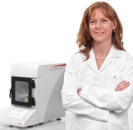
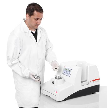


1 MATERIAL RESEARCH LIFE SCIENCE RESEARCH Application Booklet MICRO-CT ANALYSIS Related instrument: Leica EM CPD300 INDUSTRIAL MEDICAL MANU RESEARCH FACTURING NATURAL RESOURCES
2
3 2. 3 X-RAY MICRO-COMPUTED TOMOGRAPHY (MICRO-CT) IS A ROUTINELY APPLIED NON- INVASIVE TECHNIQUE FOR THE INVESTIGA TION OF THE INTERNAL ANATOMY AND MOR PHOLOGY OF ORGANISMS. AS A RESULT OF A MICRO-CT SCAN A STACK OF GREY-SCALE IMAGES IS GENERATED FROM A SERIES OF PROJECTIONS TAKEN AT DEFINED ANGLES DURING SAMPLE ROTATION. SINCE SEVERAL YEARS THE NUMBER OF LAB- BASED MICRO-CT IMAGING SYSTEMS IS CONSTANTLY GROWING MAKING THIS TECH NIQUE AVAILABLE TO A BROAD SPECTRUM OF RESEARCHERS AND APPLICATIONS. CRITICAL-POINT DRYING FOR THE PREPARATION OF BIOLOGICAL SAMPLES FOR MICRO-CT ANALYSIS Peter Michalik and Elisabeth Lipke Zoological Institute and Museum, Ernst-Moritz-Arndt University Greifswald, Germany Similar to other imaging techniques such as scanning electron microscopy, micro-ct allows to study biological samples in nearly every condition (e.g. fresh, dried or within preservatives). Micro-CT is the ideal technique for studying bones, teeth and shells in 3D, but the analysis of soft tissue is significantly influenced and hindered by its low absorption contrast based on the presence of compounds with low-atomic number elements. In order to overcome this limitation, several approaches can be applied including different staining and/or drying techniques as well as phase-related contrast imaging. However, whenever possible samples should be analyzed in dry condition as it provides a significantly higher signal to noise ratio compared to samples scanned in liquid. In order to dry delicate biological samples, critical point drying was proofed to be the best method compared to e.g., chemical or air drying as it preserves the structures while minimizing artifacts such as shrinkage of tissue and distortion.
4 MICRO-COMPUTER TOMOGRAPHY PROTOCOLS 1. Micro-CT of Book Scorpion Musculature Introduction Fixation and dehydration Species: Book scorpion (Neobisium sp.) Critical point drying of book scorpion with subsequent X-ray micro-computed tomography (micro-ct) to detect anatomical features with special regard to the musculature. > > Ethanol (70%): overnight > > Ethanol series (80 %, 90 %, 96 %, 100 %): 2 10 min. > > Iodine staining (1 % iodine solution in 100 % ethanol): overnight > > Ethanol (100 %): 2 10 min. Procedure Mounting and scanning Sample holder: Sample was transferred to microporous specimen pot and placed into chamber of filter discs and porous pots holder. Dried sample was glued on an insect pin and scanned with an Xradia MicroXCT-200 X-ray imaging system (Carl Zeiss X-ray Microscopy Inc., Pleasanton, USA). CPD300 auto program
. Courtesy of Elisabeth Lipke and Dr.")
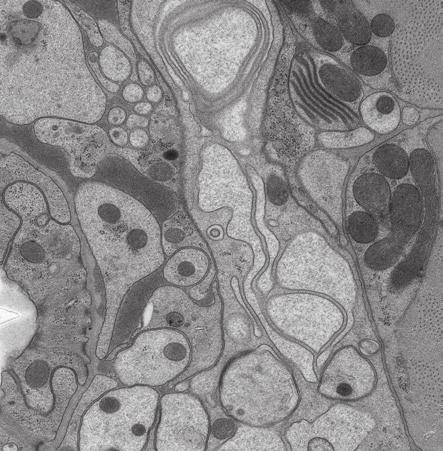
5 4. 5 Results Volume reconstruction of a book scorpion prosoma (outer view and inner view to visualize the musculature). Courtesy of Elisabeth Lipke and Dr. Peter Michalik, University of Greifswald, Germany.
6 MICRO-COMPUTER TOMOGRAPHY PROTOCOLS 2. Micro-CT of Insect Brain Protocol Introduction Fixation and dehydration Species: Blow fly (Lucilia caesar) Critical point drying of the blow fly with subsequent X-ray micro-computed tomography (micro-ct) to detect neuroanatomical features. Procedure > > Bouin s fixative: overnight > > 0.1 M phosphate buffer (1.8 % Sucrose, ph 7.2): 3 10 min. > > Ethanol series (60 %, 70 %, 80 %, 90 %, 96 %, 100 %): 2 10 min. > > Iodine staining (1 % iodine solution in 100 % ethanol): overnight > > Ethanol (100 %): 2 x 10 min. Sample holder: Samples were placed individually in the chambers of Arthropoda holder. Mounting and scanning Dried samples were glued on insect pins and scanned with an Xradia MicroXCT-200 X-ray imaging system (Carl Zeiss X-ray Microscopy Inc., Pleasanton, USA). CPD300 auto program

7 Results Volume reconstructions and virtual sections of the head and brain of a blow fly. Courtesy of Elisabeth Lipke and Dr. Peter Michalik, University of Greifswald, Germany.
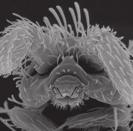
8 MICRO-COMPUTER TOMOGRAPHY PROTOCOLS 3. Micro-CT of Insect Larva Protocol Introduction Fixation and dehydration Species: Red blood worm (midge larva) Critical point drying of midge larvae with subsequent X-ray micro-computed tomography (micro-ct) to reconstruct the inner anatomy. Procedure > > 2.5 % Glutardialdehyde (in 0.1 M phosphate buffer): overnight > > 0.1 M phosphate buffer (1.8 % Sucrose, ph 7.2): 3 10 min. > > Ethanol series (60 %, 70 %, 80 %, 90 %, 96 %, 100 %): 2 10 min. > > Iodine staining (1 % iodine solution in 100 % ethanol): overnight > > Ethanol (100 %): 2 10 min. Sample holder: Sample was placed individually in the chambers of Arthropoda holder. Mounting and scanning Dried sample was glued on an insect pin and scanned with an Xradia MicroXCT-200 X-ray imaging system (Carl Zeiss X-ray Microscopy Inc., Pleasanton, USA). CPD300 auto program

9 8. 9 Results Volume reconstructions and virtual section of the red blood worm showing a variety of organ systems. Courtesy of Elisabeth Lipke and Dr. Peter Michalik, University of Greifswald, Germany.
10 Leica EM ACE200/EM ACE600 Brochure Engilsh 05/2015 Copyright by Leica Mikrosysteme GmbH, Vienna, Austria, Subject to modifications. LEICA and the Leica Logo are registered trademarks of Leica Microsystems IR GmbH. Leica Mikrosysteme GmbH Vienna Austria T F CONNECT WITH US!
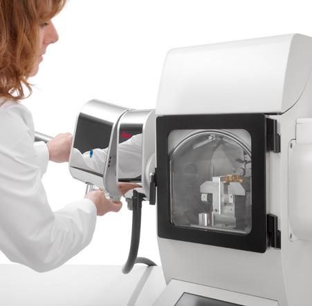
Application Booklet. Leica EM CPD300 Automated Critical Point Dryer
 Application Booklet Leica EM CPD300 Automated Critical Point Dryer Foreword This Application Booklet is intended to provide standard protocols to facilitate the optimizing process of critical point drying
From Eye to Insight. Leica EM ACE. Coater Family
 From Eye to Insight Leica EM ACE Coater Family EM ACE COATERS A Coater Family to cover all your needs. Developed in cooperation with leading scientists, the EM ACE coaters cover all the requirements for
Application Booklet. Leica EM CPD300 Automated Critical Point Dryer
 Application Booklet Leica EM CPD300 Automated Critical Point Dryer Foreword This Application Booklet is intended to provide standard protocols to facilitate the optimizing process of critical point drying
Application Note. Thin metal foils with coatings. Sample Preparation for SEM. related instrument Leica EM RES102.
 Material Life Science Application Note Thin metal foils with coatings Sample Preparation for SEM related instrument Leica EM RES102 Medical Industrial Manufacturing Natural Resources 2 Ion beam slope cutting
Application Note. Sample Protection prior to FIB Processing. related instruments: Leica EM ACE600. Material Research. Life Science Research
 Material Life Science Application Note Sample Protection prior to FIB Processing related instruments: Leica EM ACE600 Medical Industrial Manufacturing Natural Resources 2 Each Atom Really Counts: Protect
GENESIS Edition. Transforming CT
 GENESIS Edition Transforming CT Transforming clinical confidence Transforming patient experience Transforming your workspace GENESIS Edition Transforming CT Brought to you by the leaders in area detector
ThermoBrite Elite. Feasibility/Time Savings Study. Blood/Bone Marrow Protocol
 ThermoBrite Elite Feasibility/Time Savings Study Blood/Bone Marrow Protocol For this study, WaveSense, Inc. provided twelve U266 myeloma cell line slides prepared with their patented EpiSep cell enrichment
PREPARATION OF HISTOLOGICAL SPECIMENS
 PREPARATION OF HISTOLOGICAL SPECIMENS Histo-techniques Preparation of tissue for microscopic examination Series of processes Ultimate aim to make tissue visible as it is Pathology Vs Anatomy Steps vary
Chapter 2 Methods of Collecting and Studying Microinsects
 Chapter 2 Methods of Collecting and Studying Microinsects 2.1 Introduction Microinsects are one of the least thoroughly studied groups of insects and quite objective reasons are largely associated with
GENESIS Edition Transforming CT
 GENESIS Edition Transforming CT 2 Transforming clinical confidence Transforming patient experience Transforming your workspace GENESIS Edition Transforming CT Brought to you by the leaders in area detector
PROTOCOL. Introduction. Method. Page 1
 Page 1 Introduction Following fixation a variety of different cytological stains can be used to visualise cell components in detail. Here we describe the use of Haematoxylin and Eosin, a general structural
Diffraction Contrast Tomography. Unlocking Crystallographic Information from Laboratory X-ray Microscopy. Technical Note
 Diffraction Contrast Tomography Unlocking Crystallographic Information from Laboratory X-ray Microscopy Technical Note Diffraction Contrast Tomography Unlocking Crystallographic Information from Laboratory
PLASTINATION OF OLD FORMALIN-FIXED SPECIMENS
 1 J Int Soc Plastination, Vol 5:11-15, 1991 PLASTINATION OF OLD FORMALIN-FIXED SPECIMENS Mario Cannas and Paolo Fuda University of Torino, Department of Human Anatomy and Physiology, C.so M. D' Azeglio
Damage and damage accumulation in fiber reinforced composites by X-ray CT
 Damage and damage accumulation in fiber reinforced composites by X-ray CT Phil Withers Regius Professor of Materials Science Henry Moseley X-ray Imaging Facility, BP Int. Centre for Advanced Materials,
ab EXPOSE Rabbit Specific HRP/DAB Detection IHC Kit
 Version 3 Last updated 3 November 2017 ab80437 - EXPOSE Rabbit Specific HRP/DAB Detection IHC Kit For the detection of a specific antibody bound to an antigen in tissue sections. This product is for research
ab Mouse and Rabbit Specific HRP/AEC IHC Detection Kit - Micropolymer
 Version 4 Last updated 21 June 2018 ab236467 Mouse and Rabbit Specific HRP/AEC IHC Detection Kit - Micropolymer For the detection of a specific antibody bound to an antigen in tissue sections. This product
Materials and Methods Materials Required for Fixing, Embedding and Sectioning
 Page 1 Introduction Immunofluorescence uses the recognition of cellular targets by fluorescent dyes or antigenspecific antibodies coupled to fluorophores. Depending on the antibody or dye used, proteins,
ab EXPOSE Mouse and Rabbit Specific HRP/DAB Detection IHC Kit
 Version 7 Last updated 17 January 2018 ab80436 - EXPOSE Mouse and Rabbit Specific HRP/DAB Detection IHC Kit For the detection of a specific antibody bound to an antigen in tissue sections. This product
Materials and Methods Materials Required for Fixing, Embedding and Sectioning. OCT embedding matrix (Thermo Scientific, LAMB/OCT)
") Page 1 Introduction Tissue freezing and sectioning is a rapid method of generating tissue samples (cryosections) for histological analysis, and obviates the need for wax embedding. The method is popular
Page 1 Introduction Tissue freezing and sectioning is a rapid method of generating tissue samples (cryosections) for histological analysis, and obviates the need for wax embedding. The method is popular
ab64254 Liquid Fast-Red Substrate Kit (75X)
") Version 1 Last updated 6 June 2018 ab64254 Liquid Fast-Red Substrate Kit (75X) For the immunohistochemical staining. This product is for research use only and is not intended for diagnostic use. Table
Version 1 Last updated 6 June 2018 ab64254 Liquid Fast-Red Substrate Kit (75X) For the immunohistochemical staining. This product is for research use only and is not intended for diagnostic use. Table
Evolution of the microstructure of frozen foods during storage: Application on plant tissue
 Evolution of the microstructure of frozen foods during storage: Application on plant tissue Victor Vicent Graciela Alvarez Fatou Toutie Ndoye Prof. Bart Nicolaï Pieter Verboven Outline Introduction Objectives
Stellaris RNA FISH Protocol for D. Melanogaster Wing Imaginal Discs
 Stellaris RNA FISH Protocol for D. Melanogaster Wing Imaginal Discs General Protocol & Storage Product Description A set of Stellaris RNA FISH Probes is comprised of up to 48 singly labeled oligonucleotides
Quantifying the tissue shrinkage caused by sample preparation for micro-ct and LSFM
 Quantifying the tissue shrinkage caused by sample preparation for micro-ct and LSFM J. Goyens 1,2, J. Buytaert 1, D. De Greef 1, P. Aerts 2,3 and J. Dirckx 2 1 University of Antwerp, Laboratory of BioMedical
Intracranial stent visualization for image guided interventions and therapy
 Intracranial stent visualization for image guided interventions and therapy Daniel Ruijters, Peter van de Haar, Ruben Roijers, Niels J. Noordhoek, Jan Timmer, and Drazenko Babic interventional X-Ray, Philips
Trusted Performance. Smart Investment. 80 detector row Ultra Helical CT
 TM Trusted Performance. Smart Investment. 80 detector row Ultra Helical CT 2 High performance, highly economical Increased productivity and patient safety Maximum clinical capabilities Are you looking
> 3d imaging of ceramic CMC for numerical modelling - 91st annual meeting of the DKG - 8. March 2016
 www.dlr.de Chart 1 > 3d imaging of ceramic CMC for numerical modelling - 91st annual meeting of the DKG - 8. March 2016 3-dimensional microstructure characterization of porous ceramic matrix composites
Abstract. 1.0 Background
 NON DESTRUCTIVE CHARACTERIZATION, INSPECTION, FAILURE ANALYIS OF ADVANCED COMPONENTS AND SENSORS WITH A HIGH RESOLUTION & HIGH CONTRAST MICROTOMOGRAPHY (microct) SYSTEM S H Lau*, Hauyee Chang, Joanna Cheong,
INNOVATING INFRARED SPECTROSCOPY PRODUCT BROCHURE ATR CRYSTALS
 INNOVATING INFRARED SPECTROSCOPY PRODUCT BROCHURE ATR CRYSTALS November 2018 IRUBIS ATR Crystal Single Reflection ATR Crystal SEIRAS Optimized ATR Crystal Signal Enhanced ATR Crystal Planned release: 2019
EMS MICROSCOPY ACADEMY BIOLOGICAL TEM WORKSHOP: A COMPLETE PICTURE
 Examples of the endless possibilities in the field of Microscopy Bone Marrow: Transmission electron microscope image of a thin section cut through an area of bone marrow area near the cartilage/bone interface
PATIENT PATHOLOGIST CEREBRO SPECIMEN TRACKING AND WORKFLOW MANAGEMENT. Patient Safety. Productivity. Flexibility.
 HISTOLOGY PATIENT GROSSING CENTRIFUGING PROCESSING EMBEDDING PATHOLOGIST SMEARING SECTIONING REPORTING STAINING SEND-OUT CEREBRO SPECIMEN TRACKING AND WORKFLOW MANAGEMENT Patient Safety. Productivity.
SOP# version e1.0 Material Handling and Documentation Preservation of Tissue: Paraffin Embedding
 CTRNet Standard Operating Procedure SOP Number: 8.3.005 Version e1.0 Supersedes: SR 001.001 Effective Date 09 Jan 08 Subject: Preservation of Tissue: Paraffin Embedding Category Material Handling and Documentation
PATIENT PATHOLOGIST CEREBRO SPECIMEN TRACKING AND WORKFLOW MANAGEMENT. Patient Safety. Productivity. Flexibility.
 HISTOLOGY PATIENT ACCESSIONING CYTOLOGY ACCESSIONING GROSSING CENTRIFUGING PROCESSING EMBEDDING PATHOLOGIST SMEARING SECTIONING REPORTING STAINING SEND-OUT CEREBRO SPECIMEN TRACKING AND WORKFLOW MANAGEMENT
HISTOPATHOLOGY INTRODUCTION
 HISTOPATHOLOGY INTRODUCTION Surgical, anatomical and consultative pathology services are available through pathologists in the Department of Pathology. The services available include: Routine surgical
ab DoubleStain IHC Kit: R&Rt on Human/Mouse Tissue (Green/HRP & AP/Red)
") ab183285 DoubleStain IHC Kit: R&Rt on Human/Mouse Tissue (Green/HRP & AP/Red) Instructions for Use For the detection of Rat and Rabbit Primary antibodies on Human/Mouse Tissue. This product is for research
ab183285 DoubleStain IHC Kit: R&Rt on Human/Mouse Tissue (Green/HRP & AP/Red) Instructions for Use For the detection of Rat and Rabbit Primary antibodies on Human/Mouse Tissue. This product is for research
ZEISS Microscopy Labs Scientific Support, Training and LabService. AutoLPC from glass slides results in good quality RNA
 ZEISS Microscopy Labs Scientific Support, Training and LabService AutoLPC from glass slides results in good quality RNA Front figure: AutoLPC The power of multiple single pulses Harvesting glass-mounted
X-ray imaging of synchrotrons & lab sources. Janos Kirz
 X-ray imaging of bio-matter @ synchrotrons & lab sources Janos Kirz EuXFEL 11/2017 W. C. Röntgen 1895 Origins Nov 8 first observation Nov 9 Dec 27 experimentation, write-up Dec 28 manuscript submitted
Stellaris RNA FISH Protocol for C. elegans
 Stellaris RNA FISH Protocol for C. elegans General Protocol & Storage Product Description A set of Stellaris RNA FISH Probes is comprised of up to 48 singly labeled oligonucleotides designed to selectively
KCC Path-Core Page 1 of 5
 Instructions for Sample preparation for Paraffin embedding PLEASE NOTE: There is no one-size-fits-all method of tissue preparation for all experimental designs. Before harvesting tissue, you need to assess
Web Based Promotion! 10% Off any initial product order. Mention promo code 1204!
 Web Based Promotion! 10% Off any initial product order. Mention promo code 1204! Volume 2, Number 2 1998 FROM PATIENT TO EMBEDDING CENTER IN TWO HOURS OR LESS The single biggest factor in health care today
ab StayGreen/AP (Alcohol and Xylene Substitute Compatible) Stain Kit
 Stain Kit") ab156428 StayGreen/AP (Alcohol and Xylene Substitute Compatible) Stain Kit Instructions for Use An immunohistochemical chromogen substrate for staining tissue sections This product is for research use
ab156428 StayGreen/AP (Alcohol and Xylene Substitute Compatible) Stain Kit Instructions for Use An immunohistochemical chromogen substrate for staining tissue sections This product is for research use
PLASTINUM Foam Injection Moulding solutions.
 PLASTINUM Foam Injection Moulding solutions. Combining the benefits of chemical foaming with the efficiency gains of physical foaming. 02 03 Injection moulding machines Big bag with plastic granulate electric
Table of Contents. Adaptive Diagnostics...7 Integrated Dose Reduction Streamlined Workflow Clinical Images... 26
 2 Table of Contents Adaptive Diagnostics...7 Integrated Dose Reduction... 17 Streamlined Workflow... 21 Clinical Images... 26 3 AQUILION TM PRIME PROVIDES CLINICAL FLEXIBILITY, ENHANCED WORKFLOW FEATURES
The role of thermal spray in medical applications
 The role of thermal spray in medical applications Rajan Bamola Traditionally, thermal spray in the medical industry has been linked to the spraying of either titanium or hydroxyapatite on dental, spinal
EM Coupons. Coupon - Installation
 EM Coupons Tim Jenkins Coupon - Installation Installation Locations of coupons Typically near any potentially corrosive inputs to the system or where liquids can collect Critical angle of inclination NACE
Extending quality care to more people. Brivo CT315
 GE Healthcare Extending quality care to more people. Brivo CT315 Big impact. Improving the quality of healthcare around the world is a goal we all share. But this can be a daunting challenge, with patients
BioMater Centre. - the equipment and services at the university - Virpi Tiitu Translational research-kuh and UEF opportunities
 BioMater Centre - the equipment and services at the university - Virpi Tiitu 21.1. 2011 Translational research-kuh and UEF opportunities Basic Role of BioMater Centre "BioMater Centre acts as an independent
Introduction to histology and its methods of study
 Introduction to histology and its methods of study Li shulei lishulei@tom.com Department of Histology & Embryology 1 What is histology Definition Cell: smallest units functions in the human body Tissue
X-RAY COMPUTER TOMOGRAPHY INVESTIGATION FOR POLYMER COMPOSITE MATERIAL UNDER THE STATIC LOAD
 X-RAY COMPUTER TOMOGRAPHY INVESTIGATION FOR POLYMER COMPOSITE MATERIAL UNDER THE STATIC LOAD Vasiliev Sergey*, Artemiev Andrey*, Jurgenson Sergey* *Moscow Aviation Institute (National Research University)
Leksell. Vantage Stereotactic System. Advancing stereotactic neurosurgery
 Leksell Vantage Stereotactic System Advancing stereotactic neurosurgery Innovation built on strong foundations Since the founding of our company by Lars Leksell more than 45 years ago, Elekta has delivered
ZytoDot CISH Polymer Detection Kit
 ZytoDot CISH Polymer Detection Kit C-3005-40 40 C-3005-10 40 For the detection of DIG labeled probes by chromogenic in situ hybridization (CISH).... In vitro diagnostic medical device according to EU directive
Which hydrogel preparation for immunostaining protocol should I use?
 Protocol: Preparation of TissueSpec hydrogels for immunostaining This protocol may be used prior to immunostaining cells, organoids, or patient-derived xenografts cultured in TissueSpec matrix hydrogels.
Application Note. 4D Study of Silicon Anode Volumetric Changes in a Coin Cell Battery using X-ray Microscopy
 4D Study of Silicon Anode Volumetric Changes in a Coin Cell Battery using X-ray Microscopy 4D Study of Silicon Anode Volumetric Changes in a Coin Cell Battery using X-ray Microscopy Authors: Dr. Claus
PreAnalytiX Supplementary Protocol
 PreAnalytiX Supplementary Protocol Preparation of sections from PAXgene Tissue fixed, paraffin-embedded (PFPE) and PAXgene Tissue fixed, cryo-embedded (PFCE) tissues for manual or laser microdissection
Three dimensional visualisation of barley corn seed and fish tissue using Talbot Lau grating interferometer.
 Three dimensional visualisation of barley corn seed and fish tissue using Talbot Lau grating interferometer. Guruprasad Rao, Christian Gusenbauer, Sascha Senck, Johann Kastner University of Applied Sciences
Supporting Protocols
 Supporting Protocols This protocol may be used prior to immunostaining cells, organoids, or patient-derived xenografts cultured in TissueSpec ECM Hydrogels. Introduction Cells and organoids may form complex
Introduction to Histology
 Introduction to Histology Histology The term "Histology" is derived from the Greek word for a tissue "Histos", and "-logos" = the study of Histology : Is the study of tissues and how they are arranged
The Orthopaedic Research Laboratory (ORL) performs research in the field of orthopaedics.
 performs research in the field of orthopaedics.") The Orthopaedic Research Laboratory (ORL) performs research in the field of orthopaedics. The biomechanical section focusses on bone, soft tissues and on pre-clinical testing of implants. We are specialized
The Orthopaedic Research Laboratory (ORL) performs research in the field of orthopaedics. The biomechanical section focusses on bone, soft tissues and on pre-clinical testing of implants. We are specialized
Power of BRANSIST safire in Neuroendovascular Therapy
 Vascular Power of BRANSIST safire in Neuroendovascular Therapy Department of Radiology, Kinki University Hospital Suguru Ueda Mr. Suguru Ueda 1. Introduction Kinki University Hospital is located in the
Methodology for Immunohistochemistry. Learning Objectives:
 Proteomics Methodology for Immunohistochemistry Methodology for Immunohistochemistry A staining process for identifying the proteins location in cells, tissues by using antigen-antibody property. Immuno
FISH Implementation Kit
 ZytoLight FISH Implementation Kit 20 reactions For fluorescence in situ hybridization (FISH) using a FISH probe of ZytoVision FOR RESEARCH USE ONLY Product No.: Z-2028 Manufacturer: ZytoVision GmbH Fischkai
ZytoLight FISH-Cytology. Implementation Kit. For fluorescence in situ hybridization (FISH) on cytology specimens using any ZytoLight FISH probe
 on cytology specimens using any ZytoLight FISH probe") ZytoLight FISH-Cytology Implementation Kit Z-2099-20 20 For fluorescence in situ hybridization (FISH) on cytology specimens using any ZytoLight FISH probe.... In vitro diagnostic medical device according
ZytoLight FISH-Cytology Implementation Kit Z-2099-20 20 For fluorescence in situ hybridization (FISH) on cytology specimens using any ZytoLight FISH probe.... In vitro diagnostic medical device according
Study Guide Imaging Physics and Biophysics for the Master-Study Programmes
 Study Guide Imaging Physics and Biophysics for the Master-Study Programmes Imaging Physics is one of the main areas of research of the Faculty for Physics and Astronomy at the Julius-Maximilians-University
siemens.com/symbiaintevobold Symbia Intevo Bold More CT for your SPECT/CT.
 siemens.com/symbiaintevobold Symbia Intevo Bold More CT for your SPECT/CT. Symbia Intevo Bold A better image, for every body1 For quick and conclusive answers to clinical questions, you need the best image
A Comparison of Techniques Useful for Preparing Nematodes for Scanning Electron Microscopy 1
 Journal of Nematology 18(4):479-487. 1986. The Society of Nematologists 1986. A Comparison of Techniques Useful for Preparing Nematodes for Scanning Electron Microscopy 1 J. D. EISENBACK ~ Abstract: Second-stage
AAPM ACTIVITIES WITH RESPECT TO CT IMAGING Cynthia McCollough, PhD, DABR, FAAPM, FACR, FAIMBE
 AAPM ACTIVITIES WITH RESPECT TO CT IMAGING Cynthia McCollough, PhD, DABR, FAAPM, FACR, FAIMBE 1631 Prince Street, Alexandria, VA 22314 571-298-1300 www.aapm.org DISCLOSURES President-elect designate, AAPM
PROTOCOL. Histology Series Part 1: Choosing the Right Fixative to Preserve 3D Cell Cultures. Introduction. Fixation methods.
 Page 1 Introduction These histology protocols contain a series of detailed methods that will allow the user to examine the morphology of their cultured cells subsequent to 3D growth. These are not in any
ab TripleStain IHC Kit: R&R&M on human tissue (DAB, AP/Red & Green/HRP)
") ab183288 TripleStain IHC Kit: R&R&M on human tissue (DAB, AP/Red & Green/HRP) Instructions for Use For the detection of Rabbit and Mouse Primary antibodies on Human Tissue. This product is for research
ab183288 TripleStain IHC Kit: R&R&M on human tissue (DAB, AP/Red & Green/HRP) Instructions for Use For the detection of Rabbit and Mouse Primary antibodies on Human Tissue. This product is for research
ab Human on human IHC kit (HRP/DAB)
") Version 1 Last updated 13 September 2016 ab214749 Human on human IHC kit (HRP/DAB) For staining human primary antibodies on human tissues without background staining This product is for research use only
Version 1 Last updated 13 September 2016 ab214749 Human on human IHC kit (HRP/DAB) For staining human primary antibodies on human tissues without background staining This product is for research use only
Stains. for blood and bone marrow. Overview of the classic staining methods, fast stain and foil staining. Focussing your hematology targets.
 Stains for blood and bone marrow Overview of the classic staining methods, fast stain and foil staining Focussing your hematology targets. Advantages of the hematological staining methods from Merck All
Heart Cells Grown in Lab Breakthrough. by Megan Ogilvie, Health Reporter, The Toronto Star (April 24, 2008)
") August 2008 No 8 Heart Cells Grown in Lab Breakthrough by Megan Ogilvie, Health Reporter, The Toronto Star (April 24, 2008) The Objective In this section, we like to highlight application-oriented stories
August 2008 No 8 Heart Cells Grown in Lab Breakthrough by Megan Ogilvie, Health Reporter, The Toronto Star (April 24, 2008) The Objective In this section, we like to highlight application-oriented stories
Department of Microbiology, Lab 016 instructions
 Protocol for standard FISH and DOPE-FISH for prokaryotes (slightly modified from Amann, 1995, note, for other modifications or other microorganisms like eukaryotes, consult special literature or check
Histology Series Part 1. Choosing the Right Fixative to Preserve 3D Cell Cultures
 Introduction These histology protocols contain a series of detailed methods that will allow the user to examine the morphology of their cultured cells subsequent to 3D growth. These are not in any way
CT post processing and low dose scanning
 CT post processing and low dose scanning Gabor Szell GE Healthcare CT Modality Manager EE Annual Scientific and Educational Meeting Innovations in Cardiothoracic Imaging 201 13-14 May 2011, Tokuda Hospital
PROTOCOL. Wheat Germ Agglutinin Staining of 3D Cultures Grown on Alvetex Scaffold and Alvetex Strata Introduction
 Page 1 1.0. Introduction Wheat Germ Agglutinin (WGA) is a well-known lectin originally noted for its differential ability to agglutinate cancerous versus normal cells [1] and later identified as a plasma
Histology FISH Accessory Kit Step-by-Step Procedure
 PROCEDURE Histology FISH Accessory Kit Step-by-Step Procedure Code K5799 For probes diluted in Formamide based hybridization buffer Reagent Preparation Equilibration Deparaffinization/Rehydration Prepare
Technical Note. Tissue Section Imaging. Published August The most recent version of this Technical Note is posted at licor.com/bio/support.
 Technical Note Tissue Section Imaging Published August 2017. The most recent version of this Technical Note is posted at licor.com/bio/support. Page 2 - Tissue Section Imaging Table of Contents Page I.
Monday: Y42 G53 Tuesday: Y42 G53 Wednesday: Y42 J11
 Locations: Irchel building 42, Level H and F Locations: Irchel building 42, Level H and F Self-study sessions: Monday: Y42 G53 Tuesday: Y42 G53 Wednesday: Y42 J11 1 Center for Microscopy and Image Analysis
ab TripleStain IHC Kit: M&M&R on Human tissue (DAB, AP/Red & HRP/Green)
") ab183286 TripleStain IHC Kit: M&M&R on Human tissue (DAB, AP/Red & HRP/Green) Instructions for Use For the detection of Rabbit and Mouse Primary antibodies on Human Tissue. This product is for research
ab183286 TripleStain IHC Kit: M&M&R on Human tissue (DAB, AP/Red & HRP/Green) Instructions for Use For the detection of Rabbit and Mouse Primary antibodies on Human Tissue. This product is for research
Leica AS LMD. Laser Microdissection System
 Leica AS LMD Laser Microdissection System Leica AS LMD Laser Microdissection System Molecular techniques are transforming our understanding of cellular function. The sensitivity and specificity of these
ab Mouse and Rabbit Specific HRP/DAB (ABC) Detection IHC Kit
 Detection IHC Kit") ab64264 - Mouse and Rabbit Specific HRP/DAB (ABC) Detection IHC Kit Instructions for Use For the detection of a specific antibody bound to an antigen in tissue sections. This product is for research use
ab64264 - Mouse and Rabbit Specific HRP/DAB (ABC) Detection IHC Kit Instructions for Use For the detection of a specific antibody bound to an antigen in tissue sections. This product is for research use
Not for publication in the USA Erlangen, November 26, 2017
 Press Not for publication in the USA Erlangen, November 26, 2017 RSNA 2017 in Chicago: South Building, Hall A, Booth 1937 strengthens its CT portfolio by improving patient experience and expanding precision
BIOLOGICAL SAMPLE PREPARATION FOR TEM OBSERVATION. TEM Seminar Nov 16, 2017 Astari Dwiranti, Ph.D
 BIOLOGICAL SAMPLE PREPARATION FOR TEM OBSERVATION TEM Seminar Nov 16, 2017 Astari Dwiranti, Ph.D Why do we need EM for biological samples? (O'Connor and Adams, 2010) Why do we need EM for biological samples?
Supplementary Materials
 Electronic Supplementary Material (ESI) for Toxicology Research. This journal is The Royal Society of Chemistry 2014 Supplementary Materials Confocal Microscopy and 3D Image Analysis Relative Quantitative
X-ray Tomography in Industry: Current Status and Future Trends
 X-ray Tomography in Industry: Current Status and Future Trends Dr. Simon Zabler Fraunhofer Magnetic Resonance- and X-ray Imaging Outline Industrial CT Current examples and limits of ict Future applications
Immunofluorescence Staining Protocol for 3 Well Chamber, removable
 Immunofluorescence Staining Protocol for 3 Well Chamber, removable This Application Note presents a simple protocol for the cultivation, fixation, and staining of cells using the 3 Well Chamber, removable.
BME101 Introduction to Biomedical Engineering Medical Imaging Özlem BİRGÜL Ankara University Department of Biomedical Engineering
 BME101 Introduction to Biomedical Engineering Medical Imaging Özlem BİRGÜL Ankara University Department of Biomedical Engineering Outline What is Medical Imaging? History of Medical Imaging X-Ray Imaging
ab TripleStain IHC Kit: M&M&R on human tissue (DAB, Red/AP & DAB/Ni)
") ab183287 TripleStain IHC Kit: M&M&R on human tissue (DAB, Red/AP & DAB/Ni) Instructions for Use For the detection of Rabbit and Mouse Primary antibodies on Human tissue or cell samples. This product is
ab183287 TripleStain IHC Kit: M&M&R on human tissue (DAB, Red/AP & DAB/Ni) Instructions for Use For the detection of Rabbit and Mouse Primary antibodies on Human tissue or cell samples. This product is
Quick-DNA Tissue/Insect Miniprep Kit Catalog No. D6016
 INSTRUCTION MANUAL Quick-DNA Tissue/Insect Miniprep Kit Catalog No. D6016 Highlights Simple method for the isolation of DNA (up to 25 µg) from fresh, frozen, or stored insect and arthropod specimens in
Immunofluorescence Confocal Microscopy of 3D Cultures Grown on Alvetex
 Immunofluorescence Confocal Microscopy of 3D Cultures Grown on Alvetex 1.0. Introduction Immunofluorescence uses the recognition of cellular targets by fluorescent dyes or antigen-specific antibodies coupled
Quick-DNA Tissue/Insect Microprep Kit Catalog No. D6015
 INSTRUCTION MANUAL Quick-DNA Tissue/Insect Microprep Kit Catalog No. D6015 Highlights Simple method for the isolation of DNA (up to 5 µg) from fresh, frozen, or stored insect and arthropod specimens in
Cell Culture Flasks DATA SHEET
 DATA SHEET Cell Culture Flasks In research cell culture flasks are used as a matter of routine for the cultivation of eukaryotic cells. They are optimal products for adherent cells as well as for suspension
GEOLOGY 333 LAB 14. Lab Final Exam See information sheet for details
 GEOLOGY 333 LAB 14 X-RAY DIFFRACTION OF EVERYDAY MATERIALS Lab Final Exam See information sheet for details! Next week during Lab (10 am - noon, May 2, 69 CAB).! 25% of Lab grade, out of 65 points plus
Micro-CT as a tool for nanoporosity investigation of bone engineered scaffolds
 Micro-CT as a tool for nanoporosity investigation of bone engineered scaffolds K. Szlazak 1, J. Jaroszewicz 1, J. Idaszek 1, B. Ostrowska 1, Christian Hellmich 2, W. Swieszkowski 1 1 Faculty of Materials
ab Giemsa Stain Kit
 Version 3 Last updated 19 December 2018 ab150670 Giemsa Stain Kit For the histological visualization of Cells present in Hematopoietic Tissues and Certain Microorganisms. View kit datasheet: www.abcam.com/ab150670
YXLON Cougar EVO PLUS
 YXLON Cougar EVO PLUS The best small footprint X-ray inspection system for LABORATORY applications Technology with Passion Choose a custom-built EVO solution for premium inspection Why compromise? As technology
Dino-Lite knowledge & education. Fluorescence Microscopes
 Dino-Lite knowledge & education Fluorescence Microscopes Dino-Lite Fluorescence models Smallest fluorescence microscope in the world Revolution to biomedical and educational applications Flexible Easy
IncuCyte phrodo Red Phagocytosis Assay
 IncuCyte phrodo Red Phagocytosis Assay For quantification of phagocytosis of apoptotic and non-apoptotic cell This protocol is intended for the measurement of both apoptotic (efferocytosis) and non-apoptotic
PATHOLOGY LABORATORY SERVICES
 VANDERBILT PATHOLOGY LABORATORY SERVICES VANDERBILT UNIVERSITY MEDICAL CENTER Request for Neuropathology Consultation Division of Neuropathology C-2318 Medical Center North Nashville, TN 37232-2561 Phone
PARTICULATE ANALYSES
 PARTICULATE ANALYSES Analysis for Asbestos and other Fibrous Particulates Spectrophotometric Analyses for Inorganic Particulate Matter Other Analyses for Inorganic Particulates (and Vapors) Analysis for
IncuCyte Phagocytosis Assay
 IncuCyte Phagocytosis Assay For quantification of phagocytosis of apoptotic and non-apoptotic cell This protocol is intended for the measurement of both apoptotic (efferocytosis) and non-apoptotc phagocytosis