Supplemental Data. ALDH1 Is a Marker of Normal and Malignant. Human Mammary Stem Cells. and a Predictor of Poor Clinical Outcome
|
|
|
- Buck Nichols
- 6 years ago
- Views:
Transcription
1 Cell Stem Cell, Volume 1 Supplemental Data ALDH1 Is a Marker of Normal and Malignant Human Mammary Stem Cells and a Predictor of Poor Clinical Outcome Christophe Ginestier, Min Hee Hur, Emmanuelle Charafe-Jauffret, Florence Monville, Julie Dutcher, Marty Brown, Jocelyne Jacquemier, Patrice Viens, Celina Kleer, Suling Liu, Anne Schott, Dan Hayes, Daniel Birnbaum, Max S. Wicha, and Gabriela Dontu
2
3 Figure S1. Overlap between the normal mammary epithelial cell population with ALDH activity detected by the ALDEFLUOR assay and the cell population expressing ALDH1 protein or expressing cytokeratin 18 protein, as detected by immunostaining. ALDEFLUOR-negative and ALDEFLUOR-positive cells from normal breast epithelium were separated by FACS using the ALDEFLUOR assay. Cells were then fixed in RNA later, immunostained with ALDH1 antibody and analyzed by flow cytometry (A-C), or cytospun on poly-lysined slides, fixed in methanol-acetone (1:1) and immunostained with ALDH1 or CK18 antibodies (D-G). ALDEFLUOR-negative cells did not have ALDH1 protein at levels detectable by immunostaining (M1=.88%) (A), whereas ALDEFLUOR-positive cells contained the entire cell population detected by the ALDH1 antibody (B). Overlay showing a direct comparison between ALDH1 immunostaining of ALDEFLUOR-negative and ALDEFLUOR-positive cells (C). Immunostaining with ALDH1 antibody on cytospun sorted cells confirmed the FACS analysis showing that ALDEFLUOR-negative cells are negative for ALDH1 staining whereas ALDEFLUOR-positive cells comprised 15% of ALDH1-positive cells (D-E). Immunostaning with CK18 antibody on cytospun sorted cells confirmed the results of the double immunofluorescent staining in situ, showing that only ALDEFLUOR-negative cells contained CK18-positive cells (F-G)
4
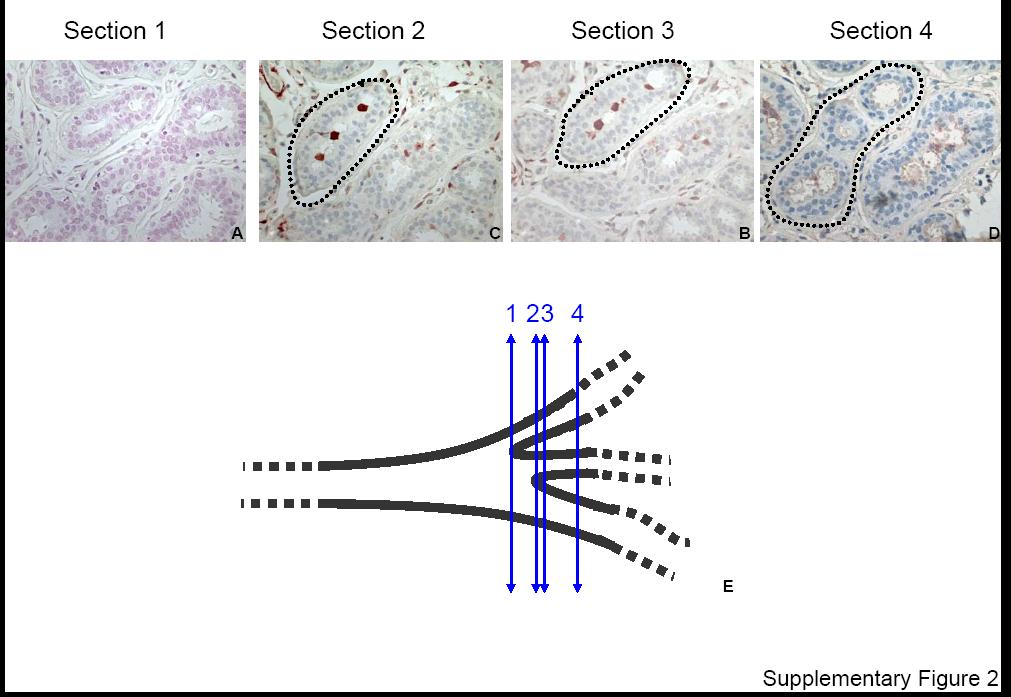
5 Figure S2. ALDH1 staining and duct morphology in a sequence of four consecutive sections of normal breast epithelium. A-D. Four consecutive sections of normal breast epithelium were stained with H&E (A) and immunostained for ALDH1 (red cytoplasmic staining) (B-C). The sequence of serial sections shows the formation of three side branches from the initial duct. The ALDH1-positive cells are located at the separation points, where side branches emerge. D. A schematic representation of the branching structure showing the position of the four consecutive sections (blue arrows).
6 UM1 UM2 UM3 Supplementary Figure 3 With DEAB Without DEAB With DEAB Without DEAB With DEAB Without DEAB 1K R1 R2 1K R1 R2 1K R1 R2 1K R1 1K R2 R1 R2 1K R1 R Side Scatter 6 4 Side Scatter 6 4 Side Scatter 6 4 Side Scatter 6 4 Side Scatter 6 4 Side Scatter BAAA BAAA BAAA BAAA BAAA BAAA Region Events %Gated R R Region Events %Gated R R Region Events %Gated R R2 9.9 Region Events %Gated R R Region Events %Gated R R Region Events %Gated R R Tum or size ( cm ) Days after injection 5, cells ALDEFLUOR + 5, cells ALDEFLUOR + 5 cells ALDEFLUOR + 5, cells ALDEFLUOR - 5, cells ALDEFLUOR - 5 cells ALDEFLUOR - 5, unsorted cells 5 unsorted cells Tumor size ( cm ) Days after injection 5, cells ALDEFLUOR + 5, cells ALDEFLUOR + 5 cells ALDEFLUOR + 5, cells ALDEFLUOR - 5, cells ALDEFLUOR - 5 cells ALDEFLUOR - 5, unsorted cells 5 unsorted cells Tumor size (cm) Days after injection 5, cells ALDEFLUOR + 5, cells ALDEFLUOR + 5 cells ALDEFLUOR + 5, cells ALDEFLUOR - 5, cells ALDEFLUOR - 5 cells ALDEFLUOR - 5, unsorted cells 5 unsorted cells
7 Figure S3. The ALDEFLUOR positive cell population has properties of cancer stem cells. Representative flow cytometry analysis of ALDH activity in cells derived from human breast tumors, orthotopically xenotransplanted in NOD/SCID mice (UM1, left panel; UM2, central panel; UM3 right panel). Cells incubated with ALDEFLUOR substrate (BAAA) and the ALDH specific inhibitor, DEAB, were used to establish the baseline fluorescence of these cells (R1) and to define the ALDEFLUOR-positive region (R2). Incubation of cells with ALDEFLUOR substrate in the absence of DEAB induced a shift in BAAA fluorescence defining the ALDEFLUOR-positive population. All the ALDEFLUOR analyses of human breast tumor cells were first gated on PI negative cells (viable cells) that represented 73.6 ± 1.8% of the total population. Tumor progression curves were plotted for the numbers of cells injected in NOD/SCID mice (5, cells; 5, cells; 5 cells) and for each cell population (ALDEFLUOR-positive, ALDEFLUOR-negative, Unseparated).
8
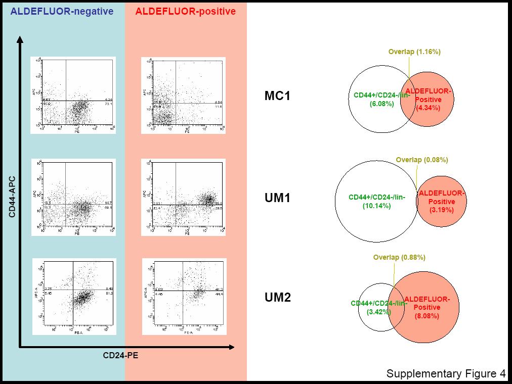
9 Figure S4. Overlap between the cell populations identified by the ALDEFLUOR assay and the CD44+/CD24 - /lin - phenotype in human breast tumors. ALDEFLUOR-negative and -positive cells from three human breast tumor xenotransplants (MC1, UM1, UM2) were separated by FACS using the ALDEFLUOR assay. In all experiments cells were first gated on PI negative cells (viable cells) that represented 73.6 ± 1.8% of the total population. Cells were then fixed in RNA later, immunostained with a CD24-PE antibody, a CD44 APC antibody, and antibodies for lineage markers, labeled Pe-Cy5. In all the flow cytometry analyses cells were first gated on lin - markers, that represented 12.3 ± 1.1% of the total population. These cell populations were gated out in the flow charts shown on the left side of the figure. The diagrams in the right side show the representation of the cell fractions defined by the ALDEFLUOR and the CD44 + /CD24-/lin - combined phenotype in the total tumor cell population (PI negative).

10 A B D Gated on dead cells stained with 7AAD PI Alde - Alde + Alde - Alde + 14% 11% 16% 1% C Supplementary Figure 5
11 Figure S5. Example of FACS analysis procedure used for the ALDEFLUOR assay in the normal and malignant human breast epithelium. A. All samples (normal breast reductions and tumors) were first gated according to the side and the forward scatter to select epithelial cells and to eliminate contaminating non-epithelial cells, clusters, and debris (gate R). The cell fraction gated on R represented 65.4 ±4.2% of the total cell population. B. Subsequently, all tumor samples were gated according to PI staining and H2Kd staining. Only the cells negative for PI staining (viable cells) and negative for H2Kd staining (human cells) were selected for the ALDEFLUOR analysis (gate R1). The PI-negative/H2Kd-negative population represented 73.6 ±1.8% of the cell population gated on R. For the normal epithelium samples, only PI staining was performed for viability, since there were no contaminating cells of mouse origin. The PI-negative population represented 93.4 ±2.4% of the cell population gated on R. C. All samples (normal epithelium and tumors) were analyzed using the ALDEFLUOR assay on the cell population gated on R and R1. D. The percentage of non-viable cells detected using PI staining was similar to that detected using 7AAD. Two aliquots from normal mammary epithelial cells treated with BAAA according to the ALDEFLUOR assay were stained in the last step with 7AAD and PI respectively, to identify potential differences in the ability of the two stains to detect non-viable cells. The two viability assays appeared to identify the same cells.
12 Compensation and gate setup Analysis Control unstained ALDEFLUOR-negative Control APC only ALDEFLUOR-positive CD44-APC CD24-PE Control PE only CD44-APC CD24-PE Supplementary Figure 6
13 Figure S6. Analysis of the cell populations defined by the ALDEFLUOR and CD44CD24lin- phenotypes in the UM2 tumor. The ALDEFLUOR-positive population in UM2 showed a considerably higher fluorescence compared to the other tumors (Figure S3). This required compensation and different gate setup for the analysis of the markers CD44CD24lin, as shown in the flow chart on the left side of the figure.
14 Table S1. Tumorigenicity in the humanized fat pad of NOD/scid mice. MC1 UM1 UM2 UM3 Tumors/injections Cells injected/fat pad 5x x1 4 5x1 3 5 ALDEFLUOR-negative 2/4 --- /2 /4 ALDEFLUOR-positive 4/ /1 4/4 Unseparated 4/ /1 3/3 ALDEFLUOR-negative /4 --- /1 /3 ALDEFLUOR-positive 3/ /1 3/3 Unseparated 3/ /1 2/3 ALDEFLUOR-negative 1/3 /1 /1 /3 ALDEFLUOR-positive 3/3 1/1 1/1 3/3 Unseparated 3/3 1/1 1/1 3/3 ALDEFLUOR-negative 1/ /4 ALDEFLUOR-positive 3/ /1 4/4 Unseparated 3/ /1 3/3
15 Characteristics Table S2. Correlation between ALDH1 protein expression and histoclinical characteristics U.M. set I.P.C. set ALDH1 ALDH1 ALDH1 ALDH1 Negative Positive Negative Positive No. of (%) No. of (%) p-value No. of (%) No. of (%) p-value patients patients patients patients All cases 122 (81) 24 (19) 243 (7) 12 (3) Age (years) 5 33 (27) 6 (25) NS 79 (35) 25 (25) NS >5 89 (73) 18 (75) 164 (65) 77 (75) Pathological tumor size PT1 61 (6) 1 (45) NS 13 (42) 37 (37) NS PT2 33 (33) 7 (32) 18 (45) 46 (47) PT3 7 (7) 5 (23) 3 (13) 16 (16) SBR grade I 25 (22) 2 (9) <.5 8 (33) 24 (24) <.1 II 55 (49) 7 (3) 12 (49) 38 (38) III 33 (29) 14 (61) 43 (18) 39 (38) Lymph node metastasis Negative 53 (56) 9 (41) NS 134 (56) 43 (44) NS Positive 42 (44) 13 (59) 17 (44) 55 (56) Estrogen receptor Negative 38 (34) 14 (61) <.5 41 (17) 39 (39) <.1 Positive 75 (66) 1 (39) 198 (83) 61 (61) Progesterone receptor Negative 5 (44) 15 (62) <.5 61 (26) 51 (52) <.1 Positive 64 (56) 9 (38) 17 (74) 47 (48) Ki-67 Negative (<2) (91) 77 (82) <.5 Positive ( 2) (9) 17 (18) ERBB2 Negative (/1+) 94 (83) 15 (62) <.5 28 (94) 76 (79) <.1 Positive (2+/3+) 19 (17) 9 (38) 14 (6) 2 (21) Cytokeratin 18 (CK18) Negative (5) 9 (9) NS Positive (95) 85 (91) Cytokeratin 5/6 (CK5/6) Negative (72) 46 (58) <.5 Positive (28) 33 (42) Cytokeratin 14 (CK14) Negative (96) 68 (86) <.1 Positive (4) 11 (14)
16 Supplemental Experimental Procedures Normal breast tissue dissociation Normal breast tissue from reduction mammoplasties was dissociated mechanically and enzymatically, as previously described (Stingl et al., 1998). Tissue was minced and dissociated in Ham s F12/Dulbecco s modified Eagle s medium (F12:DMEM; 1:1; StemCell technologies, Durham, NC, USA) supplemented with 1 mm Hepes, 2% bovine serum albumin (BSA; Fraction V; GIBCO TM INVITROGEN), 5 mg/ml insulin,.5 mg/ml hydrocortisone, 1 ng/ml cholera toxin, 3 U/ml collagenase and 1 U/ml hyaluronidase (all from Sigma, St Louis, MO, USA) at 37 C for 16h. The epithelialcell-rich pellet (95-99% purity) was collected by centrifuging the cell suspension at 8 g for 4 min followed by one wash with F12/DMEM. The supernatant from the first centrifugation contained the mammary stromal cells (fibroblasts and endothelial cells). Epithelial organoids were further digested for 5 min in.5% trypsin (Gibco)-.25% EDTA (Sigma) solution to generate a single-cell suspension. An equal volume of F12/DME/H supplemented with 5% FBS was added to stop the digestion. The cell suspension was filtered twice through a 4-mm nylon mesh (BioDesign Inc., New York, N.Y., USA). Following centrifugation at 8 g, the pellet was resuspended in F12/DMEM with a reduced calcium concentration (.6 mm, StemCell technologies) supplemented with 5 U/ml dispase (Collaborative Biomedical Products, Bedford, Md., USA). To remove red blood cells the pellets were treated with ammonium chloride solution. Flow cytometry analysis CD44/CD24/Lin staining was performed as previously described (Al-Hajj., 23). Briefly, cells were stained with primary antibodies anti-cd44 labeled APC (dilution 1:1, BD Biosciences), anti-cd24 labeled PE (dilution 1:1, BD Biosciences), and lineage markers anti-cd31, CD64, CD14b (BD Biosciences), CD2, CD3, CD1, CD16, CD18 (all labeled with PE-
17 Cy5, Jackson Labs). Fresh cells were stained with 1µg/ml PI (Sigma) for 5 min. for viability. Examples of flow charts and more details on gate setup are shown in Figures S5 and S6. Mammosphere culture Mammosphere culture was performed as previously described (Dontu et al., 23). Single cells were plated in ultra-low attachment plates (Corning, Acton, MA, USA) or plates coated with 1% agarose in PBS, at a density of 2, viable cells/ml in primary culture and 5 cells/ml in subsequent passages. For mammosphere culture, cells were grown in a serum-free mammary epithelial basal medium (MEBM) (Cambrex Bio Science Walkersville, Inc, Walkerville, MD, USA) supplemented with B27 (INVITROGEN, Carlsbad, CA, USA), 2 ng/ml EGF (BD Biosciences, San Jose, CA, USA), antibiotic-antimycotic (1 unit/ml penicillin G sodium, 1 ug/ml streptomycin sulfate and.25 µg/ml amphotericin B), 2 ug/ml Gentamycin, 1 ng/ml Hydrocortisone, 5 µg/ml Insulin and 1 µm beta-mercaptoethanol (GIBCO TM INVITROGEN) in a humidified incubator (1% CO 2 : 95% air, 37 C for 7-1 days, as previously described (Dontu et al., 23).
3D Mammary Colony-Forming Cell Assay Giusy Tornillo 1* and Sara Cabodi 2
 3D Mammary Colony-Forming Cell Assay Giusy Tornillo 1* and Sara Cabodi 2 1 Cardiff School of Biosciences, European Cancer Stem Cell Research Institute, Cardiff University, Cardiff, UK; 2 Department of
TF-1a lymphoblastic leukemia cell line: marking with GFP, phenotyping and sorting
 Supplemental Material Supplemental Methods TF-1a lymphoblastic leukemia cell line: marking with GFP, phenotyping and sorting In order to determine if the multi-parameter FACS approach would be successful
Figure S1. Phenotypic characterization of AND-1_WASKO cell lines. AND- 1_WASKO_C1.1 (WASKO_C1.1) and AND-1_WASKO_C1.2 (WASKO_C1.
 and AND-1_WASKO_C1.2 (WASKO_C1.") LEGENDS TO SUPPLEMENTARY FIGURES Figure S1. Phenotypic characterization of AND-1_WASKO cell lines. AND- 1_WASKO_C1.1 (WASKO_C1.1) and AND-1_WASKO_C1.2 (WASKO_C1.2) were stained with the antibodies oct3/4
LEGENDS TO SUPPLEMENTARY FIGURES Figure S1. Phenotypic characterization of AND-1_WASKO cell lines. AND- 1_WASKO_C1.1 (WASKO_C1.1) and AND-1_WASKO_C1.2 (WASKO_C1.2) were stained with the antibodies oct3/4
Isolation, culture, and transfection of primary mammary epithelial organoids
 Supplementary Experimental Procedures Isolation, culture, and transfection of primary mammary epithelial organoids Primary mammary epithelial organoids were prepared from 8-week-old CD1 mice (Charles River)
Supplemental Information. Materials and methods.
 Supplemental Information Materials and methods. Cell culture. hmscs were isolated from bone marrow of 3 male donors, undergoing orthopedic surgery (mean age 69.7). Cells were cultured in high glucose DMEM
All quality control test results are reported on a lot specific Certificate of Analysis which is available at or upon request.
 PRIME-XV Neural Basal Medium PRIME-XV Neural Basal Medium is a chemically-defined basal medium optimized for the culture and maintenance of neuronal cells when supplemented with PRIME-XV IS21 Supplement
Application Protocol: CD45 CK Immunostaining for patient blood
 REPRODUCTION AND USE This document is protected by copyright and it cannot be used or shared without permission from Vortex Biosciences, Inc. Such permission is given on condition that Vortex Biosciences
Engraftment of human induced pluripotent stem cell-derived hepatocytes in. immunocompetent mice via 3D co-aggregation and encapsulation
 Engraftment of human induced pluripotent stem cell-derived hepatocytes in immunocompetent mice via 3D co-aggregation and encapsulation Wei Song 1, Yen-Chun Lu 1, Angela S. Frankel 2, Duo An 1, Robert E.
Autocrine Complement Inhibits IL10-Dependent T-Cell Mediated. Antitumor Immunity to Promote Tumor Progression
 Supplemental Information Autocrine Complement Inhibits IL10-Dependent T-Cell Mediated Antitumor Immunity to Promote Tumor Progression Yu Wang, Sheng-Nan Sun, Qing Liu, Yang-Yang Yu, Jian Guo, Kun Wang,
Propagation of H7 hesc From: UW (John Stamatoyannopoulos) ENCODE group Date: 12/17/2009 Prepared By: S. Paige/S. Hansen (UW)
 ENCODE group Date: 12/17/2009 Prepared By: S. Paige/S. Hansen (UW)") Propagation of H7 hesc From: UW (John Stamatoyannopoulos) ENCODE group Date: 12/17/2009 Prepared By: S. Paige/S. Hansen (UW) Growth and Harvest Modifications Addendum to: Propagation of H7 hesc from UW
Propagation of H7 hesc From: UW (John Stamatoyannopoulos) ENCODE group Date: 12/17/2009 Prepared By: S. Paige/S. Hansen (UW) Growth and Harvest Modifications Addendum to: Propagation of H7 hesc from UW
Table of Contents. 2.1 NeuroCult NCFC Assay Kit (Rat) Components Additional Required Reagents Required Equipment...
 Components Additional Required Reagents Required Equipment...") i Table of Contents 1.0 Overview of the NeuroCult NCFC Assay 2.0 Materials 2.1 NeuroCult NCFC Assay Kit (Rat) Components... 4 2.2 Additional Required Reagents... 4 2.3 Required Equipment... 4 3.0 Preparation
i Table of Contents 1.0 Overview of the NeuroCult NCFC Assay 2.0 Materials 2.1 NeuroCult NCFC Assay Kit (Rat) Components... 4 2.2 Additional Required Reagents... 4 2.3 Required Equipment... 4 3.0 Preparation
Corning BioCoat Matrigel Matrix 6-well Plates for Embryonic Stem (ES) Cell Culture. Catalog Number Guidelines for Use
 Cell Culture. Catalog Number Guidelines for Use") Corning BioCoat Matrigel Matrix 6-well Plates for Embryonic Stem (ES) Cell Culture Catalog Number 354671 Guidelines for Use Discovery Labware, Inc., Two Oak Park, Bedford, MA 01730, Tel: 1.978.442.2200
Corning BioCoat Matrigel Matrix 6-well Plates for Embryonic Stem (ES) Cell Culture Catalog Number 354671 Guidelines for Use Discovery Labware, Inc., Two Oak Park, Bedford, MA 01730, Tel: 1.978.442.2200
Supporting Information
 Electronic Supplementary Material (ESI) for Lab on a Chip. This journal is The Royal Society of Chemistry 2014 Supporting Information Cell culture. C2C12 cells (ATCC CRL 1772) were cultured in 75 cm 2
Preparation of Mouse Bone Marrow Stromal Cells
 Preparation of Mouse Bone Marrow Stromal Cells A single-step stem cell purification method using adhesion to cell culture plastic was employed as described in the Reference. Briefly, neonatal and adult
Supplementary Figure 1 A green: cytokeratin 8
 Supplementary Figure 1 A green: cytokeratin 8 green: α-sma red: α-sma blue: DAPI blue: DAPI Panc-1 Panc-1 Panc-1+hPSC Panc-1+hPSC monoculture coculture B Suppl. Figure 1: A, Immunofluorescence staining
BD IMag. Streptavidin Particles Plus - DM. Technical Data Sheet. Product Information
 Technical Data Sheet Streptavidin Particles Plus - DM Product Information Material Number: Size: Storage Buffer: 557812 5 ml Aqueous buffered solution containing BSA and 0.09% sodium azide. Description
Large-Scale Analysis of Breast Cancer-Related. Conformational Changes in Proteins using SILAC-SPROX. *Corresponding author
 SUPPORTING INFORMATION for: Large-Scale Analysis of Breast Cancer-Related Conformational Changes in Proteins using SILAC-SPROX Fang Liu, 1 He Meng, 1 and Michael C. Fitzgerald 1, * 1 Department of Chemistry,
Mice genotyping for FAK flox, deletion (Δ), and KD alleles as well as GFP, Cre and
, and KD alleles as well as GFP, Cre and") Supplementary Methods Mice genotyping Mice genotyping for FAK flox, deletion (Δ), and KD alleles as well as GFP, Cre and PyMT alleles were performed using the following primers: FAK Flox/WT: 5 - GCTGATGTCCCAAGCTATTCC
Supplementary Methods Mice genotyping Mice genotyping for FAK flox, deletion (Δ), and KD alleles as well as GFP, Cre and PyMT alleles were performed using the following primers: FAK Flox/WT: 5 - GCTGATGTCCCAAGCTATTCC
Identification of Single Chain Antibodies to Breast Cancer Stem Cells Using Phage Display
 Identification of Single Chain Antibodies to Breast Cancer Stem Cells Using Phage Display Deniz Gur Dept. of Surgery, Vermont Comprehensive Cancer Center, University of Vermont, College of Medicine, Burlington,
In vivo BrdU Incorporation Assay for Murine Hematopioetic Stem Cells Ningfei An, Yubin Kang *
 In vivo BrdU Incorporation Assay for Murine Hematopioetic Stem Cells Ningfei An, Yubin Kang * Division of Hematology-Oncology, Department of Medicine, Medical University of South Carolina, Charleston,
Protocol Reprogramming MEFs using the Dox Inducible Reprogramming Lentivirus Set: Mouse OKSM
 STEMGENT Page 1 OVERVIEW The following protocol describes the reprogramming of one well of mouse embryonic fibroblasts (MEFs) into induced pluripotent stem (ips) cells in a 6-well format. Transduction
Quick-Spin the tetramer tubes before opening, due to the change in altitude and the low volume.
 Quick-Spin the tetramer tubes before opening, due to the change in altitude and the low volume. Staining ex vivo samples with IA g7 Ins10-23 RE#3 Tetramers, from Kappler/Marrack Laboratory Howard Hughes
B. ADM: C. D. Apoptosis: 1.68% 2.99% 1.31% Figure.S1,Li et al. number. invaded cells. HuH7 BxPC-3 DLD-1.
 A. - Figure.S1,Li et al. B. : - + - + - + E-cadherin CK19 α-sma vimentin β -actin C. D. Apoptosis: 1.68% 2.99% 1.31% - : - + - + - + Apoptosis: 48.33% 45.32% 44.59% E. invaded cells number 400 300 200
Isolation and 3-dimensional Culture of Primary Murine Intestinal Epithelial Cells Agnieszka Pastuła * and Michael Quante
 Isolation and 3-dimensional Culture of Primary Murine Intestinal Epithelial Cells Agnieszka Pastuła * and Michael Quante II. Medizinische Klinik, Klinikum rechts der Isar, Technische Universität München,
Supplementary Materials and Methods
 Supplementary Materials and Methods Reagents Supplementary Material (ESI) for Lab on a Chip RPMI medium, FBS, HEPES buffer solution, sodium pyruvate, penicillin, and streptomycin were obtained from Biological
High throughput screening: Huh-7 cells were seeded into 96-well plate (2000
 1 SUPPLEMENTARY INFORMATION METHODS 6 7 8 9 1 11 1 1 1 1 16 17 18 19 High throughput screening: Huh-7 cells were seeded into 96-well plate ( cells/well) and infected with MOI of DENV-. One hour post-infection
Mammosphere formation assay. Mammosphere culture was performed as previously described (13,
 Supplemental Text Materials and Methods Mammosphere formation assay. Mammosphere culture was performed as previously described (13, 17). For co-culture with fibroblasts and treatment with CM or CCL2, fibroblasts
HUMAN IPSC CULTURE PROTOCOLS
 HUMAN IPSC CULTURE PROTOCOLS FOR TRAINING COURSE IXCELLS BIOTECHNOLOGIES USA, LLC 10340 CAMINO SANTA FE, SUITE C, SAN DIEGO, CA 92121 TABLE OF CONTENTS CHAPTER 1 BEFORE STARTING 2-7 SECTION 1.1 COATING
All quality control test results are reported on a lot specific Certificate of Analysis which is available at or upon request.
 PRIME-XV NPC Expansion XSFM PRIME-XV NPC Expansion XSFM is a xeno-and serum-free medium optimized for the cultivation and expansion of neural progenitor cells (NPC) that are able to maintain their ability
Whole Spleen Flow Cytometry Assay Cathy S. Yam *, Adeline M. Hajjar *
 Whole Spleen Flow Cytometry Assay Cathy S. Yam *, Adeline M. Hajjar * Department of Comparative Medicine, University of Washington, Seattle, USA *For correspondence: csyam@u.washington.edu; hajjar@uw.edu
Regulatory B Cell Isolation Kit mouse
 For further information refer to our website www.miltenyibiotec.com For technical questions, please contact your local subsidiary or distributor. Technical Support Team, Germany: E-mail: macstec@miltenyibiotec.de
Single-cell suspensions of C57BL/6J splenocytes were incubated with CD5 beads
 S S Single-cell suspensions of C57BL/6J splenocytes were incubated with CD5 beads (Miltenyi Biotech) and T cells were positively selected by magnetically activated cell sorting (MACS). 50x10 6 or greater
Chemical mixtures isolated from house dust disrupt thyroid receptor β (TRβ) signaling
 signaling") SUPPORTING INFORMATION Chemical mixtures isolated from house dust disrupt thyroid receptor β (TRβ) signaling Erin M. Kollitz, Christopher D. Kassotis, Kate Hoffman, P. Lee Ferguson, Julie Ann Sosa, Heather
SUPPORTING INFORMATION Chemical mixtures isolated from house dust disrupt thyroid receptor β (TRβ) signaling Erin M. Kollitz, Christopher D. Kassotis, Kate Hoffman, P. Lee Ferguson, Julie Ann Sosa, Heather
InVERT moulding for scalable control of tissue microarchitecture
 InVERT moulding for scalable control of tissue microarchitecture Stevens KR, Ungrin MD, Schwartz RE, Ng SY, Carvalho B, Christine KS, Chaturvedi R, Li CY, Zandstra PW, Chen CS, Bhatia SN Supplementary
T ECHNICAL MANUAL. Culture of Human Mesenchymal Stem Cells Using MesenCult -XF Medium
 T ECHNICAL MANUAL Culture of Human Mesenchymal Stem Cells Using MesenCult -XF Medium i Table of Contents 1.0 Materials... 1 1.1 MesenCult -XF Medium and Required Products... 1 1.2 Additional Required
F4/80, CD11b, Gr-1, NK1.1, CD3, CD4, CD8 and CD19. A-antigen was detected with FITCconjugated
 SDC MATERIALS AND METHODS Flow Cytometric Detection of A-Antigen Expression Single cell suspensions were prepared from bone marrow, lymph node and spleen. Peripheral blood was obtained and erythrocytes
Protocol Using a Dox-Inducible Polycistronic m4f2a Lentivirus to Reprogram MEFs into ips Cells
 STEMGENT Page 1 OVERVIEW The following protocol describes the transduction and reprogramming of one well of Oct4-GFP mouse embryonic fibroblasts (MEF) using the Dox Inducible Reprogramming Polycistronic
Welcome to the CORES Flow Cytometry Sorter Facility
 Welcome to the CORES Flow Cytometry Sorter Facility The FACSAria I and FACSARIA III are twin laser high speed sorters capable of analyzing cells based on 7-8 distinct fluorescent properties (besides FSC
COMPONENT NAME COMPONENT # QUANTITY STORAGE SHELF LIFE FORMAT. Store at 2-8 C. Do not freeze.
 This document is available at www.stemcell.com/pis EasySep Mouse PE Kit II Catalog #17666 Catalog #17696 For processing 1 x 10^9 cells For processing 5 x 10^9 cells Description Isolate highly purified
TECHNICAL REPORT. Experimental Campaign on the in-vitro platelet-endothelial cells interactions
 TECHNICAL REPORT Experimental Campaign on the in-vitro platelet-endothelial cells interactions Marco Malvestiti, Paolo Gresele Department of Internal Medicine, University of Perugia, Perugia, Italy marco.malvestiti@gmail.com,
MagniSort Mouse Hematopoietic Lineage Depletion Kit Catalog Number: RUO: For Research Use Only. Not for use in diagnostic procedures.
 Page 1 of 2 MagniSort Mouse Hematopoietic Lineage Depletion Kit RUO: For Research Use Only. Not for use in diagnostic procedures. Mouse bone marrow cells were unsorted (top row) or sorted with the MagniSort
Modeling Cardiomyocyte Differentiation:
 icell Cardiac Progenitor Cells Prototype Application Protocol Wnt- and Activin/TGFβ-inhibitor Induction with Flow Cytometry Analysis Introduction The ability of cardiac progenitor cells to proliferate
PCCS Growth Media, Cell Tagging, Cell Separation Final Assignment. Igneris Rosado-Erazo. Panama College of Cell Science
 Running Head: Growth Media, Cell Tagging, Cell Separation PCCS Growth Media, Cell Tagging, Cell Separation Final Assignment Igneris Rosado-Erazo Panama College of Cell Science In partial fulfillment of
ReproRNA -OKSGM is a non-integrating, self-replicating RNA-based reprogramming vector for generating induced pluripotent stem (ips)
") Kit for generating ips cells using ReproRNA -OKSGM, a non-integrating, self-replicating RNA reprogramming vector Product Description ReproRNA -OKSGM is a non-integrating, self-replicating RNA-based reprogramming
Kit for generating ips cells using ReproRNA -OKSGM, a non-integrating, self-replicating RNA reprogramming vector Product Description ReproRNA -OKSGM is a non-integrating, self-replicating RNA-based reprogramming
Data Sheet CD137/NF-κB Reporter - HEK293 Recombinant Cell Line Catalog # 79289
 Data Sheet CD137/NF-κB Reporter - HEK293 Recombinant Cell Line Catalog # 79289 Background Human CD137 (4-1BB; TNFRS9) is an inducible co-stimulatory molecule that activates T cells. CD137:CD137L-mediated
STEMdiff Mesenchymal Progenitor Kit
 Defined culture kit for derivation and expansion of mesenchymal progenitor cells Catalog #05240 1 Kit Product Description STEMdiff Mesenchymal Progenitor Kit is a defined culture kit consisting of animal
Antibody used for FC Figure S1. Multimodal characterization of NIR dyes in vitro Figure S2. Ex vivo analysis of HL60 cells homing
 Antibody used for FC The following antibodies were used following manufacturer s instructions: anti-human CD4 (clone HI3, IgG1, k - Becton Dickinson), anti-human CD33 (clone WM3, IgG1, k- Becton Dickinson),
Standard Operating Procedure
 Standard Operating Procedure Title Subtitle NANoREG Work package/task: Owner and co-owner(s) HTS Comet Assay with and without FPG - 20 wells Comet assay with and without FPG using Trevigen 20-well slides
Flow Cytometry Immune Activation SOP
 Flow Cytometry Immune Activation SOP Purpose This SOP standardizes the procedure for measuring immune activation of T cells using flow cytometry in ACTG Immunology Laboratories. Materials 1. 12x75mm flow
Data Sheet ICOS/NFAT Reporter-Jurkat Recombinant Cell Line Catalog #: 79668
 Data Sheet ICOS/NFAT Reporter-Jurkat Recombinant Cell Line Catalog #: 79668 Product Description Recombinant Jurkat T cell expressing firefly luciferase gene under the control of NFAT response elements
Metzger Lab Protocol Book EF May 2003
 I. Preparation for cell isolation: A. Make sure the following items are autoclaved: 1. Glassware* (1) 100 ml beaker (3) wide mouthed 1 liter bottles (1) 100 mm glass petri dish (1) 60 mm sigma coated petri
PRACTICAL CELL SORTING. The following is a discussion of best practices for investigators planning experiments that involved flow sorting of cells:
 PRACTICAL CELL SORTING The following is a discussion of best practices for investigators planning experiments that involved flow sorting of cells: 1. Cell preparation 2. Pre-sort cell viability 3. Post-sort
Culturing Protocol for JM8.N4 ES Cell Clones Revised July 2014
 Culturing Protocol for JM8.N4 ES Cell Clones Revised July 2014 Cell Line Information The JM8.N4 subline is derived from the JM8 parental line and are considered to be feeder independent. These cells are
Protocol Reprogramming Human Fibriblasts using the Dox Inducible Reprogramming Polycistronic Lentivirus Set: Human 4F2A LoxP
 STEMGENT Page 1 OVERVIEW The following protocol describes the reprogramming of one well of BJ Human Fibroblasts (BJ cells) into induced pluripotent stem (ips) cells in a 6-well format. Transduction efficiency
RPCI 001 v.003 In vitro Intracellular Cytokine Staining With and Without Stimulation
 Immune Tolerance Network RPCI 001 v.003 Author: Paul Wallace and Earl Timm, Director, RPCI Laboratory of Flow Cytometry Approved by: Paul Wallace, Director, RPCI Laboratory of Flow Cytometry 1.0 Title
Supplementary Figure. S1
 Supplementary Figure. S1 Supplementary Figure S1. Correlation of phagocytic ability measured with YG and YO beads. Fresh human monocytes (2 10 6 /ml) were labelled with APC conjugated anti CD14 mab alone
Flow Cytometry SOP: Monocytes from Frozen Cells
 Flow Cytometry SOP: Monocytes from Frozen Cells Purpose This SOP standardizes the procedure for measuring immune cells using flow cytometry in ACTG Immunology Laboratories. Materials 1. 12x75mm flow tubes
Life Sciences Reporting Summary
 Life Sciences Reporting Summary Corresponding Author: Johanna A. Joyce Date: Jun 13, 2017 Nature Research wishes to improve the reproducibility of the work we publish. This form is published with all life
International Journal of Pharma and Bio Sciences
 Research Article Cell Biology International Journal of Pharma and Bio Sciences ISSN 0975-6299 EFFECT OF ANTIBIOTICS ON MESENCHYMAL STROMAL CELLS UNDER XENO-FREE CULTURE CONDITIONS FOR CLINICAL USE JAIANAND
Nature Medicine: doi: /nm.2084
 Supplementary Table 1. Description of tumor lesions by number, location, and type. Tumor # AAH (%) Adenoma (%) ADCA (%) Alveolar (%) Bronchiolar (%) 8 Wks K-ras/NE +/+ 1.47 +/- 0.38 67.7 +/- 5.1 32.3 +/-
Submitting Cells for Cell Sorting University of Colorado Cancer Center Flow Cytometry Shared Resource (FCSR)
") Submitting Cells for Cell Sorting University of Colorado Cancer Center Flow Cytometry Shared Resource (FCSR) cc.flowcyto@ucdenver.edu 303.724.3145 Scheduling: For appointments see online calendar @ www.ucccflow.calendarhost.com
Submitting Cells for Cell Sorting University of Colorado Cancer Center Flow Cytometry Shared Resource (FCSR) cc.flowcyto@ucdenver.edu 303.724.3145 Scheduling: For appointments see online calendar @ www.ucccflow.calendarhost.com
Isolation of ILC2 from Mouse Liver Tamar Mchedlidze and Stefan Wirtz *
 Isolation of ILC2 from Mouse Liver Tamar Mchedlidze and Stefan Wirtz * Medical Department 1, FAU Erlangen-Nuremberg, Erlangen, Germany *For correspondence: stefan.wirtz@uk-erlangen.de [Abstract] Group
Data Sheet PD-1 / NFAT - Reporter - Jurkat Recombinant Cell Line Catalog #: 60535
 Data Sheet PD-1 / NFAT - Reporter - Jurkat Recombinant Cell Line Catalog #: 60535 Product Description Recombinant Jurkat T cell expressing firefly luciferase gene under the control of NFAT response elements
Data Sheet GITR / NF-ĸB-Luciferase Reporter (Luc) - Jurkat Cell Line Catalog #60546
 - Jurkat Cell Line Catalog #60546") Data Sheet GITR / NF-ĸB-Luciferase Reporter (Luc) - Jurkat Cell Line Catalog #60546 Description This cell line expresses a surface human GITR (glucocorticoid-induced TNFR family related gene; TNFRSF18;
Data Sheet GITR / NF-ĸB-Luciferase Reporter (Luc) - Jurkat Cell Line Catalog #60546 Description This cell line expresses a surface human GITR (glucocorticoid-induced TNFR family related gene; TNFRSF18;
Mice TRAMP mice were maintained in a C57BL/6J background. Syngeneic UBI-GFP/BL6 mice were used for bone marrow engraftment. 2
 Antibodies Chicken IgY polyclonal α-gfp antibodies were purchased from Abcam (Cambridge, MA) and were detected using α-chicken IgY-FITC or α-chicken-hrp (also purchased from Abcam). The CD31-PE, CD11b-PE,
Materials and Reagents
 Isolation of CD34 + Cells from Human Fetal Liver and Cord Blood Institute of Molecular and Cell Biology, 138673, Singapore Qingfeng Chen 1* and Jianzhu Chen 2* 1 Humanized Mouse Unit, Institute of Molecular
Supplementary Table I: List of primers used for qpcr
 Supplementary Table I: List of primers used for qpcr Primer ABCG2 F ABCG2 R β Actin F β Actin R ALDH1A1 F ALDH1A1 R ALDH1A1 promoter F ALDH1A1 promoter R CD24 F CD24 R CD44 F CD44 R CXCR1 F CXCR1 R CXCR4
PROTOCOL. Generating Cardiomyocytes from Human Pluripotent Stem Cells using Stemgent MesoFate Differentiation Medium. Overview. Required Materials
 Page 1 Overview Analysis of mouse and human embryonic stem cell differentiation cultures indicate the existence of a cardiovascular progenitor representing one of the earliest stages in mesoderm specification
Fitness Measurements of Evolved Escherichia coli Migla Miskinyte and Isabel Gordo *
 Fitness Measurements of Evolved Escherichia coli Migla Miskinyte and Isabel Gordo * Evolutionary Biology Group, Instituto Gulbenkian de Ciência, Oeiras, Portugal *For correspondence: igordo@igc.gulbenkian.pt
Transfection of Human Naive CD4 + T Cells with PHA Activation and Neon Electroporation Amy Palin 1* and David B. Lewis 2*
 Transfection of Human Naive CD4 + T Cells with PHA Activation and Neon Electroporation Amy Palin 1* and David B. Lewis 2* 1 Department of Pediatrics and Program in Immunology, Stanford University Medical
Phagocytosis Assay Kit (IgG PE)
") Phagocytosis Assay Kit (IgG PE) Item No. 600540 www.caymanchem.com Customer Service 800.364.9897 Technical Support 888.526.5351 1180 E. Ellsworth Rd Ann Arbor, MI USA TABLE OF CONTENTS GENERAL INFORMATION
Phagocytosis Assay Kit (IgG PE) Item No. 600540 www.caymanchem.com Customer Service 800.364.9897 Technical Support 888.526.5351 1180 E. Ellsworth Rd Ann Arbor, MI USA TABLE OF CONTENTS GENERAL INFORMATION
Supporting Information
 Supporting Information of Laser Immunotherapy in Combination with Perdurable PD-1 Blocking for Treatment of Metastatic Tumor Lihua Luo, Chunqi Zhu, Hang Yin, Mengshi Jiang, Junlei Zhang, Bing Qin, Zhenyu
Measurement of peritoneal macrophage apoptosis by Celigo plate imaging cytometer
 SUPPLEMENTAL METHODS Measurement of peritoneal macrophage apoptosis by Celigo plate imaging cytometer For Celigo experiments, 0.1 ml containing 5 x 10 4 cells was seeded into 96 well plates for 30 min
Human Skeletal Muscle Myoblast Care Manual: Maintenance and Differentiation from Myoblasts to Myocytes
 Human Skeletal Muscle Myoblast Care Manual: Maintenance and Differentiation from Myoblasts to Myocytes STORAGE CONDITIONS Media: Short Term 4 C 6 months -20 C F or research use only. Not approved for human
For in vitro killing assays with lysed cells, neutrophils were sonicated using a 550 Sonic
 Supplemental Information Cell Host & Microbe, Volume 8 Statins Enhance Formation of Phagocyte Extracellular Traps Ohn A. Chow, Maren von Köckritz-Blickwede, A. Taylor Bright, Mary E. Hensler, Annelies
MagniSort Mouse B cell Enrichment Kit Catalog Number: RUO: For Research Use Only. Not for use in diagnostic procedures.
 Page 1 of 2 MagniSort Mouse B cell Enrichment Kit RUO: For Research Use Only. Not for use in diagnostic procedures. Mouse splenocytes were unsorted (left) or sorted with the MagniSort Mouse B cell Enrichment
Supplementary File 3: DNA and RNA isolation
 Supplementary File 3: DNA and RNA isolation Q-CROC-02 Biopsy protocol For the purposes of this protocol, four needle core biopsies (NCBs) of lymph node tissue are isolated from each patient using a 16G
Human Adult Mammary Fibroblast Care Manual
 Human Adult Mammary Fibroblast Care Manual INSTRUCTIONAL MANUAL ZBM0062.05 SHIPPING CONDITIONS Human Adult Mammary Fibroblast Cells Orders are delivered via Federal Express courier. All US and Canada orders
Protocols for Hematopoietic Differentiation of Murine ES Cells (in 6 or 24 well plates) Media Preparation
 Media Preparation") Protocols for Hematopoietic Differentiation of Murine ES Cells (in 6 or 24 well plates) Media Preparation A) Embryonic Stem Cell Medium (ES) Dulbecco s modified Eagle s Medium (DMEM) with high glucose
Protocols for Hematopoietic Differentiation of Murine ES Cells (in 6 or 24 well plates) Media Preparation A) Embryonic Stem Cell Medium (ES) Dulbecco s modified Eagle s Medium (DMEM) with high glucose
Normalization of Agilent Seahorse XF Data by In-situ Cell Counting Using a BioTek Cytation 5
 Normalization of Agilent Seahorse XF Data by In-situ Cell Counting Using a BioTek Cytation Application Note Authors Yoonseok Kam 1, Ned Jastromb 1, Joe Clayton, Paul Held, and Brian P. Dranka 1 1 Agilent
Protocol to detect N-Glycolylneuraminic acid (Neu5Gc) using Flow Cytometry Analysis.
 using Flow Cytometry Analysis.") Page 1 of 5 Protocol to detect N-Glycolylneuraminic acid (Neu5Gc) using Flow Cytometry Analysis. The Gc-Free Basic Pack may be used for immuno-staining cells prior to analysis by Flow Cytometry. This Basic
Page 1 of 5 Protocol to detect N-Glycolylneuraminic acid (Neu5Gc) using Flow Cytometry Analysis. The Gc-Free Basic Pack may be used for immuno-staining cells prior to analysis by Flow Cytometry. This Basic
STEMdiff Hematopoietic Kit
 For differentiation of human ES or ips cells into hematopoietic progenitor cells Catalog #05310 1 Kit Product Description Kit includes a defined, serum-free basal medium and supplements for the feeder-free
COMPONENT NAME COMPONENT # QUANTITY STORAGE SHELF LIFE FORMAT. Store at 2-8 C. Do not freeze. Store at 2-8 C. Do not freeze.
 This document is available at www.stemcell.com/pis EasySep Mouse CD90. Kit II Catalog #89 For processing x 0^9 cells Description Isolate highly purified CD90.+ (Thy.+) cells from mouse splenocytes or other
Supplemental Figure Legends
 Supplemental Figure Legends Supplemental Figure 1. (A) Reference guide for the phospho-protein array. (B) Representative fluorescent arrays for whole cell lysates from MDA-MB-231 cells cultured at ph 7.4
SUPPLEMENTAL FIGURES AND TABLES
 SUPPLEMENTAL FIGURES AND TABLES A B Flag-ALDH1A1 IP: α-ac HEK293T WT 91R 128R 252Q 367R 41/ 419R 435R 495R 412R C Flag-ALDH1A1 NAM IP: HEK293T + + - + D NAM - + + E Relative ALDH1A1 activity 1..8.6.4.2
Immunofluorescence of organoids embedded in Basement Membrane Matrix
 Immunofluorescence of organoids embedded in Basement Membrane Matrix Sol Degese 1, Gabe Benton 1 1 Organoid Resource Lab (ORL), Trevigen, Inc., 8405 Helgerman Court, Gaithersburg, MD 20877 Introduction
Xeno-Free Systems for hesc & hipsc. Facilitating the shift from Stem Cell Research to Clinical Applications
 Xeno-Free Systems for hesc & hipsc Facilitating the shift from Stem Cell Research to Clinical Applications NutriStem Defined, xeno-free (XF), serum-free media (SFM) specially formulated for growth and
Epithelial Cells cells lining the trachea Epithelium layer of epithelial cells in the tissue Many epithelial cell types exist on apical surface
 Matthew Pilewski Epithelial Cells cells lining the trachea Epithelium layer of epithelial cells in the tissue Many epithelial cell types exist on apical surface Epithelium form tight junctions that keep
Supplementary methods
 Supplementary methods Cell culture, infection, transfection, and RNA interference HEK293 cells and its derivatives were grown in DMEM supplemented with 10% FBS. Various constructs were introduced into
VDL602.2 RAPID ASSAY FOR DETERMINING ADENOVIRAL VECTOR TITER
 1. Purpose 1.1. The purpose of this protocol is to determine the number of infectious adenoviral particles. 1.2. The starting material is purified adenoviral vectors. 1.3. This protocol is based upon the
ETS-Embryo Medium. In Vitro Culture Media. Version 1.0
 ETS-Embryo Medium In Vitro Culture Media Version 1.0 3D Culture Media Product Catalogue No Storage ETS-Embryo Medium, 25 ml (5 x 5 ml) M13-25 -20 C ETS-Embryo Medium, 100 ml M13-100 -20 C Additional Reagents
Standard Operating Procedure
 Standard Operating Procedure Title Standard Operation Procedure (SOP) and background documentation for real-time label-free impedance-based nanotoxicity assessment Subtitle NANoREG Work package/task: Owner
Terrific Validation, Tech Support and Prompt Delivery. User Guide for Selleck Human ipsc Enhancer Kit Cat. No. K2010. Guidelines for Use
 Terrific Validation, Tech Support and Prompt Delivery User Guide for Selleck Human ipsc Enhancer Kit Cat. No. K2010 Guidelines for Use For Research Use Only. Not for use in diagnostic or therapeutic procedures.
Enumeration, Phenotyping, and Identification of Activation Events in Conjugates Between T Cells and Antigen-Presenting Cells by Flow Cytometry
 Enumeration, Phenotyping, and Identification of Activation Events in Conjugates Between T Cells and Antigen-Presenting Cells by Flow Cytometry Kristie M. Grebe and Terry A. Potter* (Published 10 September
Centre for Innovative Cancer Research, Ottawa Hospital Research Institute, Ottawa, Canada;
 Ex vivo Natural Killer Cell Cytotoxicity Assay Lee-Hwa Tai 1, Christiano Tanese de Souza 1, Andrew P. Makrigiannis 2 and Rebecca Ann C. Auer 3* 1 Centre for Innovative Cancer Research, Ottawa Hospital
Supplementary Figure 1
 Supplementary Figure 1 Ex2 promotor region Cre IRES cherry pa Ex4 Ex5 Ex1 untranslated Ex3 Ex5 untranslated EYFP pa Rosa26 STOP loxp loxp Cre recombinase EYFP pa Rosa26 loxp 1 kb Interleukin-9 fate reporter
Sample Preparation Demonstrated Protocol
 10x Genomics Sample Preparation Demonstrated Protocol Removal of Dead Cells from Single Cell Suspensions for Single Cell RNA Sequencing NOTICES Notices Manual Part Number CG000093 Legal Notices Rev B 2017
Mouse/Rat Neural Stem Cell Functional Identification Kit
 Mouse/Rat Neural Stem Cell Functional Identification Kit Catalog Number SC013 Reagents for the identification of mouse/rat Neural Stem Cells (NSCs) by in vitro functional differentiation. This package
Human ipsc-derived Renal Proximal Tubular Cells. Protocol version 1.0
 Human ipsc-derived Renal Proximal Tubular Cells Protocol version 1.0 Protocol version 1.0 Table of Contents Product Information 2 Preparation of Reagents 3 Culture of Human ipsc-derived Renal Proximal
Application Note. Introduction. Kerry Thompson, Jeff Partridge, Paula Flaherty, Susan Qian, and Deepa Saxena
 Page 1 BD PureCoat ECM Mimetic Cultureware Fibronectin Peptide: Novel Synthetic, Animalfree Surface for Culture of Human Bone Marrow-derived Mesenchymal Stem Cells Kerry Thompson, Jeff Partridge, Paula