Expansion of Human Pluripotent Stem Cells as Aggregates in Suspension Culture Using mtesr 3D
|
|
|
- Claud Holland
- 6 years ago
- Views:
Transcription

1 Expansion of Human Pluripotent Stem Cells as Aggregates in Suspension Culture Using mtesr 3D 1
2
3 i Table of Contents 1.0 mtesr TM 3D Workflow Introduction Materials, Reagents, and Equipment mtesr 3D (Catalog #03950) Materials Required for hpsc Suspension Culture Vessels and Coatings for hpsc Suspension Culture Equipment Required for hpsc Suspension Culture Preparation of Reagents and Materials mtesr 3D Seed Medium mtesr 3D Feed Medium Suspension Culture Vessel Parameters Initiating hpsc Suspension Culture in mtesr 3D High-Quality hpsc Cultures Dissociating 2D Culture to Initiate 3D Suspension Culture Choosing a Dissociation Method Seeding Small Clumps into Suspension Choosing an Optimal Seeding Density Culturing hpsc Suspension Aggregates Using mtesr 3D Fed-Batch mtesr 3D Feed Medium Addition hpsc Aggregate Morphology Passaging Suspension Culture hpscs Grown in mtesr 3D Passaging Schedule Choosing a Passaging Method Passaging Aggregates as Small Clumps Troubleshooting References Appendix 1: Preparing Suspension Culture Vessels Coating Polycarbonate Vessels Cleaning and Coating Glass Spinner Flasks Appendix 2: Counting Clumps... 22
4 ii 12.0 Appendix 3: Differentiating Suspension Cultures into Three Germ Layers Appendix 4: Re-Establishing 2D Colony Cultures Appendix 5: G-Banding Karyotype Appendix 6: Dissociating Aggregates to Single Cells for Flow Cytometry Appendix 7: Flow Cytometry Methods Reagents and Materials Antibodies General Reagents and Materials Preparation of a Single-Cell Suspension for Flow Cytometry Surface Antigen Labeling Protocol Intracellular Antigen Labeling Protocol for OCT


5 1 1.0 mtesr TM 3D Workflow
6 2 2.0 Introduction A significant challenge in the field of human pluripotent stem cell research is the generation of large numbers of highly pure undifferentiated cells. Although growing human pluripotent stem cells (hpscs; embryonic stem [ES] cells and induced pluripotent stem [ips] cells) in a two-dimensional (2D) adherent mtesr 1 system has long been the preferred method for maintenance of hpscs, a standardized system to scale up hpsc production has yet to be established. To meet this growing demand, we have developed mtesr 3D, a defined, serum-free formulation optimized for the expansion of undifferentiated hpscs in a three-dimensional (3D) suspension culture system. mtesr 3D uses a fed-batch strategy in which all the necessary nutrients and growth factors are added daily and spent medium is only removed prior to passaging. The mtesr 3D kit consists of a Seed Medium (Basal Medium + 5X Supplement) to initiate cultures and a Feed Medium (Supplement A + Supplement B) for daily fed-batch addition. The media contain recombinant human basic fibroblast growth factor (rh bfgf) and recombinant human transforming growth factor β (rh TGFβ). Addition of further growth factors is not required. mtesr 3D Seed Medium requires addition of 10 µm Y for optimal aggregate formation. In a traditional 2D hpsc culture system, cells are grown as adherent colonies or as a monolayer on a 2D surface that is usually coated with inactivated mouse embryonic fibroblasts (imefs) or a feeder-free extracellular matrix (e.g. Matrigel ). By contrast, in the mtesr 3D suspension culture system, cells grow as spherical aggregates in suspension without the addition of matrices or microcarriers. Compared to 2D, growing cells in a 3D fed-batch suspension system has a number of advantages: Eliminates the need to replace medium daily between passages Reduces fluctuations in ph and in the concentration of nutrients and growth factors associated with a daily medium replacement No manual selection and removal of differentiated cells required Can be easily automated using liquid handling robots and facilitates high-throughput experiments Suspension hpscs cultured in mtesr 3D have been shown to: Transition efficiently from 2D cultures Yield more cells per ml of medium used than cells grown in 2D mtesr 1 cultures Express equivalent levels of OCT4, TRA-1-60, and SSEA-3 (assessed by flow cytometry) as cells grown in 2D mtesr 1 cultures Have similar metabolic profiles compared to 2D mtesr 1 cultures (glucose-to-lactate yield, cell-specific glucose uptake rate, cell-specific lactate production rate) Maintain differentiation capacity to the 3 germ layers, similar to 2D mtesr 1 cultures Have normal hpsc colony morphology when re-plated as 2D cultures
7 3 3.0 Materials, Reagents, and Equipment 3.1 mtesr 3D (Catalog #03950) COMPONENT VOLUME STORAGE SHELF LIFE mtesr 3D Seed Basal Medium (#03951) 400 ml Store at 2-8 C. mtesr 3D Seed 5X Supplement (#03952) 100 ml Store at -20 C. mtesr 3D Feed Supplement A (#03953) 100 ml Store at -20 C. mtesr 3D Feed Supplement B (#03954) 12 ml Store at 2-8 C. Stable until expiry date (EXP) on label. Stable until expiry date (EXP) on label. Stable until expiry date (EXP) on label. Stable until expiry date (EXP) on label. 3.2 Materials Required for hpsc Suspension Culture PRODUCT TYPE PRODUCT NAME CATALOG # Materials Recommended for 2D Maintenance Culture Passaging Materials for 3D Suspension Culture Tissue culture-treated cultureware e.g. 6-well plates, Corning Matrigel hesc-qualified Matrix Corning mtesr D-PBS (Without Ca++ and Mg++) Gentle Cell Dissociation Reagent (GCDR) Cell scrapers Fisher Y (ROCK inhibitor) Gentle Cell Dissociation Reagent (GCDR) µm Reversible Strainer Falcon Conical Tubes Falcon Serological Pipettes (Large) (Small) (15 ml) OR (50 ml) (5 ml) OR (25 ml) OR (50 ml) For a complete list of products for human ES and ips cell research available from STEMCELL Technologies Inc., visit
8 4 3.3 Vessels and Coatings for hpsc Suspension Culture Note: The following vessels and coatings have been evaluated by STEMCELL Technologies. Additional culture vessels may be suitable for mtesr 3D suspension culture. PRODUCT TYPE PRODUCT NAME CATALOG # Suspension Culture Vessels 6-Well Flat-Bottom Plate, Non-Treated Corning 125 ml Polycarbonate Erlenmeyer Flask with Vent Cap Corning ml Polycarbonate Round Bottles INTEGRA CELLSPIN 100 ml Glass Spinner Flask with 1 Pendulum INTEGRA CELLSPIN 250 ml Glass Spinner Flask with 2 Pendulums Applikon MiniBio 500 bioreactor Qorpak AKM INTEGRA INTEGRA V3LP Anti-Adherence Rinsing Solution Vessel Coatings (see Appendix 1) DMEM/F-12 with 15 mm HEPES Sodium Hydroxide (NaOH) Sigma Sigmacote Sigma SL2 For a complete list of products for human ES and ips cell research available from STEMCELL Technologies Inc., visit Equipment Required for hpsc Suspension Culture Vertical laminar flow hood certified for Level II handling of biological materials Incubator with humidity and gas control to maintain 37 C and 95% humidity in an atmosphere of 5% CO 2 in air Low-speed centrifuge with a swinging bucket rotor Pipette-Aid with appropriate serological pipettes Micropipettes with appropriate tips Inverted microscope with a total magnification of 20X to 100X -20 C freezer Refrigerator (2-8 C) Orbital shaker, spinner flask platform, or alternative suspension culture system Autoclave for re-usable glass spinner flasks, if applicable Optional Viable cell counter (e.g. ChemoMetec NucleoCounter NC-250 ) and viability stain
9 5 4.0 Preparation of Reagents and Materials 4.1 mtesr 3D Seed Medium Use sterile techniques to prepare mtesr 3D Seed Medium (Basal Medium + 5X Supplement). The following example is for preparing 500 ml of mtesr 3D Seed Medium. If preparing other volumes, adjust accordingly. 1. Thaw mtesr 3D Seed 5X Supplement at room temperature (15-25 C) or overnight at 2-8 C. Do not thaw in a 37 C water bath. Mix thoroughly. Note: Once thawed, use immediately or aliquot and store at -20 C for up to 3 months. Do not exceed the shelf life of the supplement. After thawing the aliquoted supplement, use immediately. Do not re-freeze. 2. Add 100 ml of mtesr 3D Seed 5X Supplement to 400 ml of mtesr 3D Seed Basal Medium. Mix thoroughly. Note: If not used immediately, store mtesr 3D Seed Medium at 2-8 C for up to 2 weeks. Alternatively, aliquot and store at -20 C for up to 6 months. Do not exceed the shelf life of the individual components. After thawing the aliquoted medium, use immediately or store at 2-8 C for up to 2 weeks. Do not re-freeze. If desired, the medium can be filtered using a 0.2 µm low-protein binding filter. 3. Immediately before use, add 10 µm Y (section 5.2.2). 4.2 mtesr 3D Feed Medium Use sterile techniques to prepare mtesr 3D Feed Medium (Supplement A + Supplement B). The following example is for preparing 112 ml of mtesr 3D Feed Medium. If preparing other volumes, adjust accordingly. 1. Thaw mtesr 3D Feed Supplement A at room temperature (15-25 C) or overnight at 2-8 C. Do not thaw in a 37 C water bath. Mix thoroughly. Note: Once thawed, use immediately or aliquot and store at -20 C for up to 3 months. Do not exceed the shelf life of the supplement. After thawing the aliquoted supplement, use immediately. Do not re-freeze. 2. Add 12 ml of mtesr 3D Feed Supplement B to 100 ml of mtesr 3D Feed Supplement A. Mix thoroughly. Note: Store mtesr 3D Feed Medium at 2-8 C for up to 2 weeks. Alternatively, aliquot and store at -20 C for up to 3 months. Do not exceed the shelf life of the individual components. After thawing the aliquoted Feed Medium, use immediately or store at 2-8 C for up to 2 weeks. Do not re-freeze. If prepared aseptically, mtesr 3D Feed Medium is ready for use. If desired, the medium can be filtered using a 0.2 µm low-protein binding filter.
10 6 4.3 Suspension Culture Vessel Parameters A variety of culture vessels can be used for growing hpscs in suspension, depending on the desired culture scale and the available systems in each laboratory. For small-scale optimization or high-throughput experiments, we recommend non-tissue culture-treated 6-well plates (Catalog #38040). For large- scale hpsc cultures, we recommend a larger vessel such as the Applikon MiniBio 500 bioreactor. Refer to Table 1 for culture vessels evaluated by STEMCELL Technologies along with the recommended volumes and orbital/impeller speeds. For more information about preparing suspension culture vessels, refer to Appendix 1: Preparing Suspension Culture Vessels. Table 1. Suspension Culture Vessels Evaluated by STEMCELL Technologies CULTURE VESSEL CATALOG # 6-Well Flat-Bottom Plate, Non-Treated RECOMMENDED INITIAL CULTURE VOLUME (ml) RECOMMENDED ORBITAL SHAKER/PENDULUM/IMPELLER SPEED (rpm) (1.9 cm orbital diameter) Corning 125 ml Polycarbonate Erlenmeyer Flask with Vent Cap Corning (1.9 cm orbital diameter) Qorpak 300 ml Polycarbonate Round Bottles INTEGRA CELLSPIN 100 ml Glass Spinner Flask with 1 Pendulum INTEGRA CELLSPIN 250 ml Glass Spinner Flask with 2 Pendulums Applikon MiniBio 500 bioreactor Qorpak AKM INTEGRA INTEGRA Applikon V3LP (2.5 cm orbital diameter)
11 7 5.0 Initiating hpsc Suspension Culture in mtesr 3D Adherent 2D cultures grown on Corning Matrigel with mtesr 1 can be transitioned directly into dynamic suspension culture with no separate adaptation step. Note that some cell lines might expand less rapidly during the first passage than in later passages. 5.1 High-Quality hpsc Cultures To successfully expand cells and maintain pluripotency in a suspension culture system, it is crucial to begin with high-quality hpsc cultures. Typically, hpscs are maintained in 2D on Corning Matrigel with mtesr 1, and are passaged with Gentle Cell Dissociation Reagent as clumps that are µm in diameter. Cells cultured in other 2D matrix-media combinations should also transition smoothly to 3D, but some optimization may be needed. For additional information on maintaining hpscs as adherent colony cultures, refer to the Technical Manual: Maintenance of Human Pluripotent Stem Cells in mtesr 1 (Document #28315), available at or contact us to request a copy. High-quality hpsc cultures should express high levels of undifferentiated hpsc markers (> 95%) including OCT4, TRA-1-60, and SSEA-3 as assessed by flow cytometry. They should also display normal colony morphology and a low percentage of differentiated cells, have normal growth rates, and retain karyotype as assessed by G-banding analysis or other method. Note that chromosomal and genetic aberrations may appear during long-term passaging in any in vitro system. It is important to periodically check hpsc maintenance cultures to ensure maintenance of normal karyotype. We recommend checking cultures every 10 passages. 5.2 Dissociating 2D Culture to Initiate 3D Suspension Culture Choosing a Dissociation Method hpscs efficiently form aggregates in mtesr 3D in suspension whether seeded as single cells or as small clumps. One method may be preferred depending on the cell line and intended downstream applications. In Table 2, clump passaging and single-cell passaging methods are compared. A review of hpsc suspension culture literature reveals a diverse array of dissociation protocols, including using ACCUTASE 1,2,3 or trypsin/edta 4,5 to generate single cells; combinations of collagenase IV, collagenase B, and TrypLE Select 3,6,7,8 ; or mechanical dissociation to small clumps 5,6. In the mtesr 3D culture system, we recommend a non-enzymatic clump passaging protocol using Gentle Cell Dissociation Reagent (GCDR), which consistently yields undifferentiated aggregates in mtesr 3D. Refer to section for initiating a 3D suspension culture from 2D, and section 7.3 for passaging aggregates as clumps.
12 8 Table 2. Comparison of Clump Passaging and Single-Cell Passaging for hpsc Aggregate Cultures CLUMP PASSAGING Initiating suspension culture as small clumps dissociated from adherent colonies with Gentle Cell Dissociation Reagent reduces the number of single cells entering the system. Lower probability of selecting for sub-populations of cells with karyotype abnormalities. SINGLE-CELL PASSAGING Even in the presence of ROCK inhibitor Y-27632, the single-cell environment can direct cells towards apoptosis or senescence. 9 Passaging hpscs enzymatically to single cells may select for the growth of sub-populations carrying karyotype abnormalities. 9,10 hpsc aggregates may be slightly larger by the end of the passage as compared to those formed from single cells, and may require optimization of seeding density and/or passaging schedule. ROCK inhibitor Y promotes optimal aggregate formation and morphology. Passaging hpscs as single cells with enzymatic dissociation methods for up to 21 and 57 passages, respectively, with normal karyotype has been shown. 1,4 ROCK inhibitor Y supports the survival and proliferation of single cells as well as aggregate formation Seeding Small Clumps into Suspension The protocol for initiating suspension cultures with small clumps derived from 2D adherent colony cultures will be familiar to those who have passaged 2D colony cultures grown in mtesr 1 with Gentle Cell Dissociation Reagent (GCDR). GCDR is an enzyme-free reagent for passaging hpscs as clumps with manual scraping to generate small aggregates from 2D culture. The following are instructions for passaging cells from one well of a 6-well plate cultured in mtesr 1 on Corning Matrigel. If using other cultureware, adjust volumes accordingly. Six wells of a confluent 6-well plate of hpscs should generate 6-12 x 10 6 viable cells, which is sufficient to seed a ml mtesr 3D culture at a seeding density of 1-5 x 10 5 viable cells/ml. Refer to Table 3 for information about recommended seeding densities. 1. Prepare and aliquot a sufficient volume of mtesr 3D Seed Medium + 10 µm Y and warm to room temperature (15-25 C). Note: Do not warm mtesr 3D Seed Medium in a 37 C water bath. Always use fresh mtesr 3D Seed Medium + Y Use a microscope to visually identify regions of differentiation. Mark these using a felt tip or lens marker on the bottom of the plate. 3. Remove regions of differentiation by scraping with a pipette tip or by aspiration. Avoid having the culture plate out of the incubator for more than 15 minutes at a time. Note: Selection may not be required if differentiation is < 5%. Selection should not exceed 20% of the well. For further information, refer to the Technical Manual: Maintenance of Human Pluripotent Stem Cells in mtesr 1 (Document #28315), available at or contact us to request a copy. 4. Aspirate the spent medium from the well and add 1 ml of D-PBS (Without Ca++ and Mg++) at room temperature (15-25 C). 5. Aspirate D-PBS from the well and add 1 ml of GCDR at room temperature (15-25 C). 6. Incubate at room temperature (15-25 C) for 6-8 minutes.
13 9 7. Aspirate GCDR. Add 1 ml of mtesr 3D Seed Medium + 10 µm Y Gently detach the colonies by scraping with a serological glass pipette or a cell scraper (e.g. Corning Catalog #3010 or Fisher Catalog # ). Note: Take care to avoid scraping the colonies into clumps which are too small. 8. Transfer the detached cell clumps to a 15 ml conical tube. Optional: Rinse the well with an additional 1 ml of mtesr 3D Seed Medium + 10 µm Y to collect remaining clumps. 9. Carefully pipette the clump suspension up and down to break up the clumps as needed. A uniform suspension of aggregates approximately µm in size is optimal; do not create a single-cell suspension. 10. Count the clumps by manual counting or by using an automated cell counting instrument such as a ChemoMetec NucleoCounter NC-250. Refer to Appendix 2: Counting Clumps. For recommended seeding densities, refer to Table 3 in section Add concentrated clump suspension to the culture vessel to obtain the desired number of total viable cells or clumps and then top up medium with mtesr 3D Seed Medium + 10 µm Y to desired total volume. 5.3 Choosing an Optimal Seeding Density Seeding a suspension culture at an appropriate density is important for aggregate formation efficiency, growth rates, metabolite concentrations, and maintenance of pluripotency. Different cell lines have intrinsically different growth rates, and thus may have different optimal seeding densities. Typically, seeding densities range from 1-5 x 10 5 viable cells/ml, which correlates to approximately 1-5 x 10 3 clumps/ml. Final harvested cell density at the end of a suspension culture passage should not exceed 1-2 x 10 6 viable cells/ml. Some cell lines may experience an adaptation phase during which they expand slowly in the first 1-2 passages in suspension culture. In these instances, it may be desirable to seed at higher densities in early passages, and decrease the seeding density once the cells have adapted to suspension culture. It is recommended to optimize seeding densities for individual cell lines in a small-scale suspension system such as a non-tissue culture-treated 6-well plate, particularly for cell lines maintained in different 2D matrixmedium combinations. Table 3. Optimal Seeding Densities for ES and ips Cell Lines Evaluated by STEMCELL Technologies CELL LINE OPTIMAL SEEDING DENSITY (VIABLE CELLS/mL) H1 ES 1 x x 10 3 H7 ES 2 x x 10 3 H9 ES 5 x x 10 3 WLS-1C (ips) 3 x x 10 3 STiPS M001* 1 x x 10 3 STiPS F016* 2 x x 10 3 *in-house derived ips cell lines OPTIMAL SEEDING DENSITY (CLUMPS/mL)
14 Culturing hpsc Suspension Aggregates Using mtesr 3D 6.1 Fed-Batch mtesr 3D Feed Medium Addition 1. Prepare mtesr 3D Feed Medium (section 4.2). 2. For recommended daily feed volumes for various culture vessels, see Table 4. Note: The ratio of feed volume to initial culture volume has been optimized as ml of mtesr 3D Feed Medium per ml of mtesr 3D Seed Medium to provide sufficient nutrients and growth factors daily. 3. Mix mtesr 3D Feed Medium prior to use by pipetting up and down. Add the appropriate volume of mtesr 3D Feed Medium to the center of the culture. Do not remove any medium. Change pipettes between cultures to prevent cross-contamination. Note: For best results, an alternating 3-day/4-day passaging schedule is recommended, with fed-batch addition of mtesr 3D Feed Medium on non-passaging days. Table 4. Daily Volume of mtesr 3D Feed Medium for Various Culture Vessels CULTURE VESSEL 6-Well Flat-Bottom Plate, Non- Treated Corning 125 ml polycarbonate Erlenmeyer flask Qorpak 300 ml Polycarbonate Round Bottles INTEGRA CELLSPIN 100 ml glass spinner flask INTEGRA CELLSPIN 250 ml glass spinner flask VOLUME OF mtesr 3D SEED MEDIUM (ml) DAILY VOLUME OF mtesr 3D FEED MEDIUM (ml) Applikon MiniBio 500 bioreactor
15 hpsc Aggregate Morphology When culturing hpscs as aggregates in mtesr 3D, monitoring the morphology of aggregates by microscopy and imaging is an important qualitative check. Divergence from typical aggregate morphology can indicate that the aggregates may be nutrient-limited, differentiated, or necrotic. This section includes images of typical undifferentiated hpsc aggregates over the course of a passage (Figures 1-2), as well as images of hpscs with poor aggregate morphology (Figure 3). Typically, undifferentiated hpsc aggregates should not exceed 400 µm in diameter. If they grow much larger than this, they may experience nutrient deficiency in the core of the aggregate and subsequent differentiation and loss of expression of hpsc markers. See Troubleshooting (section 8.0) if aggregates are growing beyond this size in a standard passage. Cell aggregates should be mostly spherical, but not perfectly so, with some loose packing of cells around the periphery. If spheres appear to have large bulbs or fully translucent areas, these could be signs of differentiation. The appearance of shallow craters or pockmarks on the aggregate surface is associated with high expression of hpsc markers and good expansion. If aggregates appear smooth and spherical, this can indicate differentiation and loss of expression of hpsc markers. When the core of the aggregate appears slightly darkened, this has no detrimental effect on aggregate pluripotency. However, if the entire aggregate becomes very dark, this may indicate unhealthy aggregates that have been over-seeded and may be limited by low nutrient concentrations. Monitoring the morphology of aggregates over multiple passages, though qualitative, serves as an additional means to ensure high-quality aggregate cultures of pure undifferentiated hpscs.


16 12 Figure 1. Representative hpsc Aggregate Morphology of H7 ES Cell Line and STiPS-F016 Cell Line Prior to Passage in mtesr 3D. Pluripotent aggregates are roughly spherical, with visible cratering across the surfaces, and diameters between 200 and 300 µm. Images were taken using three magnifications: 20X, 40X, and 100X.


17 13 Figure 2. H9 Aggregates on Days 0, 1, 2, and 3 of Culture in mtesr 3D. Aggregates form within 24 hours and grow in size over the course of a 3-day or 4-day passage. Images were taken using 20X magnification.
, (B), & (C): Smooth or excessively large aggregates as well as translucent patches may indicate differentiated or")
: Excessive merging and aggregation of aggregates can occur if mixing is too slow or if there are stagnant zones in the")
18 14 Figure 3. Examples of hpsc Aggregates with Poor Morphology. (A), (B), & (C): Smooth or excessively large aggregates as well as translucent patches may indicate differentiated or non-pluripotent cells resulting from non-optimal seeding densities or passaging length, or poor-quality starting cultures. (D): Excessive merging and aggregation of aggregates can occur if mixing is too slow or if there are stagnant zones in the culture vessel where aggregates can collect. Images were taken using 20X magnification.
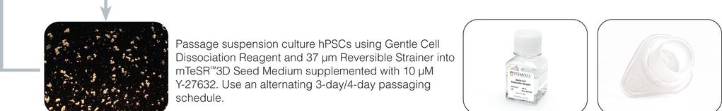
19 Passaging Suspension Culture hpscs Grown in mtesr 3D 7.1 Passaging Schedule To prevent aggregates from getting excessively large (> 400 µm), we recommend an alternating 3-day/4-day passaging schedule. If timed properly, no passaging will be required on weekends. Ensure that the mean aggregate size does not exceed 400 µm; otherwise the core of the aggregates could become nutrientdeficient, resulting in differentiation or cell death. 7.2 Choosing a Passaging Method Aggregates in suspension grow in diameter over the course of a passage and must be dissociated and re-seeded every 3-4 days. Dissociating aggregates to small clumps rather than single cells is recommended to minimize the probability of selecting for sub-populations of cells with abnormal karyotypes that can gain proliferative advantages in stressful single-cell environments. In section 7.3 a passaging method is described in which aggregates are dissociated to small clumps using Gentle Cell Dissociation Reagent and a 37 µm Reversible Strainer. 7.3 Passaging Aggregates as Small Clumps Aggregates can be dissociated to small clumps ( µm in diameter) using the following protocol in which they are first treated with Gentle Cell Dissociation Reagent (GCDR), then resuspended in mtesr 3D Seed Medium + 10 µm Y-27632, and finally pushed through a 37 µm Reversible Strainer to generate small clumps. The described protocol is for the dissociation of aggregates from an initial culture volume of 20 ml and a day 4 volume of ~26.7 ml. Volumes of GCDR and mtesr 3D Seed Medium + 10 µm Y can be scaled according to the culture volume (see Table 5). Table 5. Recommended Volumes of GCDR and Resuspension Medium When Dissociating Aggregates INITIAL CULTURE VOLUME (ml) DAY 4 CULTURE VOLUME (ml) VOLUME OF GCDR (ml) VOLUME OF mtesr 3D SEED MEDIUM + 10 µm Y FOR RESUSPENSION (ml) 1. Aliquot and warm 5 ml of GCDR to 37 C. 2. Bring mtesr 3D Seed Medium to room temperature (15-25 C). 3. Prepare enough mtesr 3D Seed Medium + 10 µm Y to resuspend and seed all conditions. See Table 5 for recommended resuspension volume. 4. Image cultures prior to passaging to assess aggregate size.
20 16 5. Filter out non-aggregated single cells by passing the entire volume of the culture through a large 37 µm Reversible Strainer into a 50 ml conical tube. Note: For suspension cultures in a 6-well plate, the small 37 µm Reversible Strainer can be used with a 15 ml conical tube. Collect aggregates on the smaller side of the strainer with the arrow pointing up. Note: If desired, cells in the filtrate can be quantified by centrifuging the collection tube, removing all but ~1 ml, and counting using a viability stain such as acridine orange (AO)/DAPI. 6. Flip the strainer onto a new 50 ml conical tube and rinse with 5 ml of warm GCDR, gently tapping the strainer to dislodge all aggregates into the new tube. 7. After the aggregates are rinsed off the strainer, flip the strainer onto a second 50 ml conical tube and set aside. This strainer can be used to dissociate aggregates into small clumps (step 13). Ensure that the strainer side that contacted the aggregates is facing up to prevent any non-dissociated aggregates from being re-seeded into the subsequent passage. 8. Incubate the conical tube containing aggregates and GCDR in a 37 C water bath for 6 minutes (undisturbed). Note: In this step, the aggregates are partially dissociated by the GCDR in preparation for generation of small clumps (step 13). Note: Optimal incubation time may vary depending on the cell line. 9. Gently remove the conical tube from the water bath without disturbing the cell pellet. Note: For larger volumes of GCDR (> 5 ml), centrifuge the tube for 2 minutes at 100 x g to collect any aggregates that have not settled. 10. Slowly aspirate the GCDR using a serological pipette, leaving ~0.5 ml to avoid removing any aggregates. 11. Add 5 ml of mtesr 3D Seed Medium + 10 µm Y to the tube. Flick or gently swirl the tube to resuspend the aggregates. Note: If the culture has expanded > 5-fold over the course of the passage and aggregate density is high, consider doubling the volume of mtesr 3D Seed Medium + 10 µm Y used to resuspend the aggregates prior to pushing through the 37 µm strainer. 12. Using a 25 ml serological pipette, remove the resuspended partially dissociated aggregates from the tube. Note: Ensure there are no bubbles at the end of the pipette tip prior to pushing the aggregates through the strainer. Prevent bubbles by aspirating the aggregate suspension from the tube slowly. Remove bubbles by shifting the pipette to a diagonal angle and tapping the pipette lightly on a tube to force the bubbles upwards and away from the tip. Note: Work quickly to prevent the aggregates from settling to the bottom of the pipette before pushing through the strainer. 13. Using the strainer and new conical tube from step 7, place the pipette containing aggregates directly on top of the strainer in a vertical orientation so that the pipette is level, with no gaps between it and the strainer. With the slowest setting on the Pipette-Aid, pass the partially dissociated aggregates through the strainer (0.5 ml/second flow rate). This will generate clumps of appropriate size to initiate the subsequent passage. For larger culture volumes, a stepwise procedure is recommended (see Table 6). Note: If the strainer appears clogged, slide the pipette laterally on the strainer while maintaining direct contact with it. Alternatively, increasing the flow rate slightly can help to prevent clogging, but use the lowest flow rate possible to minimize damage to the cells.
21 17 Table 6. Scaled Protocols for Pushing GCDR-Treated Aggregates Through a 37 µm Reversible Strainer INITIAL CULTURE VOLUME (ml) SIZE OF 37 µm REVERSIBLE STRAINER STEPWISE VOLUMES OF RESUSPENDED AGGREGATES FORCED THROUGH 37 µm REVERSIBLE STRAINER 2 Small (15 ml tube) 1 x 1 ml (5 ml serological pipette) 20 Large (50 ml tube) 1 x 5 ml (25 ml serological pipette) 50 Large (50 ml tube) 3 x 5 ml (25 ml serological pipette) 100 Large (50 ml tube) 3 x 10 ml (50 ml serological pipette) 14. Optional: Rinse the tube from step 12 with 5 ml of additional mtesr 3D Seed Medium + 10 µm Y Pass through the strainer into the tube used in step 13 to collect any remaining aggregates from the conical tube after GCDR dissociation. Note: hpsc clumps may remain on the surface of the strainer after passing the partially dissociated aggregates through it. Minimize loss by adjusting the flow rate and volume of flow-through for individual cell lines. To increase yield, rinse the strainer with an additional 1-5 ml of mtesr 3D Seed Medium + 10 µm Y Gently resuspend the clump suspension. Remove a sample for counting viable cells or clumps (see Appendix 2: Counting Clumps). 16. Seed the clumps into a new culture vessel by adding the concentrated clump suspension to mtesr 3D Seed Medium + 10 µm Y at room temperature (15-25 C) and topping up with additional medium to the desired final volume and cell density. Note: Clumps may aggregate if left for extended periods of time (> 15 minutes) as a pellet. If very large clumps have formed (> µm), gently triturate the entire clump suspension 1-2 times using a 25 ml serological pipette at a flow rate of ~1 ml per second immediately before seeding.
22 Troubleshooting PROBLEM Aggregates do not form after 24 hours Aggregates are still small on day 4 Aggregates are merging into large conglomerates Many single cells have not formed aggregates Aggregates are getting too big by end of passage Aggregates will not dissociate to small clumps at passage Aggregates will not dissociate to single cells at passage (if using single-cell passaging strategy) Too many single cells post-dissociation Medium is very yellow by end of passage Cells are sticking to the vessel wall Aggregates look dense and dark SOLUTION Increase seeding density Decrease rpm Increase initial clump size Ensure Y is present in Seed Medium Increase seeding density Decrease rpm Increase initial clump size Increase length of early passages Ensure 2D maintenance culture is high quality Decrease seeding density Use less shear during passaging Ensure 2D maintenance culture is high quality Monitor over more passages (may improve) Decrease seeding density On day 1 (t = 24 h), use 37 µm Reversible Strainer to remove single cells from culture Decrease seeding density Decrease length of passage Decrease clump size on day 0 Decrease length of passage Increase incubation time during dissociation Increase shear force during dissociation Decrease length of passage Increase incubation time during dissociation Increase shear force during dissociation Use 37 µm Reversible Strainer to exclude undissociated aggregates from the culture Ensure 2D maintenance culture is high quality Decrease incubation time during dissociation Decrease shear force during dissociation Decrease length of passage Decrease seeding density Ensure 2D maintenance culture is high quality Decrease seeding density Coat polycarbonate vessel with Anti-Adherence Rinsing Solution Coat glass vessel with Sigmacote Decrease rpm Ensure maintenance culture is high quality Decrease length of passage Decrease seeding density
23 19 PROBLEM Aggregates are irregular in shape SOLUTION Ensure 2D maintenance culture is high quality Start with more consistent clump size on day 0 Optimize culture conditions such as rpm, initial culture volume, seeding density Aggregates are non-uniform in size Start with more consistent clump size on day 0 Cells in aggregates lose expression of undifferentiated hpsc markers Cells in aggregates develop karyotype abnormalities Cells from aggregates differentiate at lower efficiency than 2D mtesr 1-derived cells Optimize culture conditions such as rpm, initial culture volume, seeding density Ensure 2D maintenance culture is high quality Decrease length of passage Decrease seeding density Passage when the aggregates are no larger than µm in size Ensure maintenance culture is high quality Passage as clumps rather than as single cells Optimize passaging to maximize percentage of cells remaining in clumps See Cells in aggregates lose expression of undifferentiated hpsc markers Try lower seeding densities in monolayer differentiation protocols, as reseeding from 3D may be more efficient than from standard 2D cultures
24 References 1. Chen VC et al. (2012) Scalable GMP compliant suspension culture system for human ES cells. Stem Cell Res 8(3): Wang Y et al. (2013) Scalable expansion of human induced pluripotent stem cells in the defined xenofree E8 medium under adherent and suspension culture conditions. Stem Cell Res 11(3): Krawetz R et al. (2010) Large-scale expansion of pluripotent human embryonic stem cells in stirredsuspension bioreactors. Tissue Eng Part C Methods 16(4): Larijani MR et al. (2011) Long-term maintenance of undifferentiated human embryonic and induced pluripotent stem cells in suspension. Stem Cells Dev 20(11): Amit M et al. (2010) Suspension culture of undifferentiated human embryonic and induced pluripotent stem cells. Stem Cell Rev Reports 6(2): Steiner D et al. (2010) Derivation, propagation and controlled differentiation of human embryonic stem cells in suspension. Nat Biotechnol 28(4): Singh H et al. (2010) Up-scaling single cell-inoculated suspension culture of human embryonic stem cells. Stem Cell Res 4(3): Olmer R et al. (2012) Suspension culture of human pluripotent stem cells in controlled, stirred bioreactors. Tissue Eng Part C Methods 18(10): Bai Q et al. (2014) Temporal analysis of genome alterations induced by single-cell passaging in human embryonic stem cells. Stem Cells Dev 00(00): Garitaonandia I et al. (2015) Increased risk of genetic and epigenetic instability in human embryonic stem cells associated with specific culture conditions. PLoS One 10(2): 1 26.
25 Appendix 1: Preparing Suspension Culture Vessels Coating Polycarbonate Vessels If cells are sticking to the vessel walls of your polycarbonate culture vessel, it is recommended to coat the vessels with Anti-Adherence Rinsing Solution prior to inoculation. Anti-Adherence Rinsing Solution contains a hydrophobic surfactant that prevents cells from sticking to the vessel walls. Cell sticking can also be minimized by decreasing the shear in the system. 1. Obtain a new Corning 125 ml Polycarbonate Erlenmeyer Flask or Qorpak 300 ml Polycarbonate Round Bottle. 2. Add 20 ml Anti-Adherence Rinsing Solution and close the lid tightly. Note: If culturing > 20 ml, increase the volume of AggreWell Rinsing Solution to equal the initial culture volume. 3. Incubate at 37 C for 15 minutes on a shaker at 145 rpm. 4. Remove from the incubator and remove the Anti-Adherence Rinsing Solution. 5. Add 10 ml of warm (room temperature; C) DMEM/F-12. Close lid tightly, then swirl and rotate the bottle to rinse all surfaces without allowing DMEM/F-12 to go into the lid. Remove DMEM/F Repeat step 5 two more times. 7. Close the lid tightly until ready to use Cleaning and Coating Glass Spinner Flasks Cleaning and coating reusable glass spinner flasks (such as the INTEGRA CELLSPIN models) prior to adding cells is recommended to remove debris and to prevent cells from sticking to the vessel walls. Note: The full cleaning and coating procedure does not need to be repeated prior to each use. At a minimum, clean the glass with warm distilled water and a gentle brush between uses followed by autoclaving. Monitor the glass over the course of each experiment for cell and debris adherence. Repeat the full cleaning and coating procedure when the cell debris adherence is noticeable. Do not use detergents, or cleaning tools that have come in contact with detergents, on glass spinner flasks. 1. Ensure that the glass surface of the spinner flask is clean and free of debris. Note: Use warm distilled water and soft brush to remove debris if necessary. 2. Prepare a 0.1 M solution of NaOH in distilled water. 3. Add 0.1 M NaOH to the spinner flask and soak for minutes at room temperature (15-25 C). Note: Volume of NaOH should be greater than or equal to the anticipated volume of suspension culture by the end of the culture passage. 4. Discard NaOH. Rinse thoroughly 3 times with an equal volume of distilled water. 5. Allow the glass to air dry until no moisture remains on surface. 6. Add 2 ml of Sigmacote to the spinner flask. Using a serological pipette, rinse all surfaces that will contact cells, including the pendulum or impeller. Note: Work in a fume hood when using Sigmacote. Excess Sigmacote can be removed and reused. If disposing of excess Sigmacote, do so in accordance with your institution's policies. 7. Leave the flask in a chemical fume hood overnight to allow the glass surface to dry completely. 8. Rinse with a large volume (to the level of the side-arms) of distilled water 3 times. 9. Autoclave the glass spinner flask in a standard dry autoclave cycle (121 C for 30 minutes). 10. Rinse the surface three times with DMEM/F-12 prior to use. Note: Rinse volume should be approximately 50% of the volume anticipated for cell culture.
26 Appendix 2: Counting Clumps Accurate and precise quantification of clumps dissociated from 2D and 3D cultures is critical for measuring expansion and accurately seeding subsequent passages. Two methods for counting clumps are described below. The first method, Manual Clump Counting, can be performed by any lab with an inverted microscope and is identical to the method used to count clumps for mtesr 1 colony passaging. The second method, Viable Nuclei Counting, requires an image-based cell counting instrument such as the ChemoMetec NucleoCounter NC-250 in combination with a viability stain. Manual Clump Counting Count clumps 50 µm in diameter, as these are the most likely to form aggregates of ideal size. 1. Draw a + centered on the bottom of 2 wells of a 96-well flat-bottom plate (e.g. Catalog #38022) to serve as a counting grid. 2. Aliquot 40 µl of DMEM/F-12 into each well. 3. Add 5 µl of a freshly dissociated and resuspended clump suspension to each well. Ensure that the clump suspension is well-mixed prior to removing the sample for counting. 4. Count the clumps in each well that are 50 µm in diameter. Average the results from the two wells to obtain the average number of clumps (N A ) in the 5 µl sample. 5. Calculate the concentration of clumps (C) and the total number of clumps (N T ) using the total volume of the clump suspension in µl (V T ): C = (N A / 5 µl) N T = C x V T 6. Determine the target number of clumps to seed (N p ) by referring to Table 3 for the optimal seeding density and multiplying by volume. 7. Calculate the volume (in µl) of clump suspension to seed (V p ) for each condition in your experiment: V P = N P / C 8. Gently mix the clump suspension prior to seeding to ensure a uniform suspension. 9. Add calculated volume of clump suspension (V P ) to the culture vessel and add mtesr 3D Seed Medium + Y to desired volume. Viable Nuclei Counting Alternatively, clump cultures can be quantified as viable cells/ml using an image-based cell counting instrument such as the ChemoMetec NucleoCounter NC-250 in combination with a viability stain. Contact us at techsupport@stemcell.com for further information.
27 Appendix 3: Differentiating Suspension Cultures into Three Germ Layers hpscs grown in suspension culture with mtesr 3D have the ability to differentiate to the three germ layers mesoderm, definitive endoderm, and ectoderm. Cells derived from hpsc suspension culture tend to have a higher plating efficiency than those derived from 2D mtesr 1 cultures when seeded as a monolayer for differentiation protocols. If cultures appear overgrown compared to 2D controls on day 1 of a differentiation protocol, consider lowering the seeding density to achieve desired confluency for differentiation. Generate a single-cell suspension using an enzymatic reagent, then use STEMdiff Trilineage Differentiation Kit (Catalog #05230) to differentiate cells to the three germ layers.
28 Appendix 4: Re-Establishing 2D Colony Cultures For suspension cultures that have been passaged as clumps: 1. Dissociate aggregates to small clumps following the protocol outlined in section Count dissociated clumps according to the protocol outlined in Appendix Plate clumps/well onto a Corning Matrigel -coated 6-well plate in 2 ml of mtesr µm Y Maintain as a standard 2D hpsc culture. Note: For additional information on coating plates, maintaining 2D cultures, and passaging 2D to 2D, refer to the Technical Manual: Maintenance of Human Pluripotent Stem Cells in mtesr 1 (Document #28315), available at or contact us to request a copy. 4. After one or two 2D clump passages, typical mtesr 1 colony morphology should be restored. For suspension cultures that have been passaged as single cells: 1. Plate 4 x 10 5 cells/well onto a Corning Matrigel -coated 6-well plate in 2 ml of mtesr 3D Seed Medium + 10 µm Y When wells are ~80% confluent (3-4 days), clump passage cells using GCDR. Count dissociated clumps according to the protocol outlined in Appendix Plate clumps/well onto a Corning Matrigel -coated 6-well plate in 2 ml of mtesr µm Y Maintain as a standard 2D hpsc culture. Note: For additional information on coating plates, maintaining 2D cultures, and passaging 2D to 2D, refer to the Technical Manual: Maintenance of Human Pluripotent Stem Cells in mtesr 1 (Document #28315), available at or contact us to request a copy. 4. After one or two 2D clump passages, typical mtesr 1 colony morphology should be restored.
29 Appendix 5: G-Banding Karyotype To assess whether chromosomal abnormalities have been acquired or have increased in prevalence in suspension cultures, it is recommended to assess the karyotype at the beginning and end of an experiment. All STEMCELL Technologies G-banding karyotypes are analyzed by WiCell Research Institute in Wisconsin, USA. The resolution of G-banding karyotype is limited to > 5 Mb. For higher resolution of variations in gene copy number, deletions, duplications, and other genotypic changes, consider using fluorescent in situ hybridization (FISH), SNP genotyping, RT-PCR, or microarrays. G-banding karyotypes are also limited in the number of spreads analyzed. Due to the laborious nature of the protocol, only cells are analyzed per culture. Low-frequency sub-populations of abnormal cells can go undetected in both maintenance and experimental cultures. Improper culture techniques may increase the frequency of genetically abnormal cells in culture.
30 Appendix 6: Dissociating Aggregates to Single Cells for Flow Cytometry The following are instructions for preparing a single-cell suspension from aggregate cultures grown in mtesr 3D in non-tissue culture-treated 6-well plates. If using other cultureware, adjust volumes accordingly. Cultures should be harvested at the time they would normally be ready for passaging. 1. Aliquot and warm an appropriate enzymatic cell dissociation reagent to 37 C. 2. Warm medium (DMEM/F-12 or mtesr 3D Seed Medium) and D-PBS (Without Ca++ and Mg++) to room temperature (15-25 C) before use. 3. Filter out non-aggregated single cells by transferring the entire volume of culture through a small 37 µm Reversible Strainer into a 15 ml conical tube. Note: Collect aggregates on the smaller side of strainer with the arrow pointing up. 4. Flip the strainer onto a new 15 ml conical tube and rinse with 2 ml of D-PBS (Without Ca++ and Mg++), tapping strainer to release all aggregates. 5. Allow aggregates to settle for 3-5 minutes, then remove D-PBS. 6. Add 1 ml of warm enzymatic reagent. Incubate at 37 C for 7 minutes. Note: The incubation time may vary for different cell lines and enzymatic reagents. 7. Dissociate the aggregates by pipetting up and down to create a single-cell suspension. 8. Add 2-4 ml of medium (DMEM/F-12 or mtesr 3D Seed Medium). 9. Centrifuge cells at 300 x g for 5 minutes. 10. Resuspend cells in an appropriate medium for desired downstream applications (e.g. 2% FBS/PBS for flow cytometry).
31 Appendix 7: Flow Cytometry Methods 16.1 Reagents and Materials Antibodies Antibodies can be used to characterize hpscs by flow cytometry. The tables below list a selection of antibodies available from STEMCELL Technologies that can be used to characterize undifferentiated hpsc cultures. For a complete list of antibodies, including other conjugates, sizes, and clones, visit Surface Antigen Labeling PRIMARY ANTIBODY* SPECIES REACTIVITY ISOTYPE CATALOG # Anti-Mouse SSEA-1 Antibody, Clone MC-480 Human, Mouse, Rat IgM, kappa (Mouse) Anti-Mouse SSEA-3 Antibody, Clone MC-631 Human, Mouse, Rat, Rhesus IgM, kappa (Rat) Anti-Human SSEA-4 Antibody, Clone MC Human, Mouse, Rat, Rhesus, Cat, Chicken, Dog, Rabbit IgG3, kappa (Mouse) Anti-Human SSEA-5 Antibody, Clone 8e11 Human IgG1, kappa (Mouse) Anti-Human TRA-1-60 Antibody, Clone TRA-1-60R Human, Rhesus, Rabbit IgM, kappa (Mouse) Anti-Human TRA-1-81 Antibody, Clone TRA-1-81 Human, Rat, Rhesus IgM, kappa (Mouse) Anti-Human TRA-2-49 Antibody, Clone TRA-2-49/6E Anti-Human TRA-2-54 Antibody, Clone TRA-2-54/2J Human, Chimpanzee, Gibbon, Gorilla, Orangutan, Owl Monkey, Squirrel Monkey, Cat, Pig, Rabbit, Tiger Human, Chimpanzee, Gibbon, Gorilla, Orangutan, Owl Monkey, Squirrel Monkey, Cat, Pig, Rabbit, Tiger *Optimal working dilutions of the antibodies should be determined by the end user. Intracellular Antigen Labeling IgG1, kappa (Mouse) IgG1, kappa (Mouse) PRIMARY ANTIBODY* SPECIES REACTIVITY ISOTYPE CATALOG # Anti-Human OCT4 (OCT3) Antibody, Clone 3A2A20 Human IgG2b, kappa (Mouse) *Optimal working dilutions of the antibodies should be determined by the end user.
32 General Reagents and Materials REAGENTS AND MATERIALS CATALOG # D-PBS (Without Ca++ and Mg++) DMEM/F-12 with 15 mm HEPES Trypan Blue Gentle Cell Dissociation Reagent Dulbecco s Phosphate Buffered Saline with 2% Fetal Bovine Serum (2% FBS/PBS) Costar Microcentrifuge Tubes, 1.7 ml Falcon Round-Bottom Tubes, 5 ml Falcon Conical Tubes, 15 ml Optional: Nuclear stain (e.g. 1 mg/ml propidium iodide diluted 1 in 1000 in 2% FBS/PBS) Additional Reagents Required for Intracellular Antigen Labeling Saponin Permeabilization Buffer (SPB)* COMPONENT CATALOG # FINAL CONCENTRATION Saponin e.g. Fluka Biochemika mg/ml 10% BSA Solution % D-PBS (Without Ca++ and Mg++) to final volume *Mix well and store at 2-8 C for up to 1 month. 2% Paraformaldehyde Solution* COMPONENT CATALOG # FINAL CONCENTRATION Paraformaldehyde e.g. Affymetrix % D-PBS (Without Ca++ and Mg++) to final volume *Mix well and store at 2-8 C Preparation of a Single-Cell Suspension for Flow Cytometry Prepare a single-cell suspension as indicated in Appendix 6: Dissociating Aggregates to Single Cells for Flow Cytometry. Perform a viable cell count using either Trypan Blue and a hemocytometer or AO/DAPI and a ChemoMetec NucleoCounter NC-250. The single-cell suspension may now be used for surface antigen and/or intracellular antigen labeling (see sections 16.3 and 16.4 for detailed protocols).
33 Surface Antigen Labeling Protocol Note: Optimal concentrations of antibodies must be predetermined by titration for each antibody. 1. Determine the number of samples required to perform flow cytometry, including labeling controls. 2. Aliquot approximately 2 x 10 5 cells per sample into a 5 ml tube or a 1.7 ml tube and place on ice. 3. Centrifuge cells at 300 x g for 5 minutes. 4. While the samples are centrifuging, prepare a sufficient quantity of the primary antibody mix or the directly conjugated antibody mix (100 µl/sample) using the appropriate antibody at the predetermined optimal working dilution. 5. Carefully remove the supernatant without disrupting the cell pellet and resuspend cells in the primary antibody mix. Gently mix and incubate on ice for minutes. Note: If using a directly conjugated antibody, protect samples from exposure to direct light. 6. Add 1 ml of 2% FBS/PBS to each tube, gently mix, and centrifuge at 300 x g for 5 minutes. If using an unconjugated primary antibody: While the samples are centrifuging, prepare a sufficient quantity of the secondary antibody mix (100 µl/sample) using the appropriate secondary antibody at the predetermined optimal working dilution. Proceed to step 7. If using a directly conjugated antibody, proceed to step Carefully remove the supernatant without disturbing the cell pellet and resuspend the cells in the secondary antibody mix. Gently mix and incubate on ice for minutes. Protect samples from exposure to direct light. 8. Add 1 ml of 2% FBS/PBS to each tube. Gently mix and centrifuge at 300 x g for 5 minutes. 9. Carefully remove the supernatant without disturbing the cell pellet and resuspend the cells in µl of 2% FBS/PBS. Transfer to a 5 ml tube if necessary. Optional: Propidium iodide (PI) can be added at a final concentration of 1 µg/ml to assess viability. 10. Place samples on ice, while avoiding exposure to direct light, and analyze by flow cytometry as soon as possible Intracellular Antigen Labeling Protocol for OCT4 Note: Optimal concentrations of antibodies must be predetermined by titration for each antibody. 1. Determine the number of samples required to perform flow cytometry, including labeling controls. 2. Aliquot approximately 4 x 10 5 cells per sample into a 5 ml tube or a 1.7 ml tube. 3. Centrifuge cells at 300 x g for 5 minutes. 4. Carefully remove the supernatant without disrupting the cell pellet and resuspend cells in 250 µl of 2% Paraformaldehyde Solution/tube. Gently mix and incubate on ice for minutes. 5. Add 1 ml of 2% FBS/PBS per tube. Gently mix and centrifuge at 300 x g for 5 minutes. 6. Carefully remove the supernatant without disrupting the cell pellet and resuspend cells in 500 µl of SPB/tube. Gently mix and incubate at room temperature (15-25 C) for 15 minutes. Note: Cells should remain in SPB until the final resuspension step, prior to flow cytometric analysis. 7. While the samples are incubating, prepare a sufficient quantity of the primary antibody mix (100 µl/sample) at the predetermined optimal working dilution, using SPB as the diluent. 8. Centrifuge cells at 300 x g for 5 minutes.
STEMdiff Mesenchymal Progenitor Kit
 Defined culture kit for derivation and expansion of mesenchymal progenitor cells Catalog #05240 1 Kit Product Description STEMdiff Mesenchymal Progenitor Kit is a defined culture kit consisting of animal
STEMdiff Hematopoietic Kit
 For differentiation of human ES or ips cells into hematopoietic progenitor cells Catalog #05310 1 Kit Product Description Kit includes a defined, serum-free basal medium and supplements for the feeder-free
Maintenance of Human Pluripotent Stem Cells in TeSR -E8
 Maintenance of Human Pluripotent Stem Cells in TeSR -E8 i Critical Parameters for Successful Cell Culture with TeSR E8 Preparation and Storage of Complete TeSR -E8 Medium It is critical to store complete
Propagation of H7 hesc From: UW (John Stamatoyannopoulos) ENCODE group Date: 12/17/2009 Prepared By: S. Paige/S. Hansen (UW)
 ENCODE group Date: 12/17/2009 Prepared By: S. Paige/S. Hansen (UW)") Propagation of H7 hesc From: UW (John Stamatoyannopoulos) ENCODE group Date: 12/17/2009 Prepared By: S. Paige/S. Hansen (UW) Growth and Harvest Modifications Addendum to: Propagation of H7 hesc from UW
Propagation of H7 hesc From: UW (John Stamatoyannopoulos) ENCODE group Date: 12/17/2009 Prepared By: S. Paige/S. Hansen (UW) Growth and Harvest Modifications Addendum to: Propagation of H7 hesc from UW
Maintenance of Human Pluripotent Stem Cells in mtesr 1
 Maintenance of Human Pluripotent Stem Cells in mtesr 1 1 i Critical Parameters for Successful Cell Culture with mtesr 1 Choosing an Appropriate Matrix for Use with mtesr 1 Cells may be cultured in mtesr
Generation and Culture of Neural Progenitor Cells using the STEMdiff Neural System
 Generation and Culture of Neural Progenitor Cells using the STEMdiff Neural System i Table of Contents 1.0 Introduction... 1 2.0 Materials, Reagents and Equipment... 2 2.1 Materials Required for Neural
Maintenance of Human Pluripotent Stem Cells in TeSR 2
 Maintenance of Human Pluripotent Stem Cells in TeSR 2 1 i Critical Parameters for Successful Cell Culture with TeSR 2 Choosing an Appropriate Matrix for Use with TeSR 2 Cells may be cultured in TeSR 2
Protocols for Neural Progenitor Cell Expansion and Dopaminergic Neuron Differentiation
 Protocols for Neural Progenitor Cell Expansion and Dopaminergic Neuron Differentiation In vitro neurological research presents many challenges due to the difficulty in establishing high-yield neuronal
T ECHNIC AL MANUAL VERSIO N Maintenance of Human Pluripotent Stem Cells in mtesr 1 and TeSR 2
 T ECHNIC AL MANUAL VERSIO N 3. 0.0 Maintenance of Human Pluripotent Stem Cells in mtesr 1 and TeSR 2 i Table of Contents 1.0 Introduction... 1 1.1 Development of Serum-Free and Feeder-Free Culture Systems
HUMAN IPSC CULTURE PROTOCOLS
 HUMAN IPSC CULTURE PROTOCOLS FOR TRAINING COURSE IXCELLS BIOTECHNOLOGIES USA, LLC 10340 CAMINO SANTA FE, SUITE C, SAN DIEGO, CA 92121 TABLE OF CONTENTS CHAPTER 1 BEFORE STARTING 2-7 SECTION 1.1 COATING
T ECHNICAL MANUAL. Culture of Human Mesenchymal Stem Cells Using MesenCult -XF Medium
 T ECHNICAL MANUAL Culture of Human Mesenchymal Stem Cells Using MesenCult -XF Medium i Table of Contents 1.0 Materials... 1 1.1 MesenCult -XF Medium and Required Products... 1 1.2 Additional Required
STEMdiff Astrocyte Differentiation Kit and STEMdiff Astrocyte Maturation Kit
 Kit and Kit Catalog #08540 Catalog #08550 1 Kit 1 Kit Product Description Kit (Catalog #08540) is used to rapidly and efficiently generate astrocyte precursors from neural progenitor cells (NPCs) derived
ReproRNA -OKSGM is a non-integrating, self-replicating RNA-based reprogramming vector for generating induced pluripotent stem (ips)
") Kit for generating ips cells using ReproRNA -OKSGM, a non-integrating, self-replicating RNA reprogramming vector Product Description ReproRNA -OKSGM is a non-integrating, self-replicating RNA-based reprogramming
Kit for generating ips cells using ReproRNA -OKSGM, a non-integrating, self-replicating RNA reprogramming vector Product Description ReproRNA -OKSGM is a non-integrating, self-replicating RNA-based reprogramming
NutriStem V9 XF Medium
 Stem Cells NutriStem V9 XF Medium A defined, xeno-free (XF), serum-free (SF) culture medium for hpsc using vitronectin Instructions for Use Product Description NutriStem V9 XF medium is a defined, xeno-free,
Guidelines for WTC Derived AICS hipsc Lines Scale Up & Banking (upto 80 vials)
") Version 1.2 2017 Allen Institute for Cell Science Guidelines for WTC Derived AICS hipsc Lines Scale Up & Banking (upto 80 vials) Note: The following protocol is for banking ~60-80 vials of cells (1 million/vial)
Version 1.2 2017 Allen Institute for Cell Science Guidelines for WTC Derived AICS hipsc Lines Scale Up & Banking (upto 80 vials) Note: The following protocol is for banking ~60-80 vials of cells (1 million/vial)
Cardiomyocyte Differentiation Methods
 Version 1.0 2018 Allen Institute for Cell Science Cardiomyocyte Differentiation Methods The methods described below were adapted from the small molecule protocol described in Lian et al. 1, and have been
Corning BioCoat Matrigel Matrix 6-well Plates for Embryonic Stem (ES) Cell Culture. Catalog Number Guidelines for Use
 Cell Culture. Catalog Number Guidelines for Use") Corning BioCoat Matrigel Matrix 6-well Plates for Embryonic Stem (ES) Cell Culture Catalog Number 354671 Guidelines for Use Discovery Labware, Inc., Two Oak Park, Bedford, MA 01730, Tel: 1.978.442.2200
Corning BioCoat Matrigel Matrix 6-well Plates for Embryonic Stem (ES) Cell Culture Catalog Number 354671 Guidelines for Use Discovery Labware, Inc., Two Oak Park, Bedford, MA 01730, Tel: 1.978.442.2200
Culture and freezing methods for AICS cell lines
 Allen Institute for Cell Science Culture and freezing methods for AICS cell lines Required Reagent List: Complete mtesr1 culture media, referred to in this protocol as simply mtesr1 : 400 ml basal media
Table of Contents. 2.1 NeuroCult NCFC Assay Kit (Rat) Components Additional Required Reagents Required Equipment...
 Components Additional Required Reagents Required Equipment...") i Table of Contents 1.0 Overview of the NeuroCult NCFC Assay 2.0 Materials 2.1 NeuroCult NCFC Assay Kit (Rat) Components... 4 2.2 Additional Required Reagents... 4 2.3 Required Equipment... 4 3.0 Preparation
i Table of Contents 1.0 Overview of the NeuroCult NCFC Assay 2.0 Materials 2.1 NeuroCult NCFC Assay Kit (Rat) Components... 4 2.2 Additional Required Reagents... 4 2.3 Required Equipment... 4 3.0 Preparation
Guide to Induced Pluripotent Stem Cell Culture
 Cellular Engineering Technologies, Inc. Guide to Induced Pluripotent Stem Cell Culture User Manual 2500 Crosspark Road E110 Coralville, IA 52241 Phone: 319-665-3000 Email: orders@celleng-tech.com Table
STEMdiff Cerebral Organoid Kit
 Kit Culture medium kit for establishment and maturation of human cerebral organoids Catalog #08570 Catalog #08571 1 Kit 1 Kit Product Description Kit (Catalog #08570) is a defined, serum-free cell culture
Human Pluripotent Stem Cell Growth Medium DXF
 Human Pluripotent Stem Cell Growth Medium DXF Instruction Manual Product Size Catalog Number hpsc Growth Medium DXF 500 ml C-28060 Recommended for Human Pluripotent Stem Cells (hpsc) Product Description
Genome Edited ipscs APOE -/- Knockout
 Applied StemCell, Inc. (866) 497-4180 www.appliedstemcell.com Datasheet Genome Edited ipscs APOE -/- Knockout Product Information Catalog Number Description Amount Parental Cell Line Gene Knockout Generated
Adaptation of hpscs from Different Culture Systems to Essential 8 Medium or Essential 8 Flex Medium
 Adaptation of hpscs from Different Culture Systems to Essential 8 Medium or Essential 8 Flex Medium Publication Part Number MAN0013768 Revision A.0 Introduction This protocol describes the transition of
All quality control test results are reported on a lot specific Certificate of Analysis which is available at or upon request.
 PRIME-XV NPC Expansion XSFM PRIME-XV NPC Expansion XSFM is a xeno-and serum-free medium optimized for the cultivation and expansion of neural progenitor cells (NPC) that are able to maintain their ability
Neural Stem Cells (ipsc from Blood Cells; Male)
") Applied StemCell, Inc. (866) 497-4180 www.appliedstemcell.com Datasheet Neural Stem Cells (ipsc from Blood Cells; Male) Product Information Catalog Number ASE-9234 (Male) Description Applied StemCell's
Applied StemCell, Inc. (866) 497-4180 www.appliedstemcell.com Datasheet Neural Stem Cells (ipsc from Blood Cells; Male) Product Information Catalog Number ASE-9234 (Male) Description Applied StemCell's
Lineage-specific Reporter Knock-in ipscs: MAP2-Nanoluc-Halotag ipsc (Heterozygous)
") Lineage-specific Reporter Knock-in ipscs: MAP2-Nanoluc-Halotag ipsc (Heterozygous) Applied StemCell, Inc. (866) 497-4180 www.appliedstemcell.com Datasheet Product Information Catalog Number Description
Lineage-specific Reporter Knock-in ipscs: MAP2-Nanoluc-Halotag ipsc (Heterozygous) Applied StemCell, Inc. (866) 497-4180 www.appliedstemcell.com Datasheet Product Information Catalog Number Description
Lineage-specific Reporter Knock-in ipscs: AAVS-DCX-GFP ipsc (Heterozygous)
") Lineage-specific Reporter Knock-in ipscs: AAVS-DCX-GFP ipsc (Heterozygous) Applied StemCell, Inc. (866) 497-4180 www.appliedstemcell.com Datasheet Product Information Catalog Number Description Amount
Lineage-specific Reporter Knock-in ipscs: AAVS-DCX-GFP ipsc (Heterozygous) Applied StemCell, Inc. (866) 497-4180 www.appliedstemcell.com Datasheet Product Information Catalog Number Description Amount
Safe Harbor Knock-in ipscs: AAVS1-CAG-GFP Chr19 ipsc (Heterozygous)
") Safe Harbor Knock-in ipscs: AAVS1-CAG-GFP Chr19 ipsc (Heterozygous) Applied StemCell, Inc. (866) 497-4180 www.appliedstemcell.com Datasheet Product Information Catalog Number Description Amount Parental
Safe Harbor Knock-in ipscs: AAVS1-CAG-GFP Chr19 ipsc (Heterozygous) Applied StemCell, Inc. (866) 497-4180 www.appliedstemcell.com Datasheet Product Information Catalog Number Description Amount Parental
Lineage-specific Reporter Knock-in ipscs: MAP2-Nanoluc-Halotag ipsc (Heterozygous)
") Lineage-specific Reporter Knock-in ipscs: MAP2-Nanoluc-Halotag ipsc (Heterozygous) Applied StemCell, Inc. (866) 497-4180 www.appliedstemcell.com Datasheet Product Information Catalog Number Description
Lineage-specific Reporter Knock-in ipscs: MAP2-Nanoluc-Halotag ipsc (Heterozygous) Applied StemCell, Inc. (866) 497-4180 www.appliedstemcell.com Datasheet Product Information Catalog Number Description
Contents Cat. No. Amount Storage Shelf life [2] 2. Mix the thawed supplement by gently inverting 3 5 times.
![Contents Cat. No. Amount Storage Shelf life [2] 2. Mix the thawed supplement by gently inverting 3 5 times.](/thumbs/72/66661577.jpg "Contents Cat. No. Amount Storage Shelf life [2] 2. Mix the thawed supplement by gently inverting 3 5 times.") USER GUIDE StemFlex Kit Catalog Number A3349401 Pub. No. MAN0016431 Rev. 2.0 WARNING! Read the Safety Data Sheets (SDSs) and follow the handling instructions. Wear appropriate protective eyewear, clothing,
USER GUIDE StemFlex Kit Catalog Number A3349401 Pub. No. MAN0016431 Rev. 2.0 WARNING! Read the Safety Data Sheets (SDSs) and follow the handling instructions. Wear appropriate protective eyewear, clothing,
Cardiomyocyte Differentiation Methods
 Version 1.1 2018 Allen Institute for Cell Science Cardiomyocyte Differentiation Methods The methods described below were adapted from the small molecule protocol described in Lian et al. 1, and have been
WOOD CELL PASSAGING PROTOCOL OVERVIEW
 CELL MODEL NAME: Wood Derived from an infiltrating ductal and lobular carcinoma of the breast. Product Category: Cellini Breast Models Catalog #: CB040-000001 Record your lot number here: Please refer
Liquid. This product is shipped with dry ice. Upon receipt, store it immediately at the temperature recommended below.
 PRIME-XV MSC Expansion SFM PRIME-XV MSC Expansion SFM is a serum-free complete medium optimized for the maintenance and expansion of purified human MSCs. This product does not contain antibiotics. Catalog
CAROUSEL CELL PASSAGING PROTOCOL OVERVIEW
 CELL MODEL NAME: Carousel Derived from an endometrioid adenocarcinoma of the ovary. Record your lot number here: Product Category: Raphael Ovarian Models Catalog #: AMS.CB010-000001 Please refer to Certificate
Protocol Reprogramming Human Fibroblasts into ips Cells using the Stemgent Reprogramming Lentivirus Set: Human OKSM
 STEMGENT Page 1 Reprogramming Lentivirus Set: Human OKSM OVERVIEW The following procedure describes the reprogramming of BJ Human Fibroblasts (BJ cells) to induced pluripotent stem (ips) cells using the
All quality control test results are reported on a lot specific Certificate of Analysis which is available at or upon request.
 PRIME-XV Neural Basal Medium PRIME-XV Neural Basal Medium is a chemically-defined basal medium optimized for the culture and maintenance of neuronal cells when supplemented with PRIME-XV IS21 Supplement
Protocol Reprogramming Human Fibriblasts using the Dox Inducible Reprogramming Polycistronic Lentivirus Set: Human 4F2A LoxP
 STEMGENT Page 1 OVERVIEW The following protocol describes the reprogramming of one well of BJ Human Fibroblasts (BJ cells) into induced pluripotent stem (ips) cells in a 6-well format. Transduction efficiency
PluriQ TM Serum Replacement (PluriQ TM SR)
") PluriQ TM Serum Replacement (PluriQ TM SR) (PluriQ SR) is a defined, serum-free supplement formulated as a direct replacement for FBS (fetal bovine serum) used in the growth and maintenance of undifferentiated
PluriQ TM Serum Replacement (PluriQ TM SR) (PluriQ SR) is a defined, serum-free supplement formulated as a direct replacement for FBS (fetal bovine serum) used in the growth and maintenance of undifferentiated
Cellartis ipsc Single-Cell Cloning DEF-CS Culture Media Kit
 Takara Bio Europe AB Cellartis ipsc Single-Cell Cloning DEF-CS Culture Media Kit Cat. No. Y30021 (050517) Takara Bio Europe AB A Takara Bio Company Arvid Wallgrens backe 20, SE-413 46 Göteborg, Sweden
Peri.4U TM Application Protocol Multiwell-MEA
 Peri.4U TM Application Protocol Multiwell-MEA Measuring Neuronal Electrical Activity of Peri.4U TM (Axiogenesis AG) on the 24-well Multiwell-MEA System (Multi Channel Systems MCS GmbH). Version 1.0: July
Human Pluripotent Stem Cell Functional Identification Kit
 Human Pluripotent Stem Cell Functional Identification Kit Catalog Number SC027B Reagents for the identification of human pluripotent stem cells by in vitro functional differentiation. This package insert
Protocol Using the Reprogramming Ecotropic Retrovirus Set: Mouse OSKM to Reprogram MEFs into ips Cells
 STEMGENT Page 1 OVERVIEW The following protocol describes the reprogramming of one well of mouse embryonic fibroblast (MEF) cells into induced pluripotent stem (ips) cells in a 6-well format. Transduction
StemXVivo. Mesoderm Kit. Catalog Number SC030B. Reagents for the differentiation of human pluripotent stem cells into mesoderm.
 StemXVivo Mesoderm Kit Catalog Number SC030B Reagents for the differentiation of human pluripotent stem cells into mesoderm. This package insert must be read in its entirety before using this product.
General Guidelines for Handling Feeder Free Human ipscs. Cells provided were cultured in the presence of Penicillin and Streptomycin.
 General Guidelines for Handling Feeder Free Human ipscs This document provides guidance on how to resuscitate, culture and cryopreserve human induced pluripotent stem cells (ipscs) supplied by the Human
RNP Transfection in WTCs
 Version 1.0 2016 Allen Institute for Cell Science RNP Transfection in WTCs Required reagent list: Complete mtesr1 culture media, referred to in this protocol as simply mtesr1 : 400 ml basal media with
Cortical Neural Induction Kit. Protocol version 1.0
 Cortical Neural Induction Kit Protocol version 1.0 Protocol version 1.0 Table of Contents Product Information 2 Preparation of Reagents 3 Protocol Overview 4 Seeding ipscs 4 Cortical Neural Induction
MiraCell Endothelial Cells (from ChiPSC12) Kit
 Kit") Cat. # Y50055 For Research Use MiraCell Endothelial Cells (from ChiPSC12) Kit Product Manual Table of Contents I. Description...3 II. III. IV. Components...4 Storage...4 Preparation Before Use...4 V. Protocol...5
Cat. # Y50055 For Research Use MiraCell Endothelial Cells (from ChiPSC12) Kit Product Manual Table of Contents I. Description...3 II. III. IV. Components...4 Storage...4 Preparation Before Use...4 V. Protocol...5
Protocol for Small-Scale Microcarrier Culture
 Protocol for Small-Scale Microcarrier Culture Hillex II Microcarriers This document provides a general protocol for initiation of small-scale mammalian cell spinner cultures. This procedure is based upon
Protocol Using a Dox-Inducible Polycistronic m4f2a Lentivirus to Reprogram MEFs into ips Cells
 STEMGENT Page 1 OVERVIEW The following protocol describes the transduction and reprogramming of one well of Oct4-GFP mouse embryonic fibroblasts (MEF) using the Dox Inducible Reprogramming Polycistronic
hpsc Maintenance Media
 hpsc Maintenance Media Brigitte Arduini, version 2, 2013-Jun-12 Initially, it was found that pluripotency of human pluripotent stem cells (hpscs) can be maintained when plated in co-culture with mouse
Required Materials. Stemgent Reprogramming-Qualified Nuff Cells GSC-3006G 4 x 10 6 cells/vial Liquid Nitrogen
 Page 1 Overview This protocol for microrna-enhanced mrna reprogramming describes the combined use of the Stemgent mrna Reprogramming Kit, the Stemgent microrna Booster Kit, and the Stemfect RNA Transfection
Protocol. Culture for human ips cells under the feeder-free condition
 Protocol Culture for human ips cells under the feeder-free condition 1 Table of contents PASSAGE AND MAINTENANCE... 3 FREEZE PROTOCOL... 6 THAW PROTOCOL... 8 TRANSFER IPSCS FROM ON-FEEDER TO ON-LAMININ...
Ready to use: Serum free culture in Insectomed SF express
 Ready to use: Serum free culture in Insectomed SF express Information from Biochrom AG, 19.05.2010 Serum free culture in Insectomed SF express is a proprietary formulation which has successfully been used
Tube Formation Assays in µ-slide Angiogenesis. 1. General Information. Application Note 19
 Tube Formation Assays in µ-slide Angiogenesis Contents 1. General Information... 1 2. Material... 2 3. Work Flow Overview... 3 4. Preparation of the Gel and the Slide... 4 4.1. Gel Application... 4 4.2.
All quality control test results are reported on a lot specific Certificate of Analysis which is available at or upon request.
 PRIME-XV MSC Expansion XSFM PRIME-XV MSC Expansion XSFM is a serum-free and xeno-free complete medium optimized for the maintenance and expansion of purified human MSCs. This product does not contain antibiotics.
GENOME EDITING OF HUMAN PLURIPOTENT STEM CELLS
 TECHNICAL BULLETIN GENOME EDITING OF HUMAN PLURIPOTENT STEM CELLS Using the ArciTect CRISPR-Cas9 System Introduction The ability of human embryonic stem (ES) and induced pluripotent stem (ips) cells to
Protocol Reprogramming MEFs using the Dox Inducible Reprogramming Lentivirus Set: Mouse OKSM
 STEMGENT Page 1 OVERVIEW The following protocol describes the reprogramming of one well of mouse embryonic fibroblasts (MEFs) into induced pluripotent stem (ips) cells in a 6-well format. Transduction
All quality control test results are reported on a lot specific Certificate of Analysis which is available at or upon request.
 PRIME-XV MSC Expansion XSFM Catalog # Product Size 91149 PRIME-XV MSC Expansion XSFM 250 ml and 1 L liquid Intended Use This product is for research use or further manufacturing use only. This product
Cellartis MSC Xeno-Free Culture Medium
 Cat. # Y50200 For Research Use Cellartis MSC Xeno-Free Culture Medium Product Manual Table of Contents I. Description... 3 II. Contents... 3 III. Storage... 3 IV. Precautions... 3 V. Materials Required
Culturing Pluripotent Stem Cells (PSCs) in Essential 8 Medium
 in Essential 8 Medium") Culturing Pluripotent Stem Cells (PSCs) in Essential 8 Medium Publication Part Number MAN0007035 Revision B.0 Introduction Essential 8 Medium is a fully defined, feeder-free medium formulated for the growth
Culturing Pluripotent Stem Cells (PSCs) in Essential 8 Medium Publication Part Number MAN0007035 Revision B.0 Introduction Essential 8 Medium is a fully defined, feeder-free medium formulated for the growth
Protocol Reprogramming Human Fibroblasts into ips Cells using the Stemgent VSVg Retrovirus Reprogramming Set: Human OKSM
 STEMGENT Page 1 VSVg Retrovirus Reprogramming Set: Human OKSM OVERVIEW The following protocol describes the reprogramming of BJ Human Fibroblasts (BJ cells) into induced pluripotent stem (ips) cells in
Kit Components (Included) Cat # # of vials Product Name Quantity Storage Human Renal Cortical Epithelial Cells (HRCEpiC)
 Cat # # of vials Product Name Quantity Storage Human Renal Cortical Epithelial Cells (HRCEpiC)") 3D Renal Tubule Formation Kit 3D-RTF Cat # 3D-4110 Product Description The human kidney is frequently exposed to drugs and toxic compounds, which can result in nephrotoxicity. A drug s uptake and clearance
3D Renal Tubule Formation Kit 3D-RTF Cat # 3D-4110 Product Description The human kidney is frequently exposed to drugs and toxic compounds, which can result in nephrotoxicity. A drug s uptake and clearance
PROTOCOL PluriQ Serum Replacement
 (PluriQ SR) is a serum free supplement formulated as a direct replacement for FBS (fetal bovine serum) used in the growth and maintenance of undifferentiated human embryonic stem cells (hesc) and induced
Transfection of pluripotent stem cells with Lipofectamine Stem reagent in mtesr1 Medium on Geltrex matrix
 TRANSFECTION PROTOCOL Lipofectamine Stem Transfection Reagent Transfection of pluripotent stem cells with Lipofectamine Stem reagent in mtesr1 Medium on Geltrex matrix PSC growth medium, passaging reagents,
Components Neural induction basal medium (Cat. # ) 100 ml x 5 Neural induction medium supplement 50x (Cat. # ) 2 ml x 5
 100 ml x 5 Neural induction medium supplement 50x (Cat. # ) 2 ml x 5") HPSC Neural Induction Medium (PSCNIM) Catalog #5931 Introduction Human embryonic stem cells (hesc) and induced pluripotent stem cells (hipsc) possess the capability of differentiating into all derivatives
HPSC Neural Induction Medium (PSCNIM) Catalog #5931 Introduction Human embryonic stem cells (hesc) and induced pluripotent stem cells (hipsc) possess the capability of differentiating into all derivatives
StemXVivo TM. Endoderm Kit. Catalog Number SC019. Reagents for the differentiation of human pluripotent stem cells into definitive endoderm.
 StemXVivo TM Endoderm Kit Catalog Number SC019 Reagents for the differentiation of human pluripotent stem cells into definitive endoderm. This package insert must be read in its entirety before using this
Part 1: Human ipsc (hipsc) culture
 culture") Direct imn (dimns) differentiation in monolayer from hipscs * Please refer to tables for media compositions and reagent catalog numbers at the end of the document Part 1: Human ipsc (hipsc) culture 1.1
Direct imn (dimns) differentiation in monolayer from hipscs * Please refer to tables for media compositions and reagent catalog numbers at the end of the document Part 1: Human ipsc (hipsc) culture 1.1
Cellartis DEF-CS 500 Xeno-Free 3D Spheroid Culture Medium w/o Antibiotics User Manual
 Takara Bio Europe AB Cellartis DEF-CS 500 Xeno-Free 3D Spheroid Culture Medium w/o Antibiotics Cat. Nos. Y30046, Y30047, Y30048 (061616) Takara Bio Europe AB A Takara Bio Company Arvid Wallgrens backe
PROTOCOL. Generating Cardiomyocytes from Human Pluripotent Stem Cells using Stemgent MesoFate Differentiation Medium. Overview. Required Materials
 Page 1 Overview Analysis of mouse and human embryonic stem cell differentiation cultures indicate the existence of a cardiovascular progenitor representing one of the earliest stages in mesoderm specification
B-27 Plus Neuronal Culture System
 USER GUIDE B-27 Plus Neuronal Culture System Catalog Number A3653401 Pub. No. MAN0017319 Rev. 1.0 WARNING! Read the Safety Data Sheets (SDSs) and follow the handling instructions. Wear appropriate protective
3D Endothelial Pericyte Coculturing Kit Product Description Kit Components
 3D Endothelial Pericyte Coculturing Kit (3D-EPC) Cat #8728 Product Description Blood vessels are responsible for transporting blood throughout the body as part of the circulatory system. Their main components
Human Gastric Organoid Culture Protocol
 PROTOCOL Human Gastric Organoid Culture Protocol This protocol provides a procedure for subculturing normal human gastric organoids. This protocol was modified from the submerged method described in Bartfeld,
Terrific Validation, Tech Support and Prompt Delivery. User Guide for Selleck Human ipsc Enhancer Kit Cat. No. K2010. Guidelines for Use
 Terrific Validation, Tech Support and Prompt Delivery User Guide for Selleck Human ipsc Enhancer Kit Cat. No. K2010 Guidelines for Use For Research Use Only. Not for use in diagnostic or therapeutic procedures.
PSC Dopaminergic Neuron Differentiation Kit (Prototype)
") USER GUIDE PSC Dopaminergic Neuron Differentiation Kit (Prototype) Catalog Number A30416SA Publication Number MAN0014654 Revision 1.0 For Research Use Only. Not for use in diagnostic procedures. This product
USER GUIDE PSC Dopaminergic Neuron Differentiation Kit (Prototype) Catalog Number A30416SA Publication Number MAN0014654 Revision 1.0 For Research Use Only. Not for use in diagnostic procedures. This product
Xeno-Free Culture and Differentiation of Neural Stem Cells into Neurons
 Xeno-Free Culture and Differentiation of Neural Stem Cells into Neurons Publication Part Number MAN0007497 Revision 1.0 Introduction As the field of neuroscience moves closer to the reality of neural stem
OPPF-UK Standard Protocols: Mammalian Expression
 OPPF-UK Standard Protocols: Mammalian Expression Joanne Nettleship joanne@strubi.ox.ac.uk Table of Contents 1. Materials... 3 2. Cell Maintenance... 4 3. 24-Well Transient Expression Screen... 5 4. DNA
Transfection of neural stem cells with Lipofectamine Stem Transfection Reagent in StemPro medium
 TRANSFECTION PROTOCOL Lipofectamine Stem Transfection Reagent Transfection of neural stem cells with Lipofectamine Stem Transfection Reagent in StemPro medium NSC media, passaging reagents, and complexation
icell DopaNeurons Kit
 icell DopaNeurons Application Protocol Measuring Synchronous Neuronal Activity on the Maestro Multielectrode Array Introduction icell DopaNeurons, human induced pluripotent stem cell (ipsc)-derived neurons,
Stem cell transfection guide
 APPLICATION NOTE Stem cell transfection guide Gene delivery solutions Introduction Stem cells continue to show immense promise for the future of regenerative medicine and personalized therapeutic treatments.
MimEX TM GI Human Descending Colon Stem Cells
 MimEX TM GI Human Descending Colon Stem Cells Adult Gastrointestinal Stem Cells for Use With MimEX GI Reagents Catalog Number: MIM006 Size: 1 vial, ~ 100,000 cells PRODUCT DESCRIPTION The MimEX GI Tissue
COMPONENT NAME COMPONENT # QUANTITY STORAGE SHELF LIFE FORMAT. Store at 2-8 C. Do not freeze. Store at 2-8 C. Do not freeze.
 This document is available at www.stemcell.com/pis EasySep Anti-Rat IgGa Kit Catalog #8990 For processing x 0^9 cells Description EasySep Anti-Rat IgGa Kit contains all of the necessary components to create
User Manual. OriCell TM Strain 129 Mouse Embryonic Stem Cells With GFP (ESCs/GFP) Cat. No. MUAES IMPI0066A3 MUAES Page 1 of 14
 Cat. No. MUAES IMPI0066A3 MUAES Page 1 of 14") User Manual OriCell TM Strain 129 Mouse Embryonic Stem Cells With GFP (ESCs/GFP) Cat. No. MUAES-01101 IMPI0066A3 MUAES-01101 Page 1 of 14 Table of Contents Contents and Storage 3 Product Introduction 3
User Manual OriCell TM Strain 129 Mouse Embryonic Stem Cells With GFP (ESCs/GFP) Cat. No. MUAES-01101 IMPI0066A3 MUAES-01101 Page 1 of 14 Table of Contents Contents and Storage 3 Product Introduction 3
WiCell Feeder Based (MEF) Pluripotent Stem Cell Protocols
 Pluripotent Stem Cell Protocols") WiCell Feeder Based (MEF) Pluripotent Stem Cell Protocols Preface This booklet of protocols is intended to serve as a primer for culturing pluripotent stem cells on mouse embryonic fibroblast (MEF) feeder
WiCell Feeder Based (MEF) Pluripotent Stem Cell Protocols Preface This booklet of protocols is intended to serve as a primer for culturing pluripotent stem cells on mouse embryonic fibroblast (MEF) feeder
Rat Cortical Stem Cells
 Rat Cortical Stem Cells Catalog Number NSC001 For cortical stem cell expansion and differentiation into neurons, astrocytes, and oligodendrocytes. This package insert must be read in its entirety before
BD IMag. Streptavidin Particles Plus - DM. Technical Data Sheet. Product Information
 Technical Data Sheet Streptavidin Particles Plus - DM Product Information Material Number: Size: Storage Buffer: 557812 5 ml Aqueous buffered solution containing BSA and 0.09% sodium azide. Description
Modeling Cardiomyocyte Differentiation:
 icell Cardiac Progenitor Cells Prototype Application Protocol Wnt- and Activin/TGFβ-inhibitor Induction with Flow Cytometry Analysis Introduction The ability of cardiac progenitor cells to proliferate
Tube Formation Assays in µ-slide Angiogenesis
 Tube Formation Assays in µ-slide Angiogenesis Related topics: Application Note 27 Data Analysis of Tube Formation Assays. Contents 1. General Information... 1 2. Material... 2 3. Work Flow Overview...
User Manual. OriCell TM Dog Adipose-Derived Mesenchymal. Stem Cells (ADSCs) Cat. No. CAXMD-01001
 Cat. No. CAXMD-01001") User Manual OriCell TM Dog Adipose-Derived Mesenchymal Stem Cells (ADSCs) Cat. No. CAXMD-01001 Table of Contents Contents and Storage 3 Product Introduction... 3 Cell Characteristics and Identity. 3 Product
User Manual OriCell TM Dog Adipose-Derived Mesenchymal Stem Cells (ADSCs) Cat. No. CAXMD-01001 Table of Contents Contents and Storage 3 Product Introduction... 3 Cell Characteristics and Identity. 3 Product
ClonaCell -CHO. Semi-Solid Cloning Testing Guidelines
 ClonaCell -CHO Semi-Solid Cloning Testing Guidelines Table of Contents 1.0 Introduction... 3 2.0 Before Planning a Semi-Solid Cloning Protocol... 4 3.0 Equipment and Materials... 5 4.0 Storage of Semi-Solid
HiMesoXL TM Mesenchymal Stem Cell Expansion Medium
 HiMesoXL TM Mesenchymal Stem Cell Expansion Medium Product Code: AL512 Product description: HiMesoXL TM Mesenchymal Stem Cell Expansion Medium is designed for in vitro cultivation and expansion of Human
Kit Components Product # (50 samples) Wash Solution A Elution Buffer B
 Wash Solution A Elution Buffer B") 3430 Schmon Parkway Thorold, ON, Canada L2V 4Y6 Phone: 866-667-4362 (905) 227-8848 Fax: (905) 227-1061 Email: techsupport@norgenbiotek.com Cells and Tissue DNA Isolation Kit Product # 53100 Product Insert
3430 Schmon Parkway Thorold, ON, Canada L2V 4Y6 Phone: 866-667-4362 (905) 227-8848 Fax: (905) 227-1061 Email: techsupport@norgenbiotek.com Cells and Tissue DNA Isolation Kit Product # 53100 Product Insert
mtesr 1 Medium kit (STEMCELL Technologies, cat. no ; Maintenance Medium for Human Embryonic Stem Cells).
.") BD Biosciences Assay Methods Protocol: Human Embryonic Stem Cell Culture Basement membranes are continuous sheets of specialized extracellular matrix that are found at the dermal-epidermal junction, at
BD Biosciences Assay Methods Protocol: Human Embryonic Stem Cell Culture Basement membranes are continuous sheets of specialized extracellular matrix that are found at the dermal-epidermal junction, at
Accumax Cell Dissociation Solution
 http://www.accutase.com/accumax.html 2015/01/30 Accumax Cell Dissociation Solution Accumax is a ready to use non-mammalian, non-bacterial replacement for all applications of trypsin and collagenase in
Cellartis Definitive Endoderm Differentiation Kit with DEF-CS Culture System User Manual
 Takara Bio Europe AB Cellartis Definitive Endoderm Differentiation Kit with DEF-CS Culture System User Cat. No. Y30035 (121515) Takara Bio Europe AB A Takara Bio Company Arvid Wallgrens backe 20, SE-413
Monoclonal antibodies
 Monoclonal antibodies 1. Boost the best-responded mouse 3-5 days before fusion. 2. Build up frozen stock of myeloma cells (NS-1 or P 3 U 1 etc.) in MEM + 10% FCS containing 0.1
icell DopaNeurons Application Protocol
 icell DopaNeurons Application Protocol Measuring Synchronous Neuronal Activity on the Maestro Multielectrode Array Introduction icell DopaNeurons, human induced pluripotent stem cell (ipsc)-derived neurons,
WiCell Feeder Free Pluripotent Stem Cell Protocols
 WiCell Feeder Free Pluripotent Stem Cell MEF Conditioned Medium Preface This booklet of protocols is intended to serve as a primer for culturing pluripotent stem cells using MEF Conditioned Medium. MEF
Feeder-Independent Pluripotent Stem Cell Protocols. E8 Medium WiCell
 Feeder-Independent Pluripotent Stem Cell Protocols E8 Medium 2003 WiCell Preface This booklet of protocols is intended to serve as a primer for culturing pluripotent stem cells using E8 medium. E8 medium
Supplementary File 3: DNA and RNA isolation
 Supplementary File 3: DNA and RNA isolation Q-CROC-02 Biopsy protocol For the purposes of this protocol, four needle core biopsies (NCBs) of lymph node tissue are isolated from each patient using a 16G