CloneSelect Imager. Objective, quantitative assessment of cell growth
|
|
|
- Veronica May
- 6 years ago
- Views:
Transcription


1 CloneSelect Imager Objective, quantitative assessment of cell growth


2 KEY BENEFITS Assess cell confluence objectively and quantitatively Streamline workflow: image, analyze, report Image every well anytime to track colony formation and verify monoclonality Assess cell confluence objectively and quantitatively Rapid determination of the growth of cell lines is important for a number of processes, such as optimization of cell culture conditions and verification of monoclonality. However, conventional techniques are time-consuming, subjective and may risk interference with cell growth: Tracking cell growth in 96-well plates is challenging and labor-intensive 2


3 CONSISTENT RESULTS Produce consistent results in less time Save time and produce objective, quantitative, and consistent results by using the CloneSelect Imager system to overcome the challenges associated with conventional techniques. Label-free white light imaging of living cells Suitable for adherent and settled suspension cells Growth rates accurately determined in every well of a 96-well plate Consistent results Visual readouts for label-free, cell migration assays Determine migration rates, maximum migration rate and total migration area Screen one microplate within 3 minutes Easy to read, numerical and graphical output mm mm mm hrs 6.1 hrs 23.9 hrs Wound healing time course Wound healing heat map CloneSelect Imager 3


4 Track and record cell growth CloneSelect Imager system estimates cell confluence and cell number Automatically scans every well in every plate Streamlined workflow: images, analyzes, reports Imaging Use adherent or settled suspension cells in microwell plate Generates growth curves for each well Enables viewing and tracking of every well in every plate Reveals additional information on cellular morphology and an understanding of growth characteristics Conventional technique: subjective, time-consuming Inconsistent results: cannot determine whole well confluence well after well Sample by eye within wells across each plate CloneSelect Imager CloneSelect Imager: objective, automatic Quantitative, whole well cell confluence for every well White light image Scan every well in every plate 4 OPTIMIZE CLONAL OUTGROWTH The system is particularly useful for optimizing clonal outgrowth strategies when platform approaches are not suitable e.g. when investigating new cell lines or variants.


5 Analysis Cell confluence and cell number estimation displayed for each well Growth curves calculated and displayed A A B B1: 43% B B1: C C D D E E F F G G Well selection Cell distribution is highlighted by software overlay H Cell confluence & cell number estimation for each well displayed H Repeated over several days Report Make confident, image-driven decisions throughout plate history Track and view growth of every cell line Growth curves calculated and displayed Electronically track and store plate data: cell confluence, cell number estimation, and growth curve VIEW EVERY GROWTH CURVE IN EVERY WELL Well of interest Automate with robotic solutions Electronic data tracking ensures control of high throughput processes 3% Day 1 5% Day 2 15% Day 3 55% Day 7 Process up to 75 lidded plates in a single run. automate-it scara robot is recommended and supplied through Molecular Devices optimized for CloneSelect Imager. CloneSelect Imager integrated with robotics from Beckman Coulter. Photo courtesy of Beckman Coulter Corp., shows first generation CloneSelect Imager system. CloneSelect Imager 5


6 Verify monoclonality After initial seeding, CloneSelect Imager system can image every well, at any time point, using a loci of growth functionality to highlight those wells that contain a single colony. Seed one cell per well and image at any point Focus on wells with a single loci of growth and view image history to verify monoclonality CLONESELECT IMAGER HAS BECOME AN ESSENTIAL SYSTEM FOR VERIFICATION OF MONOCLONALITY WITHIN OUR CELL LINE DEVELOPMENT WORKFLOW Dr. Howard Clarke, Senior Staff Scientist in Process Development, CMC ICOS Biologics Inc., USA Verify colony origin by tracking image history of each well Day 0 Day 3 Day 9 Day 11 Single loci Yes! View image history of colony development Is colony monoclonal? Colony forming assay After seeding in a matrix that enhances colony formation, such as a semi-solid media, cells are typically incubated with compounds that may affect colony growth. The CloneSelect Imager system is used to image every well to count colony number, estimate colony area and track colony growth. Image every well at any time point Analyze wells of interest e.g. showing inhibition of colony growth Export colony number and colony area for each well Colony counting and colony area 6


,")


7 Optimize cell culture conditions CloneSelect Imager has been used to rapidly screen culture variables to identify optimal culture conditions for low density or clonal outgrowth. Identify low-cell density growth conditions over a two-week period Achieve a robust and extended growth range over base-case data MAXIMIZE SUCCESS RATE FOR SERUM-FREE COLONY OUTGROWTH IN CHEMICALLY-DEFINED MEDIA BY PRIOR OPTIMIZATION OF GROWTH CONDITIONS Ben Hughes, Senior Bioprocess Engineer, NCRIS Biologics Facility, Australian Institute for Bioengineering & Nanotechnology (AIBN), University of Queensland Base-case Day 7 data. Additional information gained on cellular morphology and understanding of growth characteristics. Identify multiple nucleation points versus edge only growth Identify sub-optimal environmental conditions or edge-effects ASSESS CELL VIABILITY Replace cumbersome colorimetric MTT assays with a non-invasive technique that enables monitoring over time* Direct overview of initial results per well Screen one microplate within 3 minutes Avoid costly colorimetric kits no staining required ACCELERATE CELL LINE DEVELOPMENT Monitor and evaluate outgrowth and productivity of cell lines that have been screened and selected using a ClonePix system. * Accurate non-invasive image-based cytotoxicity assays for cultured cells, Marques-Gallego et al., BMC Biotechnology 2010, 10:43 CloneSelect Imager 7

 selection and cloning of hybridomas are accomplished in one step, which minimizes both time and materials required Not only does the semi-solid CloneMedia")


 Images of hybridomas were captured with the ClonePix 2 System in white light (left panel) and FITC (right panel), after 7 days growth, to determine growth and expression of IgGs, respectively.")
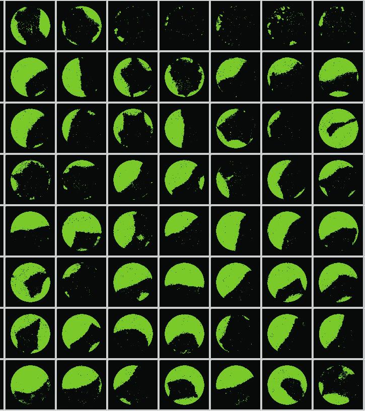
 Software detection of cell confluency, indicated by the green overlay, across a 96-well plate allowing for a quick visualization of plating efficiency.")
8 Maximize your hybridoma yield with our complete set of culture media Culture media for every stage of hybridoma cell line generation Stable hybridoma cell lines are critical for monoclonal antibody production. Our XP Media and CloneMedia portfolio of products is a complete solution that supports all stages of hybridoma cell line development from fusion to scale up. Optimized to support the selection and growth of hybridoma clones using our ClonePix 2 System, the kit is also compatible with other appropriate methods. HAT (hypoxanthine-aminopterin-thymidine) selection and cloning of hybridomas are accomplished in one step, which minimizes both time and materials required Not only does the semi-solid CloneMedia method eliminate the possible masking of potentially valuable slow-growing clones by fast-growing clones, but it also reduces or eliminates sub-cloning steps Reduce hands-on time when the workflow is combined with the ClonePix 2 System Optimized growth conditions result in stable antibody production A Hybridoma fusion from 5 separate immunizations of BALB/c mice B C Confluency across 96-well plate D Average lgg production Fusion efficiency lgg production (ng/ml) >90% plating efficiency clones (CV < 5%) < 5%) Hybridomas were generated, selected, and screened using our XP Media and CloneMedia suite of hybridoma media. (A) 5 individual hybridoma fusion experiments were conducted on BALB/c mice, immunized to the same antigen, to assess reproducibility of fusions in XP Media suite of products. Fusion efficiency was calculated by dividing the number of hybridoma colonies detected on the ClonePix 2 System by the number of splenocytes grown in XP Media Pre-Fusion Myeloma Growth Medium and Hybridoma Expansion Medium (without HT). (B) Images of hybridomas were captured with the ClonePix 2 System in white light (left panel) and FITC (right panel), after 7 days growth, to determine growth and expression of IgGs, respectively. Colonies grown in the presence of CloneDetect were ranked according to their FITC intensity (indicating total IgG production), with the highest producers picked for further characterization. (C) Software detection of cell confluency, indicated by the green overlay, across a 96-well plate allowing for a quick visualization of plating efficiency. Images were collected on the CloneSelect Imager. 87 out of 96 wells grew to a confluency >5% after 7 days (the initial confluency of all wells was <1%) for a >90% plating efficiency. The real plating efficiency may be even higher because slow growing clones may be classified as non-growing using the >5% confluency criteria. (D) IgG production plotted per well (show in blue) with red lines indicating 2 s.d. away from the mean. Because these are stable hybridomas, we don t expect a large variation in the total amount of IgG produced per cell, which is confirmed by <5% CV across all clones tested. 8
. XP Media Hybridoma Fusion Medium, P/N K8863 Used to wash cells before fusion and during fusion process.")
.")

9 The full kit contains*: XP Media Pre-Fusion Myeloma Growth Medium and Hybridoma Expansion Medium (without HT), P/N K8862 Used to support the growth of myeloma cells before fusion. Also supports expansion of hybridoma clones. Does not contain hypoxanthine or thymidine (HT). XP Media Hybridoma Fusion Medium, P/N K8863 Used to wash cells before fusion and during fusion process. Does not contain supplements to support growth. XP Media Hybridoma Fusion Recovery Medium, P/N K8864 Used to promote hybridoma viability after the fusion process but before clone selection. Does not contain hypoxanthine, aminopterin, and thymidine (HAT). CloneMedia Hybridoma Semi-Solid Selection and Cloning Medium (with HAT), P/N K8865 Used after fusion of splenocytes and myeloma cells to select and clone hybridomas in one step. Optimized for colony formation. Equally suitable for fresh fusions and for stable hybridoma cell lines. XP Media Hybridoma Growth Medium (with HT), P/N K8866 Optimized for hybridoma expansion following clone selection and colony picking. Contains hypoxanthine and thymidine (HT) and is used to wean hybridomas off aminopterin from the selection process. Hybridoma Polyethylene Glycol (PEG) for Cell Fusion, P/N K8868 Used for the fusion of mouse splenocytes and parental myeloma cells to generate hybridomas. PEG is present as a 50% solution in DMEM. CloneMedia Hybridoma Semi-Solid Selection and Cloning Medium (without HAT), P/N K8867 Does not contain any selection reagents. If appropriate selective reagent has been added, then the medium can be used after fusion to select and clone hybridomas in one step. Optimized for colony formation. * Components can be ordered separately. If you are using alternate hybridoma selection methods, then CloneMedia Hybridoma Semi-Solid Selection and Cloning Medium (without HAT) (P/N K8867) is available. You must add agents for hybridoma selection to this medium before use. CloneSelect Imager 9

 ClonePix 2 System, Growth Medium with HT (K8866) Accurate and gentle picking")

10 Accelerate your hybridoma cell line development with a complete set of platforms and culture media Pre-Fusion Culture myeloma cells in pre-fusion growth medium. Prepare splenocytes for fusion. Fuse splenocytes and myeloma cells. Fusion Clone Selection Clone screening and selection in semi-solid medium. Pick selected, highproducing clones from semi-solid medium and transfer to liquid medium. Clone Picking Expansion Medium without HT (K8862), Fusion Medium (K8863) Fusion Medium (K8863), Polyethylene Glycol (K8868), Fusion Recovery Medium (K8864) Hybridoma fusion VIEW EVERY GROWTH CURVE IN EVERY WELL ClonePix 2 System, CloneDetect, CloneMedia Selection and Cloning Medium with and without HAT (K8865, K8867) ClonePix 2 System, Growth Medium with HT (K8866) Accurate and gentle picking Transfer of colony to destination plates well of interest 3% Day 1 Clone Stability Monitor growth of picked clones. Functional Characterization Perform secondary screening on clone supernatants (e.g. ELISA). CloneSelect Imager 5% Day 2 15% Day 3 55% Day 7 FL FL Anti-species IgG-FL Primary antibody Cells or beads with antigen FL SpectraMax i3x or other Molecular Devices microplate readers FL Scale-Up and Wean Expand clones producing desired antibodies Scale up clones producing desired antibodies and wean off HT selection. Growth Medium with HT (K8866) and Expansion Medium without HT (K8862) at 1:1 ratio Expansion Expansion Medium without HT (K8862)


11 Accelerate cell line development with a range of Molecular Devices platforms ClonePix 2 Mammlian Colony Picker Automatically screen more clones in less time than conventional techniques, select cells with optimal expression levels, and pick colonies with accuracy with the ClonePix 2 System. ClonePix systems are now used in over 100 laboratories around the world to increase workflow productivity, leaving more time to better characterize target proteins and run new projects. QPix 400 Series Microbial Colony Picker The QPix 400 series of microbial colony pickers offer you the unique option to simultaneously detect colonies and quantify fluorescent markers in a prescreening step before picking. Our QPix systems are used worldwide in over 600 installations in research institutes, biotech, and pharmaceutical companies. QPix robotics developed a famous reputation for reliability and accuracy in sequencing centers during the Human Genome project. SpectraMax i3x Multi-Mode Microplate Reader The SpectraMax i3x multi-mode microplate reader measures spectral-based absorbance, fluorescence, and luminescence with the added functionality of modular upgrades for western blot, imaging, and fast kinetics with injectors. CloneSelect Imager 11
12 Unrivalled solutions based on excellent imaging and intelligent image analysis Products from Molecular Devices offer scientists unrivalled solutions that utilize imaging and intelligent image analysis to support basic research, pharmaceutical and biotherapeutic development. The company s systems continue to establish industry standards in areas such as picking microbial colonies for genomic studies or screening and selection of mammalian cell lines. Other systems use imaging platforms to monitor cell growth, evaluate cellular responses and quantify protein production. Through its expertise in robotics, cell and molecular biology, image analysis and interpretation, supported by a strong IP portfolio, the company is committed to the continual development of innovative solutions for life science applications. CloneSelect Imager - System Specifications Imaging Software Dedicated imaging software pre-installed on a high specification PC, Microsoft Windows 7 White light imaging Trans-illumination Data tracking 1 x internal barcode reader for data tracking of each plate Camera High-resolution CCD camera Imaging speed 96-well microplate: 90 sec Objective 4x Resolution Standard: 3.6 micron; Maximum: 1.8 micron Instrumentation Source plate type Range of 6-, 12-, 24-, 96-well SBS microplates Source plate capacity 1 x plates Instrument dimensions 720 mm (width) x 428 mm (height) x 575 mm (depth) Instrument weight 45 kg Regulatory approval Compliance CE Quality ISO9001:2008 certified Automation compatibility API suite available for robotic integration. Please contact us for details. For more information, visit Contact Us Phone: Web: info@moldev.com Check our website for a current listing of worldwide distributors Regional Offices USA and Canada Brazil China (Beijing) China (Shanghai) Germany Japan (Osaka) Japan (Tokyo) South Korea United Kingdom The trademarks used herein are the property of Molecular Devices, LLC or their respective owners. Specifications subject to change without notice. Patents: FOR RESEARCH USE ONLY. NOT FOR USE IN DIAGNOSTIC PROCEDURES Molecular Devices, LLC 5/ C Printed in USA
Accelerate cell line development. Get to your high value hits faster
 Accelerate cell line development Get to your high value hits faster Transform your workflows to elevate productivity Fast screening and selection of secretory cell lines with ClonePix Systems Screen 10x
CloneSelect Imager. Objective, quantitative assessment of cell growth. Genetix Now part of Molecular Devices.
 CloneSelect Imager Objective, quantitative assessment of cell growth www.moleculardevices.com / genetix Genetix Now part of Molecular Devices Assess cell confluence objectively and quantitatively Rapid
Get to your high-value clones faster with a complete hybridoma media solution
 Get to your high-value clones faster with a complete hybridoma media solution ENEFITS Complete solution from hybridoma fusion to expansion Simultaneous cloning and selection of hybridomas Optimized for
ClonePix 2. Screen and select more clones in less time. Genetix Now part of Molecular Devices.
 ClonePix 2 Screen and select more clones in less time www.moleculardevices.com/genetix Genetix Now part of Molecular Devices Screen more clones in less time Cut cell line and antibody development times
CloneMedia Reagent. Item Quantity Part Number. CloneMedia Semi-Solid Media for Hybridoma and Myeloma cells, 90 ml 6 bottles K8600
 CloneMedia Reagent Table 1-1: Available CloneMedia Reagent Item Quantity Part Number CloneMedia Semi-Solid Media for Hybridoma and Myeloma cells, 90 ml 1 bottle K8610 CloneMedia Semi-Solid Media for Hybridoma
QPix Systems. An industry standard for microbial colony picking. Genetix Now part of Molecular Devices.
 QPix Systems An industry standard for microbial colony picking www.moleculardevices.com/genetix Genetix Now part of Molecular Devices Setting an industry standard Although an essential step in many different
CloneMedia and XP Media CHO Growth A Reagents Table 0-1: Available Reagents
 CloneMedia and XP Media CHO Growth A Reagents Table 0-1: Available Reagents Item Quantity Part Number CloneMedia CHO Growth A with L-Gln, 90 ml 1 bottle K8810 CloneMedia CHO Growth A with L-Gln, 90 ml
Cyto-Mine. The Single Cell Analysis and Monoclonality Assurance System
 Cyto-Mine The Single Cell Analysis and Monoclonality Assurance System Biopharmaceutical discovery and cell line development workflows streamlined like never before w w w. s p h e ref l u i d i c s. c o
Select high-value cell lines like a pro
 Select high-value cell lines like a pro ccelerate cell line development for recombinant proteins and monoclonal antibody production eook contents Cell line development workflow... 2 Hybridoma selection
MetaXpress High-Content Image Acquisition and Analysis Software
 High-Content Image Acquisition and Analysis Software BENEFITS Meet high throughput requirements with a scalable, streamlined workflow Adapt your analysis tools to tackle your toughest problems, including
CloneMedia-CHO Semi-solid media for the growth of CHO-S Colonies
 Protocol CloneMedia-CHO Semi-solid media for the growth of CHO-S Colonies Control #: 03PCT1010.A0 Effective Date: 01-Jul-11 ECO #: 4002 Content Page Introduction 3 Protocol 4 1. Media Preparation 4 2.
SpectraMax Paradigm Microplate Reader
 SpectraMax Paradigm Microplate Reader User-upgradeable Multi-Mode Detection Platform BENEFITS Flexible design allows for single or dual excitation and emission High speed detection and excellent sensitivity
SpectraMax Multi-Mode Microplate Readers
 SpectraMax Multi-Mode Microplate s Your applications, your modes, your choice > upgradeable platform for changing lab needs > three-mode cuvette port FOR ASSAY DEVELOPMENT > Dual monochromator tunability
EarlyTox Cell Integrity Kit
 EarlyTox Cell Integrity Kit The EarlyTox Cell Integrity Kit from Molecular Devices is an optimized set of reagents that simplifies the measurement of live and dead cells in a single well. The assay uses
Cell biology workflows EMPOWERED BY TECAN
 Cell biology workflows EMPOWERED BY TECAN 02 Cell engineering Tedious manual processes? automation can help AUTOMATED COLONY PICKING SciRobotics Pickolo Colony-Picker is an add-on for Tecan s liquid handling
Measuring cell proliferation using the CyQUANT Cell Proliferation Assay with SpectraMax Microplate Readers
 APPLICATION NOTE Measuring cell proliferation using the CyQUANT Cell Proliferation Assay with SpectraMax Microplate Readers Introduction Quantitation of cell proliferation using fluorescence allows one
Innovative Solutions for Drug Discovery and Life Sciences Research
 Innovative Solutions for Drug Discovery and Life Sciences Research Unleash your brilliance Advancing cell and protein analysis technologies for your landmark scientific achievements Molecular Devices is
Cell Processing Workstation. In Situ Laser-Mediated Cell Purification and Processing
 LEAP Cell Processing Workstation Cell Processing Workstation In Situ Laser-Mediated Cell Purification and Processing Expanding the Capabilities of Cell Processing Consistent, High-Yield, High-Purity Processing
Accurate and Automated cell confluence assessment in microplates
 Accurate and Automated cell confluence assessment in microplates TECAN S SPARK 20M MICROPLATE READER WITH INTEGRATED CELL IMAGING SIMPLIFIES CELL CULTURE QC AND SIGNAL NORMALIZATION TO CELL CONFLUENCE
AUTOMATED HIGH THROUGHPUT CELL LINE GENERATION
 AUTOMATED HIGH THROUGHPUT CELL LINE GENERATION TABLE OF CONTENTS 3 4 6 8 10 11 ClonaCell EasyPick Platform Hamilton Robotics The Advantage of ClonaCell Media Automated High Throughput Colony Isolation
ClonePix TM FL. Application Guide for New Users. 07MAN1056.A1 Effective Date: 05-Oct-09
 ClonePix TM FL Application Guide for New Users 07MAN1056.A1 Effective Date: 05-Oct-09 Contents Table of Figures... 4 Introduction... 6 Setting up mammalian cell line selection experiments... 6 Principles
MetaXpress High-Content Image Acquisition and Analysis Software
 High-Content Image Acquisition and Analysis Software BENEFITS Meet high throughput requirements with a scalable, streamlined workflow Adapt your analysis tools to tackle your toughest problems, including
Application Note AN001
 Testing hybridoma supernatants with the Spots On Dots Antibody Screening Kit Application Note AN1 Table of Contents Overview... 2 Figure 1. Screening of hybridomas raised against peptide antigens... 3
SoftMax Pro 5 Software
 SoftMax Pro 5 Software THE INDUSTRy STANDARD IN MICROPLATE DATA ANALySIS > SIMPLE AND CUSTOMIZABLE ASSAy SETUP > flexible AND POWERfUL DATA ANALySIS > COMPLETE fda 21 CfR PART 11 COMPLIANCE TOOLS > INTEGRATION
Cell Health and Viability Assays Real-time automated measurements of cell health and viability inside your incubator
 INCUCYTE LIVE-CELL ANALYSIS SYSTEM Cell Health and Viability Assays Real-time automated measurements of cell health and viability inside your incubator See what your cells are doing and when they do it
Applications of rapid immunoassays (FAST ILA) in cell culture/fermentation and process development using the Threshold System
 in cell culture/fermentation and process development using the Threshold System") ILA Application Note Applications of rapid immunoassays (FAST ILA) in cell culture/fermentation and process development using the Threshold System INTRODUCTION Accurate measurement of the concentration
ILA Application Note Applications of rapid immunoassays (FAST ILA) in cell culture/fermentation and process development using the Threshold System INTRODUCTION Accurate measurement of the concentration
Faster Chemiluminescent camp detection by HitHunter camp XS+ on the SpectraMax L Luminescence Microplate Reader
 SpectraMax Systems Faster Chemiluminescent camp detection by HitHunter camp XS+ on the SpectraMax L Luminescence Microplate Reader By Rajini Bompelli and Sunitha Ambikapathi, DiscoveRx Corporation, 42501
Redefining Automation
 Redefining ELISA. Redefining Automation The DYNEX Agility utilizes the proven versatility of ELISA s core technology in a powerful, fully automated platform, for true ELISA optimization. State-of-the-art
Service solutions for peak performance. Flexible service plans, maximum productivity, expert engineers.
 Service solutions for peak performance Flexible service plans, maximum productivity, expert engineers www.moleculardevices.com Choose the right service plan for your specific laboratory priorities Performance
Measuring Wound Healing and Cell Migration using Celigo Imaging Cytometer
 Measuring Wound Healing and Cell Migration using Celigo Imaging Cytometer Nexcelom Bioscience LLC. 360 Merrimack Street, Building 9 Lawrence, MA 01843 T: 978.327.5340 F: 978.327.5341 E: info@nexcelom.com
Automation Supporting Single Cell Cloning Experiments and QbD-Based Bioprocess Development
 Automation Supporting Single Cell Cloning Experiments and QbD-Based Bioprocess Development Dr. Roland Schaefer Roche Diagnostics, Mannheim, Germany ELRIG High Throughput BioProcess Development, June 22,
THE BEACON OPTOFLUIDIC PLATFORM. 1000s of cells 100X the insights 10X faster 1/X total cost BERKELEY LIGHTS. DIGITAL CELL BIOLOGY AT LIGHT SPEED.
 THE BEACON OPTOFLUIDIC PLATFORM 1000s of cells 100X the insights 10X faster 1/X total cost BERKELEY LIGHTS. DIGITAL BIOLOGY AT LIGHT SPEED. FINDING THE MOST IMPORTANT INDIVIDUAL S REQUIRES THE MOST TIME,
Fluent Laboratory Automation Workstation for cell-based assays THE SIMPLE WAY TO TAKE THE COMPLEXITY OUT OF CELL BIOLOGY RESEARCH
 Fluent Laboratory Automation Workstation for cell-based assays THE SIMPLE WAY TO TAKE THE COMPLEXITY OUT OF CELL BIOLOGY RESEARCH 02 FLUENT FOR CELL-BASED ASSAYS Designed specifically for cell-based applications
NEW INSIGHTS. NEW DISCOVERIES. Real-time automated measurements of cell health, movement and function inside your incubator.
 THE NEXT GENERATION HAS ARRIVED IncuCyte S3 Live-Cell Analysis System Real-time automated measurements of cell health, movement and function inside your incubator. NEW INSIGHTS. NEW DISCOVERIES. See what
CloneMedia-CHOK1SV Semi-solid media for the growth of CHOK1SV colonies
 Protocol CloneMedia-CHOK1SV Semi-solid media for the growth of CHOK1SV colonies Control #: 03PCT1011.A0 Effective Date: 01-Jul-11 ECO #: 4002 Content Page Introduction 3 Protocol 4 1. Media Preparation
Redefining Automation
 Redefining Automation The DYNEX Agility utilizes the proven versatility of ELISA s core technology in a powerful, fully automated platform, for true ELISA optimization. State-of-the-art robotic processing
NEW INSIGHTS. NEW DISCOVERIES. Real-time automated measurements of cell health, movement and function inside your incubator.
 THE NEXT GENERATION HAS ARRIVED IncuCyte S3 Live-Cell Analysis System Real-time automated measurements of cell health, movement and function inside your incubator. NEW INSIGHTS. NEW DISCOVERIES. See what
Advanced Therapeutic Antibody Discovery with Multiplexed Screening
 Advanced Therapeutic Antibody Discovery with Multiplexed Screening White Paper Scientists need powerful tools that can deliver results to fully understand the ability of candidate antibodies to interrupt
FlexStation 3 Microplate Reader
 FlexStation 3 Microplate Reader A five-mode microplate reader with integrated fluid transfer > five-mode reader with wide range of applications > flexible liquid transfer enables more assay conditions
Acetyl-p53 (K381) Cell-Based Colorimetric ELISA Kit
 Cell-Based Colorimetric ELISA Kit") Acetyl-p53 (K381) Cell-Based Colorimetric ELISA Kit Catalog No. KA8015C Detection and Quantification of Acetyl-p53 (K381) Protein Concentration in Cell. Research Purposes Only. Not Intended for Diagnostic
Acetyl-p53 (K381) Cell-Based Colorimetric ELISA Kit Catalog No. KA8015C Detection and Quantification of Acetyl-p53 (K381) Protein Concentration in Cell. Research Purposes Only. Not Intended for Diagnostic
BR-9516B. SNP genotyping analysis for medium to ultra-high throughput. GENOMELAB TM SNPSTREAM GENOTYPING SERIES
 BR-9516B SNP genotyping analysis for medium to ultra-high throughput. GENOMELAB TM SNPSTREAM GENOTYPING SERIES Where power meets flexibility. The GenomeLab SNPstream Genotyping System provides an automated,
Cell Culture Automation in Biopharma
 Cell Culture Automation in Biopharma Cell Line Optimization and Monoclonal Antibody Production Dr. Christoph Krüll Product Manager Automated Solutions Beckman Coulter Europe Agenda Cell Line Optimization
Custom Antibody Services. Antibodies Designed Just for You. HuCAL Recombinant Monoclonal Antibody Generation Service
 Custom Antibody Services Antibodies Designed Just for You HuCAL Recombinant Monoclonal Antibody Generation Service Antibodies Designed Just for You What if there was a technology that would allow you to
The BioFlux 200 System Using Well Plate Microfluidics for Live Cell Assays Product Overview and Tutorial
 The BioFlux 200 System Using Well Plate Microfluidics for Live Cell Assays Product Overview and Tutorial Introduction to the BioFlux System Enables live-cell assays with precisely-controlled shear flow
FLIPR Potassium Assay Kit
 FLIPR Potassium Assay Kit Table A-1: Available Kits Assay Kit Explorer Kit Bulk Kit FLIPR Potassium Assay Kit R8222 R8223 The FLIPR Potassium Assay Kit from Molecular Devices provides a fast, simple, and
mirrorball accelerating antibody discovery
 mirrorball accelerating antibody discovery meet mirrorball efficient antibody discovery is within your reach TTP Labtech s mirrorball fluorescence cytometer revolutionises the productivity of antibody
Total PFK-2 liv/tes Cell-Based Colorimetric ELISA Kit
 Total PFK-2 liv/tes Cell-Based Colorimetric ELISA Kit Catalog No. KA3774C Detection and Quantification of PFK-2 liv/tes Protein Concentration in Cell. Research Purposes Only. Not Intended for Diagnostic
Faster ELISA results with CatchPoint SimpleStep ELISA kits
 Application note Faster ELISA results with CatchPoint SimpleStep ELISA kits Kristen Pennington (Associate Scientist - Abcam) Jonathan Wagner (Sr. Research Associate - Abcam) Yen-Wen Chen (Sr. Scientist
BECAUSE CAN T WAIT BREAKTHROUGHS. Drug Discovery Screening Solutions SCREENING SOLUTIONS. Automated Liquid Handling. Assays and Reagents
 BECAUSE BREAKTHROUGHS CAN T WAIT SCREENING SOLUTIONS Automated Liquid Handling Assays and Reagents Imaging and Detection Systems Informatics Drug Discovery Screening Solutions 2 As the premier provider
SpectraMax Quant AccuBlue Pico dsdna Assay Kit
 SpectraMax Quant AccuBlue Pico dsdna Assay Kit The SpectraMax Quant AccuBlue Pico dsdna Assay Kit is a part of the SpectraMax Quant family of double-stranded DNA (dsdna) assay products. Each product is
Accelerate Your Antibody Discovery and Cell Line Development Workflows with Cyto-Mine
 Accelerate Your Antibody Discovery and Cell Line Development Workflows with Cyto-Mine Cyto-Mine The Single Cell Analysis Platform Cyto-Mine is the world s first fully integrated platform for high-throughput
BR-16994A BIOMEK 4000 LABORATORY AUTOMATION WORKSTATION INTUITIVE SOFTWARE. MAXIMUM VERSATILITY.
 BR-16994A BIOMEK 4000 LABORATORY AUTOMATION WORKSTATION INTUITIVE SOFTWARE. MAXIMUM VERSATILITY. FAST AND SIMPLE SETUP EASY DEVICE INTEGRATION APPLICATIONS EXCELLENCE ICON-DRIVEN USER INTERFACE EXPANDABLE
Comparison of Luminex 200 to MAGPIX using the Poultry Serology Assay Technical Notes
 Comparison of Luminex 200 to MAGPIX using the Poultry Serology Assay Technical Notes Background and Rationale: Luminex xmap Technology affords many advantages over traditional assay formats, namely the
You ll never look at cells the same way again.
 E S S E N I N C U C Y T E T M You ll never look at cells the same way again. The number of cell-based experiments conducted every day is growing dramatically. Through these experiments, and leveraging
Tecan Microplates ASSURED PERFORMANCE FOR ABSORBANCE, FLUORESCENCE AND LUMINESCENCE MEASUREMENTS PLUS CELL IMAGING IN ANSI/SLAS FORMAT.
 Tecan Microplates ASSURED PERFORMANCE FOR ABSORBANCE, FLUORESCENCE AND LUMINESCENCE MEASUREMENTS PLUS CELL IMAGING IN ANSI/SLAS FORMAT. 02 Tecan Microplates Tecan Microplates are the latest addition to
Innovative Solutions for Drug Discovery and Life Sciences Research
 Innovative Solutions for Drug Discovery and Life Sciences Research Unleash your brilliance Advancing cell and protein analysis technologies for your landmark scientific achievements Molecular Devices is
FlexStation 3 microplate reader
 FlexStation 3 microplate reader A five-mode microplate reader with integrated fluid transfer > five-mode reader with wide range of applications > flexible liquid transfer enables more assay conditions
Drug Discovery Research Clinical Screening. Comparison of ELISA and AlphaScreen Assay Technologies for Measurement of Protein Expression Levels
 Drug Discovery Research Clinical Screening FLAG Expression Measurement Application Note Comparison of ELISA and AlphaScreen Assay Technologies for Measurement of Protein Expression Levels ALPHASCREEN APPLICATION
A Comparison of AlphaLISA and TR-FRET Homogeneous Immunoassays in Serum-Containing Samples
 application Note A Comparison of and Homogeneous Immunoassays in Serum-Containing Samples Authors Anuradha Prasad, PhD, Catherine Lautenschlager, PhD, Stephen Hurt, PhD, David Titus, PhD and Stéphane Parent,
Application Note. Introduction. Robbie Narang, Zhaoping Liu, Kim Luu IntelliCyt Corporation
 pplication Note High Throughput Flow ssays using the MultiCyt Cell Proliferation Reagent Dye Panel: Multiplexing with Membrane Integrity, Cell Cycle, and Immunophenotyping Endpoints Robbie Narang, Zhaoping
A Bridging Immunogenicity Assay Using SPARCL TM Technology
 A Bridging Immunogenicity Assay Using SPARCL TM Technology Wenhua F. Xie Lumigen Inc., a Beckman Coulter Company INTRODUCTION Evaluating the immune response in patients is an important aspect associated
An ELISA-based assay using fluorogenic substrates to measure total HIF-1 in the context of a whole cell.
 Cell-Based ELISA Human/Mouse Total HIF-1 Immunoassay Catalog Number KCB1935 An ELISA-based assay using fluorogenic substrates to measure total HIF-1 in the context of a whole cell. This package insert
An ELISA-based assay using fluorogenic substrates to measure total Cyclooxygenase-2 (COX-2) in whole cells.
 in whole cells.") Cell-Based ELISA Human/Mouse Total COX-2 Immunoassay Catalog Number KCB4198 An ELISA-based assay using fluorogenic substrates to measure total Cyclooxygenase-2 (COX-2) in whole cells. This package insert
Cell-Based ELISA Human/Mouse Total COX-2 Immunoassay Catalog Number KCB4198 An ELISA-based assay using fluorogenic substrates to measure total Cyclooxygenase-2 (COX-2) in whole cells. This package insert
Androgen Receptor (Phospho-Tyr363) Colorimetric Cell-Based ELISA Kit. Catalog #: OKAG02138
 Colorimetric Cell-Based ELISA Kit. Catalog #: OKAG02138") Androgen Receptor (Phospho-Tyr363) Colorimetric Cell-Based ELISA Kit Catalog #: OKAG02138 Please read the provided manual entirely prior to use as suggested experimental protocols may have changed. Research
Androgen Receptor (Phospho-Tyr363) Colorimetric Cell-Based ELISA Kit Catalog #: OKAG02138 Please read the provided manual entirely prior to use as suggested experimental protocols may have changed. Research
Comparison of the Ca2+-Sensitive Dyes Fluo-3 and Fluo-4 Used with the FLIPR Fluorometric Imaging Plate Reader System
 FLIPR Application Note Comparison of the Ca2+-Sensitive Dyes Fluo-3 and Fluo-4 Used with the FLIPR Fluorometric Imaging Plate Reader System INTRODUCTION In the FLIPR System, fluorescence-based measurement
An ELISA-based assay using fluorogenic substrates to measure total inducible Nitric Oxide Synthase (inos) in whole cells.
 in whole cells.") Cell-Based ELISA Human Total inos Immunoassay Catalog Number KCB9502 An ELISA-based assay using fluorogenic substrates to measure total inducible Nitric Oxide Synthase (inos) in whole cells. This package
Cell-Based ELISA Human Total inos Immunoassay Catalog Number KCB9502 An ELISA-based assay using fluorogenic substrates to measure total inducible Nitric Oxide Synthase (inos) in whole cells. This package
Automated Optimization of IgG Production in CHO Cells
 APPLICATION NOTE Automated Optimization of IgG Production in CHO Cells Michael Kowalski, Staff Applications Scientist Beckman Coulter Life Sciences, Indianapolis, IN David Apiyo, Marketing Applications
QuantiSir General Gene Knockdown Quantification Kit
 QuantiSir General Gene Knockdown Quantification Kit Base Catalog # PLEASE READ THIS ENTIRE USER GUIDE BEFORE USE The QuantiSir General Gene Knockdown Quantification Kit is suitable for quantifying gene
INOS. Colorimetric Cell-Based ELISA Kit. Catalog #: OKAG00807
 INOS Colorimetric Cell-Based ELISA Kit Catalog #: OKAG00807 Please read the provided manual entirely prior to use as suggested experimental protocols may have changed. Research Purposes Only. Not Intended
FLIPR Calcium 6 Assay Explorer Kit (R8190) FLIPR Calcium 6-QF Assay Explorer Kit (R8192)
 FLIPR Calcium 6-QF Assay Explorer Kit (R8192)") FLIPR Calcium 6 Assay Explorer Kit (R8190) FLIPR Calcium 6-QF Assay Explorer Kit (R8192) About the FLIPR Calcium 6 and 6-QF Assay Kits The FLIPR Calcium 6 Assay Kits from Molecular Devices, LLC (hereafter
FLIPR Calcium 6 Assay Explorer Kit (R8190) FLIPR Calcium 6-QF Assay Explorer Kit (R8192) About the FLIPR Calcium 6 and 6-QF Assay Kits The FLIPR Calcium 6 Assay Kits from Molecular Devices, LLC (hereafter
CytoGLOW. IKK-α/β. Colorimetric Cell-Based ELISA Kit. Catalog #: CB5358
 CytoGLOW IKK-α/β Colorimetric Cell-Based ELISA Kit Catalog #: CB5358 Please read the provided manual entirely prior to use as suggested experimental protocols may have changed. Research Purposes Only.
EarlyTox Live Cell Assay Kit
 EarlyTox Live Cell Assay Kit The EarlyTox Live Cell Assay Kit enables the detection of the live population of mammalian cells based on the measurement of intracellular esterase activity using a fluorescence
EGFR (Phospho-Ser695)
") Assay Biotechnology Company www.assaybiotech.com Tel: 1-877-883-7988 Fax: 1-877-610-9758 EGFR (Phospho-Ser695) Colorimetric Cell-Based ELISA Kit Catalog #: OKAG02090 Please read the provided manual entirely
Assay Biotechnology Company www.assaybiotech.com Tel: 1-877-883-7988 Fax: 1-877-610-9758 EGFR (Phospho-Ser695) Colorimetric Cell-Based ELISA Kit Catalog #: OKAG02090 Please read the provided manual entirely
Cytotoxicity LDH Assay Kit-WST
 Cytotoxicity LDH Assay Kit-WST Supplementary Information Notice to Users This instruction complements the Technical Manual in the product. Please use this instruction as supplements of the Technical Manual.
MAGPIX. Multiplexed Genomic, and Proteomic Biomarker Analysis
 MAGPIX Multiplexed Genomic, and Proteomic Biomarker Analysis Less Sample Input. More Results. The MAGPIX system is a versatile multiplexing platform capable of performing qualitative and quantitative analysis
Kinase Activity: a total solution
 Kinase Activity: a total solution Protein arrays for multiplex kinetic measurements Robust Instrumentation User friendly software Kinetic data at your fingertips! PamGene s novel platform for profiling
SpectraMax Quant AccuClear Nano
 SpectraMax Quant AccuClear Nano dsdna Assay Kit The SpectraMax Quant AccuClear Nano dsdna Assay Kit is a part of the SpectraMax Quant family of double-stranded DNA (dsdna) assay products. Each product
High-Throughput Screening for Antibody Discovery Using Mirrorball
 High-Throughput Screening for Antibody Discovery Using Mirrorball Christyne Kane Senior Scientist I AbbVie Bioresearch Center Antibody Engineering & Therapeutics December 12, 2017 Christyne Kane is an
Label-free, real-time live-cell assays for spheroids: IncuCyte bright-field analysis
 Introduction APPLICATION NOTE IncuCyte Live-Cell Analysis System Label-free, real-time live-cell assays for spheroids: IncuCyte bright-field analysis Susana L. Alcantara, Miniver Oliver, Kalpana Patel,
SpectraMax DuoLuc Reporter Assay Kit
 SpectraMax DuoLuc Reporter Assay Kit The SpectraMax DuoLuc Reporter Assay Kit enables highly sensitive quantitation of both firefly and Renilla luciferases in mammalian cells. Serial injection of two optimized
Automated Liquid Handling for Cell Staining and Imaging
 Automated Liquid Handling for Cell Staining and Imaging Abstract High content imaging is a powerful way to acquire per-cell data for adherent cell cultures. As the throughput increases for this method,
Automated Protocol for ANTI-FLAG High Sensitivity, M2 coated 96-well plate Using the Sciclone ALH 3000 Workstation (Caliper Life Sciences)
") Automated Protocol for ANTI-FLAG High Sensitivity, M2 coated 96-well plate Using the Sciclone ALH 3000 Workstation (Caliper Life Sciences) ANTI-FLAG High Sensitivity, M2 coated 96-well plate P 2983 Automation
Automated Protocol for ANTI-FLAG High Sensitivity, M2 coated 96-well plate Using the Sciclone ALH 3000 Workstation (Caliper Life Sciences) ANTI-FLAG High Sensitivity, M2 coated 96-well plate P 2983 Automation
HOST CELL PROTEIN & BIOPROCESSING REAGENT DEVELOPMENT
 HOST CELL PROTEIN & BIOPROCESSING REAGENT DEVELOPMENT INTRODUCTION Biopharmaceuticals require products to be free of residual host cell protein (HCP) contaminants from the bioprocessing workflow. To evaluate
Advance the potential of your cell culture Cedex Bio and Cedex Bio HT Analyzers
 Advance the potential of your cell culture Cedex Bio and s The value of certainty Cedex Bio and s Understanding the challenges of bioprocess monitoring Whether you are working in the pharmaceutical or
+ M III. IMAP Screening Express Kit Product #8073 Quantity: 8000, 20 µl reactions (80 µl final volumes) Low FP. M III High FP.
 Low FP. M III High FP.") Product Insert IMAP Screening Express Kit Product #8073 Quantity: 8000, 20 µl reactions (80 µl final volumes) Introduction About the IMAP Screening Express Kit The IMAP Screening Express Kit is used to
Product Insert IMAP Screening Express Kit Product #8073 Quantity: 8000, 20 µl reactions (80 µl final volumes) Introduction About the IMAP Screening Express Kit The IMAP Screening Express Kit is used to
DNAse I qualification and sample treatment
 DNA Application Note DNAse I qualification and sample treatment INTRODUCTION The Threshold Total DNA Assay quantitates contaminant DNA in biopharmaceutical drugs. DNase I can be used in conjunction with
Optiblot ECL Ultra Detect Kit (1.2pg 2ng)
") ab133409 Optiblot ECL Ultra Detect Kit (1.2pg 2ng) Instructions for Use For enhanced chemiluminescent Western blotting. This product is for research use only and is not intended for diagnostic use. 1 Table
ab133409 Optiblot ECL Ultra Detect Kit (1.2pg 2ng) Instructions for Use For enhanced chemiluminescent Western blotting. This product is for research use only and is not intended for diagnostic use. 1 Table
Quantifiler Human DNA Quantification Kit Quantifiler Y Human Male DNA Quantification Kit
 Product Bulletin Human Identification Quantifiler Human DNA Quantification Kit Quantifiler Y Human Male DNA Quantification Kit The Quantifiler kits produce reliable and reproducible results, helping to
Application note. Guideline for validation of analytical methods using Cedex Bio, Cedex Bio HT, and Cedex HiRes Analyzers.
 Application note Guideline for validation of analytical methods using Cedex Bio, Cedex Bio HT, and Cedex HiRes Analyzers April 216 Doerthe Druhmann, Sabrina Schuette, Dr. Dirk Ponsel Pharma Biotech, Penzberg,
Monoclonal Antibodies
 Monoclonal Antibodies Homogeneous antibody preparations produced in the laboratory by hybridomas. Produced by identical type of B cell clones. Recognize a signal epitope on an antigen. Hybridoma Technology
WHEN MULTIMODE MEETS IMAGING YOU GAIN A WHOLE NEW PERSPECTIVE. EnSight Multimode Plate Reader
 WHEN MULTIMODE MEETS IMAGING YOU GAIN A WHOLE NEW PERSPECTIVE EnSight Multimode Plate Reader TRANSLATING RELEVANT RESULTS INTO REAL INSIGHTS 2 THE WAY TO GREATER CONFIDENCE IN YOUR RESULTS Today s leading
Productivity Barriers
 LumiLux Cellular Screening Platform Break Through the Productivity Barriers in Cellular Screening Imagine an instrument that will cellular screening productivity Introducing a high impact instrument whose
Calcein AM Cell Viability Kit
 Instructions For Research Use Only. Not For Use In Diagnostic Procedures Calcein AM Cell Viability Kit Catalog# 4892-010-K 1000 Tests* * Calculated based on using 1 μm final concentration of Calcein AM;
Biacore System description. GE Healthcare. Biacore label-free interaction analysis. Optional software packages
 GE Healthcare Biacore label-free interaction analysis Biacore 4000 Biacore 4000 offers a complete solution for large-scale, label-free molecular interaction analysis, delivering high throughput without
Cytotoxicity LDH Assay Kit-WST
 Cytotoxicity LDH Assay Kit-WST Supplementary Information Notice to Users Preparation of Reagent This instruction complements the Technical Manual in the product. Please use this instruction as supplements
IgG Purity/Heterogeneity and SDS-MW Assays with High- Speed Separation Method and High Throughput Tray Setup
 IgG Purity/Heterogeneity and SDS-MW Assays with High- Speed Separation Method and High Throughput Tray Setup High Throughput Methods to Maximize the Use of the PA 800 Plus system Jose-Luis Gallegos-Perez
PCR SYSTEMS. a new era in high-productivity qpcr. Applied Biosystems ViiA 7 Real-Time PCR System
 PCR SYSTEMS a new era in high-productivity qpcr Applied Biosystems ViiA 7 Real-Time PCR System a new era in high-productivity qpcr The ViiA 7 Real-Time PCR System delivers the proven reliability, sensitivity,
PKA α/β CAT (Phospho-Thr197)
") Assay Biotechnology Company www.assaybiotech.com Tel: 1-877-883-7988 Fax: 1-877-610-9758 PKA α/β CAT (Phospho-Thr197) Colorimetric Cell-Based ELISA Kit Catalog #: OKAG01634 Please read the provided manual
Assay Biotechnology Company www.assaybiotech.com Tel: 1-877-883-7988 Fax: 1-877-610-9758 PKA α/β CAT (Phospho-Thr197) Colorimetric Cell-Based ELISA Kit Catalog #: OKAG01634 Please read the provided manual
Technical Note Detection of post-immunoprecipitation proteins by Western blot using the Quick Western Kit IRDye 680RD
 Technical Note Detection of post-immunoprecipitation proteins by Western blot using the Quick Western Kit IRDye 680RD Developed for: Aerius, Odyssey Classic, Odyssey CLx and Odyssey Sa Imaging Systems
Contaminant bovine IgG assay
 ILA Application Note Contaminant bovine IgG assay INTRODUCTION Bovine IgG, a 16, Dalton protein, is one of the constituents of bovine serum. Fetal calf serum containing bovine IgG is commonly used in mammalian