Partha Roy
|
|
|
- Solomon Charles
- 6 years ago
- Views:
Transcription
1 Fluorescence microscopy Partha Roy 1
2 Lecture Outline Definition of fluorescence Common fluorescent reagents Construction ti of a fluorescence microscope Optical considerations Biological applications of fluorescence microscopy - Immunofluorescence - FRAP (Fluorescence Recovery After Photobleaching) - FRET (Fluorescence Resonance Energy Transfer) 2
3 Definition of fluorescence: Property of a molecule to absorb light (photons) at one wavelength and emit light at a different (higher) wavelength. Molecules capable of fluorescing fluorescent molecule fluorescent dye fluorochrome Fluorescent dye Protein/lipid/DNA Dye conjugated to macromolecule 3
4 λex max λem max - Stoke s shift t / i /i d 4
5 Properties of fluorescent molecule 1) Stoke s shift ( λ emission >λλ excitation ) - useful flfor microscopy 2) Extinction coefficient (ε) measure of efficiency to absorb photons 3) Quantum efficiency or yield (Q): Efficiency to emit photons = photons emitted /photons absorbed 5
6 4) Quenching : Loss of fluorescence Dynamic quenching: collision with a second non-fluorescent molecule results in transfer of energy (Reduces Q) Non-excited surrounding fluorophore or another molecule Excited fuorophore Increases with proximity(concentration) of fluorescent molecules (can reduce fluorescence protein protein by quenching) Static quenching : conjugation with a second molecule (quencher) reduces the absorption o of photons (protein can act as a quencher) e 6
7 5) Photobleaching: Fading of fluorescence as a result of repeated excitation of fluorochrome Example of photobleaching due to prolonged excitation A Compare B vs A B Requires interaction of the fluorophore with light and oxygen in the environment (fluorophore undergoes irreversible chemical modification) Reduction of photobleaching : 1) by reducing environment (oxyrase); antifade agents (n-propyl-galate) OK for fixed cells 2) by cutting down the excitation light 7 (slows down the rate) - Live cells
8 Common fluorophores and their ex/em characteristics DAPI (diamidino-2-phenylindole) 358 (UV) /461 (purplish blue) Binds to DNA (nuclear probe) Fluorescein isothiocynate FITC 494 (blue) /520 (green) High QE, photobleaches quickly Aromatic molecules Rhodamine 543 (yellow) /567 (red) Good photostability Texas Red ~ (red) /~615 (far red) 8
9 Problem with labeling cellular proteins with fluorophores: fixing the cell Fluorescent proteins GFP (green fluorescent protein): ajell jellyfish fishaequorea protein that absorbs blue light and emit green light Variants a : B(blue)FP; C(cyan)FP, Y(yellow)FP, R(red)F or DsRed RFP source: Discosoma coral E - enhanced Advantage: GFP protein Genetically engineered fluorescent protein (GFP-fusion) DNA into cell GFP is expressed when your protein expressed 9
10 Principle of fluorescence microscopy object Emission light Excitation light 1) Excite the specimen at the right wavelength 2) Collect ONLY the fluorescence emission and block the excitation light (very important) Possible because of stoke s shift (important) 10
11 Has peaks (365, 400, 440, 546, 580 nm) Good for UV excitation, excitation of DAPI (358), FITC (494), Rhodamine (543) Uniform spectral response in the visible range (useful for ion imaging) 11
12 excitation emission FITC: blue/green Principles of a fluorescence microscope 1) Excite the specimen with blue light White light P B Dichroic mirror (45 o ) Reflects most of blue (<500), Transmits higher wavelengths Excitation filter Filter passes blue (say upto 495) White light P B specimen 12
13 2) Collect the emitted light ONLY by the specimen in the green wavelength Dichroic mirror fluorescence G Y/G/B G Emission/barrier filter (allows green and higher wavelength) White light excitation emission Y/G/B G fluorescence Excitation filter specimen B G greenish blue FITC: blue/green Note : stoke s shift becomes handy 13
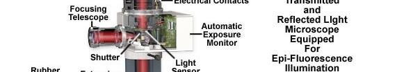
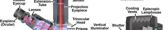
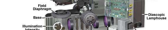
14 Construction of a fluorescence microscope (reflection type) eye eyepiece Objective also acts as a condenser No condenser needed 14
15 15
16 Same principle: field diaphragm controls the field of view aperture diaphragm controls the NA of illumination i Same principle on conjugate planes 16
17 What determines the brightness of fluorescence images n α Remember light gathering power of objective NA 2 objective acts as both exciter and collector of fluorescence Excitation efficiency NA 2 Efficiency to gather emission NA 2 Brightness NA 4 Image Brightness NA 4 /M 2 Brightness 1/M 2 (M magnification) 17
18 RIGHT CHOICE of filters and dichroic is key to proper fluorescence microscopy EX DM EM Biht Brighter image, but not good dfor imaging i more than one fluorophores at a time because of wide spectral characteristics (can excite multiple fluorophores simultaneously; can collect emission from multiple fluorophores simultaneously) Rhodamine SP ex For FITC LP em For FITC FITC Rhodamine FITC Rhodamine FITC excitation emission Also loss of contrast from autofluorescence (from flavins, fatty acid etc) 18
19 Alternative: bandpass filters Bandpass ex Rhodamine For FITC Bandpass em for FITC FITC FITC Rhodamine excitation FITC Rhodamine emission Adv: specificity (good for imaging multi-fluorophore, more contrast Disadv: weaker signal Widening the bandwidth causes Excitation and Emission bleedthrough 19
20 Use of mutipass dichroic and emission filters dichroic emission DAPI (ex) Single filter cube has the Dichroic and emission filter Change exciters on the filterwheel. FITC (ex) cy5 (ex) Cy3/ Rhodamine/ Tx red (ex) 1- DAPI (em) 2-FITC (em) 3- Rh/cy3 (em) 4- Tx-red (em) 5-Cy5 (em) Chroma corp. website 20
21 Applications of fluorescence microscopy 1) To visualize molecules (structures) in cells 2) To study dynamics (mobility) of molecules in cells 3) To study protein-protein interactions in cells in real-time 21
 b) Coupling a fluorescent dye")
 Nuclear staining by DAPI fluorophore phalloidin actin filaments 22 http://micro.magnet.fsu.")
22 1) To visualize molecules (structures) in cells a) Using a fluorescent dye that t directly binds to the molecule l of interest t Chromosome staining by DAPI (a common dye for nuclei) b) Coupling a fluorescent dye to a probe that directly binds to the molecule of interest FITC-conjugated phalloidin (binds to actin filaments) Nuclear staining by DAPI fluorophore phalloidin actin filaments 22
 Integrin immunostaining")
23 C) Immunofluorescence (using an antibody that binds to a specific molecule) 1 o antibody Molecule of interest cell X Fluorochrome attached to antibody Ex: 1 o - rabbit-anti X 2 o - goat-anti rabbit 1 o antibody Molecule of interest Fluorochrome cell 2 o antibody Direct Indirect Rhodaminephalloidin labeled actin filaments Actin filaments Cell-substrate adhesion (integrin) Integrin immunostaining showing cell-substrate adhesion (FITC staining) 23
24 2) To study molecular dynamics Time-lapse imaging of fluorescent proteins in living cell SEE the movie: microtubule dynamics (cells injected with fluorescent tbli tubulin- building block of microtubule) n[tubulin] = microtubule Shrinking microtubule Growing microtubule Inject fluor. tubulin Endogenous tubulin Fluor. tubulin is incorporated in microtubule 24
25 Measuring mobility (diffusion) of a molecule cell (high intensity) Protein conjugated with fluorochrome Exchange between bleached and Unbleached region Near Immobile (restricted movement) Diffusion constant =f (τ 1/2, area of bleached region) Freely mobile: F =F i ; Immobile molecule: F << F i 25
26 Example: FRAP of FITC-conjugated thymosinβ4 (cytoplasmic) in cell Time: -60 ms ms 1000 ms Pre-bleach Bleach Recovery Cytoplasmic proteins more diffusible Membrane proteins - often restricted in diffusion 26
27 3) To study molecular interaction in real-time FRET (Fluoroscence Resonance Energy Transfer) Principle: Transfer of energy from one fluorophore (donor) to a second fluorphore (acceptor) when two fluorophores come close in that case excitation of donor leads to emission from acceptor. (prereq: spectral overlap between donor emission and acceptor excitation) FRET efficiency 1/R 6 Donor emission (decreases) D- donor fluorophore A- acceptor fluorophore 12 1,2 interacting protein pair R o (forster distance) typically < 100 nm Donor emission Acceptor excitation Spectral overlap 27 (causing FRET)
28 Common FRET pairs Donor Acceptor BFP GFP BFP YFP BFP RFP CFP YFP CFP RFP GFP Rhodamine FITC Rhodamine FITC Cy3 Cy3 Cy5 Alexa-488 Rhodamine 28
29 Microscopic imaging: Excite Donor fluorophore (donor excitation filter) Collect emission from acceptor fluorophore (acceptor emission filter) If 1 and 2 bind: FRET between D and A fluorescence in A channel If 1 and 2 do not bind: No FRET between D and A No fluorescence in A channel Filter comb (Ex/Em): Donor/Donor: Donor distribution Acceptor/Acceptor : Acceptor distribution Donor/Acceptor: Interaction between donor and acceptor 29
30 FRET design Intramolecular FRET (donor and acceptor on the same molecule) Bimolecular FRET (donor and acceptor on different molecules) 30
31 Example: Using intermolecular FRET as a biosensor Rac a molecular switch (binds to either GTP or GDP) Active when it is bound to GTP; Inactive when it is bound to GDP Use FRET to determine when and where Rac is active When active it can bind to PAK (p21 activated kinase): No binding if it is inactive. Rac PAK Ex/Em: GFP/Alexa PBD (PAK binding domain) GFP/GFP Higher activity of Rac Higher conc. Of Rac GFP donor; A- alexa 546 (acceptor) Blue: low; red - high 31 Science 2000:290 ( )


32 Intramolecular l FRET as a biosensor (ex: monitor protease activity) i excitation I D Protease recognition sequence I A donor acceptor Protease activity: loss of FRET excitation I D I A=0 X donor acceptor 32
33 Intramolecular FRET as a biosensor (ex: receptor activation) EGFR EGF (epidermal (p growth factor) (EGF receptor) EGFR activation phosphorylation (py) of EGFR py py by EGFR itself py- phosphotyrosine Phosphatase converts pegfr to dpegfr (receptor inactivation) shc No binding between dp-egfr and shc binding between p-egfr and shc FRET construct Use FRET to determine the activation status of EGFR linker CFP YFP donor acceptor Shc EGFR Phosphorylation (binding region) Substrate (EEAEYMNMAPQ) 33
34 Inactive EGFR Shc can t bind EGFR phosphorylation substrate Longer distance between CFP and YFP: Low FRET CFP Shc (binding region) YFP EGFR Phosphorylation Substrate (EEAEYMNMAPQ) (No phosphorylation of Y) Active EGFR Shc binds to (p)egfr substrate Smaller distance between CFP and YFP: high FRET CFP YFP shc py by EGFR YFP/CFP ratio Fibroblast with EGFR construct and EGF stimulation Ting et al.,
Fluorescence Microscopy. Terms and concepts to know: 10/11/2011. Visible spectrum (of light) and energy
 and energy") Fluorescence Microscopy Louisiana Tech University Ruston, Louisiana Microscopy Workshop Dr. Mark DeCoster Associate Professor Biomedical Engineering 1 Terms and concepts to know: Signal to Noise Excitation
Fluorescence Microscopy Louisiana Tech University Ruston, Louisiana Microscopy Workshop Dr. Mark DeCoster Associate Professor Biomedical Engineering 1 Terms and concepts to know: Signal to Noise Excitation
Dino-Lite knowledge & education. Fluorescence Microscopes
 Dino-Lite knowledge & education Fluorescence Microscopes Dino-Lite Fluorescence models Smallest fluorescence microscope in the world Revolution to biomedical and educational applications Flexible Easy
More on fluorescence
 More on fluorescence Last class Fluorescence Absorption emission Jablonski diagrams This class More on fluorescence Common fluorophores Jablonski diagrams to spectra Properties of fluorophores Excitation
Fluorescence Light Microscopy for Cell Biology
 Fluorescence Light Microscopy for Cell Biology Why use light microscopy? Traditional questions that light microscopy has addressed: Structure within a cell Locations of specific molecules within a cell
Special Techniques 1. Mark Scott FILM Facility
 Special Techniques 1 Mark Scott FILM Facility SPECIAL TECHNIQUES Multi-photon microscopy Second Harmonic Generation FRAP FRET FLIM In-vivo imaging TWO-PHOTON MICROSCOPY Alternative to confocal and deconvolution
Fluorescence Microscopy
 Fluorescence Microscopy Dr. Arne Seitz Swiss Institute of Technology (EPFL) Faculty of Life Sciences Head of BIOIMAGING AND OPTICS BIOP arne.seitz@epfl.ch Fluorescence Microscopy Why do we need fluorescence
FLUORESCENCE. Matyas Molnar and Dirk Pacholsky
 FLUORESCENCE Matyas Molnar and Dirk Pacholsky 1 Information This lecture contains images and information from the following internet homepages http://micro.magnet.fsu.edu/primer/index.html http://www.microscopyu.com/
Contact Details. Dr Alexander Galkin. Office: MBC Room 186. Tel: (028) Frequency and wavelength.
 Frequency and wavelength.") Contact Details The electromagnetic spectrum Biological Spectroscopy Dr Alexander Galkin Email: a.galkin@qub.ac.uk Dr Alexander Galkin MSc Biomolecular Function - BBC8045 Office: MBC Room 186 Tel: (028)
Contact Details The electromagnetic spectrum Biological Spectroscopy Dr Alexander Galkin Email: a.galkin@qub.ac.uk Dr Alexander Galkin MSc Biomolecular Function - BBC8045 Office: MBC Room 186 Tel: (028)
Introduction to Fluorescence Jablonski Diagram
 ntroduction to Fluorescence Jablonski Diagram Excited Singlet Manifold S1 internal conversion S2 k -isc k isc Excited riplet Manifold 1 S0 k nr k k' f nr fluorescence k p phosphorescence Singlet round
Widefield Microscopy Bleed-Through
 In widefield microscopy the excitation wavelengths which illuminate the sample, and the emission wavelengths which reach the CCD camera are selected throughout a filter cube. A filter cube consists of
F* techniques: FRAP, FLIP, FRET, FLIM,
 F* techniques: FRAP, FLIP, FRET, FLIM, FCS Antonia Göhler March 2015 Fluorescence explained in the Bohr model Absorption of light (blue) causes an electron to move to a higher energy orbit. After a particular
MICROSCOPY. "micro" (small) "scopeo" (to watch)
 scopeo (to watch)") MICROSCOPY "micro" (small) "scopeo" (to watch) THE RELATIVE SIZES OF MOLECULES, CELLS AND ORGANISMS THE RELATIVE SIZES OF MOLECULES, CELLS AND ORGANISMS MICROSCOPY 1590 2012 MICROSCOPY THE LIGHT Light:
MICROSCOPY "micro" (small) "scopeo" (to watch) THE RELATIVE SIZES OF MOLECULES, CELLS AND ORGANISMS THE RELATIVE SIZES OF MOLECULES, CELLS AND ORGANISMS MICROSCOPY 1590 2012 MICROSCOPY THE LIGHT Light:
Visualizing mechanical tension across membrane receptors with a fluorescent sensor
 Nature Methods Visualizing mechanical tension across membrane receptors with a fluorescent sensor Daniel R. Stabley, Carol Jurchenko, Stephen S. Marshall, Khalid S. Salaita Supplementary Figure 1 Fabrication
Fluorescence Microscopy
 Fluorescence Microscopy Dr. Arne Seitz Swiss Institute of Technology (EPFL) Faculty of Life Sciences Head of BIOIMAGING AND OPTICS BIOP arne.seitz@epfl.ch Fluorescence Microscopy Why do we need fluorescence
Confocal Microscopy & Imaging Technology. Yan Wu
 Confocal Microscopy & Imaging Technology Yan Wu Dec. 05, 2014 Cells under the microscope What we use to see the details of the cell? Light and Electron Microscopy - Bright light / fluorescence microscopy
What to look for in a fluorophore. What to do with a fluorophore. Types of fluorochromes
 What to do with a fluorophore Intracellular localization (ER, Golgi, PM, nuclear, lysosome, MT, actin,...) Dynamic processes (protein synthesis, trafficking, turnover, DNA replication, cytoskeletal remodeling,
Fluorescence quenching, Fluorescence anisotropy, Fluorescence resonance energy transfer (FRET)
") Fluorescence quenching, Fluorescence anisotropy, Fluorescence resonance energy transfer (FRET) Timescale of fluorescence processes The excited electron decay possibilities k f k ph k q k t k ic Biophysics
Fluorescence quenching, Fluorescence anisotropy, Fluorescence resonance energy transfer (FRET) Timescale of fluorescence processes The excited electron decay possibilities k f k ph k q k t k ic Biophysics
Visualizing Cells Molecular Biology of the Cell - Chapter 9
 Visualizing Cells Molecular Biology of the Cell - Chapter 9 Resolution, Detection Magnification Interaction of Light with matter: Absorbtion, Refraction, Reflection, Fluorescence Light Microscopy Absorbtion
Reminder: absorption. OD = A = - log (I / I 0 ) = ε (λ) c x. I = I ε(λ) c x. Definitions. Fluorescence quenching and FRET.
 = ε (λ) c x. I = I ε(λ) c x. Definitions. Fluorescence quenching and FRET.") Reminder: absorption Special fluorescence applications I 0 I Fluorescence quenching and FRET Miklós Nyitrai; 24 th of Februry 2011. substance OD = A = - log (I / I 0 ) = ε (λ) c x optical density I = I
Reminder: absorption Special fluorescence applications I 0 I Fluorescence quenching and FRET Miklós Nyitrai; 24 th of Februry 2011. substance OD = A = - log (I / I 0 ) = ε (λ) c x optical density I = I
FLUORESCENT PEPTIDES. Outstanding Performance and Wide Application Range
 FLUORESCENT PEPTIDES Peptides and amino acids labeled with and Tide Quencher TM We offer peptides and amino acids tagged with fluorescent dyes. They meet highest demands in fluorescence intensity and photo-stability,
Fluorescence Spectroscopy. Student: Marin Cristina Antonia Coordinator:S.l. Preda Liliana
 Fluorescence Spectroscopy Student: Marin Cristina Antonia Coordinator:S.l. Preda Liliana Fluorescence Electron in the ground state is excited to a higher energy state After loss of some energy in vibrational
Nodes of regulation in cellular systems
 Nodes of regulation in cellular systems cell membrane signal transduction ligands receptors oligomerization transport signal transduction modified protein Golgi transcription factor transport ER transport
Con-focal and Multi-photon Microscope Experiment Fundamental. Qian Hu, Lab of Laser Scanning Confocal & Two-Photon Microscopy, ION, CAS
 Con-focal and Multi-photon Microscope Experiment Fundamental Qian Hu, Lab of Laser Scanning Confocal & Two-Photon Microscopy, ION, CAS 1. Light is Electromagnetic Wave ν = c / λ 2. Image of a Point Source
Lab 1: Ensemble Fluorescence Basics
 Lab 1: Ensemble Fluorescence Basics This laboratory module is divided into two sections. The first one is on organic fluorophores, and the second one is on ensemble measurement of FRET (Fluorescence Resonance
Contents. SCHOOL of FLUORESCENCE. For more information, go to lifetechnologies.com/imagingbasics
 MPSF educator packet This packet contains illustrations and figures from the Molecular Probes School of Fluorescence website. They illustrate concepts from the basic physical properties that underlie fluorescence
BIO 315 Lab Exam I. Section #: Name:
 Section #: Name: Also provide this information on the computer grid sheet given to you. (Section # in special code box) BIO 315 Lab Exam I 1. In labeling the parts of a standard compound light microscope
cell and tissue imaging by fluorescence microscopy
 cell and tissue imaging by fluorescence microscopy Steven NEDELLEC Plateforme Micropicell SFR Santé François Bonamy Nantes 1 A matter of size Limit of resolution 0.15mm aims: building the image of an object
FRET and FRET based Microscopy Techniques
 Big Question: We can see rafts in Model Membranes (GUVs or Supported Lipid Bilayers, LM), but how to study in cells? Do rafts really exist in cells? Are they static large structures? Are they small transient
BIO 315 Lab Exam I. Section #: Name:
 Section #: Name: Also provide this information on the computer grid sheet given to you. (Section # in special code box) BIO 315 Lab Exam I 1. In labeling the parts of a standard compound light microscope
Resolution of Microscopes Visible light is nm Dry lens(0.5na), green(530nm light)=0.65µm=650nm for oil lens (1.4NA) UV light (300nm) = 0.13µm f
, green(530nm light)=0.65µm=650nm for oil lens (1.4NA) UV light (300nm) = 0.13µm f") Microscopes and Microscopy MCB 380 Good information sources: Alberts-Molecular Biology of the Cell http://micro.magnet.fsu.edu/primer/ http://www.microscopyu.com/ Approaches to Problems in Cell Biology
Microscopes and Microscopy MCB 380 Good information sources: Alberts-Molecular Biology of the Cell http://micro.magnet.fsu.edu/primer/ http://www.microscopyu.com/ Approaches to Problems in Cell Biology
Confocal Microscopes. Evolution of Imaging
 Confocal Microscopes and Evolution of Imaging Judi Reilly Hans Richter Massachusetts Institute of Technology Environment, Health & Safety Office Radiation Protection What is Confocal? Pinhole diaphragm
Quality Matters: Bethyl DyLight Antibody Conjugates
 Quality Matters: Bethyl DyLight Antibody Conjugates Criteria for Fluorescent Probe Selection The use of fluorescent probes has become a mainstream technique to detect speci ic molecular targets in both
Concept review: Fluorescence
 16 Concept review: Fluorescence Some definitions: Chromophore. The structural feature of a molecule responsible for the absorption of UV or visible light. Fluorophore. A chromophore that remits an absorbed
Using Quantum Dots in Fluorescence Resonance Energy Transfer Studies
 p.1/31 Using Quantum Dots in Fluorescence Resonance Energy Transfer Studies Rajarshi Guha Pennsylvania State University p.2/31 Introduction Using organic fluorophores as labels A brief overview of fluorescence
Spectral Separation of Multifluorescence Labels with the LSM 510 META
 Microscopy from Carl Zeiss Spectral Separation of Multifluorescence Labels with the LSM 510 META Indians living in the South American rain forest can distinguish between almost 200 hues of green in their
Confocal Microscopy Analyzes Cells
 Choosing Filters for Fluorescence A Laurin Publication Photonic Solutions for Biotechnology and Medicine November 2002 Confocal Microscopy Analyzes Cells Reprinted from the November 2002 issue of Biophotonics
Supplementary Figure 1. The normalized absorption and emission spectra of 605QD
 1..8 65Q Absorbance 65Q Emission Cy5 Absorbance Cy5 Emission 1..8 Extiction Absorption Coefficient.6.4.2. 45 5 55 6 65 7 75 8 Wavelength (nm).6.4.2. Fluorescence Emission Intensity Supplementary Figure
CF Dyes Next Generation Fluorescent Dyes Secondary antibody
 CF Dyes Next Generation Fluorescent Dyes Secondary antibody OZYME 10 AVENUE AMPÈRE - CS 30268-78053 ST QUENTIN EN YVELINES CEDEX Tél. : 01 34 60 24 24 - Fax : 01 34 60 92 12 - www.ozyme.fr/info CF Dyes
The Green Fluorescent Protein. w.chem.uwec.edu/chem412_s99/ppt/green.ppt
 The Green Fluorescent Protein w.chem.uwec.edu/chem412_s99/ppt/green.ppt www.chem.uwec.edu/chem412_s99/ppt/green.ppt Protein (gene) is from a jellyfish: Aequorea victoria www.chem.uwec.edu/chem412_s99/ppt/green.ppt
Practical light microscopy: an introduction
 Practical light microscopy: an introduction Dr. Mark Leake, Oxford University www.physics.ox.ac.uk/users/leake Aim of today s talk: Explanation of the very (very) basics of how a light microscope works
Chapter 4. the biological community to assay for protein-protein interactions. FRET describes the
 31 Chapter 4 Determination of nachr stoichiometry using Normalized Försters Resonance Energy Transfer (NFRET) Försters resonance energy transfer (FRET) has become a technique widely used in the biological
Biochemistry. Biochemical Techniques. 18 Spectrofluorimetry
 Description of Module Subject Name Paper Name 12 Module Name/Title 1. Objectives 1.1 To understand technique of Spectrofluorimetry. 1.2 To explain instrumentation design 1.3 What are applications of Spectrofluorimetry?
ATFM 2015 ATFM Sample preparation guide
 ATFM 2016 Sample preparation guide The workshop is organized in the framework of the Czech-BioImaging research infrastructure supported by MEYS (LM2015062) 1 Table of Contents General recommendations...
THE BASICS OF IMMUNOHISTOCHEMISTRY
 THE BASICS OF IMMUNOHISTOCHEMISTRY Introduction Immunohistochemistry (IHC) identifies specific tissue components by means of a specific antigen/antibody reaction tagged with a visible label. IHC makes
FRET measurement between YFP and CFP
 FRET measurement between YFP and CFP EYFP and ECFP function as a donor-acceptor pair for fluorescence resonance energy transfer (FRET), in which excitation of the donor (cyan) molecule leads to emission
FLIM Fluorescence Lifetime IMaging
 FLIM Fluorescence Lifetime IMaging Fluorescence lifetime t I(t) = F0 exp( ) τ 1 τ = k f + k nr k nr = k IC + k ISC + k bl Batiaens et al, Trends in Cell Biology, 1999 τ τ = fluorescence lifetime (~ns to
Selected Topics in Electrical Engineering: Flow Cytometry Data Analysis
 Selected Topics in Electrical Engineering: Flow Cytometry Data Analysis Bilge Karaçalı, PhD Department of Electrical and Electronics Engineering Izmir Institute of Technology Outline Experimental design
Lecture 4: Fluorescence Microscopy
 Lecture 4: Fluorescence Microscopy Basic concepts Fluorescence: a phenomenon that some molecules absorb energy from light and emit another light of longer wavelength. FL-Microscopy: use fluorescence light
Advanced fluorescence microscopy techniques
 Practice-oriented, student-friendly modernization of the biomedical education for strengthening the international competitiveness of the rural Hungarian universities TÁMOP-4.1.1.C-13/1/KONV-2014-0001 Advanced
Immunostaining Protocols
 Immunostaining Protocols Lula L. Hilenski, Ph.D. Director Microscopy in Medicine Core Emory University Variables in standard immunostaining protocol 2-step or indirect immunofluorescence 1. Substrate on
2004 Debye Lecture 4 C. B. Murray. Quantum Dot Applications: Sun Screen. Solar Cells. Bio-tagging. Solid State Lighting?
 2004 Debye Lecture 4 C. B. Murray Quantum Dot Applications: Sun Screen Solar Cells Bio-tagging Solid State Lighting? Quantum Dot Solar cells Nanocrystal Solar Cells Double-labeling of mitochondria
Challenges to measuring intracellular Ca 2+ Calmodulin: nature s Ca 2+ sensor
 Calcium Signals in Biological Systems Lecture 3 (2/9/0) Measuring intracellular Ca 2+ signals II: Genetically encoded Ca 2+ sensors Henry M. Colecraft, Ph.D. Challenges to measuring intracellular Ca 2+
The analysis of fluorescence microscopy images for FRET detection
 The analysis of fluorescence microscopy images for FRET detection Ela Claridge, Dale J. Powner and Michael J.O. Wakelam School of Computer Science, The University of Birmingham B5 2TT Institute for Cancer
Imaging facilities at WUR
 Imaging facilities at WUR Advanced light microscopy facilities at Wageningen UR Programme Thursday 13 June 2013 Lunch meeting organized by Cat-Agro Food 12.00 Welcome and sandwich lunch 12.10 Introduction
Fluorescence Resonance Energy Transfer (FRET) Microscopy
 Microscopy") Applications in Confocal Microscopy Fluorescence Resonance Energy Transfer (FRET) Microscopy Product Info Brochures Confocal Theory Java Tutorials Glossary Applications Image Gallery Resource Links The
Applications in Confocal Microscopy Fluorescence Resonance Energy Transfer (FRET) Microscopy Product Info Brochures Confocal Theory Java Tutorials Glossary Applications Image Gallery Resource Links The
Design for Manufacturability (DFM) in the Life Sciences
 in the Life Sciences") T E C H N I C A L N O T E Design for Manufacturability (DFM) in the Life Sciences Fluorescence Spectroscopy Product Platform Realized with TracePro TM Suite of Opto-Mechanical Design Software Tools Authors:
T E C H N I C A L N O T E Design for Manufacturability (DFM) in the Life Sciences Fluorescence Spectroscopy Product Platform Realized with TracePro TM Suite of Opto-Mechanical Design Software Tools Authors:
Absorption of an electromagnetic wave
 In vivo optical imaging?? Absorption of an electromagnetic wave Tissue absorption spectrum Extinction = Absorption + Scattering Absorption of an electromagnetic wave Scattering of an electromagnetic wave
Principles of flow cytometry: overview of flow cytometry and its uses for cell analysis and sorting. Shoreline Community College BIOL 288
 Principles of flow cytometry: overview of flow cytometry and its uses for cell analysis and sorting Shoreline Community College BIOL 288 Flow Cytometry What is Flow Cytometry? Measurement of cells or particles
Multiplexed 3D FRET imaging in deep tissue of live embryos Ming Zhao, Xiaoyang Wan, Yu Li, Weibin Zhou and Leilei Peng
 Scientific Reports Multiplexed 3D FRET imaging in deep tissue of live embryos Ming Zhao, Xiaoyang Wan, Yu Li, Weibin Zhou and Leilei Peng 1 Supplementary figures and notes Supplementary Figure S1 Volumetric
Imaging Quantum Dots using FUJIFILM LAS 4000
 Imaging Quantum Dots using FUJIFILM LAS 4000 Application Note John Pizzonia, Ph.D. 9-28-07 Quantum dots (also known as nanocrystals) are a special class of materials known as semiconductors, which are
D e c N o. 2 8
 D e c. 2 0 0 7 N o. 2 8 CONFOCAL APPLICATION LETTER resolution FRET Acceptor Photobleaching LAS AF Application Wizard FRET with Leica TCS SP5 LAS AF Version 1.7.0 Introduction Fluorescence Resonance Energy
Localization Microscopy
 Localization Microscopy Theory, Sample Prep & Practical Considerations Patrina Pellett & Ann McEvoy Applications Scientist GE Healthcare, Cell Technologies May 27 th, 2015 Localization Microscopy Talk
Advanced fluorescence microscopy techniques
 Practice-oriented, student-friendly modernization of the biomedical education for strengthening the international competitiveness of the rural Hungarian universities TÁMOP-4.1.1.C-13/1/KONV-2014-0001 Advanced
Fluorescence spectroscopy
 Fluorescence spectroscopy The light: electromagnetic wave Zoltán Ujfalusi Biophysics seminar Dept. of Biophysics, University of Pécs 14-16 February 2011 Luminescence: light is not generated by high temperatures!!!
Live cell microscopy
 Live cell microscopy 1. Why do live cell microscopy? 2. Maintaining living cells on a microscope stage. 3. Considerations for imaging living cells. 4. Fluorescence labeling of living cells. 5. Imaging
Imaging of Cells using fluorescents dyes. By: Josué A. Benjamín Rivera September 27, 2018
 Imaging of Cells using fluorescents dyes By: Josué A. Benjamín Rivera September 27, 2018 1 History Sir William Henry Perkin BRITISH CHEMIST In 1856, at the age of 18, William Henry Perkin set out with
Experiment 3: Fluorescence Spectroscopy I (continued) All life appears to be nurtured by the excitation of electrons by light in photosynthesis.
 All life appears to be nurtured by the excitation of electrons by light in photosynthesis.") Experiment 3: Fluorescence Spectroscopy I (continued) Last week: Part I.A: Introduction to steady state spectra Today: Part 1.B: Fluorescence Quenching and the Stern-Volmer Relation Prelab Lecture 9feb17
Experiment 3: Fluorescence Spectroscopy I (continued) Last week: Part I.A: Introduction to steady state spectra Today: Part 1.B: Fluorescence Quenching and the Stern-Volmer Relation Prelab Lecture 9feb17
Goat Anti Rabbit IgG Antibodies
 Goat Anti Rabbit IgG Antibodies Table 1. Contents and storage information. Material Amount Concentration Storage Upon Receipt Stability Whole antibodies 0.5 ml F(ab ) 2 fragments 250 µl 2 mg/ml in 0.1
Microscopy from Carl Zeiss
 Microscopy from Carl Zeiss LSM 710 In Tune with Your Application Enjoy new freedom in selecting fluorescent dyes with In Tune, the new laser system for the LSM 710. Whatever the wavelength, you can match
BASICS OF FLOW CYTOMETRY
 BASICS OF FLOW CYTOMETRY AUTHOR: Ana Isabel Vieira APPROVAL: Henrique Veiga Fernandes Ana Sílvia Gonçalves SOP.UCF.002 03-09-2015 Pag. 1/9 Overview Flow: Fluid Cyto: Cell Metry: Measurement Flow cytometry
If you like us, please share us on social media. The latest UCD Hyperlibrary newsletter is now complete, check it out.
 Sign In Forgot Password Register username username password password Sign In If you like us, please share us on social media. The latest UCD Hyperlibrary newsletter is now complete, check it out. ChemWiki
SUPPORTING INFORMATION
 Electronic Supplementary Material (ESI) for Dalton Transactions. This journal is The Royal Society of Chemistry 2015 Terbium-Based Time-Gated Förster Resonance Energy Transfer Imaging for Evaluating Protein-Protein
1. The fluorescence process.
 1. The fluorescence process. 1.1 introduction Fluorescence is the result of a three-stage process that occurs in certain molecules (generally polyaromatic hydrocarbons or heterocycles) called fluorophores
ab CytoPainter ER Staining Kit Red Fluorescence
 ab139482 CytoPainter ER Staining Kit Red Fluorescence Instructions for Use Designed to detect Human endoplasmic reticulum by microscopy. This product is for research use only and is not intended for diagnostic
Lab 5: Optical trapping and single molecule fluorescence
 Lab 5: Optical trapping and single molecule fluorescence PI: Matt Lang Lab Instructor: Jorge Ferrer Summary Optical tweezers are an excellent experimental tool to study the biophysics of single molecule
Fluorescence spectroscopy
 Fluorescence spectroscopy The light: electromagnetic wave Tamás Huber Biophysics seminar Dept. of Biophysics, University of Pécs 05-07. February 2013. Luminescence: light emission of an excited system.
Workshop advanced light microscopy
 Workshop advanced light microscopy Multi-mode confocal laser scanning microscope Jan Willem Borst Laboratory of Biochemistry Biomolecular Networks www.bic.wur.nl MicroSpectroscopy Centre Wageningen Microspectroscopy
24.1 Reference Standards and Antifade Reagents
 24.1 Reference Standards and Antifade Reagents To obtain accurate and reproducible results from fluorescence imaging applications, it is essential to maximize the performance of the optical system. Careful
Welcome! openmicberkeley.wordpress.com. Open Berkeley
 Welcome! openmicberkeley.wordpress.com Agenda Jen Lee: Introduction to FRET Marla Feller: Using FRET sensors to look at time resolved measurements Becky Lamason: Using FRET to determine if a bacterial
August 15, 2018 $ " % ' & OMe. NEt2 MeO
 Taking a deep look: a near infrared fluorescent dye for long term bioimaging ~ Promising photostable tool for single molecule tracking and multicolor imaging ~ August 15, 2018 A team of researchers at
Beta3 integrin promotes long-lasting activation and polarization of Vascular Endothelial Growth Factor Receptor 2 by immobilized ligand
 SUPPLEMENTAL FIGURES Beta3 integrin promotes long-lasting activation and polarization of Vascular Endothelial Growth Factor Receptor 2 by immobilized ligand C. Ravelli et al. FIGURE S. I Figure S. I: Gremlin
single-molecule fluorescence spectroscopy
 single-molecule fluorescence spectroscopy 5 dynamics of a single molecule by FRET michael börsch 18/07/2003 topics theory of fluorescence resonance energy transfer solvent effects and fluorescence quenching
Two-Photon Microscopy for Deep Tissue Imaging of Living Specimens
 for Deep Tissue Imaging of Living Specimens Tilman Franke* and Sebastian Rhode TILL Photonics GmbH, an FEI company, Lochhamer Schlag 21, D-82166 Gräfelfing, Germany *tilman.franke@fei.com Introduction
ANAT 3231 Cell Biology Lab12 Stem Cell Analysis
 ANAT 3231 Cell Biology Lab12 Stem Cell Analysis 2 June 2010 Dr Antonio Lee Neuromuscular & Regenera9ve Medicine Unit School of Medical Sciences, UNSW Introduction to Flow Cytometry Contributed by Vittoria
Five steps for publication-quality cell imaging the first time
 Five steps for publication-quality cell imaging the first time Follow this proven guide to capture the best possible fixed-cell images Introduction We are all driven by great scientific innovation and
Fluorescence Microscopy: A Biological Perspective
 Fluorescence Microscopy: A Biological Perspective From nanometre to metre: the scale of life Instrumentation and accessible scale limits the questions that can be addressed in biology Why are there limits?
Fluorescence spectroscopy
 Fluorescence spectroscopy The light: electromagnetic wave Tamás Huber Biophysics seminar Dept. of Biophysics, University of Pécs 05-06. February 2014. 1 Luminescence: light emission of an excited system.
Fluorescence microscopy
 Fluorescence microscopy 1 Fluorescence microscopies basic fluorescence, fluorophores Deconvolution Confocal Two-photon/multi-photon 4Pi Light sheet Total internal reflection STED FRAP/FLIP/FCS FRET PALM/STORM/iPALM
11/19/2013. Janine Zankl FACS Core Facility 13. November Cellular Parameters. Cellular Parameters. Monocytes. Granulocytes.
 DEPARTEMENT BIOZENTRUM Janine Zankl FACS Core Facility 13. November 2013 Cellular Parameters Granulocytes Monocytes Basophils Neutrophils Lymphocytes Eosinophils Cellular Parameters 1 What Is Flow Cytometry?
Supporting Information for. Electrical control of Förster energy transfer.
 1 Supporting Information for Electrical control of Förster energy transfer. Klaus Becker 1, John M. Lupton 1*, Josef Müller 1, Andrey. L. Rogach 1, Dmitri V. Talapin, Horst Weller & Jochen Feldmann 1 1
Application of Quantum Mechanics to Biology
 Application of Quantum Mechanics to Biology How can we apply quantum mechanics to biology? Polymers of nucleotides and amino acids - millions of atoms bounded into a large molecule Visual System Must turn
Image-iT FX Kits with Alexa Fluor Secondary Detection Conjugates
 Image-iT FX Kits with Alexa Fluor Secondary Detection Conjugates Table 1. Contents and Storage Information. Material Amount Concentration Storage Stability Alexa Fluor IgG conjugates Alexa Fluor streptavidin
A Brief History of Light Microscopy And How It Transformed Biomedical Research
 A Brief History of Light Microscopy And How It Transformed Biomedical Research Suewei Lin Office: Interdisciplinary Research Building 8A08 Email: sueweilin@gate.sinica.edu.tw TEL: 2789-9315 Microscope
The best and brightest
 Labeling and Detection The best and brightest Alexa Fluor 488 dye Labeling and Detection A superior alternative to FITC Brighter conjugate fluorescence Unequalled photostability Perfect spectral match
A photoprotection strategy for microsecond-resolution single-molecule fluorescence spectroscopy
 Nature Methods A photoprotection strategy for microsecond-resolution single-molecule fluorescence spectroscopy Luis A Campos, Jianwei Liu, Xiang Wang, Ravishankar Ramanathan, Douglas S English & Victor
Aoki et al.,
 JCB: SUPPLEMENTAL MATERIAL Aoki et al., http://www.jcb.org/cgi/content/full/jcb.200609017/dc1 Supplemental materials and methods Calculation of the raw number of translocated proteins First, the average
CD93 and dystroglycan cooperation in human endothelial cell adhesion and migration
 /, Supplementary Advance Publications Materials 2016 CD93 and dystroglycan cooperation in human endothelial cell adhesion and migration Supplementary Materials Supplementary Figure S1: In ECs CD93 silencing
Rice/TCU REU on Computational Neuroscience. Fundamentals of Molecular Imaging
 Rice/TCU REU on Computational Neuroscience Fundamentals of Molecular Imaging June 2, 2009 Neal Waxham 713-500-5621 m.n.waxham@uth.tmc.edu Objectives Introduction to resolution in light microscopy Brief
Sapphire. Biomolecular Imager THE NEXT GENERATION OF LASER-BASED IMAGING
 Sapphire Biomolecular Imager THE NEXT GENERATION OF LASER-BASED IMAGING Breakthrough image capture and analysis The Sapphire Biomolecular Imager is a next generation laser scanning system that provides
Organelle Atlas APPENDIX TO CHAPTER 4 4A.1
 APPENDIX TO CHAPTER 4 This atlas contains images of cellular organelles visualized using a wide variety of reagents and techniques and demonstrates the diversity of methods for exploring the cell. The