Basic Fluorescence Microscopy and Sample Preparation. Eva Wegel
|
|
|
- Julius Simon
- 6 years ago
- Views:
Transcription


1 Basic Fluorescence Microscopy and Sample Preparation Eva Wegel


2 Visible Light nm visible to the human eye White light is split into its components through a prism Reason: different λ refract at different angles
 Stokes Shift Jablonski diagram Alexa Fluor 488 Photoluminescence: Fluorescence - spontaneous emission of light during transition of the system from its lowest")
3 What is fluorescence? George Gabriel Stokes ( ) Stokes Shift Jablonski diagram Alexa Fluor 488 Photoluminescence: Fluorescence - spontaneous emission of light during transition of the system from its lowest vibrational energy level of an excited singlet state S 1 back to the ground state S 0 (10-9 to 10-6 s) Phosphorescence a non-radiative transition into an isoenergetic vibrational level of a triplet state T 1, which lasts for 10-3 to 1000 s before it decays to the ground state IC: internal conversion ISC: intersystem crossing


4 Photobleaching and Phototoxicity Photobleaching: photochemical destruction of the fluorophore In an excited triplet state, fluorophores may interact with another molecule to produce irreversible covalent modifications Phototoxicity: illumination of a fluorophore causes damage to the cell expressing it, eventually leading to cell death Common situation: the excited dye molecule passes its excess energy on to O 2, creating reactive oxygen species (ROS): - ROS reacts with dye dye bleaches - ROS diffuses away and reacts with other dyes or cell components Solution: - Reduce the intensity of the excitation light and frequency of illumination - Close down the field aperture in order to restrict the illuminated area
 Fluorescence")


5 Basic principle of an (Inverted) Fluorescence Microscope Ideal filter cube properties Fluorescence microscope: Fluorescence light source Excitation filter Dichroic mirror Objective Emission filter Camera/eye pieces Excitation Dichroic Emission

6 Upright Fluorescence Microscope Used for fixed samples on slides and for live imaging where the objective is immersed in the medium

7 Inverted Fluorescence Microscope Used for live imaging through a coverslip and for fixed samples on slides More versatile but danger of oil running down the objective
8 Fluorescence Light Sources Mercury lamps (old-fashioned): 400 h life time, manual adjustment necessary Xenon lamps: 800 h life time Metal halide lamps: 2000 h life time, easy to fit


9 Spectra of Mercury, Metal Halide and Xenon lamps
10 Fluorescence light sources cont. LED lights (state of the art): bandwidth 10 nm, λ above 360 nm, 5000 h life time Lasers (specialised wide-field fluorescence applications, confocal, multiphoton): Narrow beams of highly monochromatic, coherent and collimated light Fluorescence detectors Cooled CCD cameras (Lecture 10) EMCCD cameras (Lecture 10) scmos cameras (Lecture 10) PMTs (Lecture 8)

11 Preparation of Fixed Samples Why use fixed samples at all? - Primary cells cannot easily be transfected and transgenic animals are time-consuming to produce and not always possible - Brighter than fluorescent fusion proteins - Injection of antibodies only possible with big cells (e.g. oocytes) - Can detect four different labels or even more at the same time - High-throughput screening

12 Typical Immunocytochemistry Protocol Fixation Permeabilisation Washes Blocking 1 antibody Washes 2 antibody Washes Mounting
13 Antibodies for Immunocytochemistry Basic Fluorescence Microscopy and Sample Preparation Preferred: IgG isotype, more consistent generation and binding All constant domains are recognised by 2 Abs Polycolonal antibodies contain multiple clones of antibodies produced to different epitopes of the antigen Monoclonal antibodies, originally from one mouse, contain a single antibody from one clone of B-cells to a single epitope on the antigen Affinity-purified Abs best in theory because they have bound to the antigen, but some of the strongest binding Abs cannot be eluted from the affinity columns and are lost.
14 Polyclonal antibodies Advantage: High levels of labelling because they bind several epitopes on the same protein Disadvantages: Can label multiple proteins that share epitopes Different batches have different antibodies Monoclonal antibodies Advantages: Single epitope selected for high specificity Different clones can be generated to different epitopes on the same antigen Single clone can recognise post-transcriptionally modified protein (e.g. phosphorylation) Same clone can be generated indefinitely Disadvantages: Low levels of labelling possible Mostly from mice
15 How to choose primary antibodies: 1. Published literature recommendation 2. Product recommended for immunocytochemistry 3. High specificity for the antigen of interest in your species 4. Species the Ab was raised in compatible with other Abs in your experiment How to store antibodies: 10 µl aliquots in -70 freezer, after defrosting: in fridge for short-term


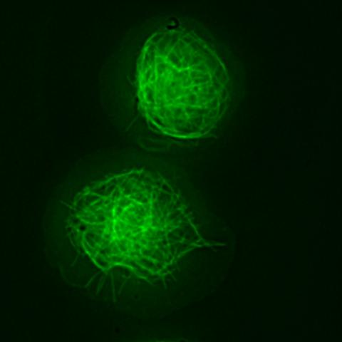
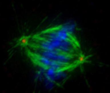
16 Fixation: preservation of cells or tissue in a life-like state Fixed sample should appear similar to living sample Uniform fixation throughout the sample Cells and organelles not swollen or shrunken No loss of proteins, lipids and other molecules Microtubules in Drosophila macrophages Left : Live cells expressing Jupiter-GFP Right: PFA fixed cell stained with anti-tubulin antibody and Alexa Fluor 488
17 Two Types of Fixation Denaturing fixation: Cold methanol or cold acetone stored at -20 C, samples submerged at -20 C for 10 to 20 min destroys 3D protein structure dissolves lipids into micelles poor morphological preservation and poor protein retention makes some epitopes accessible best used after cross-linking fixation MeOH Cross-linking fixation: aldehyde groups cross-link molecules in cells and tissues extensive cross-linking prevents antibody penetration Formaldehyde used for immunocytochemistry in light microscopy - cross-links 1 amines of Lys and Arg, sulfhydryl groups of Cys, OH groups, double bonds - binds to amino acids, peptides, proteins and some lipids, but not RNA, DNA or most sugars - retention of DNA and RNA due to protein cross-linking - for cultured cells fixation usually for 20 min in 2-4% formaldehyde PFA
18 Fixation cont. Sources of formaldehyde: Formalin Produced by oxidation of methanol, contains 37% formaldehyde and impurities including 14% methanol Some proteins are washed out during fixation Causes high levels of autofluorescence DO NOT USE except for human clinical pathology Paraformaldehyde (PFA) Powder of polymerised formaldehyde, converted into soluble monomers by heating and adding NaOH Buy as 16% stock from Polysciences, freeze aliquots at -20 C Glutaraldehyde Fixation Can form long polymers, single most effective cross-linking chemical fixative Inhibits diffusion of antibodies into cells and tissues Generates autofluorescence Used for electron microscopy
19 Effects of Fixatives on Ultrastructure MeOH fixation 4% PFA Schnell U et al, Nature Methods, Vol 9 (2), 2012, % glutaraldehyde
20 Buffers for fixation pk range must be Maintain stable ph and have to have the same tonicity as the cells (same conc. of solutes) Usually phosphate buffer but specialist buffers possible: MOPS, TES, HEPES, PIPES How to prepare cells for fixation Grow adherent cells on coverslips for fixation in multiwell plates Fix non-adherent cells in suspension after pelleting and resuspending or fix on poly-lysine coated coverslips (0.1mg/mL)
21 Blocking Aim: to allow binding of antibodies only to appropriate sites Sources of nonspecific binding: Charged groups Occur on proteins (esp. histones) or lipids Also generated by fixation in formalin or glutaraldehyde To block use bovine serum albumin at 10-30mg/mL (fraction V) Fc receptors On macrophages and other immune cells, which bind any antibody To block whole IgG 1 and 2 antibodies from binding to Fc receptors, incubate cells in buffer containing 5-10% normal serum from the host species of the 2 antibody Endogenous antibodies Only a problem for 2 antibodies recognising the same species as your tissue/ cells and only at inflammation sites or in cell cultures of immune system cell types To block use Fab fragments raised in the same species as the 2 antibody that recognise the species of your tissue/cells as part of the blocking procedure For general blocking can also try MAXblock (Active Motif): protein based, non-mammalian blocking agent, no cross-reactivity with 2 antibodies
22 Permeabilisation Aim: to allow fixative to enter the cells/tissue more quickly if necessary to allow antibodies to penetrate fixed cells/tissue done by removing lipids with detergents Detergents: polar lipids with a hydrophilic (water soluble) end and a hydrophobic end that binds the hydrophobic moieties of water insoluble compounds and renders them hydrophilic Nonionic detergents: contain methyl groups that participate in hydrogen bonds and are able to solubilise membranes but do not destroy protein-protein interactions Triton X-100: used to permeabilise unfixed or lightly fixed eukaryotic cell membranes (0.1% in PBS) Tween 20: milder than Triton X-100, used to reduce surface tension in blocking, antibody incubation and wash steps (0.1%) Nonidet P-40 (Igepal Ca-630 from Sigma-Aldrich): used to permebilise unfixed cells (0.1% in PBS for 5-10s) Ionic detergents: have highly charged hydrophilic groups and are very effective at solubilising membranes, but also destroy native three dimensional protein structures SDS, deoxycholate, CHAPS Not used for immunocytochemistry
23 Permeabilisation Saponins: mild detergents that preserve ultrastructural integrity permeabilize membranes by reversibly forming complexes with cell membrane cholesterol leading to pore formation sufficient for the detection of cytoplasmic antigens not effective for permeabilization of cholesterol-poor membranes such as the inner mitochondrial membrane and the nuclear envelope add at conc. of % to blocking solution (block for 1h) and in antibody incubations, some protocols also include saponin in wash steps


, 2012,152-158")
24 Effects of Fixatives and Permeabilisation on Ultrastructure MeOH fixation 4% PFA + MeOH 4% PFA 4% PFA + Triton X-100 Schnell U et al, Nature Methods, Vol 9 (2), 2012, % glutaraldehyde
 after fixation with 4% PFA (30 min) and permeabilized with either methanol (MeOH; 20 C) for 1 min or 0.")
25 Effects of different types of permeabilisation CLDN7-EGFP Anti-CLDN7 Merge PFA and Triton PFA and MeOH Images of 293T cells expressing tight junction protein Claudin-7 EGFP (CLDN7-EGFP) immunostained with an antibody to Claudin-7 (anti-cldn7) after fixation with 4% PFA (30 min) and permeabilized with either methanol (MeOH; 20 C) for 1 min or 0.1% Triton X-100 (Triton) for 15 min at room temperature. Scale bar, 10 µm. Schnell U et al, Nature Methods, Vol 9 (2), 2012,
26 Washes Wash with agitation (unless your cells dislodge easily) for 5-10 min for each wash step Wash 7 times leaving 10-20% of the buffer each time to prevent drying of your cells/tissue Or wash 3 times removing all buffer and replacing it immediately If cells/tissue dry out in between washes background is increased and cannot be removed Washes after the 1 antibody Incomplete removal of the 1 antibody does not increase background but lowers the amount of specific labelling because the 2 antibody reacts with the 1 in solution decreasing its conc. Washes after the 2 antibody Incomplete removal of the 2 antibody increases background
27 Antibody incubation 1 antibody: Use a series of antibody dilutions: 1:50, 1:500, 1:5000 High concs. background or very little label 2 antibody: Low conc. of antibody usually sufficient: 1: 500, 1:1000 High concs. background


28 Two 1 antibodies from the same species rabbit anti-x goat anti-rabbit IgG Alexa 488 IgG from normal rabbit serum Fab fragment goat antirabbit IgG rabbit anti-y goat anti-rabbit IgG Alexa 568
29 Mounting media Prolong Gold (Life Technologies) curing anti-fade mountant, sets over 24h to RI 1.47 (glass RI 1.52) Slides can be stored at 4 C for several months Vectashield (Vector Labs) Non-setting anti-fade mountant, RI 1.44 Slides need to be sealed with nail polish and can be stored for a month at 4 C Do not buy either with added DAPI, stain with 1-5 µg/ml for 5-10 min in a separate step
30 1 Antibody Controls check localisation of fluorescent fusion proteins in live imaging compare tissue sections from a normal animal/cells and a knockout animal/cells not often possible, knockout might not be complete single band on Western blot or better immunoprecipitation followed by gel and silver staining immunocytochemical comparison with known antibody against same target or fluorescent fusion protein
31 2 Antibody Controls omit the 1 antibody and block with normal serum if you see background purchase 2 antibodies from reliable manufacturers when choosing a 2 antibody for a 1 mouse antibody the 2 frequently needs to be able to bind to a specific class of the 1 antibody
32 Labelling Controls omit all antibodies all fluorescence seen is autofluorescence irregular or particulate autofluorescence: elastin, lipofuscin, NADH, flavins, chlorophyll, haemoglobin etc. increase signal from 1 antibody or shift label to far red diffuse or uniform autofluorescence: aldehydes from formalin or glutaraldehyde fixation reduce by blocking with glycine or sodium borohydride
33 Further reading: Richard W. Burry, Immunocytochemistry a practical guide for biomedical research, Springer 2010
2-step or indirect immunofluorescence 1. Substrate on which cells are plated: plastic vs. glass; coating vs. non
 Variables in standard immunostaining protocol 2-step or indirect immunofluorescence 1. Substrate on which cells are plated: plastic vs. glass; coating vs. non 2. Plating density: sparse vs. confluent 3.
Immunostaining Protocols
 Immunostaining Protocols Lula L. Hilenski, Ph.D. Director Microscopy in Medicine Core Emory University Variables in standard immunostaining protocol 2-step or indirect immunofluorescence 1. Substrate on
Immunofluorescence and phalloidin labeling of mammalian cells
 Immunofluorescence and phalloidin labeling of mammalian cells 2 Contents Materials for immunofluorescence and phalloidin labeling of mammalian cells...1 Immunofluorescence-labelling on cultivated adherent
Immunofluorescence Confocal Microscopy of 3D Cultures Grown on Alvetex
 Immunofluorescence Confocal Microscopy of 3D Cultures Grown on Alvetex 1.0. Introduction Immunofluorescence uses the recognition of cellular targets by fluorescent dyes or antigen-specific antibodies coupled
Biosensis P1TM Polar Lipid Tracing Reagent. Catalogue Number: TR-600-P1. For research use only, not for use in clinical and diagnostic procedures.
 Biosensis P1TM Polar Lipid Tracing Reagent Catalogue Number: TR-600-P1 For research use only, not for use in clinical and diagnostic procedures. Version: TR-600-P1/v2/Sep2018 Biosensis Pty Ltd., 51 West
Zenon Goat IgG Labeling Kits
 Product Information Revised: 19 June 2007 Quick Facts Storage upon receipt: 2 6 C Protect from light Abs/Em: See Table 1 Unlabeled IgG antibody Zenon labeling reagent (labeled Fab fragment) Introduction
ApoTrack Cytochrome c Apoptosis ICC Antibody
 ab110417 ApoTrack Cytochrome c Apoptosis ICC Antibody Instructions for Use For the Immunocytochemistry analysis of cytochrome c and a mitochondrial marker (Complex Vα) in apoptotic cells and nonapoptotic
ApoTrack Cytochrome c Apoptosis ICC Antibody Kit: 2 color immunocytochemistry of cytochrome c and mitochondria.
 PROTOCOL ApoTrack Cytochrome c Apoptosis ICC Antibody Kit 1850 Millrace Drive, Suite 3A Eugene, Oregon 97403 MSA07 Rev.1 DESCRIPTION ApoTrack Cytochrome c Apoptosis ICC Antibody Kit: 2 color immunocytochemistry
ApoTrack Cytochrome c Apoptosis ICC Antibody Kit
 ab110417 ApoTrack Cytochrome c Apoptosis ICC Antibody Kit Instructions for Use For the Immunocytochemistry analysis of cytochrome c and a mitochondrial marker (Complex Vα) in apoptotic cells and non-apoptotic
IHC staining protocol. Paraffin, frozen and free-floating sections
 IHC staining protocol Paraffin, frozen and free-floating sections IHC staining protocol Contents Paraffin and frozen sections Immunostaining free-floating sections Signal amplification Paraffin and frozen
T-cell response. Taken from NIAID: s.aspx
 T-cell receptor T-cell response 1. Macrophage or dendritic cell digest antigen bacteria, virus 2. Fragments of Ag bind to major histo-compatiblity (MHC) proteins in macrophage. 3. MHC I-Ag fragment expressed
THE BASICS OF IMMUNOHISTOCHEMISTRY
 THE BASICS OF IMMUNOHISTOCHEMISTRY Introduction Immunohistochemistry (IHC) identifies specific tissue components by means of a specific antigen/antibody reaction tagged with a visible label. IHC makes
The Dot-Spot Test. a simple method to monitor immunoreagent activity and influence of fixation on antigen recognition. Aurion Newsletter 4
 Aurion Newsletter 4 The Dot-Spot Test a simple method to monitor immunoreagent activity and influence of fixation on antigen recognition Peter F.E.M. van de Plas AURION Costerweg 5 6702 AA Wageningen The
Overview of Immunohistochemistry
 Overview of Immunohistochemistry Immunohistochemistry (IHC) combines anatomical, immunological and biochemical techniques to identify discrete tissue components by the interaction of target antigens with
IR-Blot Secondary antibodies Rev01
 0 About us Cyanagen is a biotech company located in Bologna, dedicated to research, development and production of reagents for molecular diagnostic since 2003 and one of the leading companies in the field
Image-iT FX Kits with Alexa Fluor Secondary Detection Conjugates
 Image-iT FX Kits with Alexa Fluor Secondary Detection Conjugates Table 1. Contents and Storage Information. Material Amount Concentration Storage Stability Alexa Fluor IgG conjugates Alexa Fluor streptavidin
Fluorescence spectroscopy
 Fluorescence spectroscopy The light: electromagnetic wave Zoltán Ujfalusi Biophysics seminar Dept. of Biophysics, University of Pécs 14-16 February 2011 Luminescence: light is not generated by high temperatures!!!
IR-Blot Secondary antibodies Rev00
 0 About us Cyanagen is a biotech company located in Bologna, dedicated to research, development and production of reagents for molecular diagnostic since 2003 and one of the leading companies in the field
Anti-HB-EGF (Human) mab
 mab") Page 1 For Research Use Only. Not for use in diagnostic procedures. CODE No. D308-3 Anti-HB-EGF (Human) mab CLONALITY CLONE ISOTYPE QUANTITY SOURCE IMMUNOGEN FORMURATION STORAGE Monoclonal 3H4 Mouse IgG1
Page 1 For Research Use Only. Not for use in diagnostic procedures. CODE No. D308-3 Anti-HB-EGF (Human) mab CLONALITY CLONE ISOTYPE QUANTITY SOURCE IMMUNOGEN FORMURATION STORAGE Monoclonal 3H4 Mouse IgG1
Immunohistochemistry guide
 Immunohistochemistry guide overview immunohistochemistry Overview Immunohistochemistry is a laboratory technique utilized for the visual detection of antigens in tissue. When working with cells this technique
A guide to selecting control, diluent and blocking reagents
 Specializing in Secondary Antibodies and Conjugates A guide to selecting control, diluent and blocking reagents Optimize your experimental protocols with Jackson ImmunoResearch Secondary antibodies and
A guide to selecting control, diluent and blocking reagents
 Specializing in Secondary Antibodies and Conjugates A guide to selecting control, diluent and blocking reagents Optimize your experimental protocols with Jackson ImmunoResearch Secondary antibodies and
Stellaris FISH Probes Protocols and Storage
 Stellaris FISH Probes Protocols and Storage Catalog No. SMF-2035-1 Product Name Stellaris FISH Probes, Human MALAT1 with Quasar 570 Dye Product Description Product consists of Quasar 570-labeled oligos
For Research Use Only. Not for use in diagnostic procedures.
 Printed December 13, 2011 Version 1.0 For Research Use Only. Not for use in diagnostic procedures. DDDDK-tagged Protein PURIFICATION GEL with Elution Peptide (MoAb. clone FLA-1) CODE No. 3326 / 3327 PURIFICATION
BIO 315 Lab Exam I. Section #: Name:
 Section #: Name: Also provide this information on the computer grid sheet given to you. (Section # in special code box) BIO 315 Lab Exam I 1. In labeling the parts of a standard compound light microscope
ab Optiblot Fluorescent Western Blot Kit
 ab133410 Optiblot Fluorescent Western Blot Kit Instructions for Use For quantitative, multi-color fluorescent Western blotting. This product is for research use only and is not intended for diagnostic
Propidium Iodide. Catalog Number: Structure: Molecular Formula: C 27H 34I 2N 4. Molecular Weight: CAS #
 Catalog Number: 195458 Propidium Iodide Structure: Molecular Formula: C 27H 34I 2N 4 Molecular Weight: 668.45 CAS # 25535-16-4 Physical Description: Dark red crystals Description: Reagent used for the
1. Cross-linking and cell harvesting
 ChIP is a powerful tool that allows the specific matching of proteins or histone modifications to regions of the genome. Chromatin is isolated and antibodies to the antigen of interest are used to determine
Supporting Information
 Supporting Information Stavru et al. 0.073/pnas.357840 SI Materials and Methods Immunofluorescence. For immunofluorescence, cells were fixed for 0 min in 4% (wt/vol) paraformaldehyde (Electron Microscopy
Flow Cytometry - The Essentials
 Flow Cytometry - The Essentials Pocket Guide to Flow Cytometry: 1. Know your Cytometer 2. Understanding Fluorescence and Fluorophores 3. Gating Process 4. Controls 5. Optimization 6. Panel Building 7.
Preparation of thin slices for light microscopy
 Preparation of thin slices for light microscopy Optical light microscopy course 23.10.2012 Kirsi Rilla Shortly: Histological sample preparation for microscopy 1. Fixation: To fix the tissue components
ALP (alkaline phosphatase) calibrators were analyzed manually in microtiter plates to find the linearity range by following this protocol:
 calibrators were analyzed manually in microtiter plates to find the linearity range by following this protocol:") Exam Mol 3008 May 2009 Subject 1 (15p) ALP (alkaline phosphatase) calibrators were analyzed manually in microtiter plates to find the linearity range by following this protocol: Reaction solutions: 50
Exam Mol 3008 May 2009 Subject 1 (15p) ALP (alkaline phosphatase) calibrators were analyzed manually in microtiter plates to find the linearity range by following this protocol: Reaction solutions: 50
Product Datasheet. Histone H4 [Dimethyl Lys20] Antibody NB SS. Unit Size: mg
![Product Datasheet. Histone H4 [Dimethyl Lys20] Antibody NB SS. Unit Size: mg](/thumbs/88/115416970.jpg "Product Datasheet. Histone H4 [Dimethyl Lys20] Antibody NB SS. Unit Size: mg") Product Datasheet Histone H4 [Dimethyl Lys20] Antibody NB21-2089SS Unit Size: 0.025 mg Store at 4C short term. Aliquot and store at -20C long term. Avoid freeze-thaw cycles. Protocols, Publications, Related
Product Datasheet Histone H4 [Dimethyl Lys20] Antibody NB21-2089SS Unit Size: 0.025 mg Store at 4C short term. Aliquot and store at -20C long term. Avoid freeze-thaw cycles. Protocols, Publications, Related
IMMUNOPRECIPITATION TROUBLESHOOTING TIPS
 IMMUNOPRECIPITATION TROUBLESHOOTING TIPS Creative Diagnostics Abstract Immunoprecipitation (IP) is the technique of precipitating a protein antigen out of solution using an antibody that specifically binds
Fluorescence spectroscopy
 Fluorescence spectroscopy The light: electromagnetic wave Tamás Huber Biophysics seminar Dept. of Biophysics, University of Pécs 05-07. February 2013. Luminescence: light emission of an excited system.
ab CytoPainter ER Staining Kit Red Fluorescence
 ab139482 CytoPainter ER Staining Kit Red Fluorescence Instructions for Use Designed to detect Human endoplasmic reticulum by microscopy. This product is for research use only and is not intended for diagnostic
For Research Use Only. Not for use in diagnostic procedures. Anti-NRF2 mab
 Page 1 For Research Use Only. Not for use in diagnostic procedures. Anti-NRF2 mab CODE No. M200-3 CLONALITY CLONE ISOTYPE QUANTITY SOURCE IMMUNOGEN FORMURATION STORAGE Monoclonal 1F2 Mouse IgG1 100 L,
TRIPLE (Insulin, Glucagon and EGFP) Immunofluorescence Staining Protocol in Pancreas Woogyun Choi 1, Randal J. Kaufman 2 and Sung Hoon Back 3*
 Immunofluorescence Staining Protocol in Pancreas Woogyun Choi 1, Randal J. Kaufman 2 and Sung Hoon Back 3*") TRIPLE (Insulin, Glucagon and EGFP) Immunofluorescence Staining Protocol in Pancreas Woogyun Choi 1, Randal J. Kaufman 2 and Sung Hoon Back 3* 1 School of Biological Sciences, University of Ulsan, Ulsan,
TRIPLE (Insulin, Glucagon and EGFP) Immunofluorescence Staining Protocol in Pancreas Woogyun Choi 1, Randal J. Kaufman 2 and Sung Hoon Back 3* 1 School of Biological Sciences, University of Ulsan, Ulsan,
Contents. 11 The Use of Epitope Tags in Histochemistry References... 98
 Contents 1 Antibodies for Immunohistochemistry... 1 1.1 Structure of Antibodies... 2 1.2 Polyclonal Antibodies... 4 1.3 Mouse Monoclonal Antibodies... 4 1.4 Rabbit Monoclonal Antibodies... 5 1.5 Protein
Whole Mount IHC Protocol
 Whole Mount IHC Protocol Authors: Ruth Sullivan, Ryan Trevena and Kyle Wegner Creation Date: 03/17/2016 All steps should be conducted with gentle agitation on an orbital shaker, unless otherwise instructed.
Technical Manual No Version
 TUNEL Apoptosis Detection Kit Cat. No. L00301 (For Cryopreserved Tissue Sections, FITC-labled POD) Technical Manual No. 0269 Version 01132011 I Description. 1 II Key Features.... 1 III Kit Contents.. 1
How to perform-control immunostaining experiment - microscopist subjective point of view. Pawel Pasierbek
 How to perform-control immunostaining experiment - microscopist subjective point of view. Pawel Pasierbek Immunolabeling and fluorescent detection became such a standard procedure in the biomedical research
Anti-Tilapia (Oreochromis niloticus) IgM monoclonal antibody. Product no: F04
 IgM monoclonal antibody. Product no: F04") Anti-Tilapia (Oreochromis niloticus) IgM monoclonal antibody Product no: F04 Product Description This monoclonal antibody (Mab) reacts with Anti-Tilapia (Oreochromis niloticus) immunoglobulin M (IgM).
Anti-Tilapia (Oreochromis niloticus) IgM monoclonal antibody Product no: F04 Product Description This monoclonal antibody (Mab) reacts with Anti-Tilapia (Oreochromis niloticus) immunoglobulin M (IgM).
Combined Digoxigenin-labeled in situ hybridization/ Immunohistochemistry protocol (for fixed frozen cryostat sections)
") Combined Digoxigenin-labeled in situ hybridization/ Immunohistochemistry protocol (for fixed frozen cryostat sections) A. Digoxigenin-UTP labeling of crna antisense probe Refer to laboratory protocol and
Combined Digoxigenin-labeled in situ hybridization/ Immunohistochemistry protocol (for fixed frozen cryostat sections) A. Digoxigenin-UTP labeling of crna antisense probe Refer to laboratory protocol and
Protein A Agarose Immunoprecipitation Kit
 Protein A Agarose Immunoprecipitation Kit Catalog Number KA0568 20 Reactions Version: 01 Intended for research use only www.abnova.com Table of Contents Introduction... 3 Background... 3 General Information...
Detection of protein expression
 Detection of protein expression by immunocytochemistry Dennis Brown, Ph. D. Program in Membrane Biology/Renal Unit MGH East Brown@receptor.mgh.harvard.edu http://membranebiology.mgh.harvard.edu Level of
BIO 315 Lab Exam I. Section #: Name:
 Section #: Name: Also provide this information on the computer grid sheet given to you. (Section # in special code box) BIO 315 Lab Exam I 1. In labeling the parts of a standard compound light microscope
Cdc42 Activation Assay Kit
 A helping hand for your research Product Manual Configuration-specific Monoclonal Antibody Based Cdc42 Activation Assay Kit Catalog Number: 80701 20 assays 1 Table of Content Product Description 3 Assay
Fluorescence spectroscopy
 Fluorescence spectroscopy The light: electromagnetic wave Tamás Huber Biophysics seminar Dept. of Biophysics, University of Pécs 05-06. February 2014. 1 Luminescence: light emission of an excited system.
Five steps for publication-quality cell imaging the first time
 Five steps for publication-quality cell imaging the first time Follow this proven guide to capture the best possible fixed-cell images Introduction We are all driven by great scientific innovation and
0.5% Triton X-100 for 5 min at room temperature. Fixed and permeabilized cells were
 1 Supplementary Methods Immunohistochemistry EBC-1 cells were fixed in 4% paraformaldehyde for 15 min at room temperature, followed by 0.5% Triton X-100 for 5 min at room temperature. Fixed and permeabilized
phab Amine and Thiol Reactive Dyes
 TECHNICAL MANUAL phab Amine and Thiol Reactive Dyes Instructions for Use of Products G9831, G9835, G9841 and G9845 4/15 TM451 phab Amine and Thiol Reactive Dyes All technical literature is available at:
2 mg/ml solution in PBS, ph 7.2, 5 mm azide. 2 mg/ml solution in PBS, ph 7.2, 5 mm azide
 Anti-GFP Antibodies Table 1. Contents and storage information. Material Amount Concentration Storage Stability Anti-GFP rabbit polyclonal serum (A6455) 100 μl, with 0.01% thimerosal Not applicable When
Technical Note. Tissue Section Imaging. Published August The most recent version of this Technical Note is posted at licor.com/bio/support.
 Technical Note Tissue Section Imaging Published August 2017. The most recent version of this Technical Note is posted at licor.com/bio/support. Page 2 - Tissue Section Imaging Table of Contents Page I.
PROCEDURE FOR USE NICKEL NTA Magnetic Agarose Beads (5%)
") 1 AFFINITY HIS-TAG PURIFICATION PROCEDURE FOR USE NICKEL NTA Magnetic Agarose Beads (5%) DESCRIPTION Nickel NTA Magnetic Agarose Beads are products that allow rapid and easy small-scale purification of
1 AFFINITY HIS-TAG PURIFICATION PROCEDURE FOR USE NICKEL NTA Magnetic Agarose Beads (5%) DESCRIPTION Nickel NTA Magnetic Agarose Beads are products that allow rapid and easy small-scale purification of
ab CytoPainter ER Staining Kit Red Fluorescence
 ab139482 CytoPainter ER Staining Kit Red Fluorescence Instructions for Use Designed to detect Human endoplasmic reticulum by microscopy. This product is for research use only and is not intended for diagnostic
phab Amine and Thiol Reactive Dyes
 TECHNICAL MANUAL phab Amine and Thiol Reactive Dyes Instructions for Use of Products G9831, G9835, G9841 and G9845 Revised 12/17 TM451 phab Amine and Thiol Reactive Dyes All technical literature is available
Product Datasheet. PIEZO1 Antibody NBP Unit Size: 0.1 ml
 Product Datasheet PIEZO1 Antibody NBP1-78537 Unit Size: 0.1 ml Store at 4C short term. Aliquot and store at -20C long term. Avoid freeze-thaw cycles. Publications: 3 Protocols, Publications, Related Products,
Solutions to 7.02 Quiz II 10/27/05
 Solutions to 7.02 Quiz II 10/27/05 Class Average = 83 Standard Deviation = 9 Range Grade % 87-100 A 43 74-86 B 39 55-73 C 17 > 54 D 1 Question 1 (56 points) While studying deep sea bacteria, you discover
Lecture 5 Part 2 Visualisation. Oct 2011 SDMBT 1
 Lecture 5 Part 2 Visualisation 1 Staining vs Western blotting Western blot Use of antibodies to recognise protein(s) Use of enzymes (AP and HRP) linked to antibodies to visualise Staining Direct, specific
Immunoprecipitation (IP)
") BlueGene Biotech Co.,Ltd. Tel: 0086-21-61471242 Fax: 0086-21-61471242 ext 806 E-mail: sales@bluegene.cc tech@bluegene.cc www.elisakit.cc www.bluegene.cc Immunoprecipitation (IP) Immunoprecipitation is
BlueGene Biotech Co.,Ltd. Tel: 0086-21-61471242 Fax: 0086-21-61471242 ext 806 E-mail: sales@bluegene.cc tech@bluegene.cc www.elisakit.cc www.bluegene.cc Immunoprecipitation (IP) Immunoprecipitation is
RNA was isolated using NucleoSpin RNA II (Macherey-Nagel, Bethlehem, PA) according to the
 according to the") Supplementary Methods RT-PCR and real-time PCR analysis RNA was isolated using NucleoSpin RNA II (Macherey-Nagel, Bethlehem, PA) according to the manufacturer s protocol and quantified by measuring the
Supplementary Methods RT-PCR and real-time PCR analysis RNA was isolated using NucleoSpin RNA II (Macherey-Nagel, Bethlehem, PA) according to the manufacturer s protocol and quantified by measuring the
For Research Use Only. Not for use in diagnostic procedures. Anti-NRF2 mab
 Page 1 For Research Use Only. Not for use in diagnostic procedures. Anti-NRF2 mab CODE No. M200-3 CLONALITY CLONE ISOTYPE QUANTITY SOURCE IMMUNOGEN FORMURATION STORAGE Monoclonal 1F2 Mouse IgG1 κ 100 µl,
phab Amine and Thiol Reactive Dyes for Antibody Internalization Studies Nidhi Nath, Ph.D. Group Leader, Protein Analysis Promega Corporation
 phab Amine and Thiol Reactive Dyes for Antibody Internalization Studies Nidhi Nath, Ph.D. Group Leader, Protein Analysis 1 Outline 1. phab Dyes 2. Protocols for conjugating phab Dyes to antibodies 3. Applications:
Day 3 - Al incubation steps to be performed with gentile agitation on an orbital shaker, unless otherwise indicated.
 Title: RNA in situ hybridization (from Dr. Eric Domyan) Rationale and background: To visualize gene expression by binding a tagged probe to target mrna Protocol: DAY 0 Dissect tissue, fix in 4% paraformaldehyde
Stellaris RNA FISH Protocol for Simultaneous IF + FISH in Adherent Cells
 Stellaris RNA FISH Protocol for Simultaneous IF + FISH in Adherent Cells General Protocol & Storage Product Description A set of Stellaris RNA FISH Probes is comprised of up to 48 singly labeled oligonucleotides
Immuno-Labelling Cryosections
 Thin sections of biological material, mounted on nickel or gold grids, can be labelled by floating them, section-side down, on small, 10 µl, droplets of antibody. This process is conveniently carried out
Arf6 Activation Assay Kit
 A helping hand for your research Product Manual Configuration-specific Monoclonal Antibody Based Arf6 Activation Assay Kit Catalog Number: 82401 20 assays NewEast Biosciences 1 Table of Content Product
CD93 and dystroglycan cooperation in human endothelial cell adhesion and migration
 /, Supplementary Advance Publications Materials 2016 CD93 and dystroglycan cooperation in human endothelial cell adhesion and migration Supplementary Materials Supplementary Figure S1: In ECs CD93 silencing
Supplemental Materials and Methods
 Supplemental Materials and Methods In situ hybridization In situ hybridization analysis of HFE2 and genin mrna in rat liver tissues was performed as previously described (1). Briefly, the digoxigenin-labeled
1. QUANTITY OF LYSATE 2. LYSIS BUFFER
 SAMPLE PREPARATION 1. QUANTITY OF LYSATE The amount of protein requested for the Kinex KAM-880 Antibody Microarray service is 100 µg per sample at an approximate concentration of 2 mg/ml. If your samples
Lecture 5: 8/31. CHAPTER 5 Techniques in Protein Biochemistry
 Lecture 5: 8/31 CHAPTER 5 Techniques in Protein Biochemistry Chapter 5 Outline The proteome is the entire set of proteins expressed and modified by a cell under a particular set of biochemical conditions.
QImaging Camera Application Notes Multicolor Immunofluorescence Imaging
 QImaging Camera Application Notes Multicolor Immunofluorescence Imaging In order to image localization of intracellular proteins with high specificity, it is frequently necessary to multiplex antibody
USER GUIDE. Introduction. Catalog No. C10730, C10731
 USER GUIDE CellEvent Caspase-3/7 Red Detection Reagent Catalog No. C10730, C10731 Pub. No. MAN0016090 Rev. B.0 Table 1. Contents and storage Material C10730 Amount C10731 Concentration Storage* CellEvent
Materials and Methods Materials Required for Fixing, Embedding and Sectioning
 Page 1 Introduction Immunofluorescence uses the recognition of cellular targets by fluorescent dyes or antigenspecific antibodies coupled to fluorophores. Depending on the antibody or dye used, proteins,
Stellaris RNA FISH Protocol for Adherent Cells in 96 Well Glass Bottom Plates
 Stellaris RNA FISH Protocol for Adherent Cells in 96 Well Glass Bottom Plates This protocol is specifically designed for high throughput applications of Stellaris in 96 well glass bottom plates. General
RheB Activation Assay Kit
 A helping hand for your research Product Manual Configuration-specific Monoclonal Antibody Based RheB Activation Assay Kit Catalog Number: 81201 20 assays NewEast Biosciences 1 FAX: 610-945-2008 Table
Fluorescence Microscopy
 Fluorescence Microscopy Dr. Arne Seitz Swiss Institute of Technology (EPFL) Faculty of Life Sciences Head of BIOIMAGING AND OPTICS BIOP arne.seitz@epfl.ch Fluorescence Microscopy Why do we need fluorescence
Rab5 Activation Assay Kit
 A helping hand for your research Product Manual Configuration-specific Monoclonal Antibody Based Rab5 Activation Assay Kit Catalog Number: 83701 20 assays 24 Whitewoods Lane 1 Table of Content Product
Segments of the obstructed intestinal loops were fixed in 4% paraformaldehyde
 Supplementary text Supplementary materials and methods Histopathological examination Segments of the obstructed intestinal loops were fixed in 4% paraformaldehyde (PFA) and embedded in paraffin wax with
Supplementary Figure S1
 Supplementary Figure S1 Supplementary Figure S1 Subcellular localization of Rab6a and LIS1 in the DRG neurons. (a) Exogenous expression of EGFP-Rab6a(wild-type) in the DRG neurons by the Neon transfection
Overview of Immunohistochemistry. (with a focus on wax-embedded sections)
") Overview of Immunohistochemistry (with a focus on wax-embedded sections) Overview of Immunohistochemistry (with a focus on wax-embedded sections) Overview of Immunohistochemistry IHC is like cooking. There
Overview of Immunohistochemistry (with a focus on wax-embedded sections) Overview of Immunohistochemistry (with a focus on wax-embedded sections) Overview of Immunohistochemistry IHC is like cooking. There
Immunohistochemistry. How does it look like? When do we need IHC? When do we need IHC? In clinic: In research:
 Introduction How does it look like? Immunohistochemistry Smooth muscle actin Parvalbumin Distrophyn Sandrine Bichet Head of Molecular Histology Platform Signal versus background 06.03.2012 IHC basics Introduction
EdU Click FC ROTI kit for Flow Cytometry
 USER MANUAL EdU Click FC EdU Click FC Introduction and product description: The detection of cell proliferation is of utmost importance for assessing cell health, determining genotoxicity or evaluating
USER GUIDE. Introduction. Catalog No. C10423, C10723
 USER GUIDE CellEvent Caspase-3/7 Green Detection Reagent Catalog No. C10423, C10723 Pub. No. MAN0003556 Rev. B.0 Table 1. Contents and storage Material C10423 Amount C10723 Concentration Storage* CellEvent
Four different active promoter genes were chosen, ATXN7L2, PSRC1, CELSR2 and
 SUPPLEMENTARY MATERIALS AND METHODS Chromatin Immunoprecipitation for qpcr analysis Four different active promoter genes were chosen, ATXN7L2, PSRC1, CELSR2 and IL24, all located on chromosome 1. Primer
mcherry Rat Monoclonal Antibody
 mcherry Rat Monoclonal Antibody Catalog no. M11217 Table 1 Contents and storage Material Amount Concentration Storage Stability mcherry Rat Monoclonal Antibody, unconjugated 100 μl 2 mg/ml in 1X PBS, 0.09%
Technical Overview Cross-linking fixatives: What they are, what they do, and why we use them
 Technical Overview Cross-linking fixatives: What they are, what they do, and why we use them Focus on: Formaldehyde, Glutaraldehyde, and Osmium tetroxide M. Kuwajima/Kristen Harris Lab EM processing and
Gα i Activation Assay Kit
 A helping hand for your research Product Manual Configuration-specific Monoclonal Antibody Based Gα i Activation Assay Kit Catalog Number 80301 20 assays NewEast Biosciences, Inc 1 Table of Content Product
EdU Flow Cytometry Kit. User Manual
 User Manual Ordering information: (for detailed kit content see Table 2) EdU Flow Cytometry Kits for 50 assays: Product number EdU Used fluorescent dye BCK-FC488-50 10 mg 6-FAM Azide BCK-FC555-50 10 mg
Dino-Lite knowledge & education. Fluorescence Microscopes
 Dino-Lite knowledge & education Fluorescence Microscopes Dino-Lite Fluorescence models Smallest fluorescence microscope in the world Revolution to biomedical and educational applications Flexible Easy
ab TripleStain IHC Kit: R&R&M on human tissue (DAB, AP/Red & Green/HRP)
") ab183288 TripleStain IHC Kit: R&R&M on human tissue (DAB, AP/Red & Green/HRP) Instructions for Use For the detection of Rabbit and Mouse Primary antibodies on Human Tissue. This product is for research
ab183288 TripleStain IHC Kit: R&R&M on human tissue (DAB, AP/Red & Green/HRP) Instructions for Use For the detection of Rabbit and Mouse Primary antibodies on Human Tissue. This product is for research
N- SIM. Choose a primary antibody that localises strongly to the relevant structure and has low background fluorescence.
 Specifications: Resolution: approx. 100nm in XY and 300nm in Z 3D Axial Range: up to 20μm Speed: up to 0.6 sec/frame (2D- SIM/TIRF- SIM) or up to 1sec/frame (3D- SIM) Light sources: Lasers for N- SIM:
ab TripleStain IHC Kit: M&M&R on human tissue (DAB, Red/AP & DAB/Ni)
") ab183287 TripleStain IHC Kit: M&M&R on human tissue (DAB, Red/AP & DAB/Ni) Instructions for Use For the detection of Rabbit and Mouse Primary antibodies on Human tissue or cell samples. This product is
ab183287 TripleStain IHC Kit: M&M&R on human tissue (DAB, Red/AP & DAB/Ni) Instructions for Use For the detection of Rabbit and Mouse Primary antibodies on Human tissue or cell samples. This product is
ab CytoPainter Phalloidin-iFluor 488
 Version 3 Last updated 23 May 2017 ab176753 CytoPainter Phalloidin-iFluor 488 Reagent For staining actin filaments (F-actin) in formaldehyde-fixed cells and tissues. This product is for research use only
QUICK GUIDE TO STED SAMPLE PREPARATION
 Page 1 13 Author Wernher Fouquet Date 2014-01-23 Quick Guide to STED Sample Preparation This document provides you with information about sample preparation for STED microscopy. It contains some tips and
His Tag Western and LumiBlot Reagents Sensitive detection of His Tag fusion proteins
 His Tag Western and LumiBlot Reagents Sensitive detection of His Tag fusion proteins The His Tag Reagents are kits containing optimized components for blot detection using the His Tag Monoclonal Antibody.
SUPPLEMENTAL MATERIAL. Supplemental Methods:
 SUPPLEMENTAL MATERIAL Supplemental Methods: Immunoprecipitation- As we described but with some modifications [22]. As part of another ongoing project, lysate from human umbilical vein endothelial cells
Supplemental data. Supplemental Materials and Methods
 Supplemental data Supplemental Materials and Methods Transfection of plasmid. Transfection of plasmids into FRTL5 cells was performed using Lipofectamine LTX with Plus reagent (Invitrogen) according to
CytoPainter Golgi Staining Kit Green Fluorescence
 ab139483 CytoPainter Golgi Staining Kit Green Fluorescence Instructions for Use Designed for the detection of Golgi bodies by microscopy This product is for research use only and is not intended for diagnostic