Diversity in DNA recognition by p53 revealed by crystal structures with Hoogsteen base pairs
|
|
|
- Luke Doyle
- 6 years ago
- Views:
Transcription
1 SUPPLEMENTARY INFORMATION Diversity in DNA recognition by p53 revealed by crystal structures with Hoogsteen base pairs Malka Kitayner 1, 3, Haim Rozenberg 1, 3, Remo Rohs 2, 3, Oded Suad 1, Dov Rabinovich 1, 4, Barry Honig 2, 5 and Zippora Shakked 1, 5 1 Department of Structural Biology, Weizmann Institute of Science, Rehovot, Israel 2 Howard Hughes Medical Institute, Center for Computational Biology and Bioinformatics and Department of Biochemistry and Molecular Biophysics, Columbia University, New York, NY, USA 3 These authors contributed equally to this work. 4 Deceased 6 September Correspondence should be addressed to: B.H. bh6@columbia.edu or Z.S. zippi.shakked@weizmann.ac.il

2 Supplementary Figure 1. Crystal packing of type II p53-dna complexes. View along the crystallographic b axis (based on complex 3). The protein is shown in green, the DNA in blue. The asymmetric unit containing a p53 dimer and a double-stranded DNA halfsite is shown in magenta and the unit-cell axes in red. The view highlights the pseudotranslational symmetry along the c axis. The packing of complex 3 is very similar to that of complexes 1 and 2 where the asymmetric unit contains a p53 monomer and a singlestranded DNA half-site (see Methods for detailed description).
.")

3 Supplementary Figure 2. Stereo views of the inter-dimer protein-protein interface in type II complexes (a and b). The two p53 monomers are shown in green and cyan. Water molecules are shown as red spheres. Secondary structure elements are labeled in boldface. (c) Amino acid sequence and secondary structure of the p53 core domain used in the present study.
 for type I complex 2 and by an in-house version of Curves adapted for Hoogsteen base pairs for the type II complex.")
4 Supplementary Figure 3. DNA conformations in type I and type II complexes. The decameric half-sites are separated by two base pairs in type I complexes (a) and are contiguous in type II complexes (b). The global helix axis was calculated by Curves 1 (see Methods) for type I complex 2 and by an in-house version of Curves adapted for Hoogsteen base pairs for the type II complex. The view highlights the large deformation at the junction between half-sites in type I helix.
 and in red (negative).")
5 Supplementary Figure 4. Comparison between Watson-Crick (a,b) and Hoogsteen base pairs (c,d) of the two refined models of the mouse p53 tetramer cross-linked to DNA binding site 3. Electron density map, 2F o F c at 1σ level, is shown in cyan. Difference map, F o F c at 3σ level, is shown in magenta (positive) and in red (negative). See detailed description in Supplementary Methods.
6
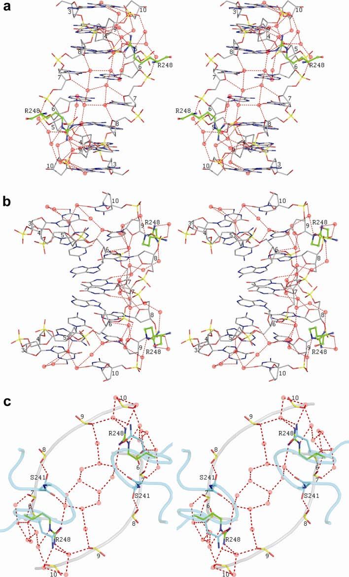
7 Supplementary Figure 5. Minor-groove hydration and interactions with Arg248 side chains. (a) View down to the minor groove of the DNA half-site and (b) View perpendicular to the first one. Only the central eight base pairs of the half-site are shown (GGCATGCC). Nucleotide numbering (5'-3' direction) is from 3 to 10 for each strand. Arg248 residues (shown in green) interact with the minor-groove hydration of the DNA represented by red spheres and dotted lines. Only the first hydration shell and some water molecules from the second hydration shell around Arg248 residues are shown. (c) Phosphate backbone of five nucleotides of each strand and protein backbone (residues ) are shown in gray and cyan, respectively. Amino acids Ser241, Asn247 (in cyan) and Arg248 (in green) are shown. Nucleotide numbering (5'-3' direction) is from 6 to 10 for each strand. Only the second hydration shell and some water molecules from the first hydration shell near the phosphate backbone are shown.
8 Supplementary Figure 6. Decomposition of the electrostatic potential in the minor groove. The electrostatic potential of DNA is caused by charged atoms of (a) the phosphates, (b) the sugar moieties, and (c) the bases. Based on the linear Poisson- Boltzmann equation, the effect of these different chemical entities on the overall potential is estimated. As illustrated in the figure, the phosphates are the main origin of the enhanced negative electrostatic potential at the Arg248 binding sites.
9 Supplementary Figure 7. Conformation of the L1 recognition loop. (a) Superposition of p53 core domains from type I and type II complexes, in magenta (based on PDB ID 2AC0 2 ) and in cyan (based on complex 1), respectively, showing the different conformations of the L1 loops from the two complexes. α-helices and loops are labeled. (b) Close-up views of the different stabilizing interactions in each complex, showing the side chains of residues Ser116, Lys120, Thr125, Arg282 and Arg283 and the backbone of residues and 124. Water atoms are shown as transparent spheres.
10 Supplementary Table 1 Direct inter-dimer polar and charged interactions 2d element of 1 st molecule Contacts 2d element of 2 nd molecule S5/S6 OE2 Glu198 OG1 Thr170 L2 2.6 S5/S6 O Gly199 N Ser96 N-ter 2.9 S5/S6 OD1 Asn200 N Ser94 N-ter 3.3 S5/S6 N Leu201 O Ser94 N-ter 2.9 S7/S8 OE2 Glu221 N Ser95 N-ter 3.1 S7/S8 OE1 Glu224 OG Ser99 N-ter 2.7 S7/S8 OE2 Glu224 NH1 Arg267 S Distance (Å) Supplementary Discussion A new conformational variant of the L1 recognition loop The reported crystal structures of the core domain in its free and DNA-bound forms have shown that, upon binding, the L1 loop undergoes a conformational change so as to allow the formation of hydrogen-bond interactions between Lys120 side chains and the second and third base pairs from each end of the decameric half-sites 2,4-6. The specific pattern of contacts made by Lys120 is sequence dependent and accommodated by adjustments in the lysine side chain 2. In several cases, mainly in the free state, the L1 loop was found to be disordered reflecting its high flexibility 7,8. In the p53 dimers and tetramers that were cross-linked to their DNA targets, this loop was shown to be either disordered or to display a dramatically different conformation in comparison to selfassembled tetrameric p53 DNA complexes. This feature is probably a result of steric hindrance caused by the covalent linkers between the two molecules 3. In type II complexes, however, the L1 loop is well defined exhibiting a new conformational variant in complexes 1 and 2 whereas complex 3 shows the previously observed L1 conformation 2. A significant change between the two conformations is observed at the five amino acid region that includes a central glycine residue (HSGTA) whereas the L1 regions flanking this pentamer are essentially identical and similarly stabilized by direct and water-mediated hydrogen bonds. Comparisons between the DNA-bound L1 conformations and the corresponding intra-molecular interactions are shown in Supplementary Fig. 7.
11 The role of the L1 loop and its highly conserved K120 residue in recognizing different response elements is uncertain in view of several recent studies. Site-directed mutagenesis studies of L1 9,10 suggest that the flexibility of this loop and hence its effect on p53 function can be significantly altered by modifications in its sequence. Other studies have shown that K120 acetylation is essential for p53-mediated activation of proapoptotic target genes 11,12. Computational analysis indicated that the energy required for desolvation of lysine side chains residues is greater than that of arginines, explaining why lysines are less likely to engage in protein-dna base contacts compared to arginines 13. The role of the single lysine residue at the p53-dna interface may well depend on the specific DNA response element and/or protein co-factors involved in the particular signaling event. Supplementary Methods Revised analysis of the published crystal structure of the mouse p53-dna complex We reanalyzed the crystal structure of the mouse p53 core-domain tetramer covalently cross-linked to two 23-mer DNA strands from Malecka et al. 3 using the deposited coordinates and structure factors (PDB code 3EXJ). In order to check the original refinement, we first used the CNS package 14,15 and calculated a sigmaa composite annealed omit map using the deposited data. Then, we submitted the model to refinement with CNS including simulated annealing with slowcooling protocol starting from 4000 C, followed by energy minimization steps, individual B-factor minimization and electron density maps calculation (sigma 2mF o DF c and mf o DF c ), without NCS restraints between the two protein molecules in the asymmetric unit. In accord with the maps from the Electron Density Server of Uppsala University ( 16, the three CNS maps exhibited several errors in the deposited model. Moreover, the composite omit map revealed that the adenine bases of the central A-T doublet of the consensus half-site sequence (GAGCATGCTC) could be better fitted into the map with a syn conformation (rotation of 180º around the glycosidic bond) which is compatible with a Hoogsteen geometry. We therefore decided to independently solve and refine the structure. We solved the structure with Phaser 17 using the deposited structure factors and molecule A from the high-resolution structure of
12 mouse p53 core domain (PDB code 2I0I) 18 as a search model. We then refined the model following the protocol used for the human p53 DNA complexes as described above. In particular, we traced manually the DNA, nucleotide by nucleotide, into the electron density maps. We further refined the whole model with the exclusion of the central two A-T base pairs. At the last stage of the refinement, we refined two independent models, one with Watson-Crick (WC) geometry and the other with Hoogsteen geometry for the central A-T base pairs of each half-site. No geometrical constraints between the bases were used. The final refinement statistics (resolution range Å and the inclusion of 420 water molecules) was similar for the two models with R work and R free being 19 and 26% respectively, and r.m.s. deviations of 0.02 Å and 2º for bond length and bond angles, respectively. The final electron density maps of the A-T base pairs calculated for the two models are shown in Supplementary Fig. 4. The hydrogen-bond distances between the adenine and thymine bases in the WC model differ by more than 0.8 Å from each other as a result of a deviation of the adenine base from a regular WC geometry, whereas the values for the Hoogsteen base pairs are similar to each other. As a result, the relative position of the glycosidic bonds and the attached sugar rings (based on N1-N9 and C1 -C1 distances across strands) in the WC model are on average shorter by 1 Å than the expected values observed in the highresolution crystal structures of type I complexes 2 (7.9 and 9.0 Å compared to 8.7 and 10.2 Å, respectively), whereas the corresponding distances in the Hoogsteen model are on average only 0.4 Å longer than the expected values based on the current crystal structures (7.0 and 8.8 Å compared to 6.7 and 8.3 Å, respectively). Also, the electron density maps (2F o F c and F o F c ) indicate a better fitting to the Hoogsteen model than to the WC one (Supplementary Fig. 4). On the basis of the electron density maps as well as the geometry of the two alternative base pairings, we conclude that the dominant form adopted by the central A-T base pairs in this crystal structure is of the Hoogsteen geometry. The crystal structure of the second published complex of the same components (PDB code 3EXL) could not be used for a revised analysis as the authors used a pseudo unit-cell dimension with c =34 Å instead of the correct unit cell, c=68 Å, as expected from the packing of 20 base-pair B-DNA helices stacked end-to-end along the c axis.
13 Supplementary References 1. Lavery, R. & Sklenar, H. Defining the structure of irregular nucleic acids: conventions and principles. J. Biomol. Struct. Dyn. 6, (1989). 2. Kitayner, M. et al. Structural basis of DNA recognition by p53 tetramers. Mol. Cell 22, (2006). 3. Malecka, K.A., Ho, W.C. & Marmorstein, R. Crystal structure of a p53 core tetramer bound to DNA. Oncogene 28, (2009). 4. Cho, Y., Gorina, S., Jeffrey, P.D. & Pavletich, N.P. Crystal structure of a p53 tumor suppressor-dna complex: understanding tumorigenic mutations. Science 265, (1994). 5. Zhao, K., Chai, X., Johnston, K., Clements, A. & Marmorstein, R. Crystal structure of the mouse p53 core DNA-binding domain at 2.7 A resolution. J. Biol. Chem. 276, (2001). 6. Joerger, A.C., Allen, M.D. & Fersht, A.R. Crystal structure of a superstable mutant of human p53 core domain. Insights into the mechanism of rescuing oncogenic mutations. J. Biol. Chem. 279, (2004). 7. Joerger, A.C., Ang, H.C., Veprintsev, D.B., Blair, C.M. & Fersht, A.R. Structures of p53 cancer mutants and mechanism of rescue by second-site suppressor mutations. J. Biol. Chem. 280, (2005). 8. Suad, O. et al. Structural basis of restoring sequence-specific DNA binding and transactivation to mutant p53 by suppressor mutations. J. Mol. Biol. 385, (2009). 9. Resnick, M.A. & Inga, A. Functional mutants of the sequence-specific transcription factor p53 and implications for master genes of diversity. Proc. Natl. Acad. Sci. USA 100, (2003). 10. Zupnick, A. & Prives, C. Mutational analysis of the p53 core domain L1 loop. J. Biol. Chem. 281, (2006). 11. Sykes, S.M. et al. Acetylation of the p53 DNA-binding domain regulates apoptosis induction. Mol. Cell 24, (2006). 12. Tang, Y., Luo, J., Zhang, W. & Gu, W. Tip60-dependent acetylation of p53 modulates the decision between cell-cycle arrest and apoptosis. Mol. Cell 24, (2006). 13. Rohs, R. et al. The role of DNA shape in protein-dna recognition. Nature 461, (2009). 14. Brunger, A.T. et al. Crystallography & NMR system: A new software suite for macromolecular structure determination. Acta Crystallogr. D Biol. Crystallogr. 54, (1998). 15. Brunger, A.T. Version 1.2 of the Crystallography and NMR system. Nat. Protoc. 2, (2007). 16. Kleywegt, G.J. et al. The Uppsala Electron-Density Server. Acta Crystallogr. D Biol. Crystallogr. 60, (2004). 17. McCoy, A.J. et al. Phaser crystallographic software. J. Appl. Crystallogr. 40, (2007).
14 18. Ho, W.C. et al. High-resolution structure of the p53 core domain: implications for binding small-molecule stabilizing compounds. Acta Crystallogr. D Biol. Crystallogr. 62, (2006).
Suppl. Figure 1: RCC1 sequence and sequence alignments. (a) Amino acid
 Amino acid") Supplementary Figures Suppl. Figure 1: RCC1 sequence and sequence alignments. (a) Amino acid sequence of Drosophila RCC1. Same colors are for Figure 1 with sequence of β-wedge that interacts with Ran in
Supplementary Figures Suppl. Figure 1: RCC1 sequence and sequence alignments. (a) Amino acid sequence of Drosophila RCC1. Same colors are for Figure 1 with sequence of β-wedge that interacts with Ran in
Molecular design principles underlying β-strand swapping. in the adhesive dimerization of cadherins
 Supplementary information for: Molecular design principles underlying β-strand swapping in the adhesive dimerization of cadherins Jeremie Vendome 1,2,3,5, Shoshana Posy 1,2,3,5,6, Xiangshu Jin, 1,3 Fabiana
SUPPLEMENTARY INFORMATION
 Structure of a tyrosyl-trna synthetase splicing factor bound to a group I intron RNA Paul J. Paukstelis 1, Jui-Hui Chen 2, Elaine Chase 2, Alan M. Lambowitz 1,*, and Barbara L. Golden 2,*,. 1 Institute
Structural Bioinformatics (C3210) DNA and RNA Structure
 DNA and RNA Structure") Structural Bioinformatics (C3210) DNA and RNA Structure Importance of DNA/RNA 3D Structure Nucleic acids are essential materials found in all living organisms. Their main function is to maintain and transmit
Structural Bioinformatics (C3210) DNA and RNA Structure Importance of DNA/RNA 3D Structure Nucleic acids are essential materials found in all living organisms. Their main function is to maintain and transmit
Basic concepts of molecular biology
 Basic concepts of molecular biology Gabriella Trucco Email: gabriella.trucco@unimi.it Life The main actors in the chemistry of life are molecules called proteins nucleic acids Proteins: many different
DNA Structures. Biochemistry 201 Molecular Biology January 5, 2000 Doug Brutlag. The Structural Conformations of DNA
 DNA Structures Biochemistry 201 Molecular Biology January 5, 2000 Doug Brutlag The Structural Conformations of DNA 1. The principle message of this lecture is that the structure of DNA is much more flexible
Non-standard base pairs Non-standard base pairs play critical roles in the varied structures observed in DNA and RNA.
 DNA ORIENTATION Non-standard base pairs Non-standard base pairs play critical roles in the varied structures observed in DNA and RNA. Non-standard base pairs Wobble and mismatched base pairs still use
MCB 110:Biochemistry of the Central Dogma of MB. MCB 110:Biochemistry of the Central Dogma of MB
 MCB 110:Biochemistry of the Central Dogma of MB Part 1. DNA replication, repair and genomics (Prof. Alber) Part 2. RNA & protein synthesis. Prof. Zhou Part 3. Membranes, protein secretion, trafficking
Hmwk # 8 : DNA-Binding Proteins : Part II
 The purpose of this exercise is : Hmwk # 8 : DNA-Binding Proteins : Part II 1). to examine the case of a tandem head-to-tail homodimer binding to DNA 2). to view a Zn finger motif 3). to consider the case
SUPPLEMENTARY INFORMATION
 Molecular basis of RNA-dependent RNA polymerase II activity Elisabeth Lehmann, Florian Brueckner, and Patrick Cramer Gene Center Munich and Center for integrated Protein Science CiPS M, Department of Chemistry
1.1 Chemical structure and conformational flexibility of single-stranded DNA
 1 DNA structures 1.1 Chemical structure and conformational flexibility of single-stranded DNA Single-stranded DNA (ssdna) is the building base for the double helix and other DNA structures. All these structures
has only one nucleotide, U20, between G19 and A21, while trna Glu CUC has two
 SPPLEMENTRY INFORMTION doi:1.138/nature9411 Supplementary Discussion The structural characteristics of trn Gln G in comparison to trn Glu 16 in trn Gln G is directed towards the G19 56 pair, or the outer
Assembly and Characteristics of Nucleic Acid Double Helices
 Assembly and Characteristics of Nucleic Acid Double Helices Patterns of base-base hydrogen bonds-characteristics of the base pairs Interactions between like and unlike bases have been observed in crystal
Structural bioinformatics
 Structural bioinformatics Why structures? The representation of the molecules in 3D is more informative New properties of the molecules are revealed, which can not be detected by sequences Eran Eyal Plant
1/4/18 NUCLEIC ACIDS. Nucleic Acids. Nucleic Acids. ECS129 Instructor: Patrice Koehl
 NUCLEIC ACIDS ECS129 Instructor: Patrice Koehl Nucleic Acids Nucleotides DNA Structure RNA Synthesis Function Secondary structure Tertiary interactions Wobble hypothesis DNA RNA Replication Transcription
NUCLEIC ACIDS. ECS129 Instructor: Patrice Koehl
 NUCLEIC ACIDS ECS129 Instructor: Patrice Koehl Nucleic Acids Nucleotides DNA Structure RNA Synthesis Function Secondary structure Tertiary interactions Wobble hypothesis DNA RNA Replication Transcription
Basic concepts of molecular biology
 Basic concepts of molecular biology Gabriella Trucco Email: gabriella.trucco@unimi.it What is life made of? 1665: Robert Hooke discovered that organisms are composed of individual compartments called cells
Nucleic acids. How DNA works. DNA RNA Protein. DNA (deoxyribonucleic acid) RNA (ribonucleic acid) Central Dogma of Molecular Biology
 RNA (ribonucleic acid) Central Dogma of Molecular Biology") Nucleic acid chemistry and basic molecular theory Nucleic acids DNA (deoxyribonucleic acid) RNA (ribonucleic acid) Central Dogma of Molecular Biology Cell cycle DNA RNA Protein Transcription Translation
Nucleic acid chemistry and basic molecular theory Nucleic acids DNA (deoxyribonucleic acid) RNA (ribonucleic acid) Central Dogma of Molecular Biology Cell cycle DNA RNA Protein Transcription Translation
X-ray structures of fructosyl peptide oxidases revealing residues responsible for gating oxygen access in the oxidative half reaction
 X-ray structures of fructosyl peptide oxidases revealing residues responsible for gating oxygen access in the oxidative half reaction Tomohisa Shimasaki 1, Hiromi Yoshida 2, Shigehiro Kamitori 2 & Koji
Structure formation and association of biomolecules. Prof. Dr. Martin Zacharias Lehrstuhl für Molekulardynamik (T38) Technische Universität München
 Technische Universität München") Structure formation and association of biomolecules Prof. Dr. Martin Zacharias Lehrstuhl für Molekulardynamik (T38) Technische Universität München Motivation Many biomolecules are chemically synthesized
Structure formation and association of biomolecules Prof. Dr. Martin Zacharias Lehrstuhl für Molekulardynamik (T38) Technische Universität München Motivation Many biomolecules are chemically synthesized
SUPPLEMENTARY INFORMATION. Supplementary Figures 1-8
 SUPPLEMENTARY INFORMATION Supplementary Figures 1-8 Supplementary Figure 1. TFAM residues contacting the DNA minor groove (A) TFAM contacts on nonspecific DNA. Leu58, Ile81, Asn163, Pro178, and Leu182
SUPPLEMENTARY INFORMATION
 doi: 10.1038/nature06147 SUPPLEMENTARY INFORMATION Figure S1 The genomic and domain structure of Dscam. The Dscam gene comprises 24 exons, encoding a signal peptide (SP), 10 IgSF domains, 6 fibronectin
Supporting Online Material. Av1 and Av2 were isolated and purified under anaerobic conditions according to
 Supporting Online Material Materials and Methods Av1 and Av2 were isolated and purified under anaerobic conditions according to published protocols (S1). Crystals of nf-, pcp- and adp-av2:av1 complexes
Supplementary Fig. 1. Initial electron density maps for the NOX-D20:mC5a complex obtained after SAD-phasing. (a) Initial experimental electron
 Initial experimental electron") Supplementary Fig. 1. Initial electron density maps for the NOX-D20:mC5a complex obtained after SAD-phasing. (a) Initial experimental electron density map obtained after SAD-phasing and density modification
Supplementary Fig. 1. Initial electron density maps for the NOX-D20:mC5a complex obtained after SAD-phasing. (a) Initial experimental electron density map obtained after SAD-phasing and density modification
Gene and DNA structure. Dr Saeb Aliwaini
 Gene and DNA structure Dr Saeb Aliwaini 2016 DNA during cell cycle Cell cycle for different cell types Molecular Biology - "Study of the synthesis, structure, and function of macromolecules (DNA, RNA,
Dr. R. Sankar, BSE 631 (2018)
") Pauling, Corey and Branson Diffraction of DNA http://www.nature.com/scitable/topicpage/dna-is-a-structure-that-encodes-biological-6493050 In short, stereochemistry is important in determining which helices
Pauling, Corey and Branson Diffraction of DNA http://www.nature.com/scitable/topicpage/dna-is-a-structure-that-encodes-biological-6493050 In short, stereochemistry is important in determining which helices
SUPPLEMENTARY DATA. DNAproDB: an interactive tool for structural analysis of DNA-protein complexes
 SUPPLEMENTARY DATA DNAproDB: an interactive tool for structural analysis of DNA-protein complexes Jared M. Sagendorf 1, Helen M. Berman 2, *, and Remo Rohs 1, * 1 Molecular and Computational Biology Program,
Supplementary Figure 1
 Supplementary Figure 1 2 Supplementary Figure 1: Sequence alignment of HsHSD17B8 and HsCBR4 of with KAR orthologs. The secondary structure elements as calculated by DSSP and residue numbers are displayed
Bioinformatics. ONE Introduction to Biology. Sami Khuri Department of Computer Science San José State University Biology/CS 123A Fall 2012
 Bioinformatics ONE Introduction to Biology Sami Khuri Department of Computer Science San José State University Biology/CS 123A Fall 2012 Biology Review DNA RNA Proteins Central Dogma Transcription Translation
RNA does not adopt the classic B-DNA helix conformation when it forms a self-complementary double helix
 Reason: RNA has ribose sugar ring, with a hydroxyl group (OH) If RNA in B-from conformation there would be unfavorable steric contact between the hydroxyl group, base, and phosphate backbone. RNA structure
Biochemistry Prof. S. Dasgupta Department of Chemistry. Indian Institute of Technology Kharagpur. Lecture - 16 Nucleic Acids - I
 Biochemistry Prof. S. Dasgupta Department of Chemistry. Indian Institute of Technology Kharagpur Lecture - 16 Nucleic Acids - I We start our discussion on Nucleic Acids and their components. Before we
Nucleotides: structure and functions. Prof. Dalė Vieželienė Biochemistry department Room No
 Nucleotides: structure and functions Prof. Dalė Vieželienė Biochemistry department Room No. 229 Email: daleveze@med.kmu.lt Composition of Nucleic Acids Nucleotide structure Two types of nucleic acids:
Supplementary Information for. Structure of human tyrosylprotein sulfotransferase-2 reveals the mechanism of protein tyrosine sulfation reaction
 Supplementary Information for Structure of human tyrosylprotein sulfotransferase-2 reveals the mechanism of protein tyrosine sulfation reaction Takamasa Teramoto, Yukari Fujikawa, Yoshirou Kawaguchi, Katsuhisa
Canonical B-DNA CGCGTTGACAACTGCAGAATC GC AT CG TA AT GC TA TA CG AT 20 Å. Minor Groove 34 Å. Major Groove 3.4 Å. Strands are antiparallel
 DNA Canonical B-DNA 20 Å GC AT CG TA CGCGTTGACAACTGCAGAATC 34 Å AT GC TA Minor Groove 3.4 Å TA CG AT Major Groove Strands are antiparallel CG GC GC Canonical B DNA First determined experimentally by fiber
 Problem: The GC base pairs are more stable than AT base pairs. Why? 5. Triple-stranded DNA was first observed in 1957. Scientists later discovered that the formation of triplestranded DNA involves a type
Conformational changes in IgE contribute to its. uniquely slow dissociation rate from receptor FcεRI
 Conformational changes in IgE contribute to its uniquely slow dissociation rate from receptor FcεRI M.D. Holdom, A.M. Davies, J.E. Nettleship, S.C. Bagby, B. Dhaliwal, E. Girardi, J. Hunt, H.J. Gould,
Improving RNA Crystallographic Models Using Rosetta. Fang-Chieh Chou (Das Lab) For RosettaCon 2011
 For RosettaCon 2011") Improving RNA Crystallographic Models Using Rosetta Fang-Chieh Chou (Das Lab) For RosettaCon 211 The number RNA crystal structures increased explosively after the first Ribosome crystal structure is solved
Improving RNA Crystallographic Models Using Rosetta Fang-Chieh Chou (Das Lab) For RosettaCon 211 The number RNA crystal structures increased explosively after the first Ribosome crystal structure is solved
Supplementary Note 1. Enzymatic properties of the purified Syn BVR
 Supplementary Note 1. Enzymatic properties of the purified Syn BVR The expression vector pet15b-syn bvr allowed us to routinely prepare 15 mg of electrophoretically homogenous Syn BVR from 2.5 L of TB-medium
Supplementary Information. Structural basis for duplex RNA recognition and cleavage by A.
 Supplementary Information Structural asis for duplex RNA recognition and cleavage y A. fulgidus C3PO Eneida arizotto 1, Edward D Lowe 1 & James S Parker 1 1 Department of Biochemistry University of Oxford
Structural Bioinformatics (C3210) Conformational Analysis Protein Folding Protein Structure Prediction
 Conformational Analysis Protein Folding Protein Structure Prediction") Structural Bioinformatics (C3210) Conformational Analysis Protein Folding Protein Structure Prediction Conformational Analysis 2 Conformational Analysis Properties of molecules depend on their three-dimensional
Structural Bioinformatics (C3210) Conformational Analysis Protein Folding Protein Structure Prediction Conformational Analysis 2 Conformational Analysis Properties of molecules depend on their three-dimensional
The Skap-hom Dimerization and PH Domains Comprise
 Molecular Cell, Volume 32 Supplemental Data The Skap-hom Dimerization and PH Domains Comprise a 3 -Phosphoinositide-Gated Molecular Switch Kenneth D. Swanson, Yong Tang, Derek F. Ceccarelli, Florence Poy,
Algorithms in Bioinformatics ONE Transcription Translation
 Algorithms in Bioinformatics ONE Transcription Translation Sami Khuri Department of Computer Science San José State University sami.khuri@sjsu.edu Biology Review DNA RNA Proteins Central Dogma Transcription
Packing of Secondary Structures
 7.88 Lecture Notes - 5 7.24/7.88J/5.48J The Protein Folding and Human Disease Packing of Secondary Structures Packing of Helices against sheets Packing of sheets against sheets Parallel Orthogonal Table:
Crystal Structure of the p53 Core Domain Bound to a Full Consensus Site as a Self-Assembled Tetramer
 Article Crystal Structure of the p53 Core Domain Bound to a Full Consensus Site as a Self-Assembled Tetramer Yongheng Chen, 1,2 Raja Dey, 1 and Lin Chen 1,2, * 1 Molecular and Computational Biology, Departments
SUPPLEMENTARY INFORMATION. Reengineering Protein Interfaces Yields Copper-Inducible Ferritin Cage Assembly
 SUPPLEMENTARY INFORMATION Reengineering Protein Interfaces Yields Copper-Inducible Ferritin Cage Assembly Dustin J. E. Huard, Kathleen M. Kane and F. Akif Tezcan* Department of Chemistry and Biochemistry,
Supplementary materials for Structure of an open clamp type II topoisomerase-dna complex provides a mechanism for DNA capture and transport
 Supplementary materials for Structure of an open clamp type II topoisomerase-dna complex provides a mechanism for DNA capture and transport Ivan Laponogov 1,2, Dennis A. Veselkov 1, Isabelle M-T. Crevel
DNA stands for deoxyribose nucleic acid
 1 DNA 2 DNA stands for deoxyribose nucleic acid This chemical substance is present in the nucleus of all cells in all living organisms DNA controls all the chemical changes which take place in cells The
Ch 10 Molecular Biology of the Gene
 Ch 10 Molecular Biology of the Gene For Next Week Lab -Hand in questions from 4 and 5 by TUES in my mailbox (Biology Office) -Do questions for Lab 6 for next week -Lab practical next week Lecture Read
Nature Structural & Molecular Biology: doi: /nsmb Supplementary Figure 1
 Supplementary Figure 1 Multiple sequence alignments of four Swi2/Snf2 subfamily proteins, ScChd1, SsoRad54 and the RNA helicase Vasa. The sequence alignments of the Swi2/Snf2 subfamily proteins, ScChd1
SUPPLEMENTARY INFORMATION
 Supplementary Table 1. Crystallographic statistics CRM1-SNUPN complex Space group P6 4 22 a=b=250.4, c=190.4 Data collection statistics: CRM1-selenomethionine SNUPN MAD data Peak Inflection Remote Native
Understanding DNA Structure
 Understanding DNA Structure I619 Structural Bioinformatics Molecular Biology Basics + Scale total length of DNA in a human cell is about 2m DNA is compacted in length by a factor of 10000 the compaction
Dina Al-Tamimi. Faisal Nimri. Ma amoun Ahram. 1 P a g e
 1 Dina Al-Tamimi Faisal Nimri Ma amoun Ahram 1 P a g e **Difference between Molecular Biology and Genetics: Molecular Biology: is a fancy term of biochemistry. It is the science that deals with DNA, RNA
Concept 5.5: Nucleic acids store and transmit hereditary information
 Concept 5.5: Nucleic acids store and transmit hereditary information The amino acid sequence of a polypeptide is programmed by a unit of inheritance called a gene Genes are made of DNA, a nucleic acid
Supporting Information Contents
 Supporting Information Choy Theng Loh, Kiyoshi Ozawa, Kellie L. Tuck, Nicholas Barlow, Thomas Huber, Gottfried Otting, and Bim Graham Lanthanide tags for site-specific ligation to an unnatural amino acid
Chapter 8 DNA Recognition in Prokaryotes by Helix-Turn-Helix Motifs
 Chapter 8 DNA Recognition in Prokaryotes by Helix-Turn-Helix Motifs 1. Helix-turn-helix proteins 2. Zinc finger proteins 3. Leucine zipper proteins 4. Beta-scaffold factors 5. Others λ-repressor AND CRO
Supporting Information
 Supporting Information van der Cruijsen et al. 10.1073/pnas.1305563110 SI Methods Structural Analysis and Validation. A homology model of KcsA- Kv1.3 in the close conductive state was generated from the
6-Foot Mini Toober Activity
 Big Idea The interaction between the substrate and enzyme is highly specific. Even a slight change in shape of either the substrate or the enzyme may alter the efficient and selective ability of the enzyme
Nucleic Acids: How Structure Conveys Information 1. What Is the Structure of DNA? 2. What Are the Levels of Structure in Nucleic Acids? 3.
 Fig. 9-CO, p.215 Nucleic Acids: How Structure Conveys Information 1. What Is the Structure of DNA? 2. What Are the Levels of Structure in Nucleic Acids? 3. What Is the Covalent Structure of Polynucleotides?
38. Inter-basepair Hydrogen Bonds in DNA
 190 Proc. Japan Acad., 70, Ser. B (1994) [Vol. 70(B), 38. Inter-basepair Hydrogen Bonds in DNA By Masashi SUZUKI*),t) and Naoto YAGI**) (Communicated by Setsuro EBASHI, M. J. A., Dec. 12, 1994) Abstract:
(Due Sept 9 th ) Problem Set 2
 Problem Set 2") Problem Set 2 (Due Sept 9 th ) 1. Consider these two polynucleotides: AAGCGT GCACTG a. Draw each molecule in the 2 deoxy form (DNA). SEE BELOW b. What is the sequence of the complementary strand? Write
Problem Set 2 (Due Sept 9 th ) 1. Consider these two polynucleotides: AAGCGT GCACTG a. Draw each molecule in the 2 deoxy form (DNA). SEE BELOW b. What is the sequence of the complementary strand? Write
Molecular Biology. Biology Review ONE. Protein Factory. Genotype to Phenotype. From DNA to Protein. DNA à RNA à Protein. June 2016
 Molecular Biology ONE Sami Khuri Department of Computer Science San José State University Biology Review DNA RNA Proteins Central Dogma Transcription Translation Genotype to Phenotype Protein Factory DNA
DNA. Branden & Tooze, Ch. 7 Deoxyribose nucleic acids are made of three parts
 DNA Branden & Tooze, Ch. 7 Deoxyribose nucleic acids are made of three parts base: adenine, cytosine, guanine, thymine sugar: deoxyribose phosphate: will form the phosphate backbone wide narrow DNA binding
Structure and Function of the First Full-Length Murein Peptide Ligase (Mpl) Cell Wall Recycling Protein
 Cell Wall Recycling Protein") Paper Presentation PLoS ONE 2011 Structure and Function of the First Full-Length Murein Peptide Ligase (Mpl) Cell Wall Recycling Protein Debanu Das, Mireille Herve, Julie Feuerhelm, etc. and Dominique
Paper Presentation PLoS ONE 2011 Structure and Function of the First Full-Length Murein Peptide Ligase (Mpl) Cell Wall Recycling Protein Debanu Das, Mireille Herve, Julie Feuerhelm, etc. and Dominique
Supplementary Information Titles
 Supplementary Information Titles Please list each supplementary item and its title or caption, in the order shown below. Note that we do NOT copy edit or otherwise change supplementary information, and
BMB/Bi/Ch 170 Fall 2017 Problem Set 1: Proteins I
 BMB/Bi/Ch 170 Fall 2017 Problem Set 1: Proteins I Please use ray-tracing feature for all the images you are submitting. Use either the Ray button on the right side of the command window in PyMOL or variations
Supplementary Figure 1. Electron microscopy of gb-698glyco/1g2 Fab complex. a)
") Supplementary Figure 1. Electron microscopy of gb-698glyco/1g2 Fab complex. a) Representative images of 2D class averages of gb-698glyc bound to 1G2 Fab. Top views of the complex were underrepresented
Supplementary Figure 1. Electron microscopy of gb-698glyco/1g2 Fab complex. a) Representative images of 2D class averages of gb-698glyc bound to 1G2 Fab. Top views of the complex were underrepresented
Nucleic Acids and the RNA World. Pages Chapter 4
 Nucleic Acids and the RNA World Pages 74-89 Chapter 4 RNA vs. Protein Chemical Evolution stated that life evolved from a polymer called a protein. HOWEVER, now many scientists question this. There is currently
The Cys 2. His 2. Research Article 451
 Research Article 451 High-resolution structures of variant Zif268 DNA complexes: implications for understanding zinc finger DNA recognition Monicia Elrod-Erickson 1, Timothy E Benson 1 and Carl O Pabo
Antibody-Antigen recognition. Structural Biology Weekend Seminar Annegret Kramer
 Antibody-Antigen recognition Structural Biology Weekend Seminar 10.07.2005 Annegret Kramer Contents Function and structure of antibodies Features of antibody-antigen interfaces Examples of antibody-antigen
Structural Bioinformatics GENOME 541 Spring 2018
 Molecular composition of a rapidly dividing Escherichia coli cell Structural Bioinformatics GENOME 541 Spring 2018 Lecture 4: Nucleic Acids Frank DiMaio (dimaio@uw.edu) The major biopolymers DNA structure
Fundamentals of Organic Chemistry. CHAPTER 10: Nucleic Acids
 Fundamentals of Organic Chemistry CHEM 109 For Students of Health Colleges Credit hrs.: (2+1) King Saud University College of Science, Chemistry Department CHEM 109 CHAPTER 10: Nucleic Acids 2 o Nucleic
What Are the Chemical Structures and Functions of Nucleic Acids?
 THE NUCLEIC ACIDS What Are the Chemical Structures and Functions of Nucleic Acids? Nucleic acids are polymers specialized for the storage, transmission, and use of genetic information. DNA = deoxyribonucleic
Electrostatic interactions between arginines and the minor groove in the nucleosome
 Journal of Biomolecular Structure & Dynamics, ISSN 0739-1102 Volume 27, Issue Number 6, (2010) Current Perspectives on Nucleosome Positioning Adenine Press (2010) Electrostatic interactions between arginines
Molecular biology (1)
") 2018/9/24 Molecular biology (1) Important. 436 Notes Original slides. 438 notes Extra information Objectives: Know the central dogma of molecular biology. Understand the composition, types and structure
2018/9/24 Molecular biology (1) Important. 436 Notes Original slides. 438 notes Extra information Objectives: Know the central dogma of molecular biology. Understand the composition, types and structure
Artificial Nucleic Acids -Their Developments and Recent Applications
 Artificial Nucleic Acids -Their Developments and Recent Applications Bioorganic Chemistry Laboratory D2 Kenichiro Ito Organic Seminar 2012/5/7 1 Nucleic acids play central roles in life Replication Transcription
Molecular biology (1)
") Molecular biology (1) Color index: Doctors slides Notes and explanations Extra information highlights Objectives Know the central dogma of molecular biology. Understand the composition, types and structure
Molecular biology (1) Color index: Doctors slides Notes and explanations Extra information highlights Objectives Know the central dogma of molecular biology. Understand the composition, types and structure
Structure and Possible Mechanism of the CcbJ Methyltransferase from Streptomyces caelestis
 Supplemental material to accompany Structure and Possible Mechanism of the CcbJ Methyltransferase from Streptomyces caelestis Jacob Bauer, a Gabriela Ondrovičová, a Lucie Najmanová, b Vladimír Pevala,
translation The building blocks of proteins are? amino acids nitrogen containing bases like A, G, T, C, and U Complementary base pairing links
 The actual process of assembling the proteins on the ribosome is called? translation The building blocks of proteins are? Complementary base pairing links Define and name the Purines amino acids nitrogen
The oestrogen receptor recognizes an imperfectly palindromic response element through an alternative side-chain conformation
 The oestrogen receptor recognizes an imperfectly palindromic response element through an alternative side-chain conformation John WR Schwabet*, Lynda Chapman and Daniela Rhodes Medical Research Council,
DNA and RNA are both composed of nucleotides. A nucleotide contains a base, a sugar and one to three phosphate groups. DNA is made up of the bases
 1 DNA and RNA are both composed of nucleotides. A nucleotide contains a base, a sugar and one to three phosphate groups. DNA is made up of the bases Adenine, Guanine, Cytosine and Thymine whereas in RNA
YOUR NAME: KEY. PLEASE PRINT your name (IN INDELIBLE INK) on the line above (& on the top right hand corner of every page).
 on the line above (& on the top right hand corner of every page).") UNIVERSITY OF CALIFORNIA, BERKELEY CHEM C130/MCB C100A MIDTERM EXAMINATION #1 SEPTEMBER 22, 2016 INSTRUCTORS: John Kuriyan and David Savage THE TIME LIMIT FOR THIS EXAMINATION: 1 HOUR 50 MINUTES SIGNATURE:
UNIVERSITY OF CALIFORNIA, BERKELEY CHEM C130/MCB C100A MIDTERM EXAMINATION #1 SEPTEMBER 22, 2016 INSTRUCTORS: John Kuriyan and David Savage THE TIME LIMIT FOR THIS EXAMINATION: 1 HOUR 50 MINUTES SIGNATURE:
DNA Glycosylase Exercise
 Name StarBiochem DNA Glycosylase Exercise Background In this exercise, you will use StarBiochem, a protein 3-D viewer, to explore the structure of a DNA repair protein found in most species, including
MBMB,BCHM, or CHEM 451A
 MBMB,BCHM, or CHEM 451A This is a team taught course Blaine Bartholomew: 1 st section Joseph Schmit: 2 nd section Peter Hardwicke:3 rd Section Text is Lehninger Principles of Biochemistry 4 th edition
doi: /nature09408 Figure S1
 doi:10.1038/nature09408 A Figure S1 www.nature.com/nature 1 RESEARCH SUPPLEMENTARY INFORMATION Figure S1 Primary sequence, and structure of NorM VC. A. Amino acid sequence alignment of NorM VC with selected
CFSSP: Chou and Fasman Secondary Structure Prediction server
 Wide Spectrum, Vol. 1, No. 9, (2013) pp 15-19 CFSSP: Chou and Fasman Secondary Structure Prediction server T. Ashok Kumar Department of Bioinformatics, Noorul Islam College of Arts and Science, Kumaracoil
Chapter 5: Nucleic Acids, etc.
 Chapter 5: Nucleic Acids, etc. Voet & Voet: Sections 1 & 3 Pages 82-84 & 88-93 Any introductory Biochemistry textbook will have an introductory chapter on nucleic acids Slide 1 Nucleotides and Derivatives
Clamping down on pathogenic bacteria how to shut down a key DNA polymerase complex
 Clamping down on pathogenic bacteria how to shut down a key DNA polymerase complex Bacterial DNA-replication machinery Pathogenic bacteria that are resistant to the current armoury of antibiotics are an
Structure of the C-cadherin ectodomain and implications for the mechanism of cell adhesion
 Structure of the C-cadherin ectodomain and implications for the mechanism of cell adhesion Titus J. Boggon, John Murray, Sophie Chappuis-Flament, Ellen Wong, Barry M. Gumbiner, and Lawrence Shapiro Ref.
BETA STRAND Prof. Alejandro Hochkoeppler Department of Pharmaceutical Sciences and Biotechnology University of Bologna
 Prof. Alejandro Hochkoeppler Department of Pharmaceutical Sciences and Biotechnology University of Bologna E-mail: a.hochkoeppler@unibo.it C-ter NH and CO groups: right, left, right (plane of the slide)
Protein Structure Prediction by Constraint Logic Programming
 MPRI C2-19 Protein Structure Prediction by Constraint Logic Programming François Fages, Constraint Programming Group, INRIA Rocquencourt mailto:francois.fages@inria.fr http://contraintes.inria.fr/ Molecules
From mechanism to medicne
 From mechanism to medicne a look at proteins and drug design Chem 342 δ δ δ+ M 2009 δ+ δ+ δ M Drug Design - an Iterative Approach @ DSU Structural Analysis of Receptor Structural Analysis of Ligand-Receptor
Nature Structural & Molecular Biology: doi: /nsmb Supplementary Figure 1. Location of the mab 6F10 epitope in capsids.
 Supplementary Figure 1 Location of the mab 6F10 epitope in capsids. The epitope of monoclonal antibody, mab 6F10, was mapped to residues A862 H880 of the HSV-1 major capsid protein VP5 by immunoblotting
Intercalated cytosine motif and novel adenine clusters in the crystal structure of the Tetrahymena telomere
 4696 4705 Nucleic Acids Research, 1998, Vol. 26, No. 20 1998 Oxford University Press Intercalated cytosine motif and novel adenine clusters in the crystal structure of the Tetrahymena telomere Li Cai,
ECS 129: Structural Bioinformatics March 15, 2016
 Notes: ES 129: Structural Bioinformatics March 15, 2016 1) The final exam is open book, open notes. 2) The final is divided into 2 parts, and graded over 100 points (with 8 points extra credit) 3) You
Chem 250 Answer Key In-class Quiz #3v1
 age 1 of 6 Quiz #3 ame. Chem 250 Answer Key In-class Quiz #3v1 This exam is composed of 20 questions. lease scan them all before starting. As discussed in the course syllabus, honesty and integrity are
Nucleic Acids. Information specifying protein structure
 Nucleic Acids Nucleic acids represent the fourth major class of biomolecules (other major classes of biomolecules are proteins, carbohydrates, fats) Genome - the genetic information of an organism Information
Information specifying protein structure. Chapter 19 Nucleic Acids Nucleotides Are the Building Blocks of Nucleic Acids
 Chapter 19 Nucleic Acids Information specifying protein structure Nucleic acids represent the fourth major class of biomolecules (other major classes of biomolecules are proteins, carbohydrates, fats)
From Gene to Protein
 8.2 Structure of DNA From Gene to Protein deoxyribonucleic acid - (DNA) - the ultimate source of all information in a cell This information is used by the cell to produce the protein molecules which are
Supplementary Information For. A genetically encoded tool for manipulation of NADP + /NADPH in living cells
 Supplementary Information For A genetically encoded tool for manipulation of NADP + /NADPH in living cells Valentin Cracan 1,2,3, Denis V. Titov 1,2,3, Hongying Shen 1,2,3, Zenon Grabarek 1* and Vamsi
The structure of a CAP DNA complex having two camp molecules bound to each monomer
 Proc. Natl. Acad. Sci. USA Vol. 94, pp. 2843 2847, April 1997 Biochemistry The structure of a CAP DNA complex having two camp molecules bound to each monomer J. M. PASSNER* AND T. A. STEITZ* *Department