SAPIENZA Università di Roma Laurea magistrale in Ingegneria delle Nanotecnologie A.A Biophotonics Laboratory Course
|
|
|
- Cameron Barber
- 6 years ago
- Views:
Transcription

1 SAPIENZA Università di Roma Laurea magistrale in Ingegneria delle Nanotecnologie A.A Biophotonics Laboratory Course Prof. Francesco Michelotti SAPIENZA Università di Roma Facoltà di Ingegneria Civile e Industriale francesco.michelotti@uniroma1.it
2 LECTURE 6 Super-resolution microscopies (STED, PALM, STORM) Test ELISA
3 Applications of optics and photonics Microscopic Techniques Conventional Wide-Field Fluorescence TIRF FLIM FRET, FRAP Confocal Two-Photon Second Harmonic Super-resolution (SNOM, STED, PALM, STORM) Non-Microscopic Label-free Surface plasmon Polaritons (SPP) Photonic crystals (PC) Raman, CARS Quantum dots Non-Microscopic Techniques Cytofluorimetry ELISA DNA-Chip Cycle-sequencing SOLID Other non Microscopic Techniques Southern Western Northern All of them make use of the emission of luminescent markers (labels)
4 STED STimulated Emission Depletion Microscopy In STED microscopy the specimen is excited and the fluorescence is collected as in conventional wide-field microscopy. A supplementary laser beam causes the relaxation of the chromophores inside an annular region around the central spot by stimulated emission.

5 STED STimulated Emission Depletion Microscopy S e t 1 S g t2>t1 t
6 STED STimulated Emission Depletion Microscopy Diffraction would limit the resolution also in this case. The real mechanism that is responsible of the increase of resolution is the saturation of fluorescence reduction obtained by means of stimulated emission. I SAT The resolution is given by: x 2nsin 1 I I SAT

7 STED STimulated Emission Depletion Microscopy

8 STED STimulated Emission Depletion Microscopy Diffraction would limit the resolution also in this case. The real mechanism that is responsible of the increase of resolution is the saturation of fluorescence reduction obtained by means of stimulated emission. I SAT The resolution is given by: x 2nsin 1 I I SAT

9 STED STimulated Emission Depletion Microscopy Diffraction would limit the resolution also in this case. The real mechanism that is responsible of the increase of resolution is the saturation of fluorescence reduction obtained by means of stimulated emission. I SAT The resolution is given by: x 2nsin 1 I I SAT
10 STED STimulated Emission Depletion Microscopy Diffraction would limit the resolution also in this case. The real mechanism that is responsible of the increase of resolution is the saturation of fluorescence reduction obtained by means of stimulated emission. I SAT The resolution is given by: x 2nsin 1 I I SAT
11 STED STimulated Emission Depletion Microscopy Diffraction would limit the resolution also in this case. The real mechanism that is responsible of the increase of resolution is the saturation of fluorescence reduction obtained by means of stimulated emission. I SAT The resolution is given by: x 2nsin 1 I I SAT

12 STED STimulated Emission Depletion Microscopy Diffraction would limit the resolution also in this case. The real mechanism that is responsible of the increase of resolution is the saturation of fluorescence reduction obtained by means of stimulated emission. I SAT The resolution is given by: x 2nsin 1 I I SAT
13 STED STimulated Emission Depletion Microscopy Diffraction would limit the resolution also in this case. The real mechanism that is responsible of the increase of resolution is the saturation of fluorescence reduction obtained by means of stimulated emission. I SAT The resolution is given by: x 2nsin 1 I I SAT
14 STED STimulated Emission Depletion Microscopy I SAT NOTE If there is no saturation of the fluorescence reduction (I SAT = ) the resolution is that given by the Abbe formula, i.e. it is limited by diffraction. x 2nsin 1 I SAT I 2nsin
15 STED STimulated Emission Depletion Microscopy Dye name (Manufacturer / Distributor) Exc. Exc. Wavelengt Pulse h Length STED Wavelengt h STED Pulse Length Repetition Rate Avg. STED Power Peak Irradiance Reported Spatial Pulse Resolution Energy (Direction) ATTO 532 (ATTO-TEC GmbH) 470 nm 80 ps 603 nm 280 ps 250 khz 0.5 mw 2 nj <25 nm (xy) Chromeo 488 (Actif Motif) 488 nm 140 ps 602 nm ~ 160 ps 250 khz 0.6 mw < 30 nm (xy) DY-485XL (Dyomics GmbH) 488 nm < 100 ps 647 nm ~ 200 ps 72 MHz (20 + 3) mw nm (xyz) GFP 490 nm 100 ps 575 nm 200 ps 80 MHz 7.2 mw ~ 70 nm (xy) ATTO 565 (ATTO-TEC GmbH) 532 nm ~ 90 ps nm ~ 90 ps 1 2 MHz nm (xy) MR 121 SE (Roche Diagnostics) 532 nm 10 ps 793 nm 107 ps 76 MHz 10.4 mw ~ 50 nm (z) NK51 (ATTO-TEC GmbH) 532 nm < 100 ps 647 nm ~ 200 ps 72 MHz Sulfonated & rigidized rhodamine derivatives (V. Boyarskiy, NanoBiophotonics, MPI Göttingen) Pyridine 2 / LDS 722 (Exciton, Radiant Dyes GmbH) 532 nm 100 ps 640 nm ~ 300 ps 80 MHz (20 + 3) mw 40 MW/cm nm (xyz) < 90 nm (xy) 554 nm 250 fs 760 nm 13 ps 76 MHz 33 nm (z) RH 414 (Invitrogen Corp.) 554 nm 250 fs 745 nm 13 ps 76 MHz 8.78 mw 30 nm (z) ATTO 590 (ATTO-TEC GmbH) 570 nm ~ 90 ps ATTO 633 (ATTO-TEC GmbH) 630 nm ~ 90 ps nm nm ~ 90 ps 1 2 MHz nm (xy) ~ 90 ps 1 2 MHz nm (xy) ATTO 647N (ATTO-TEC GmbH) 635 nm cw 750 nm cw cw 423 mw ~ 50 nm (xy) JA 26 (K.H. Drexhage, Siegen University) 635 nm 68 ps 775 nm 300 ps 76 MHz 800 MW/cm 2 16 nm (x)

16 STED STimulated Emission Depletion Microscopy By playing with the STED beam focusing one can obtain a 3D reduction of the fluorescence and increase the axial resolution.

17 STED STimulated Emission Depletion Microscopy Visualization of single vesicles in a synapsis. More precisely it is visualized the Synaptotagmin protein, which is embedded in the membrane of the vesicles. The vesicles have an average dimension of 40 nm and are filled with neurotransmitters.
 images obtained by means of a Leica")
18 STED STimulated Emission Depletion Microscopy Confocal (below) and STED (above) images obtained by means of a Leica microscope


19 STED STimulated Emission Depletion Microscopy
20 PALM Photo-Activated Localisation Microscopy STORM - STochastic Optical Reconstruction Microscopy The basic mechanisms underlying both techniques are: Label the specimen with a chromophore that is characterized by a long lifetime dark state; Bring all chromophores in such a dark state by means of a first light pulse. When in the dark state the chromophores are activated and brought again to a fluorescent state by means of a second light pulse; Due to the stochastic nature of photoactivation, only few and well separated chromophores will be lighted up. The probability to have two close chromophores is very small; The chromophore emission is fitted by Gaussian curves, retrieving the position of the emitter; At the end of the emission the few active molecules are brought back to the dark state and another activation pulse lights up randomly a different set of emitters; The process is repeated many times, reconstructing the imag molecule by molecule.
21 PALM Photo-Activated Localisation Microscopy STORM - STochastic Optical Reconstruction Microscopy These are two basically identical techniques that make use of a massive statistical analysis of the fluorescence data. CCD n The position of a fluorophore is determined with high precision, by fitting the diffraction pattern that is generated after many cycles on a CCD detector.
22 PALM Photo-Activated Localisation Microscopy STORM - STochastic Optical Reconstruction Microscopy The specimen is illuiminated with light pulses that activate stochastically few chromophores at a time. The probability that two close chromophores are activated simultaneously is minimized. The postions are fitted at every cycle. CCD In PALM and STORM we are never under the conditions in which the diffraction curves are superimposed more than the Rayleigh condition. The process is repeated up to image reconstruction.

23 PALM Photo-Activated Localisation Microscopy STORM - STochastic Optical Reconstruction Microscopy Comparison between TIRF and PALM, obtained by means of a Zeiss microscope. Labelling by means of tubulin antibodies in cultured cells.
24 STED STimulated Emission Depletion Microscopy PALM Photo-Activated Localisation Microscopy STORM - STochastic Optical Reconstruction Microscopy Summary of the Techniques
25 Applications of optics and photonics Microscopic Techniques Conventional Wide-Field Fluorescence TIRF FLIM FRET, FRAP Confocal Two-Photon Second Harmonic Super-resolution (SNOM, STED, PALM, STORM) Non-Microscopic Label-free Surface plasmon Polaritons (SPP) Photonic crystals (PC) Raman, CARS Quantum dots Non-Microscopic Techniques Cytofluorimetry ELISA DNA-Chip Cycle-sequencing SOLID Other non Microscopic Techniques Southern Western Northern All of them make use of the emission of luminescent markers (labels)
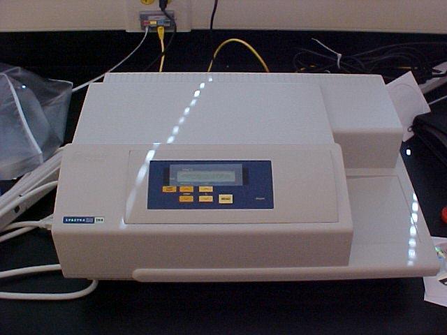
26 Non microscopic techniques ELISA - Enzyme-Linked ImmunoSorbent Assay ELISA is an immunology test used in biochemistry to reveal the presence of a given antigene, typically pertaining to a pathogenic micro-organism, and makes use of a specific antibody. There exist two variants of the ELISA test: non competitive ELISA and competitive ELISA. The non competitive ELISA test can be executed according to two different procedures: direct method and indirect method. The tests are colorimetric and the read out is performed by means of a spectrophotometer.

27 ELISA non Competitive
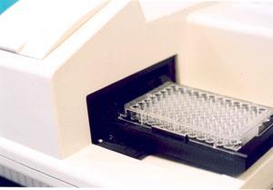
28 ELISA non Competitive Preparation of the wells of a polystyrene multi-well plate by injecting of a solution of the primary antibody, which is specific for the antigen that we intend to detect. The antibodies adhere at the base of the well by means of a layer of fish gelatin. The excess is washed out by rinsing. Addiction of the human samples in which we wish to detect the presence of the antigen that is characteristic of the pathogenic microorganism. Wash with a buffer solution. The antigen, if present, binds specifically the antibody. The excess is washed out by rinsing. Addiction of the secondary antibodies. Such antibody are conjugated to an enzyme, typically either peroxidase or alkaline phosphatase.
 will be removed during the washing step.")
29 ELISA non Competitive The secondary antibodies bind selectively to the antigen, if present. The excess is washed out. Alternatively, if the specific antigen is absent, the secondary antibody (and the conjugated enzyme) will be removed during the washing step. Addiction of a p-nitrophenyl phosphate substrate, triggers a reaction with the enzyme conjugated to the secondary antibody, that produces yellow-colored para-nitrophenol. Addiction of sodium hydroxide blocks the reaction between the enzyme and the p-nitrophenyl phosphate. Read out of the result by means of a spectrophotometer.
30 ELISA non Competitive
31 ELISA Competitive Unlabelled antibodies coming from a sample are incubated in the presence of their antigens; Such antibody/antigen complexes are injected in the wells of a plate, which were previously functionalized with the same antigens. The plate is washed so as to remove all unbound antibodies. Secondary antibodies that are specific for the first antibody are injected. Such secondary antibody are conjugated to tan enzyme. A substrate ia added and the bound enzymes cause a chromatic change. In the competitive ELISA test, the larger it is the antigen concentration in the sample the weaker it is the signal. The advantage is that we can use impure samples and that we still have the possibility to bind selectively any present antigen.

32 ELISA Competitive Quntity k-casein B The well is functionalized with k-casein B Sample pipetting Pipetting of a know quantity of the specific antibody for k- casein B The antibody binds to all k-casein B of the sample and the excess binds to the k-casein on the bottom of the well The excess antibody bound to the bottom of the well is colored k-casein B k-casein A Color Intensity The larger it is the color intensity the smaller it is the k-casein B content in the sample Antibody k-casein B Seconday antibody

33 Non microscopic techniques ELFA - Enzyme-Linked Fluorescence Assay ELFA is an evolution of the ELISA test. The enzyme that is conjugated to the secondary antibody converts the substrate to a luminescent compound The read out is carried out by means of a spectrofluorometer Figure 1: Fluorescence image of a 96-well platereader plate filled with an ELISA. The red framed wells contain known concentrations and the blue framed wells the unknowns. The wells marked with other colours contain control solutions.


34 Non microscopic techniques ELFA - Enzyme-Linked Fluorescence Assay
35 Non microscopic techniques ELFA - Enzyme-Linked Fluorescence Assay
36 Non microscopic techniques DNA sequencing Introduction to the Sanger methd The Sanger method permits to sequence DNA, making use of the biochemical techniques associated to the use of radio-active markers. It exploits the reaction of polymerization of aminoacids sequences (Nobel Chemistry 1980) and the gel electrophoresis technique (Nobel Chemistry 1958) sangerseq.exe
 and the gel electrophoresis technique (Nobel Chemistry 1958) cycseq.exe")
37 Non microscopic techniques Cycle sequencing Evolution of the Sanger method The Sanger method permits to sequence DNA, making use of the biochemical techniques associated to the use of radio-active markers. It exploits the reaction of polymerization of aminoacids sequences (Nobel Chemistry 1980) and the gel electrophoresis technique (Nobel Chemistry 1958) cycseq.exe

38 Non microscopic techniques Cycle sequencing Example


39 Non microscopic techniques Cycle sequencing Evolution of the Sanger method
 SOLID.")
40 Non microscopic techniques Cycle sequencing SOLID Further evolution (already old!) SOLID.wmv
41 Applications of optics and photonics Microscopic Techniques Conventional Wide-Field Fluorescence TIRF FLIM FRET, FRAP Confocal Two-Photon Second Harmonic Super-resolution (SNOM, STED, PALM, STORM) Non-Microscopic Label-free Surface plasmon Polaritons (SPP) Photonic crystals (PC) Raman, CARS Quantum dots Non-Microscopic Techniques Cytofluorimetry ELISA DNA-Chip Cycle-sequencing SOLID Other non Microscopic Techniques Southern Western Northern All of them make use of the emission of luminescent markers (labels)
STED microscopy with single light source. TeodoraŞcheul
 STED microscopy with single light source TeodoraŞcheul Dr. Iréne Wang, Dr. Jean-Claude Vial LIPhy, Grenoble, France Summary I. Introduction to STED microscopy II. STED with one laser source 1. Two-photon
STORM/PALM. Super Resolution Microscopy 10/31/2011. Looking into microscopic world of life
 Super Resolution Microscopy STORM/PALM Bo Huang Department of Pharmaceutical Chemistry, UCSF CSHL Quantitative Microscopy, 1/31/211 Looking into microscopic world of life 1 µm 1 µm 1 nm 1 nm 1 nm 1 Å Naked
Bi177 - Lecture 13 Microscopy Outside the Box. Fluorescence Nanoscopy TIRF 4-pi STED STORM/PALM
 Bi177 - Lecture 13 Microscopy Outside the Box Fluorescence Nanoscopy TIRF 4-pi STED STORM/PALM The diffraction limit: Abbe s law The Problem Diffraction limit 100x larger than molecular scale! Green Fluorescent
A Brief History of Light Microscopy And How It Transformed Biomedical Research
 A Brief History of Light Microscopy And How It Transformed Biomedical Research Suewei Lin Office: Interdisciplinary Research Building 8A08 Email: sueweilin@gate.sinica.edu.tw TEL: 2789-9315 Microscope
Super Resolution Microscopy - Breaking the Diffraction Limit Radiological Research Accelerator Facility
 Super Resolution Microscopy - Breaking the Diffraction Limit Radiological Research Accelerator Facility Sabrina Campelo, Dr. Andrew Harken Outline Motivation Fluorescence Microscopy -Multiphoton Imaging
Fluorescent Antibody technique Can identify microorganisms in clinical specimens and can detect the presences of a specific antibody in
 Lecture:7 Practical immunit Fluorescent Antibody technique Can identify microorganisms in clinical specimens and can detect the presences of a specific antibody in serum. These techniques combine fluorescent
ALP (alkaline phosphatase) calibrators were analyzed manually in microtiter plates to find the linearity range by following this protocol:
 calibrators were analyzed manually in microtiter plates to find the linearity range by following this protocol:") Exam Mol 3008 May 2009 Subject 1 (15p) ALP (alkaline phosphatase) calibrators were analyzed manually in microtiter plates to find the linearity range by following this protocol: Reaction solutions: 50
Exam Mol 3008 May 2009 Subject 1 (15p) ALP (alkaline phosphatase) calibrators were analyzed manually in microtiter plates to find the linearity range by following this protocol: Reaction solutions: 50
Special Techniques 1. Mark Scott FILM Facility
 Special Techniques 1 Mark Scott FILM Facility SPECIAL TECHNIQUES Multi-photon microscopy Second Harmonic Generation FRAP FRET FLIM In-vivo imaging TWO-PHOTON MICROSCOPY Alternative to confocal and deconvolution
Localization Microscopy
 Localization Microscopy Theory, Sample Prep & Practical Considerations Patrina Pellett & Ann McEvoy Applications Scientist GE Healthcare, Cell Technologies May 27 th, 2015 Localization Microscopy Talk
PALM/STORM, BALM, STED
 PALM/STORM, BALM, STED Last class 2-photon Intro to PALM/STORM Cyanine dyes/dronpa This class Finish localization super-res BALM STED Localization microscopy Intensity Bins = pixels xx 2 = ss2 + aa 2 /12
Super-resolution Microscopy
 Semr oc kwhi t epaperser i es : 1. Introduction Super-resolution Microscopy Fluorescence microscopy has revolutionized the study of biological samples. Ever since the invention of fluorescence microscopy
Confocal Microscopy & Imaging Technology. Yan Wu
 Confocal Microscopy & Imaging Technology Yan Wu Dec. 05, 2014 Cells under the microscope What we use to see the details of the cell? Light and Electron Microscopy - Bright light / fluorescence microscopy
SUPER-RESOLUTION MICROSCOPY. Dr. Nathalie Garin
 SUPER-RESOLUTION MICROSCOPY Dr. Nathalie Garin Content Motivation for superresolution Superresolution, nanoscopy, : definition Structured Illumination Microscopy (SIM) Localization microscopy STimulated
Introduction to N-STORM
 Introduction to N-STORM Dan Metcalf Advanced Imaging Manager Outline Introduction Principles of STORM Applications N-STORM overview Biological Scale Mitochondrion Microtubule Amino Acid 1Å Kinesin 1nm
Femtosecond micromachining in polymers
 Femtosecond micromachining in polymers Prof. Dr Cleber R. Mendonca Daniel S. Corrêa Prakriti Tayalia Dr. Tobias Voss Dr. Tommaso Baldacchini Prof. Dr. Eric Mazur fs-micromachining focus laser beam inside
Application Note Insulin ELISA on high binding MICROLON 600 and CELLSTAR microplates
 Application Note Insulin ELISA on high binding MICROLON 600 and CELLSTAR microplates 1 Introduction ELISA (enzyme-linked immunosorbent assay) is one of the most widely used laboratory techniques in analysis
Workshop advanced light microscopy
 Workshop advanced light microscopy Multi-mode confocal laser scanning microscope Jan Willem Borst Laboratory of Biochemistry Biomolecular Networks www.bic.wur.nl MicroSpectroscopy Centre Wageningen Microspectroscopy
MicroTime 200 STED. Super-resolution add-on for the confocal time-resolved microscopy platform
 MicroTime 200 STED Super-resolution add-on for the confocal time-resolved microscopy platform confocal STED 2 Vision The MicroTime 200... The MicroTime 200 is a high-end confocal fluorescence lifetime
Lab. 7: Serological Tests ELISA. 320 MIC Microbial Diagnosis 320 MBIO PRACTICAL. Amal Alghamdi 2018
 Lab. 7: 320 MIC Microbial Diagnosis Serological Tests ELISA. 320 MBIO PRACTICAL Amal Alghamdi 2018 1 Infection and Immunity Serology is the study of immune bodies in human blood. These are products of
Biophotonics?? Biophotonics. technology in biomedical engineering. Advantages of the lightwave
 Biophotonics - Imaging: X-ray, OCT, polarimetry, DOT, TIRF, photon migration, endoscopy, confocal microscopy, multiphoton microscopy, multispectral imaging - Biosensing: IR spectroscopy, fluorescence,
Sample region with fluorescent labeled molecules
 FLUORESCENCE IMAGING I. Fluorescence-imaging with diffraction limited spots The resolution in optical microscopy has been hampered by the smallest spot possible (~ λ/2) that can be achieved by conventional
Comparing MSD Electrochemiluminescent Detection Technology to Traditional ELISA for Clinical Applications
 Enzyme- linked immunosorbent assays (s) are widely used in clinical settings for detecting substances such as antibodies, peptides, or hormones. However, assays have been gaining in popularity due to advantages
Confocal Microscopes. Evolution of Imaging
 Confocal Microscopes and Evolution of Imaging Judi Reilly Hans Richter Massachusetts Institute of Technology Environment, Health & Safety Office Radiation Protection What is Confocal? Pinhole diaphragm
THE BASICS OF IMMUNOHISTOCHEMISTRY
 THE BASICS OF IMMUNOHISTOCHEMISTRY Introduction Immunohistochemistry (IHC) identifies specific tissue components by means of a specific antigen/antibody reaction tagged with a visible label. IHC makes
Selected Techniques Part I
 1 Selected Techniques Part I Gel Electrophoresis Can be both qualitative and quantitative Qualitative About what size is the fragment? How many fragments are present? Is there in insert or not? Quantitative
Fentanyl (Human) ELISA Kit
 ELISA Kit") Fentanyl (Human) ELISA Kit Catalog Number KA0931 96 assays Version: 06 Intended for research use only www.abnova.com Table of Contents Introduction... 3 Intended Use... 3 Background... 3 Principle of the
Fentanyl (Human) ELISA Kit Catalog Number KA0931 96 assays Version: 06 Intended for research use only www.abnova.com Table of Contents Introduction... 3 Intended Use... 3 Background... 3 Principle of the
APPLICATION OF MOLECULAR TECHNICS FOR DIAGNOSIS OF VIRAL INFECTIONS
 APPLICATION OF MOLECULAR TECHNICS FOR DIAGNOSIS OF VIRAL INFECTIONS Hossein Keyvani Basic Diagnostic Methods in Virology Immunology and serology techniques (Antigen-Antibody Reactions) 1 ELISA ( Enzyme
Symposium 20 years of nano-optics April 6th, 2004 Auditorium, Institute of Physics, St.Johanns-Ring 25
 Symposium 20 years of nano-optics April 6th, 2004 Auditorium, Institute of Physics, St.Johanns-Ring 25 9:30 9:45 Coffee and Gipfeli 9:45 10:00 Welcome address and introduction B. Hecht Uni Basel H.-J.
Introduction to Computational Fluorescence Microscopy!
 Introduction to Computational Fluorescence Microscopy! EE367/CS448I: Computational Imaging and Display! stanford.edu/class/ee367! Lecture 13! Gordon Wetzstein! Stanford University! Midterm! Tuesday, Feb
ELISA. An introduction to the basic principles and assay formats. Innova Biosciences ltd All rights reserved
 ELISA An introduction to the basic principles and assay formats Agenda Agenda: Introduction to ELISA The different assay types Assay development tips Data analysis tips Troubleshooting tips Indirect vs.
BIOMAN 2015 IMMUNOASSAYS. Barbara Bielska Northampton Community College Tannersville, PA
 BIOMAN 2015 IMMUNOASSAYS Barbara Bielska Northampton Community College Tannersville, PA 1 Immunoassays An immunoassay is a biochemical test that measures the presence or concentration of a molecules (typically
Practical Applications of Immunology (Chapter 18) Lecture Materials for Amy Warenda Czura, Ph.D. Suffolk County Community College Eastern Campus
 Lecture Materials for Amy Warenda Czura, Ph.D. Suffolk County Community College Eastern Campus") Practical Applications of Immunology (Chapter 18) Lecture Materials for Amy Warenda Czura, Ph.D. Suffolk County Community College Eastern Campus Primary Source for figures and content: Tortora, G.J. Microbiology
Practical Applications of Immunology (Chapter 18) Lecture Materials for Amy Warenda Czura, Ph.D. Suffolk County Community College Eastern Campus Primary Source for figures and content: Tortora, G.J. Microbiology
Fluorescence Nanoscopy 高甫仁 ) Institute of Biophotonics, National Yang Ming University. Outline
 Institute of Biophotonics, National Yang Ming University. Outline") Fluorescence Nanoscopy 高甫仁 ) Fu-Jen Kao ( 高甫仁 Institute of Biophotonics, National Yang Ming University Outline The Abbe s (diffraction) limit and nanoscopy Fundamentals and opportunities of FLIM/FRET Visualizing
Fluorescence Nanoscopy 高甫仁 ) Fu-Jen Kao ( 高甫仁 Institute of Biophotonics, National Yang Ming University Outline The Abbe s (diffraction) limit and nanoscopy Fundamentals and opportunities of FLIM/FRET Visualizing
Anti-MOUSE IgG (H&L) (GOAT) Antibody DyLight 488 Conjugated (Min X Bv Ch Gt GP Ham Hs Hu Rb Rt & Sh Serum Proteins)
 (GOAT) Antibody DyLight 488 Conjugated (Min X Bv Ch Gt GP Ham Hs Hu Rb Rt & Sh Serum Proteins)") Anti-MOUSE IgG (H&L) (GOAT) Antibody DyLight 488 Conjugated (Min X Bv Ch Gt GP Ham Hs Hu Rb Rt & Sh Serum Proteins) - 610-141-121 Code: 610-141-121 Size: 100 µg Product Description: Anti-MOUSE IgG (H&L)
Anti-MOUSE IgG (H&L) (GOAT) Antibody DyLight 488 Conjugated (Min X Bv Ch Gt GP Ham Hs Hu Rb Rt & Sh Serum Proteins) - 610-141-121 Code: 610-141-121 Size: 100 µg Product Description: Anti-MOUSE IgG (H&L)
An expedition to modern optical bio-diagnostic tools
 An expedition to modern optical bio-diagnostic tools Advisor Prof. James F. Rusling Dhanuka Wasalathanthri CHEM 5395 How sensitive it is? Prostate Specific Antigen (PSA) in serum (normal) - 0.5 to 2 ng
An Introduction to Immunoassay
 101 Innovation Boulevard, Suite 302, State College, PA 16803 USA Tel: 814.234.7748 Fax: 814.234.1608 www.salimetrics.com An Introduction to Immunoassay Immunoassay is an important laboratory technique
Confocal Microscopy Analyzes Cells
 Choosing Filters for Fluorescence A Laurin Publication Photonic Solutions for Biotechnology and Medicine November 2002 Confocal Microscopy Analyzes Cells Reprinted from the November 2002 issue of Biophotonics
Microarray Industry Products
 Via Nicaragua, 12-14 00040 Pomezia (Roma) Phone: +39 06 91601628 Fax: +39 06 91612477 info@lifelinelab.com www.lifelinelab.com Microarray Industry Products Page 10 NBT / BCPIP Chromogenic phosphatase
Microscopy from Carl Zeiss
 Microscopy from Carl Zeiss LSM 710 In Tune with Your Application Enjoy new freedom in selecting fluorescent dyes with In Tune, the new laser system for the LSM 710. Whatever the wavelength, you can match
1) Western Blot: It`s based on Electrophoresis.
 Western Blot: It`s based on Electrophoresis.") Today we will begin our lecture with Immunoassays. They are methods to detect proteins in samples or in cells rather than to purify them. We have two methods: 1) Western Blot 2) ELIZA 1) Western Blot:
Today we will begin our lecture with Immunoassays. They are methods to detect proteins in samples or in cells rather than to purify them. We have two methods: 1) Western Blot 2) ELIZA 1) Western Blot:
Immunological Applications. Chapter 8: Background
 Immunological Applications Chapter 8: Background The Immune System Types of Immunity Innate The natural immunity present at birth Acquired A specific response to foreign substances. Some cells remember
Fluorescence microscopy
 Fluorescence microscopy 1 Fluorescence microscopies basic fluorescence, fluorophores Deconvolution Confocal Two-photon/multi-photon 4Pi Light sheet Total internal reflection STED FRAP/FLIP/FCS FRET PALM/STORM/iPALM
E. COLI HOST CELL PROTEIN ELISA KIT ASSAY PRINCIPLE PRODUCT MANUAL HCP-002 E. COLI HOST CELL PROTEIN ELISA KIT KIT INCLUDES
 E. COLI HOST 4320 Forest Park Ave Suite 303 Saint Louis, MO 63108 +1 (314) 833-9764 CELL PROTEIN ELISA KIT ASSAY PRINCIPLE E. COLI HOST CELL PROTEIN ELISA KIT Complete kit for the determination of E. coli
BCH 462. Western Blot
 BCH 462 Western Blot Blotting Immunoassay: A test that uses antibody and antigen complexes [immuno-complexes] as a means of generating measurable results. Antigens [Ag]: A substance that when introduced
Components for Highly Advanced time-resolved fluorescence Microscopy based on Nonlinear Glass fibres
 Components for Highly Advanced time-resolved fluorescence Microscopy based on Nonlinear Glass fibres FP7 Call identifier: FP7-ICT-2011-7 Activity code: ICT-2011.3.5: Photonics Project start date and duration:
GeNei TM Dot ELISA Teaching Kit Manual
 Teaching Kit Manual Cat No. New Cat No. KT12S 106125 Revision No.: 00300305 CONTENTS Page No. Objective 3 Principle 3 Kit Description 4 Materials Provided 8 Procedure 9 Observation & Interpretation 10
Chapter 3. Clonal selection
 Chapter 3. Clonal selection I have called this principle, by which each slight variation, if useful, is preserved, by the term of Natural Selection -Charles Darwin, On the Origin of Species, 1859 4 The
Far-field fluorescence microscopy beyond the diffraction limit: Fluorescence imaging with ultrahigh resolution
 Far-field fluorescence microscopy beyond the diffraction limit: Fluorescence imaging with ultrahigh resolution James H. Rice School of Chemical Sciences and Pharmacy, University of East Anglia, Norwich
New developments in STED Microscopy
 New developments in STED Microscopy Arnold Giske*, Jochen Sieber, Hilmar Gugel, Marcus Dyba, Volker Seyfried, Dietmar Gnass Leica Microsystems CMS, Am Friedensplatz 3, 68126 Mannheim, Germany ABSTRACT
HiPer Sandwich ELISA Teaching Kit
 HiPer Sandwich ELISA Teaching Kit Product Code: HTI014 Number of experiments that can be performed: 4 Duration of Experiment: 2 days Day1-Coating of wells: 15 minutes Day2- protocol, observation and result:
OPERATING INSTRUCTIONS
 OPERATING INSTRUCTIONS SARCOPTES-ELISA 2001 DOG Enzyme immunoassay for the detection of IgG antibodies to the pathogen of sarcoptic mange, the mite Sarcoptes scabiei var. canis For in-vitro diagnosis only
Microscopy. CS/CME/BioE/Biophys/BMI 279 Nov. 2, 2017 Ron Dror
 Microscopy CS/CME/BioE/Biophys/BMI 279 Nov. 2, 2017 Ron Dror 1 Outline Microscopy: the basics Fluorescence microscopy Resolution limits The diffraction limit Beating the diffraction limit 2 Microscopy:
Fast, three-dimensional super-resolution imaging of live cells
 Nature Methods Fast, three-dimensional super-resolution imaging of live cells Sara A Jones, Sang-Hee Shim, Jiang He & Xiaowei Zhuang Supplementary Figure 1 Supplementary Figure 2 Supplementary Figure 3
Cellular imaging using Nano- Materials. A Case-Study based approach Arun Murali, Srivats V
 Cellular imaging using Nano- Materials A Case-Study based approach Arun Murali, Srivats V Agenda Discuss a few papers Explain a couple of new imaging techniques and their benefits over conventional imaging
Application Note Influence of coating buffer and incubation conditions on ELISA performance
 Application Note Influence of coating buffer and incubation conditions on ELISA performance 1. Introduction ELISA (Enzyme-Linked Immunosorbent Assay) is one of the most widely used techniques in both basic
CELL-BASED COLORIMETRIC ELISA PROTOCOL - FOR ACETYL-SPECIFIC PROTEIN
 CELL-BASED COLORIMETRIC ELISA PROTOCOL - FOR ACETYL-SPECIFIC PROTEIN Buffer Preparation and Recommendation We provide an excess of buffer components for you in order to perform two plates 96-well Cell-Based
Antigen-antibody reactions with labeled reagents
 Antigen-antibody reactions with labeled reagents Department of Immunology Faculty of Medicine University of Belgrade ANTIGEN ANTIBODY REACTIONS WITH LABELED REAGENTS Enzyme immunoassay Radioimmunoassay
Winter College on Micro and Nano Photonics for Life Sciences February General Overview
 1932-15 Winter College on Micro and Nano Photonics for Life Sciences 11-22 February 2008 General Overview Martina Havenith Ruhr University Bochum Bochum, Germany Microscopy- An Overview M. Havenith Ruhr-University
F* techniques: FRAP, FLIP, FRET, FLIM,
 F* techniques: FRAP, FLIP, FRET, FLIM, FCS Antonia Göhler March 2015 Fluorescence explained in the Bohr model Absorption of light (blue) causes an electron to move to a higher energy orbit. After a particular
7/24/2012. DNA Probes. Hybridization and Probes. CLS 420 Immunology & Molecular Diagnostics. Target Sequences. Target Sequences. Nucleic Acid Probes
 Hybridization and Probes CLS 420 Immunology & Molecular Diagnostics Molecular Diagnostics Techniques: Hybridization and Probes Nucleic acid probes: A short, known sequence of DNA or RNA Used to detect
Super-resolution imaging: early days w/ Video-enhanced DIC, TIRF, PALM, STORM, etc.
 15/05/2012 Super-resolution imaging: early days w/ Video-enhanced DIC, TIRF, PALM, STORM, etc. Prof. Dr. Rainer Duden duden@bio.uni-luebeck.de 1 Using conventional light microscopy resolution is limited
Imagerie et spectroscopie de fluorescence par excitation non radiative
 Imagerie et spectroscopie de fluorescence par excitation non radiative comment s affranchir de la limite de diffraction Rodolphe Jaffiol, Cyrille Vézy, Marcelina Cardoso Dos Santos LNIO, UTT, Troyes NanoBioPhotonics
Lecture 5: 8/31. CHAPTER 5 Techniques in Protein Biochemistry
 Lecture 5: 8/31 CHAPTER 5 Techniques in Protein Biochemistry Chapter 5 Outline The proteome is the entire set of proteins expressed and modified by a cell under a particular set of biochemical conditions.
UV MicroTime 200. Crossing the Border towards Deep UV Time-resolved Microscopy of Native Fluorophores
 UV MicroTime 200 Crossing the Border towards Deep UV Time-resolved Microscopy of Native Fluorophores Marcelle König 1, Sebastian Tannert 1, Sandra Orthaus 1, Volker Buschmann 1, Thomas Schönau 1, Kristian
User Manual. Cat. No
 User Manual ELISA Amplification System Cat. No. 19589-019 Table of Contents 1. Notices to Customer... 1 1.1 Important Information... 1 1.2 Precautions... 1 2. Overview... 2 3. Methods... 4 3.1 Components...
Fluorescence Light Microscopy for Cell Biology
 Fluorescence Light Microscopy for Cell Biology Why use light microscopy? Traditional questions that light microscopy has addressed: Structure within a cell Locations of specific molecules within a cell
Dino-Lite knowledge & education. Fluorescence Microscopes
 Dino-Lite knowledge & education Fluorescence Microscopes Dino-Lite Fluorescence models Smallest fluorescence microscope in the world Revolution to biomedical and educational applications Flexible Easy
Enzyme Linked Immunosorbent Assay for Horseradish Peroxidase Labeled Antibodies. (Cat. # )
") 115PR G-Biosciences 1-800-628-7730 1-314-991-6034 technical@gbiosciences.com A Geno Technology, Inc. (USA) brand name femtoelisa HRP Kit Enzyme Linked Immunosorbent Assay for Horseradish Peroxidase Labeled
115PR G-Biosciences 1-800-628-7730 1-314-991-6034 technical@gbiosciences.com A Geno Technology, Inc. (USA) brand name femtoelisa HRP Kit Enzyme Linked Immunosorbent Assay for Horseradish Peroxidase Labeled
Electrophoresis and transfer
 Electrophoresis and transfer Electrophoresis Cation = positively charged ion, it moves toward the cathode (-) Anion = negatively charged ion, it moves toward the anode (+) Amphoteric substance = can have
Cancer Antigen CA125 Human ELISA Kit
 ab108653 Cancer Antigen CA125 Human ELISA Kit Instructions for Use For the quantitative measurement of Human Cancer Antigen CA125 concentrations in serum This product is for research use only and is not
Absorption of an electromagnetic wave
 In vivo optical imaging?? Absorption of an electromagnetic wave Tissue absorption spectrum Extinction = Absorption + Scattering Absorption of an electromagnetic wave Scattering of an electromagnetic wave
D e c N o. 2 8
 D e c. 2 0 0 7 N o. 2 8 CONFOCAL APPLICATION LETTER resolution FRET Acceptor Photobleaching LAS AF Application Wizard FRET with Leica TCS SP5 LAS AF Version 1.7.0 Introduction Fluorescence Resonance Energy
Chapter 10 Analytical Biotechnology and the Human Genome
 Chapter 10 Analytical Biotechnology and the Human Genome Chapter Outline Enzyme tests and biosensors DNA-based tests DNA analysis technologies Human genome and genome-based analytical methods 1 Enzyme-based
Imaging facilities at WUR
 Imaging facilities at WUR Advanced light microscopy facilities at Wageningen UR Programme Thursday 13 June 2013 Lunch meeting organized by Cat-Agro Food 12.00 Welcome and sandwich lunch 12.10 Introduction
FLIM Fluorescence Lifetime IMaging
 FLIM Fluorescence Lifetime IMaging Fluorescence lifetime t I(t) = F0 exp( ) τ 1 τ = k f + k nr k nr = k IC + k ISC + k bl Batiaens et al, Trends in Cell Biology, 1999 τ τ = fluorescence lifetime (~ns to
EuBI application for access
 * Title * First name * Last name * Email address * Institution/Company URL of the Institution/Company * Phone number Country code Phone * Street address * Zip Code * City * Country * Position * Not a Principal
CHARACTERIZATION OF MOLECULAR ORIENTATION IN SUPER-RESOLUTION FLUORESCENCE MICROSCOPY
 Master Erasmus Mundus in Photonics Engineering, Nanophotonics and Biophotonics Europhotonics MASTER THESIS WORK CHARACTERIZATION OF MOLECULAR ORIENTATION IN SUPER-RESOLUTION FLUORESCENCE MICROSCOPY Yibing
االستاذ المساعد الدكتور خالد ياسين الزاملي \مناعة \المرحلة الثانية \ التحليالت المرضية \ المعهد التقني كوت
 Types of Antigen Antibody Reactions Serological tests are widely used for detection of either serum antibodies or antigens for diagnosis of a wide variety of infectious diseases. These serological tests
KLH IgM (Mouse) ELISA Kit
 ELISA Kit") KLH IgM (Mouse) ELISA Kit Catalog Number KA2461 96 assays Version: 6.1 Intended for research use only www.abnova.com Table of Contents Introduction... 3 Intended Use... 3 Principle of the Assay... 3 General
KLH IgM (Mouse) ELISA Kit Catalog Number KA2461 96 assays Version: 6.1 Intended for research use only www.abnova.com Table of Contents Introduction... 3 Intended Use... 3 Principle of the Assay... 3 General
Visualisation, Sizing and Counting of Fluorescent and Fluorescently-Labelled Nanoparticles
 Visualisation, Sizing and Counting of Fluorescent and Fluorescently-Labelled Nanoparticles Introduction Fluorescent molecules have long been used to specifically label particular structures and features
Lecture 13. Motor Proteins I
 Lecture 13 Motor Proteins I Introduction: The study of motor proteins has become a major focus in cell and molecular biology. Motor proteins are very interesting because they do what no man-made engines
Propidium Iodide. Catalog Number: Structure: Molecular Formula: C 27H 34I 2N 4. Molecular Weight: CAS #
 Catalog Number: 195458 Propidium Iodide Structure: Molecular Formula: C 27H 34I 2N 4 Molecular Weight: 668.45 CAS # 25535-16-4 Physical Description: Dark red crystals Description: Reagent used for the
Supporting Information
 Supporting Information DNA Crystals as Vehicles for Biocatalysis. Chun Geng and Paul J. Paukstelis* *Email: paukstel@umd.edu Recombinant MBP-tagged RNase inhibitor expression and purification. The porcine
Enzymes and Enzymatic Reactions Adapted with permission from the original author, Charles Hoyt
 Biol. 261 Enzymes and Enzymatic Reactions Adapted with permission from the original author, Charles Hoyt Introduction All living things use energy, give off waste, reproduce and interact with the environment.
WESTERN BLOT. BCH462- Practical
 WESTERN BLOT BCH462- Practical What is Antigen [Ag]? What is Antibody [Ab]? Immunoassay: is a test that uses the highly specific and selective antigen-antibody reactions forming antibody and antigen complexes
Luteinizing Hormone ELISA kit
 55R-IB19104 Luteinizing Hormone ELISA kit Enzyme Immunoassay for the determination of LH in human serum 1. Principle of the test: The LH ELISA kit is a solid phase direct sandwich method. The samples and
Supplementary Information. Arrays of Individual DNA Molecules on Nanopatterned Substrates
 Supplementary Information Arrays of Individual DNA Molecules on Nanopatterned Substrates Roland Hager, Alma Halilovic, Jonathan R. Burns, Friedrich Schäffler, Stefan Howorka S1 Figure S-1. Characterization
Assays for gene expression and protein production
 Assays for gene expression and protein production Module 3, Lecture 5! 20.109 Spring 2011! Topics for Lecture 5 Measuring protein levels! Measuring transcript levels! Imaging assays! 2 Module overview:
5/11/2015 MICROSCOPIC TECHNIQUES 2. Fluorescence microscopy SPECIAL TECHNIQUES BASED ON FLUORESCENCE MICROSCOPY
 UNIVERSITY OF PÉCS MEDICAL SCHOOL www.medchool.pte.hu MICROSCOPIC TECHNIQUES 2 SPECIAL TECHNIQUES BASED ON FLUORESCENCE MICROSCOPY BIOPHYSICS 2. 2015 25th March Dr. Beáta Bugyi Department of Biophyic Fluorecence
Human beta-ngf ELISA Kit. User Manual
 Human beta-ngf ELISA Kit User Manual Catalog number: GTX14737 GeneTex Table of Contents A. Product Description... 2 B. Kit Components... 3 C. Additional Required Materials (not included)... 3 D. Reagent
CENTRIFUGAL MICROFLUIDICS
 CENTRIFUGAL MICROFLUIDICS Yoon-Kyoung Cho School of Nano-Bioscience and Chemical Engineering, Ulsan National Institute of Science and Technology (UNIST), Republic of Korea ABSTRACT Lab-on-a-disc, in which
Chapter 17: Immunization & Immune Testing. 1. Immunization 2. Diagnostic Immunology
 Chapter 17: Immunization & Immune Testing 1. Immunization 2. Diagnostic Immunology 1. Immunization Chapter Reading pp. 505-511 What is Immunization? A method of inducing artificial immunity by exposing
1. Immunization. What is Immunization? 12/9/2016. Chapter 17: Immunization & Immune Testing. 1. Immunization 2. Diagnostic Immunology
 Chapter 17: Immunization & Immune Testing 1. Immunization 2. Diagnostic Immunology 1. Immunization Chapter Reading pp. 505-511 What is Immunization? A method of inducing artificial immunity by exposing
Acetyl-p53 (K381) Cell-Based Colorimetric ELISA Kit
 Cell-Based Colorimetric ELISA Kit") Acetyl-p53 (K381) Cell-Based Colorimetric ELISA Kit Catalog No. KA8015C Detection and Quantification of Acetyl-p53 (K381) Protein Concentration in Cell. Research Purposes Only. Not Intended for Diagnostic
Acetyl-p53 (K381) Cell-Based Colorimetric ELISA Kit Catalog No. KA8015C Detection and Quantification of Acetyl-p53 (K381) Protein Concentration in Cell. Research Purposes Only. Not Intended for Diagnostic
Fluorescence Microscopy. Terms and concepts to know: 10/11/2011. Visible spectrum (of light) and energy
 and energy") Fluorescence Microscopy Louisiana Tech University Ruston, Louisiana Microscopy Workshop Dr. Mark DeCoster Associate Professor Biomedical Engineering 1 Terms and concepts to know: Signal to Noise Excitation
Fluorescence Microscopy Louisiana Tech University Ruston, Louisiana Microscopy Workshop Dr. Mark DeCoster Associate Professor Biomedical Engineering 1 Terms and concepts to know: Signal to Noise Excitation
Concept review: Fluorescence
 16 Concept review: Fluorescence Some definitions: Chromophore. The structural feature of a molecule responsible for the absorption of UV or visible light. Fluorophore. A chromophore that remits an absorbed
Cancer Antigen CA19-9 Human ELISA Kit
 ab108642 Cancer Antigen CA19-9 Human ELISA Kit Instructions for Use For the quantitative measurement of Human cancer antigen CA19-9 concentrations in serum This product is for research use only and is
Fundamentals and Applications of Biofilms Analysis, Structure and Physiology of Bacterial Biofilms Ching-Tsan Huang ( 黃慶璨 ) Office: Agronomy
 Office: Agronomy") 1 Fundamentals and Applications of Biofilms Analysis, Structure and Physiology of Bacterial Biofilms Ching-Tsan Huang ( 黃慶璨 ) Office: Agronomy Building, Room 111 Tel: (02) 33664454 E-mail: cthuang@ntu.edu.tw
1 Fundamentals and Applications of Biofilms Analysis, Structure and Physiology of Bacterial Biofilms Ching-Tsan Huang ( 黃慶璨 ) Office: Agronomy Building, Room 111 Tel: (02) 33664454 E-mail: cthuang@ntu.edu.tw
RAT KIM-1 ELISA. For the quantitative determination of Kidney Injury Molecule-1 in rat serum, plasma, and urine.
 RAT KIM-1 ELISA For the quantitative determination of Kidney Injury Molecule-1 in rat serum, plasma, and urine. For Research Use Only. Not For Use In Diagnostic Procedures. Catalog Number: 41-KIMRT-E01
Easy-WESTERN-II Super
 Easy-WESTERN-II Super Primary Antibody Detection Reagent for Western Blots User Manual for High Sensitivity and Strong Signal Detection Immediately after receiving the kit, read the section titled COMPONENTS