Confocal Microscopes. Evolution of Imaging
|
|
|
- Jessie Black
- 6 years ago
- Views:
Transcription

1 Confocal Microscopes and Evolution of Imaging Judi Reilly Hans Richter Massachusetts Institute of Technology Environment, Health & Safety Office Radiation Protection


2 What is Confocal? Pinhole diaphragm in the Conjugated Focal plane = CONFOCAL Variable aperture Image by sathvi on withfriendship.com
3 History Principle of Confocal Microscopy patented by Marvin Minsky Out of focus areas are removed or suppressed Application in Material Sciences and Biology. Lasers used since end of 1980s Improvements Open beam confined use fiber optics enclosed systems Lasers new wavelengths physical large size to small size additional lasers Scanning technique mirrors spinning disc Photo detectors Marker dyes in biology Computer control and algorithms
4 Fluorescence Microscopy Material sciences: topographic imaging, photo-stimulated luminescence Biology Increase resolution Fluorescing structure, parts of cell, several parts at same time Dynamic processes in living cells movie of motion: growth & development of embryos, neural activities targeting agents: fluorescent markers The original jellyfish green fluorescent protein (GFP) Proteins Peptides RNA DNA lipids, lipoprotein drugs effects and pharmacokinetic behavior


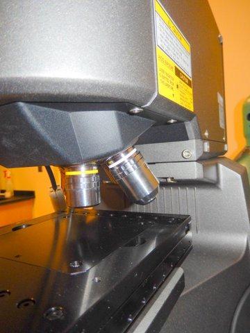
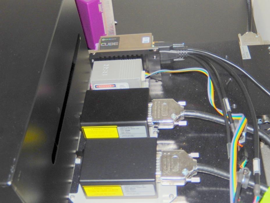
5 Laser Scanning Machine VK-9700 Class II system 408 nm laser 0.9 mw possible to focus on a target with an uneven surface at high magnification. Surface projections and depressions can be measured without damaging the target area

6


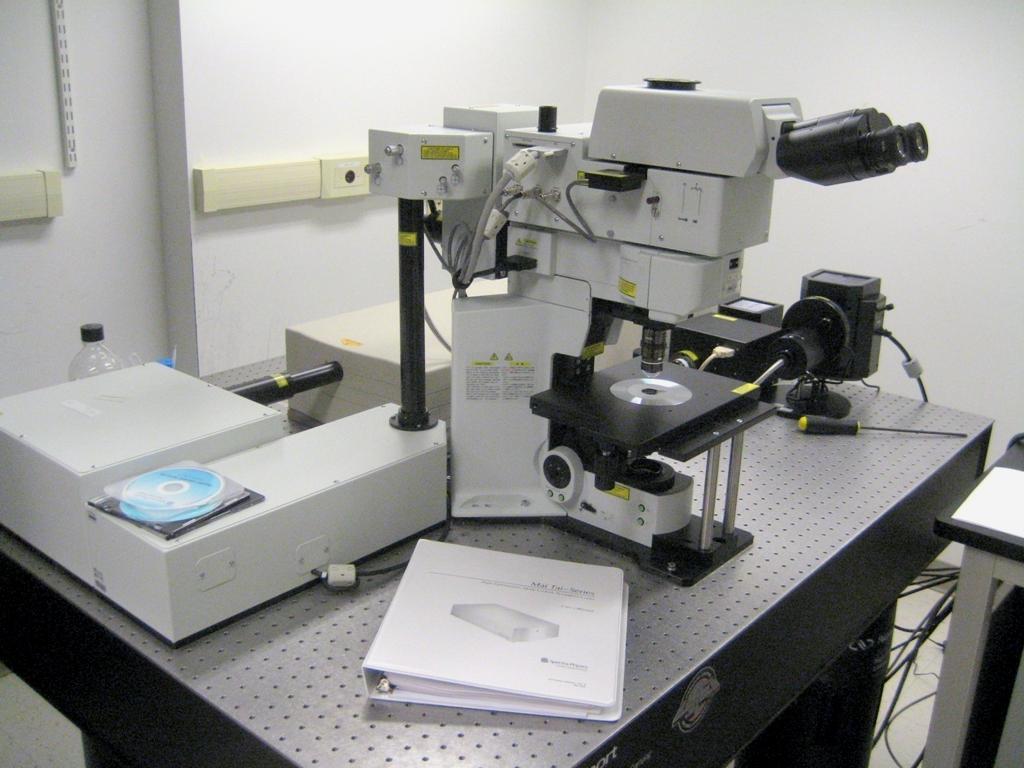
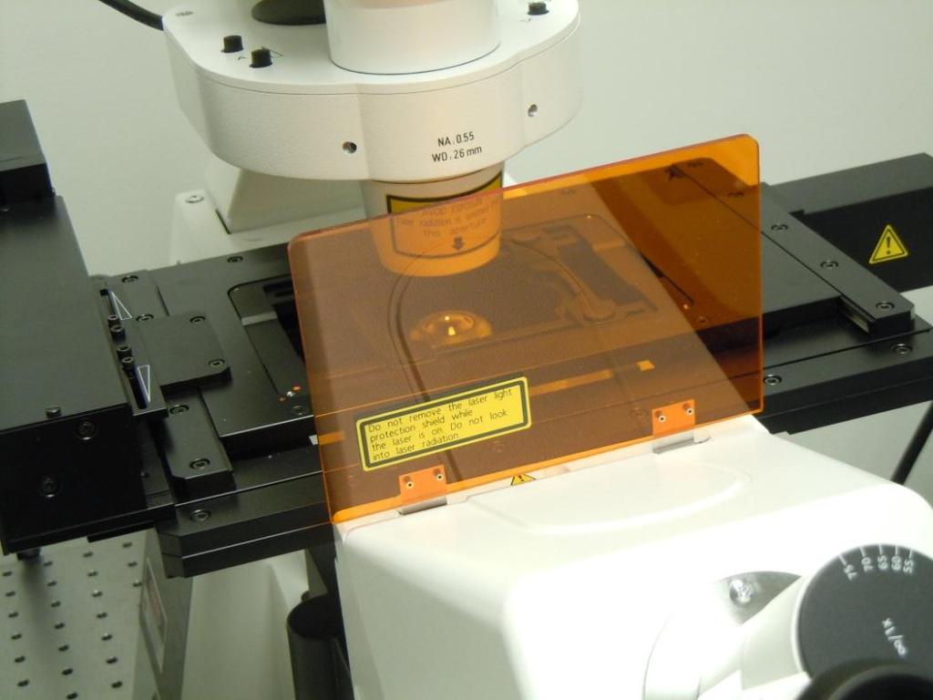
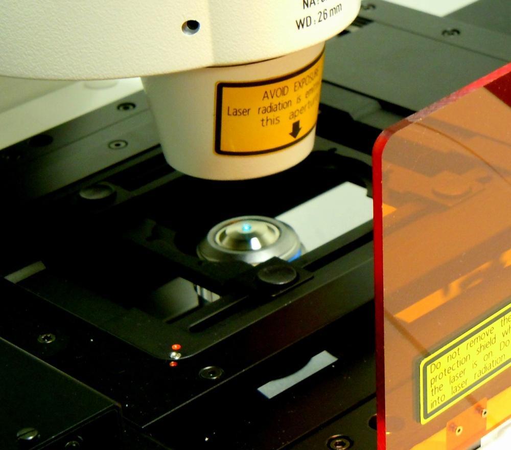
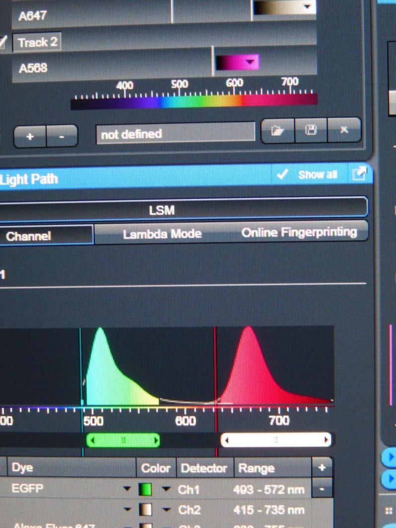
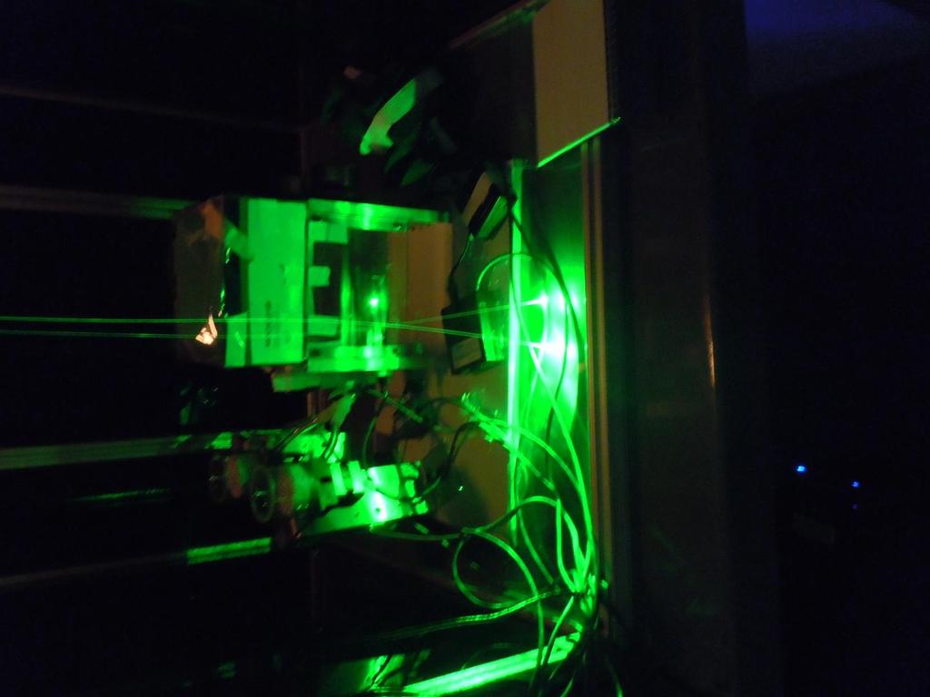
7 2.5 watts water cooled Argon-Krypton laser 350 to 647 nm Fiber optic
8
9

10 Systems that contain a Class 3B or Class 4 Laser can be classed as a Class 1 system, if the accessible light poses no potential harm

11 Signage NOTICE: Eye and skin damage will occur for direct, momentary intrabeam exposure to Class 3B lasers. Post this WARNING SIGN at the entrance to the room and close the door whenever a Class 3B laser hazard exists. Class 3B LASER flip side Each registered laser user is responsible for: 1. Complying with all requirements of the MIT Laser Safety Program. 2. Wearing appropriate laser eyewear as necessary. 3. Conducting all laser activities in accordance with accepted good safety practices.
12

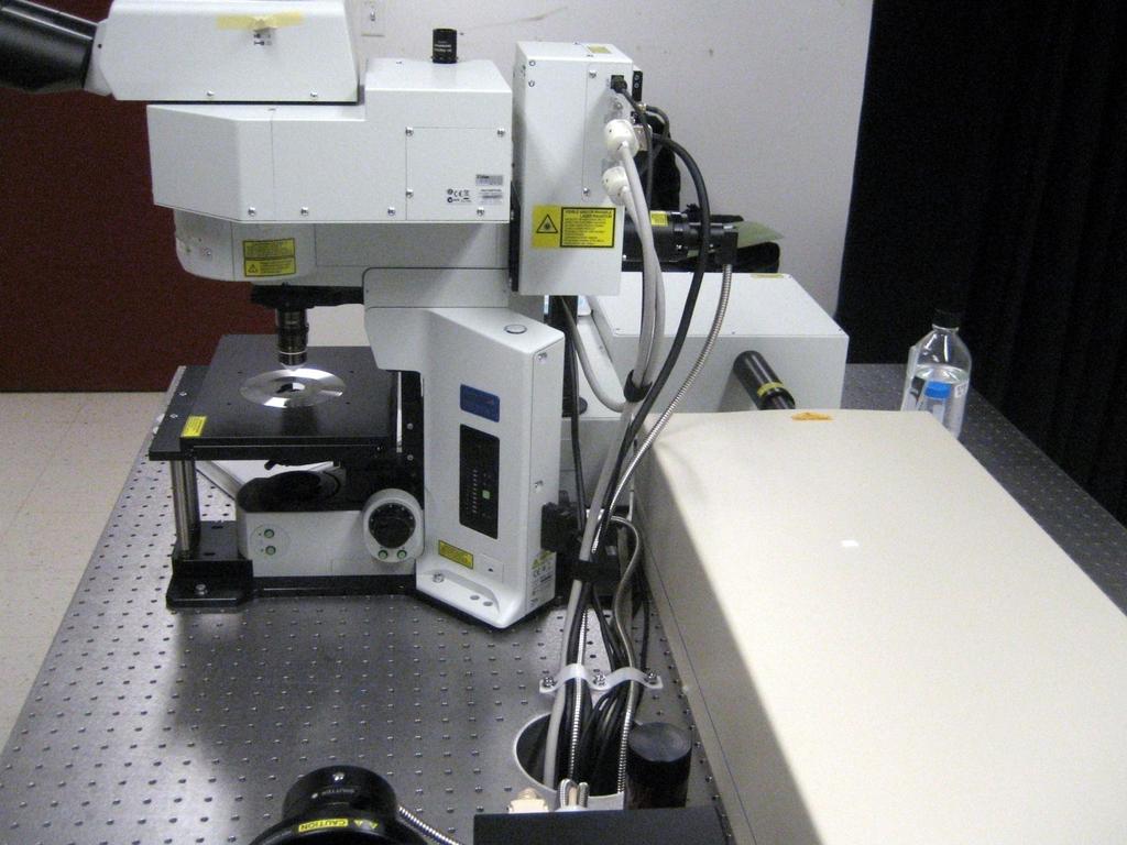
13 Laser beam is confined
14
15
16
17 A Few Examples of Fluorescent Protein Variants Protein (Acronym) Excitation Max Emission Max nm nm Green Fluorescent Protein GFP 395/ The original jellyfish green fluorescent protein (GFP) Emerald Azami Green Wasabi Blue Fluorescent Proteins EBFP Azurite Cyan Fluorescent Proteins ECFP Cerulean Yellow Fluorescent Proteins EYFP Topaz Orange Fluorescent Proteins Kusabira Orange dtomato Red Fluorescent Proteins mruby mstrawberry mraspberry

18 A Few Examples of Fluorescent Protein Variants Protein (Acronym) Excitation Max Emission Max nm nm Green Fluorescent Protein GFP 395/ The original jellyfish green fluorescent protein (GFP) Emerald Azami Green Wasabi Blue Fluorescent Proteins EBFP Azurite Cyan Fluorescent Proteins ECFP Cerulean Yellow Fluorescent Proteins EYFP Topaz Orange Fluorescent Proteins Kusabira Orange dtomato Red Fluorescent Proteins mruby mstrawberry mraspberry
19
20

21
22
23 Two photon Microscopy Two photons absorbed, photons interact with the fluorophore at the same time and one photon emitted at a different wavelength. Provides a three dimensionality to image Longer wavelength more penetrating to tissue May image not only living cells, but tissue, embryos and animals Minimization of photobleaching and photodamage And improvements in noninvasive optical biopsy 1 photon excitation 2 photon excitation
24 Details of Two Photon Microscopy: Invented in 1990 by Watt Webb and Winfried Denk Fluorescent emission following two-photon excitation is exactly the same as emission generated in normal onephoton excitation High laser power is required to generate significant two-photon-excited fluorescence Achieved by focusing mode-locked (pulsed) lasers The power during the peak of the pulse is high enough to generate significant two-photon excitation while the average laser power remains fairly low.


25 Advantage: The narrow localization of two-photon excitation to the illumination focal point is the technique's advantage over confocal microscopy. Two-photon excitation only generates fluorescence at the focal plane, and since no background fluorescence is produced, a pinhole is not required.
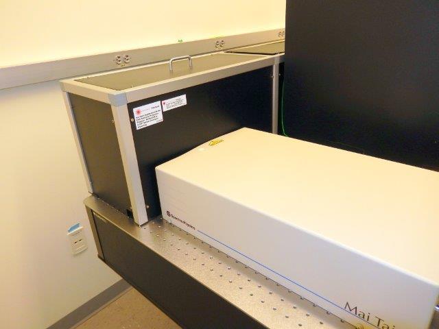
26 Two types of ultrafast mode-locked laser systems are in use with current two-photon excitation microscopes: Ti:Sapphire laser (700 to 1100 nanometers) Nd:YLF laser (1047 nanometers)
27


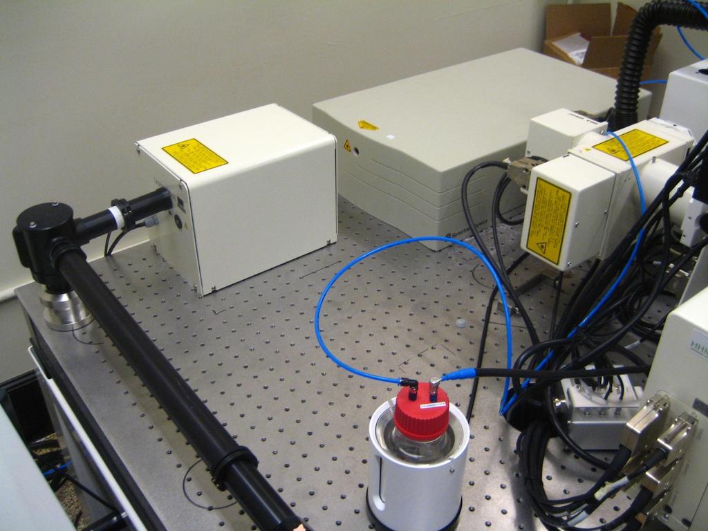
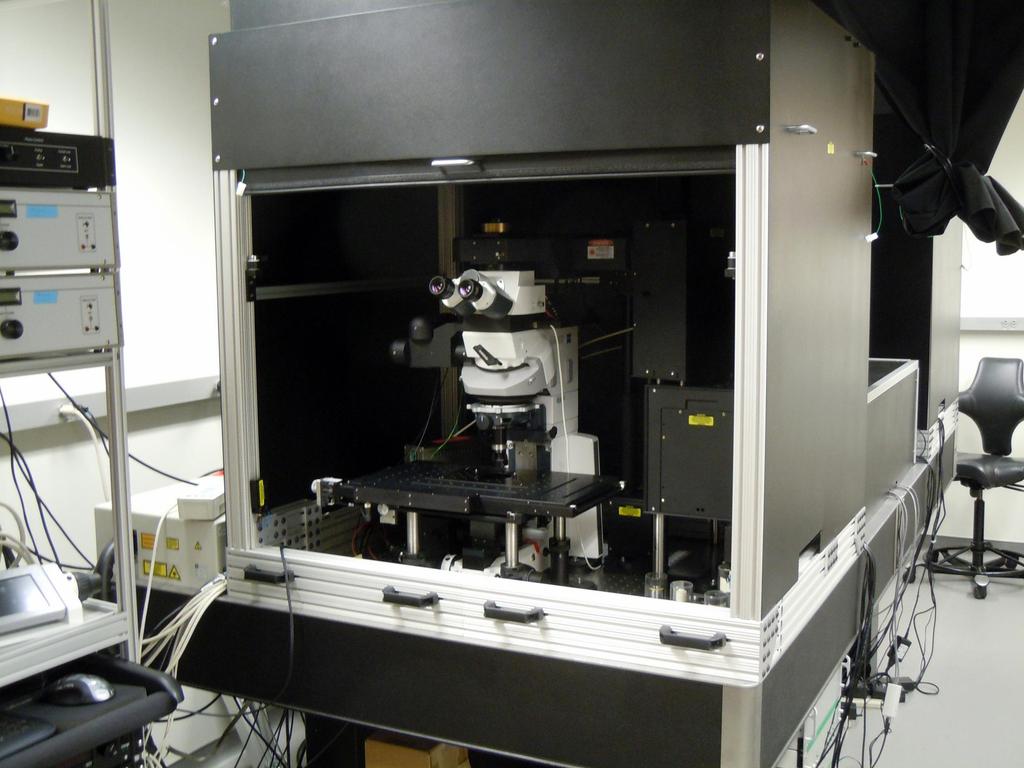
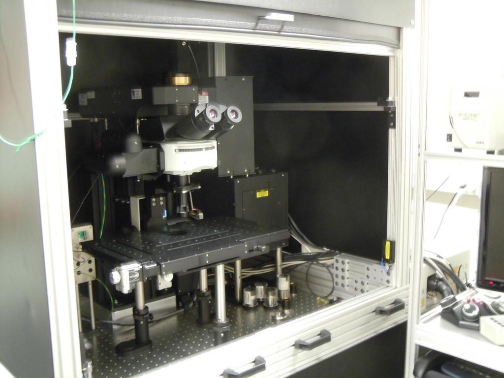
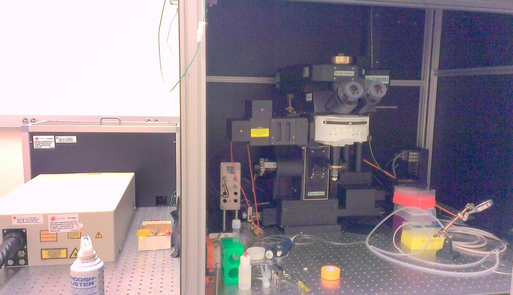
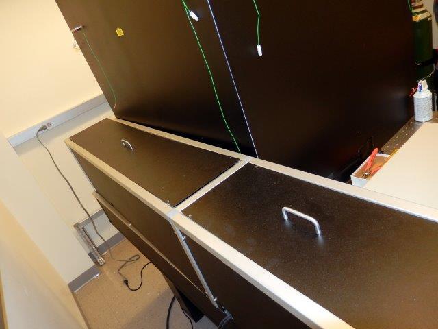
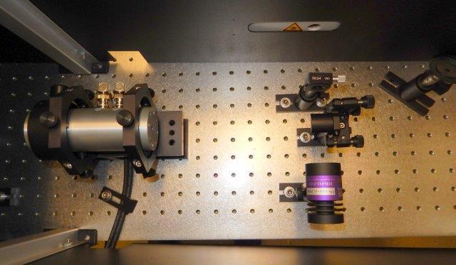
28 Two Photon Microscope Beam Evaluation Open table optics Enclosed secured table optics
29
30
31


32 Imaging system on wheels used for animal research
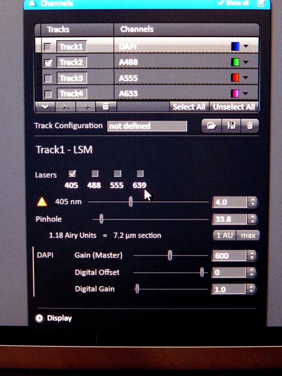
33 LASER Hazard Assessment Visible & invisible laser light Standard Operating Procedure (SOP) Beam path contained in fiber optic or light pipe If NOT: potential to human exposure accessible to insertion of items or hand unnecessary items not in or near beam path are eyepieces usable when laser on dichroic filters used beam stop used and interlocked Laser eyewear available, appropriate, good condition 405, 458, 488, 514, 561, 594, 633 common wavelengths ask about use of UV or IR beams Training Is there a supervisor Are users trained Is there documentation
34 LASER Hazard Assessment Laser controlled by computer or human User log record (best practice as it is not required) Room is often used in dim light condition Maintenance done by vendor or by trained user Warning Signs & communications Secondary hazards: infectious material chemical hazards razor blades or other sharp objects animal use electrical (grounded, power strips, extension cords)
35 LASER Hazard Assessment Each setup different Is the user comfortable in answering your questions? Evaluate, ask questions, listen to what user is not saying Key is to get into the lab, review the setup, see how it is used evaluate potential hazards, communicate potential hazard reduce or mitigate hazard

36
37


38 Images can be seen on the Koch Institute web site Public Galleries
 or Photoconvert to a new emission color upon")
39 Future: Newer fluorophores fluorescence resonance energy transfer (FRET) fluorescence recovery after photobleaching (FRAP) fluorescence lifetime imaging microscopy (FLIM) More control of pulsed lasers 3 photon techniques - deep tissue imaging Optogenetics - Neural activity and behavior Optical highlighters that are able to photoswitch (on or off) or Photoconvert to a new emission color upon stimulation with violet light Different techniques LSO to be aware of Changes/additions to initial setup New techniques Eyewear Communication

40
Special Techniques 1. Mark Scott FILM Facility
 Special Techniques 1 Mark Scott FILM Facility SPECIAL TECHNIQUES Multi-photon microscopy Second Harmonic Generation FRAP FRET FLIM In-vivo imaging TWO-PHOTON MICROSCOPY Alternative to confocal and deconvolution
A Brief History of Light Microscopy And How It Transformed Biomedical Research
 A Brief History of Light Microscopy And How It Transformed Biomedical Research Suewei Lin Office: Interdisciplinary Research Building 8A08 Email: sueweilin@gate.sinica.edu.tw TEL: 2789-9315 Microscope
Confocal Microscopy of Electronic Devices. James Saczuk. Consumer Optical Electronics EE594 02/22/2000
 Confocal Microscopy of Electronic Devices James Saczuk Consumer Optical Electronics EE594 02/22/2000 Introduction! Review of confocal principles! Why is CM used to examine electronics?! Several methods
Fluorescence Light Microscopy for Cell Biology
 Fluorescence Light Microscopy for Cell Biology Why use light microscopy? Traditional questions that light microscopy has addressed: Structure within a cell Locations of specific molecules within a cell
Cellular imaging using Nano- Materials. A Case-Study based approach Arun Murali, Srivats V
 Cellular imaging using Nano- Materials A Case-Study based approach Arun Murali, Srivats V Agenda Discuss a few papers Explain a couple of new imaging techniques and their benefits over conventional imaging
Confocal Microscopy & Imaging Technology. Yan Wu
 Confocal Microscopy & Imaging Technology Yan Wu Dec. 05, 2014 Cells under the microscope What we use to see the details of the cell? Light and Electron Microscopy - Bright light / fluorescence microscopy
Imaging facilities at WUR
 Imaging facilities at WUR Advanced light microscopy facilities at Wageningen UR Programme Thursday 13 June 2013 Lunch meeting organized by Cat-Agro Food 12.00 Welcome and sandwich lunch 12.10 Introduction
Simultaneous multi-color, multiphoton fluorophore excitation using dual-color fiber lasers
 Multiphoton Microscopy / Fiber Laser Simultaneous multi-color, multiphoton fluorophore excitation using dual-color fiber lasers Matthias Handloser, Tim Paasch-Colberg, Bernhard Wolfring TOPTICA Photonics
 Page 1 of 9 Fundamentals and Applications in Multiphoton Excitation Microscopy Two-photon excitation microscopy (also referred to as non-linear, multiphoton, or two-photon laser scanning microscopy) is
Practical light microscopy: an introduction
 Practical light microscopy: an introduction Dr. Mark Leake, Oxford University www.physics.ox.ac.uk/users/leake Aim of today s talk: Explanation of the very (very) basics of how a light microscope works
Super Resolution Microscopy - Breaking the Diffraction Limit Radiological Research Accelerator Facility
 Super Resolution Microscopy - Breaking the Diffraction Limit Radiological Research Accelerator Facility Sabrina Campelo, Dr. Andrew Harken Outline Motivation Fluorescence Microscopy -Multiphoton Imaging
Two-Photon Microscopy for Deep Tissue Imaging of Living Specimens
 for Deep Tissue Imaging of Living Specimens Tilman Franke* and Sebastian Rhode TILL Photonics GmbH, an FEI company, Lochhamer Schlag 21, D-82166 Gräfelfing, Germany *tilman.franke@fei.com Introduction
Visualizing Cells Molecular Biology of the Cell - Chapter 9
 Visualizing Cells Molecular Biology of the Cell - Chapter 9 Resolution, Detection Magnification Interaction of Light with matter: Absorbtion, Refraction, Reflection, Fluorescence Light Microscopy Absorbtion
More on fluorescence
 More on fluorescence Last class Fluorescence Absorption emission Jablonski diagrams This class More on fluorescence Common fluorophores Jablonski diagrams to spectra Properties of fluorophores Excitation
Resolution of Microscopes Visible light is nm Dry lens(0.5na), green(530nm light)=0.65µm=650nm for oil lens (1.4NA) UV light (300nm) = 0.13µm f
, green(530nm light)=0.65µm=650nm for oil lens (1.4NA) UV light (300nm) = 0.13µm f") Microscopes and Microscopy MCB 380 Good information sources: Alberts-Molecular Biology of the Cell http://micro.magnet.fsu.edu/primer/ http://www.microscopyu.com/ Approaches to Problems in Cell Biology
Microscopes and Microscopy MCB 380 Good information sources: Alberts-Molecular Biology of the Cell http://micro.magnet.fsu.edu/primer/ http://www.microscopyu.com/ Approaches to Problems in Cell Biology
Microscopy from Carl Zeiss
 Microscopy from Carl Zeiss LSM 710 In Tune with Your Application Enjoy new freedom in selecting fluorescent dyes with In Tune, the new laser system for the LSM 710. Whatever the wavelength, you can match
Final Exam, 176 points PMB 185: Techniques in Light Microscopy
 Final Exam, 176 points Name PMB 185: Techniques in Light Microscopy Point value is in parentheses at the end of each question. 1) Order the steps in setting up Köhler illumination. It is not necessary
Dino-Lite knowledge & education. Fluorescence Microscopes
 Dino-Lite knowledge & education Fluorescence Microscopes Dino-Lite Fluorescence models Smallest fluorescence microscope in the world Revolution to biomedical and educational applications Flexible Easy
Multiplexed 3D FRET imaging in deep tissue of live embryos Ming Zhao, Xiaoyang Wan, Yu Li, Weibin Zhou and Leilei Peng
 Scientific Reports Multiplexed 3D FRET imaging in deep tissue of live embryos Ming Zhao, Xiaoyang Wan, Yu Li, Weibin Zhou and Leilei Peng 1 Supplementary figures and notes Supplementary Figure S1 Volumetric
D e c N o. 2 8
 D e c. 2 0 0 7 N o. 2 8 CONFOCAL APPLICATION LETTER resolution FRET Acceptor Photobleaching LAS AF Application Wizard FRET with Leica TCS SP5 LAS AF Version 1.7.0 Introduction Fluorescence Resonance Energy
Multiphoton Microscopy: Seeing deeper and clearer
 Multiphoton Microscopy: Seeing deeper and clearer Since the invention of simple microscope by Leuwenhoek and Hooke in the 17th century, different types of light microscopy techniques (such as phase contrast,
Live cell microscopy
 Live cell microscopy 1. Why do live cell microscopy? 2. Maintaining living cells on a microscope stage. 3. Considerations for imaging living cells. 4. Fluorescence labeling of living cells. 5. Imaging
Microscopy from Carl Zeiss. DirectFRAP. News from the Cell. The New Class of Laser Manipulation for the Analysis of Cell Dynamics
 Microscopy from Carl Zeiss DirectFRAP News from the Cell The New Class of Laser Manipulation for the Analysis of Cell Dynamics DirectFRAP. New Insights into Cell Dynamics. Fluorescence breaks new ground:
Contact Details. Dr Alexander Galkin. Office: MBC Room 186. Tel: (028) Frequency and wavelength.
 Frequency and wavelength.") Contact Details The electromagnetic spectrum Biological Spectroscopy Dr Alexander Galkin Email: a.galkin@qub.ac.uk Dr Alexander Galkin MSc Biomolecular Function - BBC8045 Office: MBC Room 186 Tel: (028)
Contact Details The electromagnetic spectrum Biological Spectroscopy Dr Alexander Galkin Email: a.galkin@qub.ac.uk Dr Alexander Galkin MSc Biomolecular Function - BBC8045 Office: MBC Room 186 Tel: (028)
LASERS, LEDS AND OTHER LIGHTING SOURCES FOR LIFE SCIENCE APPLICATIONS. Wallace Latimer Coherent
 LASERS, LEDS AND OTHER LIGHTING SOURCES FOR LIFE SCIENCE APPLICATIONS Wallace Latimer Coherent IMAGING VS INTERACTION Lighting in Life Science divides into two categories Imaging UID Packaging Lab Automation
F* techniques: FRAP, FLIP, FRET, FLIM,
 F* techniques: FRAP, FLIP, FRET, FLIM, FCS Antonia Göhler March 2015 Fluorescence explained in the Bohr model Absorption of light (blue) causes an electron to move to a higher energy orbit. After a particular
FLUORESCENCE. Matyas Molnar and Dirk Pacholsky
 FLUORESCENCE Matyas Molnar and Dirk Pacholsky 1 Information This lecture contains images and information from the following internet homepages http://micro.magnet.fsu.edu/primer/index.html http://www.microscopyu.com/
Lab 5: Optical trapping and single molecule fluorescence
 Lab 5: Optical trapping and single molecule fluorescence PI: Matt Lang Lab Instructor: Jorge Ferrer Summary Optical tweezers are an excellent experimental tool to study the biophysics of single molecule
DOE Laser EFCOG: Results from SOP questionnaire
 DOE Laser EFCOG: Results from SOP questionnaire February 5, 2011 The ANSI Z136.1 description of the SOP document defines its role in describing procedures used during normal operation, maintenance and
Introduction to Computational Fluorescence Microscopy!
 Introduction to Computational Fluorescence Microscopy! EE367/CS448I: Computational Imaging and Display! stanford.edu/class/ee367! Lecture 13! Gordon Wetzstein! Stanford University! Midterm! Tuesday, Feb
Confocal Microscopy Analyzes Cells
 Choosing Filters for Fluorescence A Laurin Publication Photonic Solutions for Biotechnology and Medicine November 2002 Confocal Microscopy Analyzes Cells Reprinted from the November 2002 issue of Biophotonics
Super-resolution Microscopy
 Semr oc kwhi t epaperser i es : 1. Introduction Super-resolution Microscopy Fluorescence microscopy has revolutionized the study of biological samples. Ever since the invention of fluorescence microscopy
Workshop advanced light microscopy
 Workshop advanced light microscopy Multi-mode confocal laser scanning microscope Jan Willem Borst Laboratory of Biochemistry Biomolecular Networks www.bic.wur.nl MicroSpectroscopy Centre Wageningen Microspectroscopy
A simple introduction to multiphoton microscopy
 Journal of Microscopy, Vol. 243, Pt 3 2011, pp. 221 226 Received 29 April 2011; accepted 28 June 2011 doi: 10.1111/j.1365-2818.2011.03532.x A simple introduction to multiphoton microscopy A. USTIONE &
Fluorescence Microscopy. Terms and concepts to know: 10/11/2011. Visible spectrum (of light) and energy
 and energy") Fluorescence Microscopy Louisiana Tech University Ruston, Louisiana Microscopy Workshop Dr. Mark DeCoster Associate Professor Biomedical Engineering 1 Terms and concepts to know: Signal to Noise Excitation
Fluorescence Microscopy Louisiana Tech University Ruston, Louisiana Microscopy Workshop Dr. Mark DeCoster Associate Professor Biomedical Engineering 1 Terms and concepts to know: Signal to Noise Excitation
Partha Roy
 Fluorescence microscopy http://micro.magnet.fsu.edu/primer/index.html Partha Roy 1 Lecture Outline Definition of fluorescence Common fluorescent reagents Construction ti of a fluorescence microscope Optical
FLIM Fluorescence Lifetime IMaging
 FLIM Fluorescence Lifetime IMaging Fluorescence lifetime t I(t) = F0 exp( ) τ 1 τ = k f + k nr k nr = k IC + k ISC + k bl Batiaens et al, Trends in Cell Biology, 1999 τ τ = fluorescence lifetime (~ns to
The most extensively used technique for tissue analysis is light microscopy.
 Fluorescence Theory Quantum yield Wavelength shift Ligand interactions Membrane interactions Using quenchning effects Fluorescence in-vivo Localization Distance measurements FRET The most extensively used
The new LSM 700 from Carl Zeiss
 The new LSM 00 from Carl Zeiss Olaf Selchow, Bernhard Goetze To cite this version: Olaf Selchow, Bernhard Goetze. The new LSM 00 from Carl Zeiss. Biotechnology Journal, Wiley- VCH Verlag, 0, (), pp.. .
Lesson Plan: Fluorescence
 Lesson Plan: Fluorescence Background Fluorescence is produced when a material or substance absorbs light of a given color and then gives off light of another color. The light that is given off, or emitted,
Fluorescence Microscopy
 Fluorescence Microscopy Dr. Arne Seitz Swiss Institute of Technology (EPFL) Faculty of Life Sciences Head of BIOIMAGING AND OPTICS BIOP arne.seitz@epfl.ch Fluorescence Microscopy Why do we need fluorescence
MICROSCOPY. "micro" (small) "scopeo" (to watch)
 scopeo (to watch)") MICROSCOPY "micro" (small) "scopeo" (to watch) THE RELATIVE SIZES OF MOLECULES, CELLS AND ORGANISMS THE RELATIVE SIZES OF MOLECULES, CELLS AND ORGANISMS MICROSCOPY 1590 2012 MICROSCOPY THE LIGHT Light:
MICROSCOPY "micro" (small) "scopeo" (to watch) THE RELATIVE SIZES OF MOLECULES, CELLS AND ORGANISMS THE RELATIVE SIZES OF MOLECULES, CELLS AND ORGANISMS MICROSCOPY 1590 2012 MICROSCOPY THE LIGHT Light:
BIOCHEMIST ALL IN ONE ARTICLE
 BIOCHEMIST ALL IN ONE ARTICLE Bringing ease-of-use to microscopy From the Philosopher s Stone to the Researcher s Dream Although naturally occurring luminescence has been observed for many centuries, the
Femtosecond micromachining in polymers
 Femtosecond micromachining in polymers Prof. Dr Cleber R. Mendonca Daniel S. Corrêa Prakriti Tayalia Dr. Tobias Voss Dr. Tommaso Baldacchini Prof. Dr. Eric Mazur fs-micromachining focus laser beam inside
Absorption of an electromagnetic wave
 In vivo optical imaging?? Absorption of an electromagnetic wave Tissue absorption spectrum Extinction = Absorption + Scattering Absorption of an electromagnetic wave Scattering of an electromagnetic wave
Concept review: Fluorescence
 16 Concept review: Fluorescence Some definitions: Chromophore. The structural feature of a molecule responsible for the absorption of UV or visible light. Fluorophore. A chromophore that remits an absorbed
EuBI application for access
 * Title * First name * Last name * Email address * Institution/Company URL of the Institution/Company * Phone number Country code Phone * Street address * Zip Code * City * Country * Position * Not a Principal
High Power Diode Lasers and Multi Laser Engines, Expanding the Range of Biophotonics Applications. Konstantin Birngruber TOPTICA Photonics AG
 High Power Diode Lasers and Multi Laser Engines, Expanding the Range of Biophotonics Applications Konstantin Birngruber TOPTICA Photonics AG TOPTICA Photonics AG Company facts Founded 1998 180 employees
Visualisation, Sizing and Counting of Fluorescent and Fluorescently-Labelled Nanoparticles
 Visualisation, Sizing and Counting of Fluorescent and Fluorescently-Labelled Nanoparticles Introduction Fluorescent molecules have long been used to specifically label particular structures and features
Fluorescence Microscopy: A Biological Perspective
 Fluorescence Microscopy: A Biological Perspective From nanometre to metre: the scale of life Instrumentation and accessible scale limits the questions that can be addressed in biology Why are there limits?
Nodes of regulation in cellular systems
 Nodes of regulation in cellular systems cell membrane signal transduction ligands receptors oligomerization transport signal transduction modified protein Golgi transcription factor transport ER transport
STED microscopy with single light source. TeodoraŞcheul
 STED microscopy with single light source TeodoraŞcheul Dr. Iréne Wang, Dr. Jean-Claude Vial LIPhy, Grenoble, France Summary I. Introduction to STED microscopy II. STED with one laser source 1. Two-photon
Rice/TCU REU on Computational Neuroscience. Fundamentals of Molecular Imaging
 Rice/TCU REU on Computational Neuroscience Fundamentals of Molecular Imaging June 2, 2009 Neal Waxham 713-500-5621 m.n.waxham@uth.tmc.edu Objectives Introduction to resolution in light microscopy Brief
 Introduction to Fluorescent Proteins The discovery of green fluorescent protein in the early 1960s ultimately heralded a new era in cell biology by enabling investigators to apply molecular cloning methods,
PALM/STORM, BALM, STED
 PALM/STORM, BALM, STED Last class 2-photon Intro to PALM/STORM Cyanine dyes/dronpa This class Finish localization super-res BALM STED Localization microscopy Intensity Bins = pixels xx 2 = ss2 + aa 2 /12
Biochemistry. Biochemical Techniques. 18 Spectrofluorimetry
 Description of Module Subject Name Paper Name 12 Module Name/Title 1. Objectives 1.1 To understand technique of Spectrofluorimetry. 1.2 To explain instrumentation design 1.3 What are applications of Spectrofluorimetry?
A cost-effective fluorescence detection system for pulsed laser analysis
 Susquehanna University Scholarly Commons Chemistry Faculty Publications 2-2015 A cost-effective fluorescence detection system for pulsed laser analysis J. W. Lafferty Susquehanna University N. A. Fox Susquehanna
Boundary-breaking acoustic focusing cytometry
 Boundary-breaking acoustic focusing cytometry Introducing the Attune NxT Acoustic Focusing Cytometer a high-performance system that s flexible enough for any lab One of the main projects in my laboratory
HYPERSPECTRAL MICROSCOPE PLATFORM FOR HIGHLY MULTIPLEX BIOLOGICAL IMAGING. Marc Verhaegen
 HYPERSPECTRAL MICROSCOPE PLATFORM FOR HIGHLY MULTIPLEX BIOLOGICAL IMAGING Marc Verhaegen CMCS, MONTREAL, MAY 11 th, 2017 OVERVIEW Hyperspectral Imaging Multiplex Biological Imaging Multiplex Single Particle
Symposium 20 years of nano-optics April 6th, 2004 Auditorium, Institute of Physics, St.Johanns-Ring 25
 Symposium 20 years of nano-optics April 6th, 2004 Auditorium, Institute of Physics, St.Johanns-Ring 25 9:30 9:45 Coffee and Gipfeli 9:45 10:00 Welcome address and introduction B. Hecht Uni Basel H.-J.
MicroTime 200 STED. Super-resolution add-on for the confocal time-resolved microscopy platform
 MicroTime 200 STED Super-resolution add-on for the confocal time-resolved microscopy platform confocal STED 2 Vision The MicroTime 200... The MicroTime 200 is a high-end confocal fluorescence lifetime
BIO 315 Lab Exam I. Section #: Name:
 Section #: Name: Also provide this information on the computer grid sheet given to you. (Section # in special code box) BIO 315 Lab Exam I 1. In labeling the parts of a standard compound light microscope
Fluorescence Microscopy
 Fluorescence Microscopy Dr. Arne Seitz Swiss Institute of Technology (EPFL) Faculty of Life Sciences Head of BIOIMAGING AND OPTICS BIOP arne.seitz@epfl.ch Fluorescence Microscopy Why do we need fluorescence
Introduction to histology and its methods of study
 Introduction to histology and its methods of study Li shulei lishulei@tom.com Department of Histology & Embryology 1 What is histology Definition Cell: smallest units functions in the human body Tissue
Bioinstrumentation Light Sources Lasers or LEDs?
 Bioinstrumentation Light Sources Lasers or LEDs? A comprehensive analysis of all the factors involved in designing and building life sciences instrumentation reveals that lasers provide superior performance
Lab 1: Ensemble Fluorescence Basics
 Lab 1: Ensemble Fluorescence Basics This laboratory module is divided into two sections. The first one is on organic fluorophores, and the second one is on ensemble measurement of FRET (Fluorescence Resonance
NEWTON 7.0 BIOLUMINESCENCE & FLUORESCENCE IMAGING IN VIVO - IN VITRO IMAGING
 NEWTON 7.0 BIOLUMINESCENCE & FLUORESCENCE IMAGING IN VIVO - IN VITRO IMAGING SMART IMAGING SYSTEM The NEWTON 7.0 system combines high sensitivity with advanced animal-handling features and userfriendly
LUM. SpectraMax Paradigm Dual Color Luminescence (LUM) (BRET2 ) Detection Cartridge. User Guide
 (BRET2 ) Detection Cartridge. User Guide") LUM SpectraMax Paradigm Dual Color Luminescence (LUM) (BRET2 ) Detection Cartridge User Guide 5008542 A September 2010 This document is provided to customers who have purchased Molecular Devices, Inc.
LUM SpectraMax Paradigm Dual Color Luminescence (LUM) (BRET2 ) Detection Cartridge User Guide 5008542 A September 2010 This document is provided to customers who have purchased Molecular Devices, Inc.
New single-molecule imaging system ends prna debate over phi29 motor
 Page 1 of 5 January 30, 2007 New single-molecule imaging system ends prna debate over phi29 motor WEST LAFAYETTE, Ind. - Scientists are able to view active molecules within a biological motor of the nanometer
Methods of Characterizing Neural Networks
 Methods of Characterizing Neural Networks Ashley Nord University of Minnesota Minneapolis, MN 55414 Advisors: Katsushi Arisaka, Adrian Cheng University of California Los Angeles Los Angeles, CA 90024 September
Defense Technical Information Center Compilation Part Notice
 UNCLASSIFIED Defense Technical Information Center Compilation Part Notice ADPO 11225 TITLE: Applications of Two-Photon Fluorescence Microscopy in Deep Tissue Imaging DISTRIBUTION: Approved for public release,
Biophotonics?? Biophotonics. technology in biomedical engineering. Advantages of the lightwave
 Biophotonics - Imaging: X-ray, OCT, polarimetry, DOT, TIRF, photon migration, endoscopy, confocal microscopy, multiphoton microscopy, multispectral imaging - Biosensing: IR spectroscopy, fluorescence,
5/11/2015 MICROSCOPIC TECHNIQUES 2. Fluorescence microscopy SPECIAL TECHNIQUES BASED ON FLUORESCENCE MICROSCOPY
 UNIVERSITY OF PÉCS MEDICAL SCHOOL www.medchool.pte.hu MICROSCOPIC TECHNIQUES 2 SPECIAL TECHNIQUES BASED ON FLUORESCENCE MICROSCOPY BIOPHYSICS 2. 2015 25th March Dr. Beáta Bugyi Department of Biophyic Fluorecence
A Thin Layer Imaging with the Total Internal Reflection Fluorescence Microscopy
 Journal of Optoelectronical Nanostructures Islamic Azad University Summer 2017 / Vol. 2, No. 2 A Thin Layer Imaging with the Total Internal Reflection Fluorescence Microscopy Neda Roostaie 1, Elham Sheykhi
Sub-micron scale patterning of fluorescent. silver nanoclusters using low-power laser
 Sub-micron scale patterning of fluorescent silver nanoclusters using low-power laser Puskal Kunwar 1,*, Jukka Hassinen 2, Godofredo Bautista 1, Robin H. A. Ras 2, and Juha Toivonen 1 1 Tampere University
Cell analysis and bioimaging technology illustrated
 Cell analysis and bioimaging technology illustrated The Cell Analysis Center Scientific Bulletin Part 1 Sysmex has been studying and exploring principles of automated haematology analysers, making full
d. Bands hidden by overexposure with chemiluminescence become clear when imaged with Odyssey.
 Core Equipment ID: 15922 Description: LI COR Biosciences, Odyssey Infrared Imaging System Room: B446 (Molecular & Biochemical Core) Champion: 1.0 Purpose Standardize the process for control, maintenance,
High Throughput Whole Organ Imaging Based on Multifocal Multiphoton Microscope
 High Throughput Whole Organ Imaging Based on Multifocal Multiphoton Microscope LBRC researchers: Peter So, Jae Won Cha, Elijah Yew, Vijay Singh External technology collaborators: Prof. Hanry Yu (University
New developments in STED Microscopy
 New developments in STED Microscopy Arnold Giske*, Jochen Sieber, Hilmar Gugel, Marcus Dyba, Volker Seyfried, Dietmar Gnass Leica Microsystems CMS, Am Friedensplatz 3, 68126 Mannheim, Germany ABSTRACT
Design for Manufacturability (DFM) in the Life Sciences
 in the Life Sciences") T E C H N I C A L N O T E Design for Manufacturability (DFM) in the Life Sciences Fluorescence Spectroscopy Product Platform Realized with TracePro TM Suite of Opto-Mechanical Design Software Tools Authors:
T E C H N I C A L N O T E Design for Manufacturability (DFM) in the Life Sciences Fluorescence Spectroscopy Product Platform Realized with TracePro TM Suite of Opto-Mechanical Design Software Tools Authors:
Monitoring and Optimizing the Lipopolysaccharides-plasmid DNA interaction by FLIM-FRET
 Transactions on Science and Technology Vol. 4, No. 3-3, 342-347, 2017 Monitoring and Optimizing the Lipopolysaccharides-plasmid DNA interaction by FLIM-FRET Nur Syahadatain Abdul Razak 1#, Clarence M.
Spectra Chacracterizations of Optical Nanoparticles
 THAI NGUYEN UNIVERSITY OF EDUCATION Spectra Chacracterizations of Optical Nanoparticles Chu Viet Ha Department of Physics 18/2018 1 THAI NGUYEN UNIVERSITY OF EDUCATION Address 20 Luong Ngoc Quyen Street,
Sample region with fluorescent labeled molecules
 FLUORESCENCE IMAGING I. Fluorescence-imaging with diffraction limited spots The resolution in optical microscopy has been hampered by the smallest spot possible (~ λ/2) that can be achieved by conventional
Supplementary Table 1. Components of an FCS setup (1PE and 2PE)
") Supplementary Table 1. Components of an FCS setup (1PE and 2PE) Component and function Laser source Excitation of fluorophores Microscope with xy-translation stage mounted on vibration isolated optical
Supplementary Table 1. Components of an FCS setup (1PE and 2PE) Component and function Laser source Excitation of fluorophores Microscope with xy-translation stage mounted on vibration isolated optical
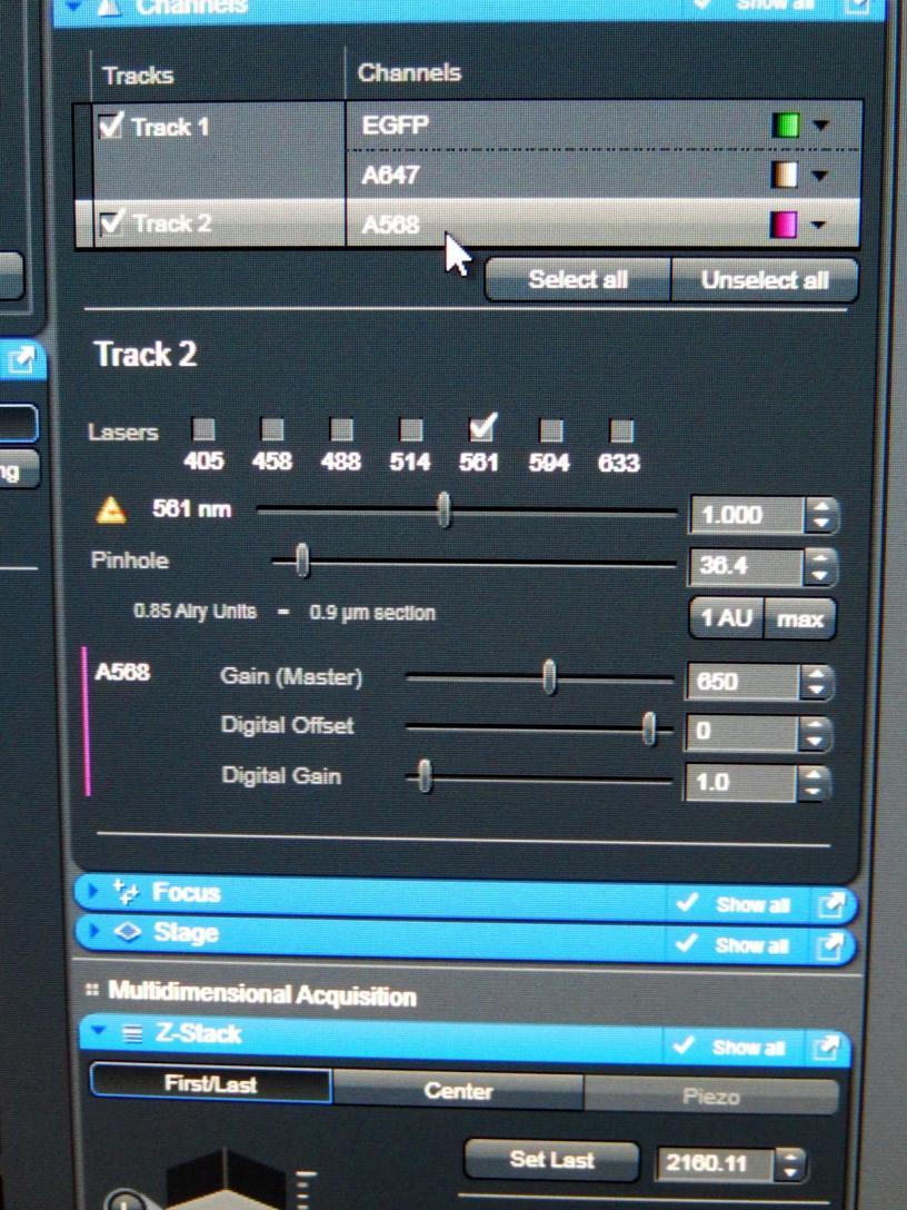
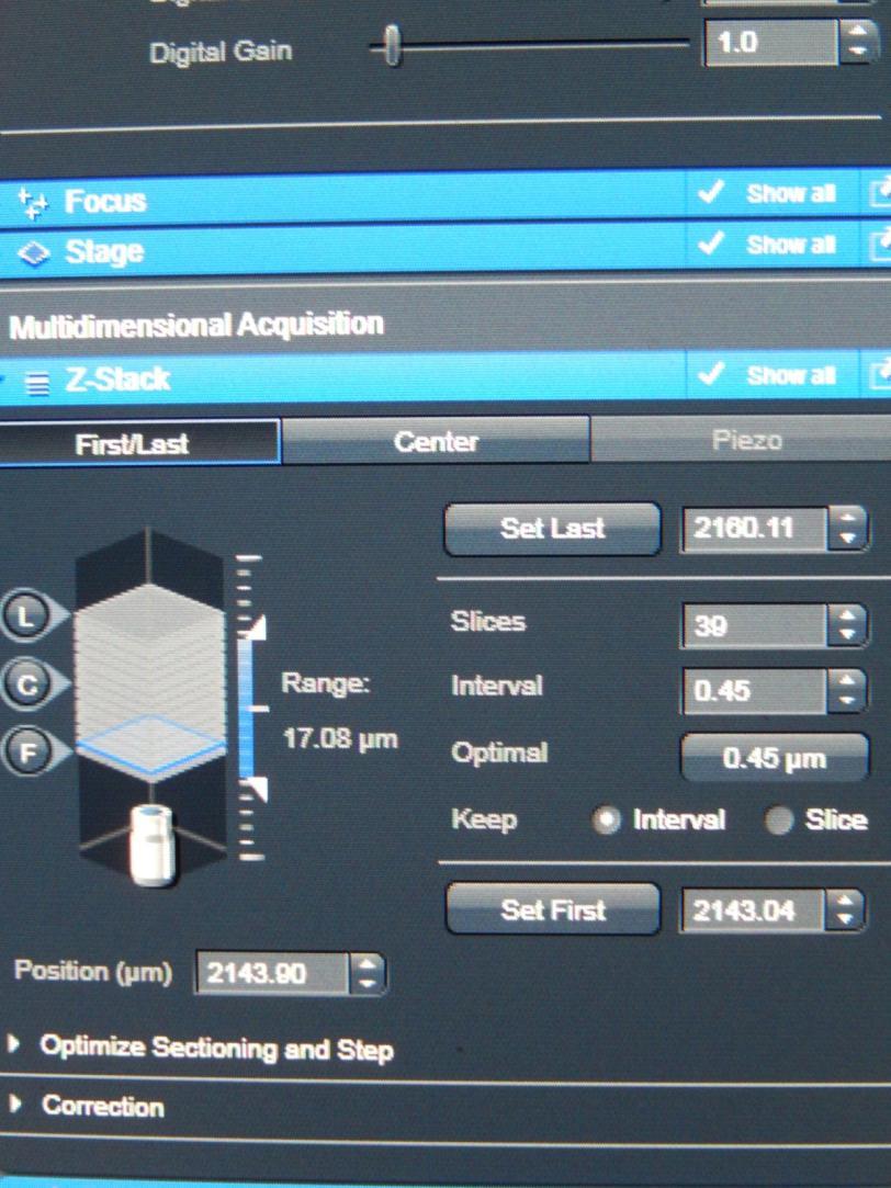
How to use the SP5 confocal microscope
 How to use the SP5 confocal microscope Mailfert Sébastien (mailfert@ciml.univ-mrs.fr) Tel : 9126 Imaging Immunity (ImagImm) photonic microscopy facility Centre d Immunologie de Marseille-Luminy 2016 /
Second Harmonic Generation Microscope Product Requirements Document Harmonigenic/ Dr Robert Hill Faculty Advisor: Dr. Wayne Knox
 Second Harmonic Generation Microscope Product Requirements Document Harmonigenic/ Dr Robert Hill Faculty Advisor: Dr. Wayne Knox James Emery (Scribe) Ava Hurlock (Document Handler) Jordan Rabinowitz (Project
Spectral imaging and its use in the measurement of Förster resonance energy transfer in living cells
 Laboratory Techniques in Biochemistry and Molecular Biology, Volume 33 FRET and FLIM Techniques T. W. J. Gadella (Editor) CHAPTER 8 Spectral imaging and its use in the measurement of Förster resonance
Principles of flow cytometry: overview of flow cytometry and its uses for cell analysis and sorting. Shoreline Community College BIOL 288
 Principles of flow cytometry: overview of flow cytometry and its uses for cell analysis and sorting Shoreline Community College BIOL 288 Flow Cytometry What is Flow Cytometry? Measurement of cells or particles
11/19/2013. Janine Zankl FACS Core Facility 13. November Cellular Parameters. Cellular Parameters. Monocytes. Granulocytes.
 DEPARTEMENT BIOZENTRUM Janine Zankl FACS Core Facility 13. November 2013 Cellular Parameters Granulocytes Monocytes Basophils Neutrophils Lymphocytes Eosinophils Cellular Parameters 1 What Is Flow Cytometry?
NEWTON 7.0 BIOLUMINESCENCE & FLUORESCENCE IMAGING IN VIVO - IN VITRO IMAGING
 NEWTON 7.0 BIOLUMINESCENCE & FLUORESCENCE IMAGING IN VIVO - IN VITRO IMAGING The NEWTON s protocol driven image acquisition is as quick as it is intuitive: adjust your exposure, save, print or quantify.
cell and tissue imaging by fluorescence microscopy
 cell and tissue imaging by fluorescence microscopy Steven NEDELLEC Plateforme Micropicell SFR Santé François Bonamy Nantes 1 A matter of size Limit of resolution 0.15mm aims: building the image of an object
Supporting Information. Two-Photon Luminescence of Single Colloidal Gold NanoRods: Revealing the Origin of Plasmon Relaxation in Small Nanocrystals
 Supporting Information Two-Photon Luminescence of Single Colloidal Gold NanoRods: Revealing the Origin of Plasmon Relaxation in Small Nanocrystals Céline Molinaro 1, Yara El Harfouch 1, Etienne Palleau
Ilya Turchin. Institute of Applied Physics of the RAS, Nizhny Novgorod, Russia.
 Fluorescence 3D imaging of small animals Ilya Turchin Institute of Applied Physics of the RAS, Nizhny Novgorod, Russia ilya@ufp.appl.sci-nnov.ru http://www.bioimaging.ru German-Russian Forum Biotechnology
Fuel Fluorescence Logging using the Optical Image Profiler (OIP)
") Fuel Fluorescence Logging using the Optical Image Profiler (OIP) Note: A Patent is Pending for this System. Daniel Pipp Chemist, Geoprobe Systems Presented May 2017 at the Battelle Bioremediation Symposium
Fuel Fluorescence Logging using the Optical Image Profiler (OIP) Note: A Patent is Pending for this System. Daniel Pipp Chemist, Geoprobe Systems Presented May 2017 at the Battelle Bioremediation Symposium
Lambda Square Mapping and FLIM
 Lambda Square Mapping and FLIM Explore Photonic Landscapes with the Leica TCS SP5 X A 620 B C Excitation/nm 470 485 Emission/nm 695 Full spectral analysis of images and lifetime measurements User guidance
Cell Structure and Function
 Cell Structure and Function Dead White Men Who Discovered (and were made of) Cells: Anton Van Leeuwenhoek Robert Hooke Where the Magic Happened Schleiden Cell Theory All plants are made of cells Schwann
Genetically targeted all-optical electrophysiology with a transgenic Credependent
 Genetically targeted all-optical electrophysiology with a transgenic Credependent Optopatch mouse Short title: Transgenic Optopatch mouse Shan Lou 1, Yoav Adam 1, Eli N. Weinstein 1,4, Erika Williams 2,
Microscopy. CS/CME/BioE/Biophys/BMI 279 Nov. 2, 2017 Ron Dror
 Microscopy CS/CME/BioE/Biophys/BMI 279 Nov. 2, 2017 Ron Dror 1 Outline Microscopy: the basics Fluorescence microscopy Resolution limits The diffraction limit Beating the diffraction limit 2 Microscopy:
Components for Highly Advanced time-resolved fluorescence Microscopy based on Nonlinear Glass fibres
 Components for Highly Advanced time-resolved fluorescence Microscopy based on Nonlinear Glass fibres FP7 Call identifier: FP7-ICT-2011-7 Activity code: ICT-2011.3.5: Photonics Project start date and duration:
Direct visualization, sizing and concentration measurement of fluorescently labeled nanoparticles using NTA
 Direct visualization, sizing and concentration measurement of fluorescently labeled nanoparticles using NTA NANOSIGHT RANGE Visualize and Measure Nanoparticle Size and Concentration PARTICLE SIZE PARTICLE