Characterization of Blood-Derived Human Progenitor Cells for Vascular Regeneration
|
|
|
- Piers Quentin Simon
- 6 years ago
- Views:
Transcription

1 Western University Electronic Thesis and Dissertation Repository August 2014 Characterization of Blood-Derived Human Progenitor Cells for Vascular Regeneration David Putman The University of Western Ontario Supervisor David Hess The University of Western Ontario Graduate Program in Physiology A thesis submitted in partial fulfillment of the requirements for the degree in Doctor of Philosophy David Putman 2014 Follow this and additional works at: Part of the Cellular and Molecular Physiology Commons Recommended Citation Putman, David, "Characterization of Blood-Derived Human Progenitor Cells for Vascular Regeneration" (2014). Electronic Thesis and Dissertation Repository This Dissertation/Thesis is brought to you for free and open access by Scholarship@Western. It has been accepted for inclusion in Electronic Thesis and Dissertation Repository by an authorized administrator of Scholarship@Western. For more information, please contact tadam@uwo.ca.
2 CHARACTERIZATION OF BLOOD-DERIVED HUMAN PROGENITOR CELLS FOR VASCULAR REGENERATION (Thesis format: Integrated Article) by David Putman Graduate Program in Physiology A thesis submitted in partial fulfillment of the requirements for the degree of Doctor of Philosophy The School of Graduate and Postdoctoral Studies The University of Western Ontario London, Ontario, Canada David Putman 2014
3 Abstract Cardiovascular disease (CVD) remains a leading cause of premature death worldwide. Despite advances in treatment of ischemic diseases including myocardial infarction and peripheral arterial disease (PAD), there remains a need for effective revascularization therapies. Although early cell therapy trials investigated use of mononuclear cells from autologous bone marrow, emerging evidence indicates that purified progenitor cell populations are required to induce optimal vascular regeneration. In addition, autologous cells show reduced efficacy due to CVD-related progenitor dysfunction. This thesis presents preclinical studies characterizing several blood-derived cell types for the development of cellular therapies to treat PAD, focusing on the use of allogeneic umbilical cord blood (UCB) hematopoietic progenitor cells (HPC) and bone marrow multipotent stromal cells (MSC). First, I demonstrated that human UCB cells prospectively purified based on high aldehyde dehydrogenase activity (ALDH hi ), promote the recovery of perfusion and vascularization in mice with acute unilateral hindlimb ischemia. Unfortunately, the rarity of ALDH hi cells impedes clinical implementation for patients with PAD. To address this issue, I developed ex vivo culture protocols to increase the number of ALDH hi cells available for therapy. I demonstrated that after a 20-fold increase in total cell number, expanded HPC maintained robust vascular regenerative functions. Indeed, expanded UCB promoted endothelial cell survival and endothelial cell tubule formation by paracrine mechanisms. Notably, intramuscular injection of HPC significantly augmented recovery of perfusion and increased vascularization in the injured limb, and increased recovery of limb usage within one week of therapy. MSC have been widely investigated for vascular regenerative cellular therapies, however substantial variability between donor sources indicating a need for better markers to select pro-angiogenic MSC subsets for clinical applications. I demonstrated that MSC retaining high ALDH activity after expansion effected increased endothelial cell proliferation, survival, and tubule formation using in vitro systems. Taken together, my work establishes the value of ALDH as a marker of cells with increased vascular regenerative potential. My work also reveals that UCB ALDH hi cells, their ex vivo expanded HPC progeny, and MSC retaining high ALDH-activity after expansion represent promising candidates for future development of vascular cell therapies for PAD and CVD. ii
4 Keywords Stem Cell, Progenitor Cell, Cardiovascular Disease, Peripheral Arterial Disease, Ischemia, Regenerative Medicine, Cell Therapy, Umbilical Cord Blood, Aldehyde Dehydrogenase, Angiogenesis, Vasculogenesis, Hematopoietic, Endothelial, Mesenchymal. iii
5 Co-Authorship Statement All studies presented in this thesis were completed by David Putman in the laboratory of Dr. David Hess with assistance from co-authors as listed below. David Hess contributed to conception, design, data analysis, interpretation, and manuscript preparation for all experiments. Chapter 1: Some of the sections in the Introduction were previously published in a book chapter coauthored with David Hess and Gillian Bell. Gillian was the primary author of the MSC cell therapy for diabetes sections. Furthermore, some figures presented in Chapter 1 are adapted from a methods paper co-authored with David Hess. Published Manuscripts: Putman DM, Bell GI, Hess DA. Blood-derived ALDH hi cells in tissue repair, in Regenerative Therapy using Blood-Derived Stem Cells. Allan DS, Strunk D (Eds.); Springer: New York; October 31, 2011 Putman DM, Hess DA. Isolation of Human Umbilical Cord Blood Aldehyde Dehydrogenase Expressing Progenitor Cells that Modulate Vascular Regenerative Functions In Vitro and In Vivo. Current Protocols in Stem Cell Biology :2A A Chapter 2: Kevin Liu assisted with in vitro assays. Heather Broughton trained me how to do femoral artery ligation surgery and performed surgeries. Gillian Bell assisted with hematopoietic colony forming assays in Chapter 2. Published Manuscript: Putman DM, Liu KY, Broughton HC, Bell GI, Hess DA. Umbilical cord blood-derived aldehyde dehydrogenase-expressing progenitor cells promote recovery from acute ischemic injury. Stem Cells Oct;30(10): Chapter 3: Ayesh Seneviratne helped develop the ex vivo expansion protocol for HPC with me, and assisted with cell surface marker flow cytometry for HPC characterization. Mark Hewitt assisted with in vitro assays. Manuscript for Submission: Putman DM, Seneviratne AK, Hewitt MK, Hess DA. Ex vivo expanded hematopoietic progenitors promote endothelial survival after ischemic injury. iv
6 Chapter 4: Erik Leci assisted with endothelial cell proliferation assays and Christine MacCauley assisted with tube forming assays in Chapter 4. Manuscript for Submission: Putman DM, Leci E, MacCauley C, Hess DA. High aldehydye dehydrogenase activity functionally identifies MSC with increased support of vascular regeneration. Appendix 1: Methods paper co-authored with David Hess elaborating on experimental models developed for Chapters 2 and 3. Putman DM, Hess DA. Isolation of Human Umbilical Cord Blood Aldehyde Dehydrogenase Expressing Progenitor Cells that Modulate Vascular Regenerative Functions In Vitro and In Vivo. Current Protocols in Stem Cell Biology :2A A v
7 Acknowledgements I would first like to thank my supervisor and mentor, Dr. David Hess for all of the support and guidance you ve provided me over the course of my studies. Your passion, dedication, patience and vision have inspired me and taught me invaluable lessons about science and life. I would like to acknowledge all of the members of the Hess lab past and present who ve helped me along the way. You profoundly enriched my experience throughout graduate school. To my advisory committee members Dr. Dean Betts, Dr. Qingping Feng, and Dr. Robert Hegele, thank you for the valuable insight and inspiration which enhanced my studies. To the funding agencies that allowed me to pursue my research and enabled me to share my work internationally, thank you for giving me this wonderful opportunity. Finally, I want to thank my friends and family for always being there to support me throughout my studies. I could not have done this without you! vi
8 Table of Contents Abstract... ii Co-Authorship Statement... iv Acknowledgements... vi Table of Contents... vii List of Tables... xiii List of Figures... xiv List of Appendices... xvii List of Abbreviations... xviii Chapter Introduction Cardiovascular disease Atherosclerosis Chronic vascular diseases Vascular biology Vascular development Angiogenesis Arteriogenesis Postnatal vasculogenesis Cell therapy for vascular regeneration Stem cell therapy Aldehyde dehydrogenase: a functional marker of stem cells for cell therapy FACS purification of multiple human progenitor subtypes using ALDH activity vii
9 1.4.2 Cells with high ALDH activity possess hematopoietic repopulating capacity Cells with high ALDH activity for vascular regeneration ALDH-purified cells stimulating islet revascularization and repair Roles of ALDH-Purified MSC in Islet Regeneration Future perspectives for use of ALDH hi cells in cell therapy Thesis Overview and Hypotheses Aim Aim Aim References Chapter Umbilical Cord Blood-Derived Aldehyde Dehydrogenase-Expressing Progenitor Cells Promote Recovery from Acute Ischemic Injury Introduction Methods Progenitor cell isolation from human umbilical cord blood Cell surface phenotype analysis Colony forming cell assays mrna isolation and microarray analyses Co-culture assays with human umbilical vein endothelial cells (HUVEC) Murine femoral artery ligation surgery and transplantation of human UCB cells Quantification of hindlimb perfusion using laser Doppler perfusion imaging Quantification of blood vessel density Quantification of human cell engraftment by flow cytometry viii
10 Detection of human cells by GUSB-activity Statistics Results UCB ALDH hi cells were enriched for myeloid progenitor cell surface phenotypes UCB ALDH hi cells demonstrated hematopoietic and endothelial colony formation UCB ALDH hi cells demonstrated a pro-angiogenic transcription profile UCB ALDH hi cells augment the survival of HUVEC in vitro UCB ALDH hi cells augment tube-like cord formation by HUVEC in vitro Transplanted UCB ALDH hi cells enhanced the recovery of hindlimb perfusion Transplanted UCB ALDH hi cells increased capillary density in the ischemic limb Transplanted UCB ALDH hi cells recruited to the ischemic muscle at low frequency Sublethal irradiation did not improve perfusion in the ischemic hindlimb Discussion Conclusion and Summary References Chapter Ex Vivo Expanded Hematopoietic Progenitor Cells Promote Endothelial Cell Survival and Ischemic Limb Revascularization Introduction Methods Purification of human umbilical cord blood cells based on aldehyde dehydrogenase activity Ex vivo expansion of hematopoietic progenitors from UCB ALDH hi cells ix
11 3.2.3 Assessment of ALDH-activity and cell surface marker expression after ex vivo expansion Quantitation of hematopoietic colony forming capacity in vitro Microarray assessment of gene transcription after ex vivo expansion HMVEC survival when exposed to HPC-secreted factors HMVEC tubule network formation when exposed to HPC-secreted factors Identification of secreted angiogenic proteins during HPC-HMVEC coculture Murine femoral artery ligation and intramuscular transplantation of expanded HPC Quantification of hindlimb perfusion using laser Doppler perfusion imaging Gait analysis to assess recovery of limb use after injury Assessment of hindlimb muscle vascularization Statistics Results Ex vivo expansion of UCB ALDH hi cells Ex vivo expansion of ALDH hi UCB cells increases the total number of hematopoietic progenitor cells Expanded HPC retain a vascular regenerative transcription profile Expanded HPC support endothelial cell tubule formation in vitro Expanded HPC support HMVEC survival under growth factor-free, serum-free conditions HMVEC co-culture with HPC increased the secretion of pro-survival signals Expanded HPC enhance recovery of limb perfusion after transplantation into ischemic limbs Intramuscular transplantation of HPC prevents loss of endogenous blood vessels after acute ischemic injury x
12 3.4 Discussion References Chapter High Aldehyde Dehydrogenase Activity Identifies a Subpopulation of Multipotent Stromal Cells with Increased Vascular Regenerative Function Introduction Methods BM MSC derivation and culture Isolation of MSC subpopulations based on ALDH activity Cell surface marker phenotype analysis in vitro adipogenesis assay in vitro osteogenesis assay in vitro chondrogenesis assay Generation of BM ALDH lo and ALDH hi MSC conditioned media HMVEC Culture CyQUANT endothelial cell proliferation assays Fluorescent cell labeling for co-culture studies HMVEC and MSC co-culture tubule formation assays on Geltrex basement membrane extract Murine femoral artery ligation and intramuscular transplantation of expanded MSC Quantification of hindlimb perfusion using laser Doppler perfusion imaging Statistics Results BM MSC phenotype by ALDH activity level Both BM MSC ALDH hi and ALDH lo subpopulations demonstrate multipotent stromal cell differentiation xi
13 4.3.3 ALDH hi MSC demonstrated increased support of endothelial cell proliferation ALDH hi and ALDH lo MSC augmented HMVEC tubule stability Intramuscular injection of BM MSC promotes accelerated recovery of perfusion after acute ischemic injury Discussion References Chapter Summary and Discussion Summary of Major Findings Chapter 2 UCB ALDH hi cells induce vascular regeneration Chapter 3 ex vivo expanded UCB HPC retain pro-angiogenic capacity Chapter 4 MSC with high ALDH activity Clinical Applications Angiogenic Hematopoietic Cells Future Directions Conclusions References Appendices Curriculum Vitae xii
14 List of Tables Table 2.1 Cell surface marker expression for the hematopoietic (gated CD45+) component of human UCB MNC, ALDH hi cells, and ALDH lo cells Table 2.2. Cell surface marker expression for the non-hematopoietic (gated CD45 - ) component of human UCB MNC, ALDH hi cells, and ALDH lo cells Table 2.3 Angiogenesis-associated transcripts encoding cell surface proteins with increased expression in UCB ALDH hi versus ALDH lo cells Table 2.4 Angiogenesis-associated transcripts encoding secreted cytokines with increased expression in UCB ALDH hi versus ALDH lo cells Table 3.1. Cell surface marker phenotype changes after ex vivo expansion of HPC Table 3.2. Transcripts for secreted proteins associated with vascular regeneration upregulated in ex vivo-expanded HPC Table 4.1. Bone marrow MSC with high and low levels of ALDH activity display MSC markers xiii
15 List of Figures Figure 1.1. Schematic overview of FACS isolation of ALDH hi cells using Aldefluor Figure 1.2. in vitro models for determination of progenitor cell contribution to endothelial cell function Figure 1.3 Transplantation model of acute unilateral hindlimb ischemia Figure 1.4. Umbilical cord blood and bone marrow-derived stem and progenitor cells support an angiogenic niche Figure 2.1 Purification of UCB ALDH hi cells enriches for hematopoietic and endothelial colony forming cells Figure 2.2 UCB ALDH hi cells promote endothelial cell survival under growth factor and serum-free conditions Figure 2.3 UCB ALDH hi cells augmented tube-like cord formation by HUVEC Figure 2.4 Transplanted UCB ALDH hi cells augmented perfusion in ischemic hindlimbs Figure 2.5. Transplanted UCB ALDH hi cells augmented blood vessel density within ischemic hindlimbs Figure 2.6 Transplanted human UCB ALDH hi cells recruited to the ischemic hindlimb Figure 2.7 Transplanted UCB ALDH hi cells induced transient recovery of perfusion in the ischemic limbs of mice that received preparative irradiation Figure 2.8 Transplanted UCB ALDH hi cells augmented blood vessel density in the ischemic hindlimb of irradiated mice Figure 2.9 Transplanted UCB ALDH hi cell showed reduced engraftment in the ischemic limb and increased engraftment in the bone marrow of irradiated mice xiv
16 Figure 3.1. Ex vivo expansion of hematopoietic progenitor cells from lineage depleted UCB ALDH hi cells Figure 3.2. Ex vivo expanded HPC retain myeloid multipotency Figure 3.3. Ex vivo-expanded HPC support tubule formation by HMVEC in serum-free, growth factor reduced conditions Figure 3.4. Ex vivo-expanded UCB HPC and conditioned media support the survival of endothelial cells under serum-starved conditions Figure 3.5. HMVEC co-culture with HPC increases secretion of pro-survival signals: Part Figure 3.6. HMVEC co-culture with HPC increases secretion of pro-survival signals: Part Figure 3.7. Direct intramuscular injection of UCB ALDH hi cells augment perfusion of ischemic limbs Figure 3.8. Intramuscular transplantation of ex vivo-expanded cord blood hematopoietic progenitor cells prevents loss of vascularization of ischemic hindlimb muscle after acute ischemic injury Figure 4.1. Bone marrow MSC purified according to high and low levels of ALDH activity using Fluorescence Activated Cell Sorting Figure 4.2. Bone marrow MSC with high and low levels of ALDH activity display MSC markers Figure 4.3. Bone marrow-derived ALDH hi MSC show increased adipogenesis Figure 4.4. Bone marrow-derived ALDH hi and ALDH lo MSC differentiate into osteocytes Figure 4.5 ALDH activity level does not affect chondrocyte differentiation by MSC xv
17 Figure 4.6. ALDH hi MSC conditioned media supports proliferation of endothelial cells Figure 4.7. Co-culture with bone marrow-derived ALDH hi and ALDH lo MSC augments HMVEC tubule formation Figure 4.8. Bone marrow MSC show differential support of vascular regeneration by donor source Figure 5.1 Summary of thesis findings xvi
18 List of Appendices Appendix 1. Isolation of Human Umbilical Cord Blood Aldehyde Dehydrogenase Expressing Progenitor Cells That Modulate Vascular Regenerative Functions in vitro and in vivo Appendix 2. Permission to Reproduce Putman et al. Springer ebook, Appendix 3. Permission to Reproduce Putman et al. Stem Cells, Appendix 4. Permission to Reproduce Putman et al. Current Protocols in Stem Cell Biology, Appendix 5. Human Research Ethics Approval Appendix 6. Animal Use Protocol Ethics Approval xvii
19 List of Abbreviations 7AAD ALDH ALDH hi ALDH lo ANG1/2 ANOVA BFU BM c-kit CAD CCL CD CFU CLI CM CVD CX3CL CXCL CXCR DAB DEAB 7-aminoactinomycin D aldehyde dehydrogenase high aldehyde dehydrogenase activity low aldehyde dehydrogenase activity angiopoietin-1/2 analysis of variance blast forming unit bone marrow Mast/stem cell growth factor receptor, CD117 coronary artery disease chemokine (C-C motif) ligand cluster of differentiation colony forming unit critical limb ischemia conditioned media cardiovascular disease chemokine (C-X3-C motif) ligand chemokine (C-X-C motif) ligand chemokine (C-X-C motif) receptor 3, 3'-diaminobenzidine diethylaminobenzaldehyde DLL4 delta-like ligand 4 EBM EC ECFC endothelial basal media endothelial cell endothelial colony forming cell xviii
20 ECM EGF EGFR EGM ENA-78 EPC FACS FBS FGF FGFR Flk1 FLT3L FMO GF GROα GUSB HCFC HIF-1α/2α HLA HMVEC HPC HSC HUVEC I-TAC IDO IGF extracellular matrix epidermal growth factor epidermal growth factor receptor endothelial growth media epithelial cell-derived neutrophil-activating peptide 78, CXCL5 endothelial progenitor cell fluorescence-activated cell sorting fetal bovine serum fibroblast growth factor fibroblast growth factor receptor fetal liver kinase 1, also known as VEGFR2 fms-related tyrosine kinase 3 ligand fluorescence minus one growth factor growth-regulated alpha protein, CXCL1 β-glucuronidase hematopoietic colony forming cell hypoxia-inducible factor-1α/2α human leukocyte antigen human microvascular endothelial cells Hematopoietic progenitor cell Hematopoietic stem cell human umbilical vein endothelial cells interferon-inducible T cell chemoattractant protein, CXCL11 indoleamine-pyrrole 2,3-dioxygenase insulin-like growth factor xix
21 IGFBP3 insulin-like growth factor binding protein 3 IL LDPI Lin - LNGFR MCP-1 MHC MMP MNC MPSVII mrna MSC MV NK NOD/SCID PAD PAI-1 PBS PDGF-β PGE2 interleukin laser Doppler perfusion imaging mature lineage-depleted low-affinity nerve growth factor receptor monocyte chemoattractant protein-1, CC2L major histocompatibility complex matrix metalloproteinase mononuclear cell mucopolysaccharidosis type VII messenger Ribonucleic acid multipotent stromal cell/mesenchymal stem cell microvascular natural killer nonobese diabetic/severe combined immunodeficient peripheral arterial disease plasminogen activator inhibitor-1 phosphate buffered saline platelet derived growth factor-β prostaglandin E2 PHD2 prolyl hydroxylase domain 2 PI3K PLGF POS PR RANTES Phosphatidylinositol-4,5-bisphosphate 3-kinase placental growth factor positive control perfusion ratio regulated upon activation, normal T-cell expressed and secreted xx
22 RNH1 ribonuclease/ angiogenin inhibitor 1 SCF SDF-1 SEM SMC STZ TGF-β TIE TIMP TNFα TPO UCB upar VE VEGF-A/B stem cell factor stromal cell-derived factor 1, CXCL12 standard error of the mean smooth muscle cells streptozotocin transforming growth factor β tyrosine kinase with immunoglobulin-like and EGF-like domains tissue inhibitor of metalloproteinases tumour necrosis factor α thrombopoietin umbilical cord blood urokinase receptor, CD87 vascular endothelial vascular endothelial growth factor-a/b VEGFR2 vascular endothelial growth factor receptor 2 vwf von Willebrand factor xxi
23 1 Chapter 1 1 Introduction i 1.1 Cardiovascular disease Despite significant progress and advances to improve treatments and management, cardiovascular disease (CVD) remains the leading cause of premature death worldwide and represents a huge burden on the health care systems in North America 1,2. Broadly, CVD includes a diverse array of conditions and affecting the heart and blood vessels. Common classifications of CVD include both chronic conditions such as ischemic heart disease, peripheral vascular disease, cerebrovascular disease and heart failure and acute conditions like myocardial infarction and stroke. CVD accounts for roughly one third of all deaths and it is estimated that CVD and stroke-related healthcare costs for patients are now in excess of $300 billion in the US alone Atherosclerosis One common risk factor of CVD is atherosclerosis 1,4-6. Atherosclerosis is characterized by the progressive development of complex lesions or plaques that protrude from the vascular lumen effectively narrowing the blood vessel 1,3-5,7. In response to stressors like dyslipidemia, hypertension and inflammation, arterial blood vessel endothelial cells (ECs), which normally do not adhere circulating white blood cells, begin to express adhesion molecules, effectively attracting monocytes to the vessel intima 8. These monocytes differentiate into tissue resident macrophages, which engulf lipoprotein from i Parts of this chapter have been previously published: Putman, D. M., Bell, G. I. & Hess, D. A. Blood-Derived ALDH hi Cells in Tissue Repair in Regenerative Therapy Using Blood-Derived Stem Cells (Allan, D. S. S. & Strunk, D.) (Humana Press, 2012). doi: / _3 Putman DM, Hess DA. Isolation of Human Umbilical Cord Blood Aldehyde Dehydrogenase Expressing Progenitor Cells that Modulate Vascular Regenerative Functions In Vitro and In Vivo. Current Protocols in Stem Cell Biology :2A A.10.19
24 2 the interstitium and become foam cells. This stage is called the fatty streak, named for the appearance of the accumulated lipid filled foam cells in the developing lesion 8. In the developing atheroma, SMC are recruited from the medial layer to the intima where they proliferate with the foam cells. Lipid pools form in the core of the lesion between the ECs and the accumulating foam cells and SMC comprising the pathologic intimal layer 9. The lipid core becomes increasingly apoptotic and necrotic as SMC and foam cells die and release accumulated intracellular lipids and cholesterol. Advancing plaques become increasingly fibrotic and calcified as the lumen of the blood vessel becomes narrowed 7. As the lesion becomes larger and more fibrotic there is increased risk of developing what is called the vulnerable plaque, characterized by a thin fibrous cap, a large lipid-rich necrotic core, more inflammation and possible neovascularization and haemorrhage. The vulnerable plaque is more prone to initiating thrombosis where a part of the plaque breaks off and enters the circulation 10, or the lesion may become complicated resulting in a hematoma if it becomes vascularized by recruiting new blood vessels 8. Plaque rupture or thrombosis can lead to the acute ischemic events of myocardial infarction or stroke 7. Chronic reduction in blood flow to the heart or peripheral tissues due to severe narrowing, or stenosis, leads to overt tissue ischemia. Broadly, it has been shown that many factors influence the risk and development of atherosclerotic CVD including physical activity, cigarette smoking, cholesterol levels and blood pressure 11, Chronic vascular diseases Ischemia is characterized by insufficient blood flow to a given tissue or organ. Chronic ischemia due to atherosclerosis in the limbs or extremities, like the lower leg, is called peripheral arterial disease (PAD) 13. In contrast, coronary artery disease (CAD) presents where there are atheromatous lesions stenosing the coronary arteries feeding the myocardium of the heart which can lead to heart failure 14. The epidemiology of vascular disease is relatively well described and is associated with increased age, and major risk factors for the development and progression of vascular disease include, smoking and diabetes mellitus 15,16. More than half of patients with CAD have type 2 diabetes mellitus associated with insulin resistance and the severity and rate of progression of vascular disease complications is increased in patients with diabetes 16. Furthermore, diabetes is
25 3 associated with increased risk for developing PAD. Patients with diabetes have higher rates of PAD and higher rates of intermittent claudication and poor wound healing in the peripheral extremities 17, Coronary artery disease CAD is a major component of CVD, contributing to millions of deaths each year due to myocardial infarction and affecting many more people who a debilitated by permanent disability due to progressive heart failure 5. The treatment of CAD has improved greatly since the Coronary Artery Surgery Study (CASS) clinical trial first demonstrated the use of coronary artery bypass surgical intervention 19. It is estimated that 1% of the Western world is affected by congestive heart failure and the mortality in late stage heart failure is very high, estimated to be above 60% within 10 years in patients above 65 14,20. Currently, the best treatment for end stage heart failure is heart transplantation 21. Unfortunately, heart transplantation is only an option for a very small subset of patients as there are too few donors to meet the demand for potential recipients. Furthermore many patients do not meet the eligibility criteria for transplantation and do not have any remaining viable medical or surgical options 22. In these patients there is a need for alternative therapeutic strategies as well as preventative therapies to reduce progression to end stage heart failure Peripheral arterial disease Peripheral vascular or peripheral arterial disease (PAD), like CAD, is generally caused by macrovascular atherosclerotic plaque progression leading to arterial stenosis 13, Other major factors related to PAD incidence are advanced age, smoking and diabetes 26,27. PAD of varying severity is extremely widespread and is estimated to affect more than 27 million people in North America and Europe 28. The most common symptom in the diagnosis of PAD that presents in patients is intermittent claudcation, or limping associated with muscle cramping after moderate exertion and tends to present first in the distal extremity of the calf muscle 24. As the arteries of the lower extremities become more severely narrowed the symptoms in the patient worsen. PAD is graded by severity based on degree of tissue ischemia, commonly using the Rutherford scale 29. The most
26 4 common therapy for PAD has focused on smoking cessation, increasing exercise to induce improved collateralization by arteriogenesis, pharmacological vasodilatation, and surgical revascularization 30. Unfortunately, the progression of PAD severity advances aggressively culminating in the most severe form of PAD, critical limb ischemia (CLI) 24,31,32. CLI is characterized by pain at rest and/or tissue loss in the ischemic limb 24. Patients with PAD and concomitant diabetes and atherosclerosis often show endothelial dysfunction in the microcirculation in addition to the stenosis further complicating treatment in CLI as vascular regeneration is impaired 23,24. In CLI there is a significant mismatch in the metabolic demand of the affected area and the ability of the vasculature to adequately perfuse the tissue 23,33. This perfusion insufficiency causes pain at rest and can lead to persistent non-healing ulcers increasing the risk for co-infection of the wounds 23. CLI patients have poor prognosis, leg amputation is required in 30% of patients after 1 year with CLI and mortality at 1 year is greater than 20% 28. Notably, CLI patients have very poor psychological quality of life indices, about the same level as terminally-ill cancer patients 32. Despite the benefit of surgical revascularization and endovascular catheterization, it is estimated that more than 50% of patients are ineligible for these treatments due to co-morbidity or anatomic complexity 34. The lack of effective therapy for many CLI patients indicates a need for improved revascularization strategies. 1.2 Vascular biology Blood vessels are the primary conduits though which blood flows to carry oxygen, carbon dioxide, nutrients, hormones, and blood cells to and from every organ of the body 35. The fundamental structural unit of the blood vessel is the EC. The endothelium comprises the single inner layer of the blood vessel surrounding the central lumen through which the blood flows. ECs play an important role in the regulation of blood flow and serve as a barrier between the blood and interstitium. Because of a short diffusion distance (0.1 to 0.2mm) the entire body must be highly vascularised with a branching network of blood vessels 36,37. Capillaries are the smallest blood vessels and consist solely of a single layer of ECs surrounded by occasional pericytes. In larger vessels, arterioles and venules, the
27 5 endothelium is surrounded by pericytes and connective tissue. Larger and more complex, in arteries and veins the inner layer of ECs is surrounded by a basement membrane and supportive perivascular cells including pericytes and a thick layer of smooth muscle cells (SMC) 37. The heart pumps oxygenated blood from the lungs at high pressure through arteries to arterioles and finally to capillaries where nutrient and gas exchange occurs. From the arterial capillaries flow continues into venous circulation and passes from capillaries to venules to larger veins and back to the heart at lower pressure and speed. To accommodate higher pressure of arterial circulation, arteries have thicker layers of SMC compared to veins 35,38,39. ECs help mediate local responses to injury and inflammation by modulating thrombosis and by permitting blood-derived immune cells to pass through to the interstitium ECs are a heterogeneous cell type and display phenotypic differences between arterial and venous circulation and between organ source and size of vessel 35,38,39,42. In the healthy adult, ECs are primarily quiescent and have low turnover within the vessel. They are protected from injury by autocrine survival and maintenance signals, such as vascular endothelial growth factor-a (VEGF-A), angiopoietin-1 (ANG-1) and fibroblast growth factors (FGFs) Due to their involvement in oxygen supply, blood vessels express oxygen sensors like prolyl hydroxylase domain 2 (PHD2) and hypoxia inducible factors like hypoxia-inducible factors 1α and 2α (HIF-1α and HIF-2α) 36, These permit the ECs to respond appropriately to changes in oxygen levels 36. In the perfused vessel, adjacent ECs form a tight monolayer interconnected by junctional molecules like VEcadherin. The pericytes that surround the vessel share a basement membrane with the ECs and express factors like VEGF-A and ANG-1 that normally maintain homeostatic quiescence in the ECs 36, Vascular development The formation of the vasculature is an important early step in organogenesis during development of the embryo because as an organism grows it needs a more advanced system for oxygen and nutrient exchange and to remove metabolic waste 37,49. In the developing embryo the vasculature forms by two primary processes, vasculogenesis and angiogenesis. In vasculogenesis, mesodermal precursor cells migrate, aggregate and
28 6 differentiate de novo into a hemangioblast, a common precursor of both blood and ECs 37,49,50. These hemangioblasts create new blood vessels and form the primary vascular plexus of capillaries. Subsequent expansion and remodeling of the primary capillary plexus occurs by angiogenesis which encompasses many steps including adhesion, migration, proliferation, differentiation and tube formation from the already existing vessel 36,44, Vasculogenesis in development Vasculogenesis is the de novo formation of blood vessels from mesodermal progenitor cells. In the mouse the earliest marker of the hemangioblast is Flk1 also known as vascular endothelial growth factor 2 (VEGFR2). VEGFR2 is the primary receptor for VEGF-A. Flk1 marks a subset of Brachyury-positive (mesodermal) cells in the primitive streak which migrate to the extra-embryonic endoderm and form the vascular plexus, part of which will form blood islands. The outer cells in the blood islands are ECs while the inner cells become hematopoietic progenitor cells 49. The hemangioblast theory has been supported by findings from human embryonic stem cell in vitro differentiation studies 51 and in vitro differentiation of isolated hemangioblasts from mouse embryos 52. After the formation of the vascular plexus, angiogenesis becomes the primary mechanism by which blood vessels develop and remodel in the embryo proper Angiogenesis Angiogenesis is defined as the growth of new blood vessels from the preexisting vasculature 44. There are two main types of angiogenesis that occur, intussusceptive and sprouting angiogenesis. The primary difference is that sprouting angiogenesis is relatively slow and involves proliferation of ECs and subsequent invasion of vascularized tissues by the growing EC pool 44. Intussusceptive angiogenesis involves preexisting, perfused blood vessels which remodel into new vessels and occurs without need for EC proliferation 53. Sprouting angiogenesis occurs when ECs become activated by increased levels of angiogenic growth factors like VEGF-A, angiopoietin-2 (ANG-2), and FGFs 43,54,55. Chemokines which are released by hypoxic or inflammatory cells will also initiate pro-angiogenic activation in ECs 56. Broadly, angiogenesis includes extracellular
29 7 matrix and basement membrane remodeling, increased EC migration, and induction of EC proliferation leading to the formation of new blood vessel branches VEGF and NOTCH signaling in angiogenesis VEGF signaling causes ECs to reduce VE-cadherin expression and EC-EC junctions loosen, increasing permeability allowing plasma proteins access to the interstitium around the vessel, laying down a scaffold for EC migration 57. Single EC are selected to become the tip cell which lead the growing endothelial tube towards the angiogenic signal 58. In response to a VEGF-A gradient, tip cells upregulate expression of Delta-like ligand 4 (DLL4) which activates NOTCH signaling in adjacent EC cells leading to downregulation of VEGFR-2 and establishment of a stalk cell phenotype 58. This makes stalk cell less responsive to VEGF signaling compared to tip cells and the stalk cells become proliferative, causing the growing vessel sprout to lengthen by cell division. The tip cells develop filopodia enabling them to sense environmental signals and guidance cues to direct the growing vessel branch towards an angiogenic stimulus. Macrophages have been shown to act as myeloid bridge cells that help fuse the sprouting vessels with other growing branches enabling contiguous blood flow in a process known as vascular anastomosis 36,59,60. Although initiated by activated ECs, complete angiogenesis is a complex process requiring the integration multiple cell types and signaling pathways Angiogenesis in hypoxia Hypoxia, or low oxygen tension, is an important mediator of angiogenesis 47,61. The HIFs are transcription factors composed of an oxygen-sensitive α-subunit (HIFα) and a stable β-subunit (HIFβ), also called the aryl hydrocarbon nuclear translocator (ARNT) which together regulate expression hypoxia-responsive elements 47,62. Under normal oxygen tension, or normoxia, HIFα is hydroxylated by prolyl-hydroxylase domain (PHD) containing enzymes and subsequently ubiquitinated, tagging it for proteasomal degredation 62. PHD requires oxygen to hydroxylate HIFα and is therefore are unable to initiate degradation of HIFα during hypoxia 63. Stable HIFα forms heterodimers with HIFβ and translocates to the nucleus where is regulates expression of hundreds of genes that regulate angiogenesis including VEGF-A 45,46,64. Many other angiogenesis-related
30 8 genes have been shown to be regulated by hypoxia, further increasing the complexity of the angiogenic process Angiopoietin and FGF signaling in angiogenesis The angiopoietins, ANG1 and ANG2, are important regulators of EC quiescence and angiogenesis 55. Homeostasis and response to angiogenic activation is regulated by angiopoietin signaling through the TIE receptors TIE-1 and TIE-2. ANG-1 is the primary agonist of TIE-2 and depending on signaling context, ANG-2 is a generally considered a competitive antagonist of ANG-1/TIE-2 signaling axis 70. Recently ANG-2 has also been shown to be a context-dependent TIE-2 agonist. Quiescence in EC is maintained by ANG-1 signaling through TIE2 71. ANG1 also promotes mural cell/pericyte coverage and increases deposition of basement membrane, further increasing vessel stability 55. In response to angiogenic stimuli, the tip cells and sprouting EC release ANG-2. ANG-2 competitively inhibits TIE-2 signaling thereby increasing pericyte detachment, vessel permeability and endothelial branch formation 70,72. In response to ANG-2, pericytes initiate proteolytic degradation of the basement membrane by releasing matrix metalloproteinases (MMPs) and detach from the vessel wall 36. FGF signaling induces angiogenesis directly in ECs or indirectly by activating secretion of other angiogenic factors from neighboring cell types 43. Furthermore, low levels of active FGF signaling is necessary in quiescent blood vessels as inhibition of FGFR signaling leads to vessel regression Extracellular matrix modification in angiogenesis In vascular homeostasis the basement membrane between the ECs and perivascular cells helps maintain EC quiescence. During angiogenesis the basement membrane is degraded and the ECM begins to be remodeled by proteases like the MMPs 44,74. Proteolytic degradation of the extracellular matrix (ECM) liberates further VEGF and FGF release from deposits in the ECM as it is remodeled to be more accommodating to migrating and proliferating ECs 44,74,75. Matrix remodeling by MMP activity promotes EC migration and tube formation by creating a pro-angiogenic microenvironment 74,76.
31 Chemokine signaling in angiogenesis Chemokines have been demonstrated to play a role in the regulation of angiogenesis in a number of pathophysiological conditions including ischemia 36,56. Chemokines can exert their role in angiogenesis both directly via actions on ECs and indirectly by recruiting leukocytes and modulating growth factor signaling. Chemokines are a family of secreted proteins classified into subgroups based on the spacing and presence of four conserved cysteine residues, C, CC, CXC, and CX3C chemokines 77. Several members of the CXCL family have been demonstrated to promote angiogenesis including CXCL1 (GRO-α), CXCL5 (ENA-78), CXCL8 (IL-8), and CXCL12 (SDF-1) The mechanism by which CXCL12 promotes angiogenesis is particularly well established 81,82. Angiogenic VEGF signaling has been demonstrated to induce EC and perivascular expression of the chemokine SDF-1/CXCL12 which in turn recruits circulating pro-angiogenic myeloid progenitor cells to the vasculature 83. These bone marrow (BM) derived angiogenic hematopoietic cells are proposed to provide critical paracrine support enhancing both inflammation and angiogenesis Of the CC family cytokines, CC2L (MCP-1, monocyte chemoattractant protein-1) 85 and CCL5 (RANTES) 86,87, have been notably demonstrated to be angiogenic 85,88. MCP-1 is highly upregulated in vascular smooth muscle cell after injury to the blood vessel 89,90. MCP-1 signaling induces recruitment and transendothelial migration of monocytes and into the intima of the injured vessel 91. MCP-1 has been demonstrated to be an important mediator of monocyte-ec adherence which were shown to increase vascular regeneration and revascularization in a balloon-mediated endothelial injury model 92. Other studies have also implicated MCP-1 in recruitment of monocytes and macrophages and mediation of angiogenesis during wound repair 93,94. MCP-1 has also been implicated in directly promoting angiogenesis by activating chemokine receptors on ECs 95. RANTES has been shown to be necessary for angiogenesis to occur in rats with peripheral ischemia through proposed recruitment of myeloid cells to the site of injury 87. RANTES, which stands for regulated upon activation, normal T-cell expressed and secreted, also plays an important role in recruitment of monocytes and circulating angiogenic hematopoietic progenitor cells which modulate angiogenesis and vascular repair 86,96,97. CX3CL1 has
32 10 been demonstrated to promote angiogenesis directly in ECs from skin by recruiting angiogenic lymphocytes 98. In a similar fashion, other chemokines have been shown to modulate vascular repair and interact differentially with endothelium in various organs further underscoring the phenotypic differences in ECs by tissue residence and location in the vascular network Vascular remodeling and resolution of angiogenesis Most of the newly formed blood vessel networks formed during angiogenesis after vascular trauma are chaotic, tortuous and form many dead ends not forming a complete loop from arterial to venous circulation 99. As such, these vessels are poorly perfused and considered immature, as they lack tight EC-EC VE-cadherin junctions and are consequently leaky. Furthermore, most newly sprouted vessels contain very few pericytes to stabilize them 100,101. For the new vessel to become functional and stable it must resume its quiescent state and recruit pericyte support 102. The recruitment of pericytes to the newly formed vessel is mediated primarily by the platelet derived growth factors (PDGFs), angiopoietins and transforming growth factor-β (TGF-β) 55, In the growing vessel branch ECs secrete PDGF-B to recruit pericytes to stabilize the nascent vessel 103. As blood flow is restored to a tissue the hypoxic stress is reduced and consequently, there is a decrease in secretion of angiogenic growth factors and activation of ECs. As the peak of vessel density is reached in the wound bed of ischemic tissue, anti-angiogenic signals begin to outweigh angiogenic stimuli and further sprouting is stopped as the vascular network begins to be pruned back to homeostatic levels 105,106. Fluid flow forces or shear stress in newly perfused branches initiates pro-survival mechanisms helping to prune the branching network to ensure optimal blood flow 107. While only a minority of vessels newly perfused by blood flow will mature and recruit perivascular support and ECM remodeling 108, the majority of vessel branches which are not adequately perfused will undergo regression 107. The generally accepted mechanism by which regression and vascular network pruning occurs is through apoptosis, programmed cell death 109,110.
33 11 During maturation and regression of the vascular network after injury or hypoxia the ECM is concurrently remodeled and new basement membrane components must be deposited surrounding the vessels to be stabilized. MMP-mediated remodeling during this stage can lead to dissociation of EC-ECM contacts and this has been postulated to promote regression of unstable non-perfused vessels 111. In contrast, tissue inhibitors of metalloproteinases (TIMPs) and plasminogen activator inhibitor-1 (PAI-1) promote the deposition of new basement membrane around vessels that undergo subsequent functional maturation Arteriogenesis During arteriogenesis pre-existing arterioles undergo considerable remodeling to form larger, functional collateral blood vessels 112. Arteriogenesis is stimulated by increased shear stress induced by narrowing in major arteries shunting increased flow into the smaller collateral arterioles 113,114. Commonly this occurs as an effect of arterial stenosis during pathological atherosclerotic arterial disease 115. The process of arteriogenesis is regulated by cytokines like MCP-1 which recruits hematopoietic support cells, and FGF which is mitogen of both ECs and smooth muscle cells 116. Collectively, the effects modulated during arteriogenesis stimulate extracellular matrix remodeling, and subsequent formation of multilayered arteries, inclusive of the intima (endothelium, basement membrane and pericytes) the media (smooth muscle cells and their ECM) and adventitia (fibroblasts and their extracellular matrix) 112. Arteriogenesis is mechanically driven by increased pressure which increases stress on the vessel wall; the vessel enlarges until the stress is normalized 117,118. The increased shear stress causes the endothelium of the arterioles to increase expression of adhesion molecules leading to increased recruitment of monocytes and macrophages which help drive arteriogenesis 119. In addition to recruitment of monocytes, circulating BM-derived hematopoietic progenitor cells are recruited to growing arteriole and promote further arteriogenesis 81, In order to accommodate the growing diameter of the arterioles the ECM must be remodeled in a process similar to that in angiogenesis 115,116,125. Despite an increase in collateral size after arteriogenesis is resolved, the collaterals still do not
34 12 recapitulate the full conductance of the artery they ve replaced allowing for the refinenent of endogenous repair by other pro-angiogenic processes 114,115, Postnatal vasculogenesis Vasculogenesis was originally thought to be restricted solely to embryonic development until 1997 when Asahara et al. demonstrated evidence for peripheral blood-derived cells which were termed endothelial precursor cells (EPC) 127. Asahara et al. postulated that since angioblasts and hematopoietic cells arise in development nearly simultaneously in extra-embryonic blood islands and share expression of many cell surface antigens that they may share a common progenitor cell (the hemangioblast), which might persist in the adult organism, as is the case with hematopoietic stem cells. They isolated cells from human blood based on CD34 expression and from murine blood by Flk-1 (VEGF-R2) expression and showed some engraftment of transplanted EPC in close association with the injured endothelium. Because integrated EPC co-stained for mature EC marker they postulated that this was evidence of a postnatal vasculogenic process. Many of the findings of this paper became some of the defining and often controversial characteristics of a circulating EPC 128. Many subsequent studies have shown that several cell types that have been termed EPC are in fact hematopoietic cells with proangiogenic function and phenotype Many of these pro-angiogenic hematopoietic cells facilitate robust vascular regeneration without stable integration into the endothelium, in effect supporting the literature on angiogenesis and arteriogenesis, and raising questions on the clinical applicability of postnatal vasculogenesis during vascular repair in the adult 81,82,124, In 2007, Yoder et al. defined endothelial colony forming cells 129 (ECFC) as putative EPC, which are rare blood-derived cells that form late-outgrowth adherent colonies of proliferative cells when propagated under strict EC growth conditions, and can be isolated at extremely low frequency from human peripheral blood, BM, and umbilical cord blood (UCB). ECFC are distinguished from pro-angiogenic hematopoietic cell types in human blood as they were exclusively CD45 -, and represented the only cell type that could integrate directly into perfused vessels in vivo 128, ,139. It has been proposed that pro-angiogenic hematopoietic cells and vessel-resident ECFC cooperatively coordinate the vascular regenerative niche 140.
35 Cell therapy for vascular regeneration The identification of molecular mediators, cells and genes involved in angiogenesis and arteriogenesis has presented the possibility of new therapeutic approaches to promote vascular regeneration during CVD 3,21,23,33,115, Many different cell types, including blood-derived angiogenic cells have been previously implicated in vascular repair and have been recently investigated in preclinical animal models and in recent human clinical trials as potential cell therapies for CVD 21, Stem cell therapy A stem cell is defined a cell that can both self renew indefinitely, creating more stem cells, and differentiate into different, more specialized cell types Stem cells can be further classified by their potency, or more specifically their differentiation potential. A pluripotent cell is capable of differentiating and forming all tissues in the adult organism. These cells are found during development in the inner cell mass of a blastocyst 147. Pluripotent cells are absent in postnatal tissues but recently Yamanaka et al. demonstrated that terminally differentiated cells like fibroblasts can be transcriptionally reprogramed to a pluripotent state 148. These cells, called induced pluripotent stem cells, hold promise in terms of potential ability to make any cell type with one s one genetics for possible therapeutic applications 149. In the postnatal, adult organism many tissues contain resident cells that demonstrate stem cell characteristics. These multipotent stem or progenitor cells, can form multiple different cell types, but are generally restricted to one tissue type or germ layer from which they are derived. An example of the best-characterized multipotent stem cell is the hematopoietic stem cell which can form all lineages of blood cells found in circulation 150. A progenitor cell can form multiple (multipotent) or one (unipotent) type of more specialized cell, but has lost the capacity to self-renew over the life of the organism. An example of a unipotent progenitor cell that has been widely studied in the context of postnatal vasculogenesis is the endothelial progenitor cell (EPC/ECFC) which can differentiate into mature ECs 129.
36 14 Bone marrow, mobilized peripheral blood and UCB have been intensely studied as easily accessible and readily-available sources of adult progenitor cells for cell therapy for CVD 21,23. Adult or postnatal progenitor cells obtained from human BM or UCB are comprised of a heterogeneous array of regenerative cell subtypes and represent transplantable cells used to reconstitute hematopoiesis in hematological malignancies or to facilitate the repair of damaged or diseased tissues via paracrine effects 128, However, with the exception of hematopoietic stem cells (HSC) with the phenotype lin - CD34 + CD38 - CD45RA - Thy1 + Rho lo CD49f +, prospective purification of infrequent BM- or UCB-derived stem and progenitor cells for use in targeted regenerative therapies is currently underutilized 129,150,154. Indeed, non-hematopoietic stem and progenitor cell subtypes from BM and UCB demonstrate a paucity of specific markers of differentiation in situ 155, and stem cell surface markers can also vary between species, source, and cell cycle progression 156. As described in terms of postnatal vasculogenesis, circulating EPC/ECFC which can integrate into repairing vasculature or support angiogenesis show much promise as potential cell therapies for vascular regeneration 128. Many hematopoietic cells types thought to be EPC in fact represent pro-angiogenic hematopoietic cells 124,140. Asahara et al. originally identified endothelial precursor cells (EPC) as a population of circulating progenitor cells in human peripheral blood that differentiate into mature endothelial cells (EC) in vitro and contribute to vessel formation after transplantation into SCID mice 127. Later studies showed that these cells expressed the primitive cell markers CD34, CD133, and KDR (VEGFR-2) 157 and can be obtained from other sources such as BM and UCB 158,159. However, this cell phenotype was shared by hematopoietic progenitor cells, making discrimination of hematopoietic versus endothelial lineage commitment controversial. Because both myeloid hematopoietic and endothelial progenitor cells have been shown to promote angiogenesis in mouse models 138,160 Yoder et al. functionally demonstrated that true EPC are plastic-adherent blood-derived cells propagated in strict EC growth conditions that form proliferative colonies of CD45 ECs capable of forming perfused vessels in gel implants in vivo, termed ECFC 129. In contrast, the nonadherent CD45 + blood-derived cells that co-expressed typical EC markers (CD31) were not actual
37 15 EC precursors but myeloid/macrophage lineage cells, which did not incorporate into newly formed vessels, yet can contribute to angiogenesis through proposed paracrine signaling to activated vessel-derived EC 124,129, ,138. With accumulating evidence indicating the promise of multiple BM-derived cell populations for vascular regeneration, clinical trials were initiated investigating the transplantation of heterogeneous BM mononuclear cells (MNCs) to treat limb ischemia 23,161,162 and to promote cardiac repair 21,153, However, while many of these trials demonstrated promising results in promoting revascularization, scientists have undertaken studies to take a closer look at the specific progenitor cell fractions that show vascular regenerative promise 166. Using nude mice and femoral artery ligation with complete excision of the femoral artery and vein, López-Holgado et al. demonstrated that treatment with both CD14 + monocytes and CD133 + cells from mobilized peripheral blood demonstrate significantly improved perfusion in the surgical limb compared to vehicle treated mice. That research group showed furthermore that these hematopoietic cell population do not transdifferentiate into true endothelial cells or EPC 167. Lai et al. demonstrated that various sources of EC support recovery of perfusion in a similar hindlimb ischemia model in SCID mice 168. They showed that human umbilical vein EC (HUVEC), EC derived from embryonic stem cells, EC derived from induced pluripotent stem cells, and EC derived from patient BM supported augmented recovery of perfusion compared to vehicle control mice. They derived EC from BM by plating MNC in endothelial growth media (EGM2) and selecting adherent cells similar to ECFC derivation as published by Yoder. Notably they also demonstrated derivation of EC from human BM was more challenging from patients with CVD indicating the value of allogeneic transplantation strategies to treat CVD 168. Recently, using a model of very severe induction of ischemic injury by burning the femoral artery whereby some untreated mice completely lose the surgical limb, Vu et al. demonstrated that treatment with heterogeneous UCB MNC or ex vivo expanded CD34 + CD133 + cells prevented limb loss and improved recovery of limb function and reduced necrosis 169. Vu et al. termed their cells EPC however they did not show that the CD34 + CD133 + cells they used were non-hematopoietic or that they could directly integrate into
38 16 newly formed vessels making it likely that their EPC were in fact a mixture of endothelial and angiogenic hematopoietic cells as shown by others 134,167. Notably, there is evidence that there is significant variation in the basal recovery rate of perfusion after femoral artery ligation surgery between different mouse strains, an important caveat for comparing the magnitude of perfusion recovery results between studies 170, Mesenchymal stromal cells Mesenchymal stromal/stem cells (MSC), also refered to as multipotent stromal cells, can be isolated from bone marrow and other tissues and have been demonstrated to be a multipotent mesenchymal cell type that differentiate in bone, fat, and cartilage. MSC are defined by their ex vivo culture method; they must be plastic adherent, express the markers CD73 (ecto 5 nucleotidase), CD90 (Thy-1), and CD105 (endoglin) after culture and must not express hematopoietic markers like CD45, CD In addition, MSC need to demonstrate the definitive multipotential differentiation into osteocytes, adipocytes and chondrocytes 173. Strictly speaking, MSC should properly be called multipotent mesenchymal progenitor cells as they have not been demonstrated to display full selfrenewal capacity to meet the full criteria of a stem cell 174. MSC are widely considered as good candidates for allogeneic cell therapy applications because of their low immunogenicity; they do not express MHC class II (HLA-DR) nor costimulatory molecules like CD40, CD80 and CD Furthermore, MSC have been shown to demonstrate low levels of engraftment and little proliferative capacity after transplantation, reducing risk of transplant malignancy, but also potentially limiting long term therapeutic efficacy 176. In light of low levels of engraftment the currently hypothesis regarding MSC contibutions towards regenerative therapy is primarily through the modulation of the tissue microenvironment as opposed to cell replacement 154,177. MSC have been shown to augment cardiomyocyte 178 and EC 176,179 survival. MSC have also been shown to secrete many angiogenic factors including, VEGF-A, FGF-2, ANG-1, and MCP-1 176, MSC have also been recently identified as a component of vascular pericytes in multiple human organs that express CD146 in situ and help stabilize newly formed vessels when co-transplanted with ECs in vivo 183,184. MSC have furthermore been
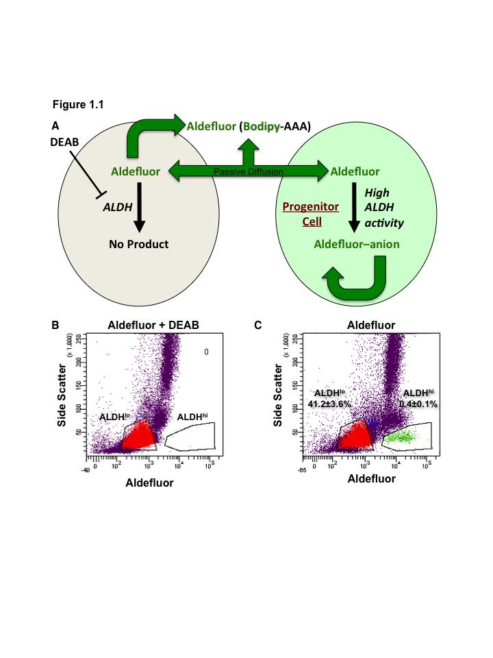
39 17 shown to play an important role in immunomodulation presenting interesting potential paracrine applications in auto-immune disease and inflammatory conditions 185. Taken together these findings show the considerable promise of MSC from human BM or UCB as a potential source for vascular regenerative cell therapies. 1.4 Aldehyde dehydrogenase: a functional marker of stem cells for cell therapy In order to simultaneously isolate stem and progenitor cells from multiple lineages, BM or UCB mononuclear cells (MNC) can be purified based on a conserved stem cell characteristic, high levels of aldehyde dehydrogenase (ALDH) activity, an intracellular enzyme first reported to be highly expressed in primitive hematopoietic 186, and neural progenitor cells 187. ALDH1A1 activity is involved in oxidation of vitamin A to retinoic acid and is predominantly implicated in the protection of long-lived cells from oxidative damage 186. Most notably, ALDH activity is downregulated as primitive cells differentiate towards maturity, making ALDH activity a unique function to distinguish essential regenerative precursors from expendable cells. Our group and others have recently shown that high ALDH activity is also a property shared by regenerative progenitors of endothelial and mesenchymal lineages 134,188. Purification of BM or UCB cells based on high ALDHactivity can be used to simultaneously isolate adult stem and progenitor cell subtypes for the preclinical development of regenerative therapies inducing tissue repair. Focusing on transplantation studies using human ALDH-expressing progenitor cells for hematopoietic reconstitution, blood vessel formation, and islet regeneration in immunodeficient mice, the Hess laboratory aims to understand how the multiple progenitor subtypes act together to formulate a regenerative niche and to coordinate complex regenerative processes FACS purification of multiple human progenitor subtypes using ALDH activity Intracellular ALDH activity can be quantified using a fluorescent substrate for ALDH, termed Aldefluor reagent 186. First synthesized by Clayton Smith s group in 1999, Aldefluor reagent is a Bodipy fluorochrome conjugated to an aminoacetaldehyde
40 18 molecule, an uncharged moiety that can freely cross through the cell membrane. Once inside the cell, cytoplasmic ALDH1A1 converts Aldefluor into a metabolized by ALDH into an anion that becomes trapped in the cell due to its negative charge. Under pharmacological inhibition of ABC transporters contained within Aldefluor buffer, cells with high ALDH activity retain Aldefluor substrate and fluoresce brightly, while cells with lower ALDH activity are more dimly fluorescent. Thus, high-speed fluorescenceactivated cell sorting (FACS) can efficiently purify UCB or BM MNC with low side scatter and low versus high ALDH activity. The integrity and function of the isolated cells are not compromised by this procedure since upon removal of the Aldefluor buffer, ATP-binding cassette transporters become reactivated, and Aldefluor is actively effluxed, returning the cell to its original state. Thus, the Aldefluor purification procedure is clinically applicable for the efficient sorting of multiple functional human progenitor cell types based on a highly conserved stem cell function. The amount of ALDH activity in all viable cells falls along a spectrum from low ALDH activity (ALDH lo ) to high ALDH activity (ALDH hi ), where ALDH lo versus ALDH hi cells are distinguished by cluster gating using diethylaminobenzaldehyde or DEAB, a pharmacological inhibitor of ALDH1A1. The basic premise of the Aldefluor assay to assess ALDH activity with FACS is summarised in Figure 1.1. It has previously been shown that purified ALDH hi cells from BM and UCB highly coexpressed stem cell-associated surface markers (CD34, CD133, c-kit) and were enriched for multipotent hematopoietic and mesenchymal stromal progenitors, as well as precursor cells with endothelial colony forming cell (ECFC) capacity in vitro 134,156,188,189. In contrast, ALDH lo cells were primarily comprised of mature leukocytes (primarily T- and B-cells) and demonstrated little progenitor function in vitro 134. Therefore, high ALDH activity simultaneously purifies multiple progenitor cell subtypes ideal for lineage-specific expansion in vitro. Subsequently, purified ALDH-purified mixed progenitor cells or their ex vivo expanded progeny can be assayed for regenerative functions after xenotransplantation into a variety of immunodeficient models of tissue damage.
41 19 Figure 1.1. Schematic overview of FACS isolation of ALDH hi cells using Aldefluor. (A) Aldefluor passively diffuses into cells where is metabolized by ALDH into an anion which can no longer diffuse out of the cell, this leads to accumulation of a fluorescent signal that can be detected and selected by FACS. (B) DEAB inhibition of ALDH activity identifies ALDH lo population for gating. (C) Aldefluor labeling allows efficient selection of ALDH hi cells from a heterogenous population of UCB MNC by FACS.
42 20
43 Cells with high ALDH activity possess hematopoietic repopulating capacity Hematopoietic stem cells (HSC) and lineage-specific hematopoietic progenitors are responsible for the replenishment and maintenance of blood after BM transplantation 190,191. These cells can be isolated from BM, cytokine-mobilized peripheral blood, or UCB based on expression of the cell surface markers CD34 and CD133, and assayed for hematopoietic repopulating function after transplantation into sublethally irradiated nonobese diabetic/severe combined immunodeficient (NOD/SCID) mice Storms et al. first established that the ALDH hi fraction of human UCB was enriched for primitive hematopoietic progenitors in vitro and was depleted of lineage-committed hematopoietic cells 186. Subsequently, prospective lineage depletion in combination with the commercially available Aldefluor reagent (Stemcell Technologies, Vancouver, Canada) was used by our group to demonstrate that transplantation of human UCB ALDH hi cells into NOD/SCID mice resulted in multilineage human hematopoietic engraftment 156. Greater than 70% of the UCB ALDH hi cell population co-expressed the HSC-associated cell surface markers CD34 and CD ALDH hi CD34 + cells were highly enriched for short-term myeloid progenitors while ALDH hi represented precursors to the CD34 + population that also demonstrated NOD/SCIDrepopulating cell (SRC) capacity CD34 cells Our laboratory has also demonstrated that while both ALDH hi CD133 and ALDH hi CD133 + cells demonstrated clonogenic hematopoietic progenitor function in vitro, only the ALDH hi CD133 + population was able to engraft the murine BM after intravenous injection 198. Furthermore, prospective selection based on both high ALDH activity and CD133 increased the frequency of SRC by tenfold compared to selection by CD133 alone. Notably, ALDH hi CD133 + cells demonstrated enhanced hematopoietic repopulating function in serial secondary transplants while maintaining primitive hematopoietic phenotypes (CD34 + CD38 ) 198. In addition to long-term hematopoietic repopulating function, human UCB ALDH hi CD133 + cells also showed previously unrecognized engraftment in nonhematopoietic tissues such as the liver, lung, heart,
44 22 brain, pancreas using the highly sensitive human cell-tracking NOD/ SCID MPSVII model 199. Later studies established that hematopoietic engraftment after human UCB transplantation in immunodeficient mice occurs faster with increasing ALDH hi cell doses 200. Similar to CD34 expression, clinical reconstitution rates following transplantation of BM or mobilized peripheral blood can be directly correlated with the number of ALDH hi cells infused 201,202. As a result of these promising preclinical data and direct potential for clinical translation, recently completed clinical trials designed to assess the safety and efficacy of transplanted allogeneic human UCB ALDH hi cells to enhance the rate of engraftment in the treatment of haematological dysfunction ( trial no. NCT ) have been reported. In summary, high ALDH activity is now well established as a functional characteristic of repopulating hematopoietic cells, and ALDH activity appears to be a superior indicator of the quality of BM or UCB samples for transplantation compared to standardized CD34 + counts Cells with high ALDH activity for vascular regeneration Although the ALDH hi population from human BM or UCB is more than 90% hematopoietic in origin, it has been shown that the ALDH hi fraction of BM is also significantly enriched for ECFC compared to ALDH lo cells and that high ALDH activity was downregulated as a result of differentiative culture in vitro 134. Thus, consistent with the classification by Yoder et al., true ECFC initially possesses elevated ALDH activity. Nagano et al. expanded EPC in culture from UCB and subsequently sorted the CD45 - EPC progeny based on ALDH activity 203. Interestingly, they found that the more differentiated ALDH lo outgrowth population was more proliferative in vitro and showed higher expression of hypoxia-inducible factors (HIF1α), VEGF, and the chemokine receptor CXCR4 under hypoxic conditions. They further showed that the cultured ALDH lo outgrowth represented more mature EC that recruited effectively to the site of ischemia and reduced necrosis in a mouse skin flap model of ischemic wound repair 203. Consistent with the idea that high ALDH activity simultaneously purifies stem and progenitor cells from multiple lineages, our group and others have shown that in addition
45 23 to HSC and tubule-forming EPC, the BM ALDH hi fraction was highly enriched for MSC that efficiently differentiated into fat, cartilage, and bone in differentiation cultures in vitro 134,189. Capoccia et al. went on further to describe that transplanted human BM ALDH hi mixed progenitor cells transiently recruited to areas of ischemia and augmented the recovery of blood flow (perfusion ratio of ischemic/control limb of 0.7±0.1) by stimulating the endogenous revascularization of ischemic limbs in immunodeficient NOD/SCID/β2microglobulin null and NOD/SCID/MPSVII mice with acute unilateral hind limb ischemia induced by femoral artery ligation compared to vehicle control treated mice at 21 days after treatment (0.3±0.1) 134. Even without permanent engraftment at the site of ischemia, a low dose of ALDH hi cells were more effective at inducing revascularization than transplantation of unsorted BM nucleated cells containing the equivalent of fourfold more ALDH hi cells. Recent work by Sondergaard et al. has shown that intravenously transplanted UCB ALDH hi cells recruited specifically to the ischemic myocardium where they augmented vascular density in a murine model of myocardial infarction 204. Although the detailed mechanisms by which ALDH hi cells induced endogenous revascularization remains an active area of investigation in the Hess lab, our working hypothesis describes that high ALDH activity simultaneously depletes for inflammatory immune cells and enriches for multiple proangiogenic progenitor subtypes. After transplantation, ALDH hi progenitor subtypes recruit transiently to areas of regional hypoxic damage and contribute to the generation of a proangiogenic microenvironment by potentially providing both structural and paracrine support. Despite the paucity of mechanistic details regarding the vascular regenerative potential of ALDH hi cells, clinical trials have been initiated to explore the safety and efficacy of the use of autologous BM ALDH hi cells in many CVD conditions 205,206. The FOCUS-Br trial was undertaken to investigate intramyocardial injection of autologous ALDH hi cells for therapeutic angiogenesis ( trial no. NCT ). The results of the trial showed evidence that transplantation intramyocardial transplantation of ALDH hi cells is safe and supported possible functional benefits in the setting of chronic
46 24 myocardial ischemia indicating need for further clinical testing in a larger cohort to fully assess efficacy 207. BM ALDH hi cells have also been investigated in clinical trials for treatment of critical limb ischemia (NCT ). Intramuscular transplantation of autologous BM ALDH hi cells were directly compared to unfractionated autologous BM MNC in PAD patients with a Rutherford score of 4 (resting pain) or 5 (ulceration or necrosis) 208. Results of the study demonstrated safety and patients that received autologous BM ALDH hi cells showed improved Rutherford category and reduced resting pain. Furthermore patients in the ALDH hi treatment group showed improved ankle to brachial index scores more rapidly than the MNC group indicating improved perfusion of the lower limb. However, patients in both cohorts reported significantly improved quality of life indices. 208 More recently, a phase 2 placebo-controlled randomized clinical trial has been initiated to investigate transplantation of BM ALDH hi cells to improve symptoms of intermittent claudication (NCT ). Another phase 2 clinical trial is currently underway to investigate intracarotid infusion of BM ALDH hi cells for patients after and ischemic stroke (NCT ) ALDH-purified cells stimulating islet revascularization and repair The Hess laboratory focuses on the development of pre-clinical models to study the potential utility of post-natal stem cells for the treatment of diabetes and its vascular complications. Indeed, the successful induction of angiogenesis or revascularization is a central process in tissue repair. In the context of regenerative therapies for diabetes, the contributions of transplanted stem cells are not limited to the direct replacement of damaged beta cells. As an alternative, the endogenous repair of damaged islets or the generation of new islets in situ has also been proposed 209. Hess and colleagues were the first to show that transplantation of murine BM-derived MNC or further purified c-kit + progenitor cells stimulate the recovery of streptozotocin (STZ)-damaged islets by inducing proliferation of recipient beta cells and augmenting glycemic control via the endogenous regeneration of beta cell function 210. Donor cells with both hematopoietic and EC phenotypes were recruited to ductal regions and surrounded damaged islets, subsequently stimulating beta cell proliferation and insulin production in recipient-
47 25 derived beta cells. Several groups have extended these findings to show that islet recovery can be induced by the induction of hematopoietic chimerism in overtly diabetic NOD mice 211 and that simultaneous infusion of murine BM MNC with allogeneic MSC optimize islet repair and protection against T-cell-mediated beta cell deletion 212. Although transplantation of murine BM-derived cells have shown proof of principle that BM-derived progenitor can impact endogenous beta cell regeneration, further purification and transplantation of human progenitor subtypes from multiple sources is underway in our lab to study the actions of specific cellular populations relevant to islet regeneration. Toward this end, Hess et al. have transplanted human ALDH-purified BM stem cells into STZ-treated hyperglycemic NOD/SCID mice to promote islet regeneration. Transplantation of BM ALDH hi cells led to a significant reduction in blood glucose and increased serum insulin due to an increase in endogenous beta cell proliferation resulting in increased islet size and total beta cell mass 213. Notably, transplanted BM ALDH hi cells recruited to damaged islets and stimulated beta cell proliferation associated with functional capillary formation in regenerating islets. Similar to hyperglycemic mice transplanted with human BM cells, mice transplanted with human UCB ALDH hi cells also demonstrated increased islet size and vascularization compared to controls. However, blood glucose reductions were transient, returning to severe hyperglycemia several weeks after transplantation. Furthermore, direct intra-pancreatic delivery of UCB ALDH hi cells increased peri-islet engraftment and stimulated a permanent reduction in hyperglycemia compare to ALDH lo cell controls via the induction of an islet proliferative and revascularization program in recipient islets 214. Unlike ALDH-purified BM progenitor populations, ALDH-purified UCB drawn by venipuncture did not consistently establish expandable MSC in culture, indicating a potential requirement for MSC to maintain islet regeneration. Fortunately, adherent MSC can be liberated from UCB after collagenase treatment 215, and inclusion of these cells within the transplanted population may further improve islet regeneration. In the past, clinical applications of UCB-derived stem cells have been limited to transplantation in the fields of hematology or oncology; however, an increasing number of studies support the use of these cells for nonhematopoietic disorders, including
48 26 diabetes 216,217. UCB MNC transplantation into diabetic mice has shown delayed onset of autoimmunity and insulitis in a model of type 1 diabetes and improved in hyperglycemia and survival rates post-transplantation 218,219. A clinical trial using autologous UCB cells is currently underway in children with recently diagnosed type 1 diabetes 217. Delayed loss of endogenous insulin production and enhanced glucose control have been reported due to a highly functional population of regulatory T-cells within UCB 220,221. However, the functional mechanisms conferring beta cell protection and potential mechanisms for the expansion of beta cell mass and vascularization after UCB transplantation require further preclinical experimentation Roles of ALDH-Purified MSC in Islet Regeneration MSC possess properties beneficial in the repair of tissues damaged by autoimmunity. MSC have been shown to modulate the microenvironment after injury and stimulate a shift from an inflammatory to a regenerative response 222, and to aid in tissue repair by exerting antifibrotic and neoangiogenic effects 223. MSC have also been shown to migrate toward areas of hypoxia and or tissue damage through their expression of a variety of chemokine receptors and adhesion molecules. Islets attract MSC in vitro and in vivo by CX3CL1-CX3CR1 and CXCL12-CXCR4 interactions 224,225. MSC may also play an important role in modulating the immune response, an important consideration in the treatment of type 1 diabetes. In damaged tissues, MSC stimulate reduced T-cell 226 and B- cell proliferation 227, inhibit maturation as well as differentiation of dendritic cells 228, and decrease the production of inflammatory cytokines by immune cells 229. MSC can exert these effects by secreting immunosuppressive effectors such as TGF-β, IDO, or PGE Collectively, these characteristics make transplanted MSC an attractive target for the development of cellular therapies for autoimmune diabetes 230. As proof of principle, Lee et al. have shown that multiple high-dose intracardiac infusion of human BM-derived MSC into STZ-treated immune-deficient NOD/ SCID mice repaired islets and improved hyperglycemia with only minimal engraftment in the pancreas 231. Transplantation of ALDH-purified MSC into the tail vein of STZ-treated hyperglycemic mice also stably reduced blood glucose and increased serum insulin. Rather than stimulating increased
49 27 islet size and vascularization, MSC transplantation stimulated an increase in the number of small islets present in the pancreas. Newly formed beta cell clusters were small in size with direct proximity to the ductal epithelium. These unique regenerative characteristics suggested the induction of a islet neogenic mechanism, whereby transplanted ALDHpurified MSC stimulated new islet formation 213. Similar to findings by Lee et al., transplanted ALDH-purified MSC showed low-level or transient recruitment to the pancreas, but initially lodge in the liver and lung capillaries 199, presumably exerting their effects on islet regeneration via the release of unknown paracrine regenerative factors into the bloodstream Future perspectives for use of ALDH hi cells in cell therapy BM- or UCB-derived ALDH-purified mixed progenitor cells and ALDH-purified MSC demonstrate a variety of regenerative functions that impact tissue repair and revascularization. However, differences in progenitor frequencies and functions should not be overlooked when considering the optimal progenitor cell populations for regenerative therapies. In addition, the regenerative mechanisms induced by ALDHexpressing progenitors and the paracrine mediators that comprise the regenerative niche require further elucidation. Provided with this foundation of critical information, scientists will be able to design rational regenerative therapies using a combination of regenerative small molecules or directed administration of multiple progenitor cell subtypes to synergize endogenous blood vessel and subsequent tissue repair. 1.5 Thesis Overview and Hypotheses Recently, our lab has shown that human BM ALDH hi mixed progenitor cells transiently recruit to areas of ischemia and augment blood flow by stimulating the endogenous revascularization of ischemic limbs in transplanted immune-deficient mice after induction of unilateral hindlimb ischemia by femoral artery ligation/transection 134. However, in patients with CVD, pro-angiogenic cell content or function may be compromised due to chronic diabetes or vascular disease-related pathologies rendering autologous BM transplantation strategies less effective 129, Readily available and early in ontogeny, human umbilical cord blood (UCB) represents a promising allogeneic
50 28 source of functional pro-angiogenic progenitor cells. During my thesis work I have investigated the vascular regenerative potential of human UCB and BM-derived hematopoietic and multipotent-stromal progenitor cells for the pre-clinical development of rational allogeneic cell-based therapies for ischemic diseases focusing on PAD. Furthermore, I have focused on global molecular approaches, and functional assays in vitro (Figure 1.2) and in vivo (Figure 1.3) to determine the paracrine signals elaborated from these pro-angiogenic cell sources that positively impact microvascular endothelial cell activation and function Our working model for transplanted progenitor cell involvement in vascular regeneration as investigated in this thesis is summarised in Figure 1.4. The central hypothesis of my studies is that high ALDH activity identifies populations of progenitor cells from blood or BM that will demonstrate augmented support of vascular regeneration. Thus, the specific objectives of my studies were as follows.
51 29 Figure 1.2. in vitro models for determination of progenitor cell contribution to endothelial cell function. Co-culture assays to assess cell-mediated effects on survival, proliferation, and tube forming capacity of endothelial cells. (A) Schematic overview of hanging transwell coculture and cell-conditioned medium assays to assess endothelial cell proliferation or survival. (B) Representative photomicrograph of human endothelial cells cultured on plastic. (C) Representative photomicrograph of human endothelial cells growing on a thin layer of Matrigel to assess modulation of tube-like structure formation. Adapted from: Putman DM, Hess DA. Isolation of human umbilical cord blood aldehyde dehydrogenase-expressing progenitor cells that modulate vascular regenerative functions in vitro and in vivo. Curr Protoc Stem Cell Biol. 2013;Chapter 2:Unit 2A.10
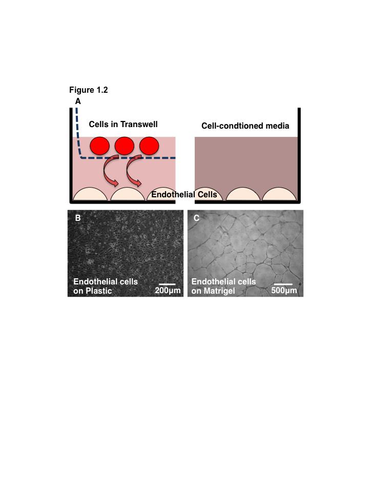
52 30
53 31 Figure 1.3 Transplantation model of acute unilateral hindlimb ischemia. (A) Diagram of the vasculature of the mouse hindlimb from the inguinal crease to the bifurcation of the femoral artery into the saphenous and popliteal arteries that descend into the calf and lower limb. Ligation sites for femoral artery ligation surgery are indicated. Representative laser Doppler perfusion images of mouse hindquarters before and after ligation and transection of femoral artery and vein. Regions of interest are selected from ankle to toe to quantify relative perfusion in the surgical limb. (B) Outline and timing for tracking recovery of perfusion in the ischemic limb after surgical induction of acute ischemia. Number in lower left quadrant of each laser Doppler perfusion image is the ratio of perfusion in the ischemic limb to the perfusion in the control limb. Adapted from: Putman DM, Hess DA. Isolation of human umbilical cord blood aldehyde dehydrogenase-expressing progenitor cells that modulate vascular regenerative functions in vitro and in vivo. Curr Protoc Stem Cell Biol. 2013;Chapter 2:Unit 2A.10
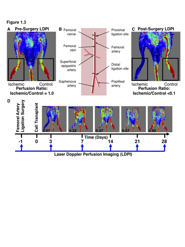
54 32
55 33 Figure 1.4. Umbilical cord blood and bone marrow-derived stem and progenitor cells support an angiogenic niche. The ALDH hi subpopulation of cells in UCB and BM is enriched for progenitors from the hematopoietic, endothelial, and mesenchymal lineages. Each of these progenitor cell types recruit to and help co-ordinate a pro-angiogenic regenerative microenvironment in ischemic tissues. Adapted from: Putman, D. M., Bell, G. I. & Hess, D. A. Blood-Derived ALDH hi Cells in Tissue Repair, in Regenerative Therapy Using Blood-Derived Stem Cells (Allan, D. S. S. & Strunk, D.) (Humana Press, 2012).

56 Figure
57 Aim 1 To characterise the capacity of umbilical cord blood-derived ALDH-purified cells to modulate endothelial cell functions in vitro To assess the effects on vascular regeneration after intra-venous transplantation of UCB ALDH hi cells into immune-deficient mice with unilateral hindlimb ischemia induced by femoral artery ligation surgery. In Aim 1, I hypothesized that selection of UCB cells based on high ALDH activity will select a population of cells with augmented capacity to promote recovery from ischemic injury Aim 2 Due to the relative rarity of ALDH hi cells from UCB or BM, in Aim 2 I sought to determine if it was possible to expand UCB-derived ALDH hi cells ex vivo to generate more cells for therapeutic applications without losing beneficial vascular regenerative effect. Therefore the objectives of Aim 2 were two-fold: To optimize the ex vivo expansion of UCB ALDH hi hematopoietic cells under clinically relevant serum-free conditions and to characterize vascular regenerative functions of the expanded progenitor cells in vitro. To investigate the paracrine mechanisms by which expanded hematopoietic progenitor cells derived from UCB ALDH hi contribute to revascularization after direct intramuscular injection into the ischemic limb. In Aim 2, I hypothesized that ex vivo expansion of UCB ALDH hi cells would increase the number of pro-angiogenic hematopoietic progenitor cells for application in vascular regeneration without decreasing EC-supportive functions Aim 3 BM MSC are a population of progenitor cells that expand readily in ex vivo cultures with underutilized potential for vascular regenerative cellular therapies. However, our lab has
58 36 demonstrated in our beta cell regeneration model studies that different MSC lines from different donors showed variable efficacy to promote regeneration 213,236. Furthermore, it has been previously shown that selection of high ALDH activity in BM cells selected for cells with increased regenerative potential. Therefore the objective in Aim 3 was as follows: To characterize the differentiative and vascular regenerative functions of MSC subpopulations selected after expansion for low versus high ALDH activity. In Aim 3, I hypothesized that selection of MSC subpopulations with high ALDH activity would identify a population of MSC with increased vascular regenerative potential.
59 References 1. Go AS, Mozaffarian D, Roger VL, et al. Executive summary: heart disease and stroke statistics update: a report from the American Heart Association. Circulation. 2014;129(3): Lackland DT, Roccella EJ, Deutsch AF, et al. Factors influencing the decline in stroke mortality: a statement from the American Heart Association/American Stroke Association. Stroke. 2014;45(1): Dotsenko O. Stem/Progenitor cells, atherosclerosis and cardiovascular regeneration. Open Cardiovasc Med J. 2010;4: Glass CK, Witztum JL. Atherosclerosis. the road ahead. Cell. 2001;104(4): Lackland DT, Roccella EJ, Deutsch AF, et al. Factors influencing the decline in stroke mortality: a statement from the American Heart Association/American Stroke Association. Stroke. 2014;45(1): Lusis AJ. Atherosclerosis. Nature. 2000;407(6801): Libby P, Ridker PM, Hansson GK. Progress and challenges in translating the biology of atherosclerosis. Nature. 2011;473(7347): Stary HC, Chandler AB, Dinsmore RE, et al. A definition of advanced types of atherosclerotic lesions and a histological classification of atherosclerosis. A report from the Committee on Vascular Lesions of the Council on Arteriosclerosis, American Heart Association. Circulation. 1995;92(5): Libby P. Inflammation in Atherosclerosis. Arteriosclerosis, Thrombosis, and Vascular Biology. 2012;32(9): Vauchez K, Marolleau J-P, Schmid M, et al. Aldehyde Dehydrogenase Activity Identifies a Population of Human Skeletal Muscle Cells With High Myogenic Capacities. Mol Ther. 2009;17(11): Rippe JM, Angelopoulos TJ. Lifestyle strategies for cardiovascular risk reduction. Curr Atheroscler Rep. 2014;16(10): Stone NJ, Robinson JG, Lichtenstein AH, et al ACC/AHA guideline on the treatment of blood cholesterol to reduce atherosclerotic cardiovascular risk in adults: a report of the American College of Cardiology/American Heart Association Task Force on Practice Guidelines. J. Am. Coll. Cardiol. 2014;63(25 Pt B): Berger JS, Hiatt WR. Medical therapy in peripheral artery disease. Circulation. 2012;126(4): Miller LW, Missov ED. Epidemilolgy of Heart Failure. Cardiol Clin. 2001;19(4):
60 Bergers G, Coussens LM. Extrinsic regulators of epithelial tumor progression: metalloproteinases. Current Opinion in Genetics & Development. 2000;10(1): Beckman JA, Paneni F, Cosentino F, Creager MA. Diabetes and vascular disease: pathophysiology, clinical consequences, and medical therapy: part II. Eur. Heart J. 2013;34(31): Lange S, Diehm C, Darius H, et al. High Prevalence of Peripheral Arterial Disease and Low Treatment Rates in Elderly Primary Care Patients with Diabetes. Exp. Clin. Endocrinol. Diabetes. 2004;112(10): Aquino R, Johnnides C, Makaroun M, et al. Natural history of claudication: longterm serial follow-up study of 1244 claudicants. J. Vasc. Surg. 2001;34(6): Killip T, Passamani E, Davis K. Coronary artery surgery study (CASS): a randomized trial of coronary bypass surgery. Eight years follow-up and survival in patients with reduced ejection fraction. Circulation. 1985;72(6 Pt 2):V Gwadry-Sridhar FH, Flintoft V, Lee DS, Lee H, Guyatt GH. A systematic review and meta-analysis of studies comparing readmission rates and mortality rates in patients with heart failure. Arch. Intern. Med. 2004;164(21): Michler RE. Stem cell therapy for heart failure. Cardiology in Review. 2014;22(3): Lenzen MJ, Boersma E, Reimer WJMSO, et al. Under-utilization of evidencebased drug treatment in patients with heart failure is only partially explained by dissimilarity to patients enrolled in landmark trials: a report from the Euro Heart Survey on Heart Failure. Eur. Heart J. 2005;26(24): Gupta NK, Armstrong EJ, Parikh SA. The Current State of Stem Cell Therapy for Peripheral Artery Disease. Curr Cardiol Rep. 2014;16(2): Wennberg PW. Approach to the patient with peripheral arterial disease. Circulation. 2013;128(20): Hirsch AT, Kalsi H, Rooke TW. Peripheral Arterial Diseases. 2007; Pande RL, Perlstein TS, Beckman JA, Creager MA. Secondary prevention and mortality in peripheral artery disease: National Health and Nutrition Examination Study, 1999 to Circulation. 2011;124(1): Howangyin KY, Silvestre J-S. Diabetes mellitus and ischemic diseases: molecular mechanisms of vascular repair dysfunction. Arteriosclerosis, Thrombosis, and Vascular Biology. 2014;34(6): Norgren L, Hiatt WR, Dormandy JA, et al. Inter-society consensus for the management of peripheral arterial disease. Int Angiol. 2007;26(2): Rutherford RB, Baker JD, Ernst C, et al. Recommended standards for reports dealing with lower extremity ischemia: revised version. J. Vasc. Surg. 1997;26(3):
61 Rowlands TE, Donnelly R. Medical Therapy for Intermittent Claudication. European Journal of Vascular and Endovascular Surgery. 2007;34(3): Eneroth M, Persson BM. Amputation for occlusive arterial disease. A prospective multicentre study of 177 amputees. Int Orthop. 1992;16(4): Albers M, Fratezi AC, De Luccia N. Assessment of quality of life of patients with severe ischemia as a result of infrainguinal arterial occlusive disease. J. Vasc. Surg. 1992;16(1): Suzuki H, Iso Y. Clinical Application of Vascular Regenerative Therapy for Peripheral Artery Disease. BioMed Research International. 2013;2013(3): Conte MS. Bypass versus Angioplasty in Severe Ischaemia of the Leg (BASIL) and the (hoped for) dawn of evidence-based treatment for advanced limb ischemia. J. Vasc. Surg. 2010;51(5 Suppl):69S 75S. 35. Pries AR, Kuebler WM. Normal endothelium. Handb Exp Pharmacol. 2006;(176 Pt 1): Carmeliet P, Jain RK. Molecular mechanisms and clinical applications of angiogenesis. Nature. 2011;473(7347): Heinke J, Patterson C, Moser M. Life is a pattern: vascular assembly within the embryo. Front Biosci (Elite Ed). 2012;4: Aird WC. Phenotypic heterogeneity of the endothelium: I. Structure, function, and mechanisms. Circ. Res. 2007;100(2): Aird WC. Phenotypic heterogeneity of the endothelium: II. Representative vascular beds. Circ. Res. 2007;100(2): Pober JS, Min W. Endothelial cell dysfunction, injury and death. 2006; Simionescu M, Antohe F. Functional ultrastructure of the vascular endothelium: changes in various pathologies. 2006; Cines DB, Pollak ES, Buck CA, et al. Endothelial cells in physiology and in the pathophysiology of vascular disorders. Blood. 1998;91(10): Beenken A, Mohammadi M. The FGF family: biology, pathophysiology and therapy : Abstract : Nature Reviews Drug Discovery. Nature reviews Drug discovery Carmeliet P. Angiogenesis in health and disease. Nature Medicine Semenza GL. Hypoxia-inducible factor 1: oxygen homeostasis and disease pathophysiology. Trends in Molecular Medicine. 2001;7(8): Maxwell PH, Dachs GU, Gleadle JM, et al. Hypoxia-inducible factor-1 modulates gene expression in solid tumors and influences both angiogenesis and tumor growth. Proceedings of the Brocato J, Chervona Y, Costa M. Molecular responses to hypoxia-inducible factor 1α and beyond. Mol. Pharmacol. 2014;85(5):
62 Betsholtz C, Lindblom P, Gerhardt H. Role of pericytes in vascular morphogenesis. Mechanisms of Angiogenesis. 2005;(Chapter 8): Coultas L, Chawengsaksophak K, Rossant J. Endothelial cells and VEGF in vascular development. Nature. 2005;438(7070): Semenza GL. Vasculogenesis, angiogenesis, and arteriogenesis: mechanisms of blood vessel formation and remodeling. J. Cell. Biochem. 2007;102(4): Choi K, Kennedy M, Kazarov A, Papadimitriou JC, Keller G. A common precursor for hematopoietic and endothelial cells. Development. 1998;125(4): Huber TL, Kouskoff V, Fehling HJ, Palis J, Keller G. Haemangioblast commitment is initiated in the primitive streak of the mouse embryo. Nature. 2004;432(7017): Burri PH, Hlushchuk R, Djonov V. Intussusceptive angiogenesis: its emergence, its characteristics, and its significance. Dev. Dyn. 2004;231(3): Ferrara N. VEGF-A: a critical regulator of blood vessel growth. Eur. Cytokine Netw. 2009;20(4): Eklund L, Saharinen P. Angiopoietin signaling in the vasculature. Experimental Cell Research. 2013;319(9): Bernardini G, Ribatti D, Spinetti G, et al. Analysis of the role of chemokines in angiogenesis. Journal of Immunological Methods. 2003;273(1-2): Dejana E, Tournier-Lasserve E, Weinstein BM. The Control of Vascular Integrity by Endothelial Cell Junctions: Molecular Basis and Pathological Implications. Developmental Cell. 2009;16(2): Phng LK, Gerhardt H. Angiogenesis: A Team Effort Coordinated by Notch. Developmental Cell. 2009;16(2): Eichmann A, Yuan L, Moyon D, et al. Vascular development: from precursor cells to branched arterial and venous networks. Int. J. Dev. Biol. 2005;49(2-3): Ribatti D. The seminal work of Werner Risau in the study of the development of the vascular system Semenza GL. Hypoxia-Inducible Factors in Physiology and Medicine. Cell Ke Q, Costa M. Hypoxia-Inducible Factor-1 (HIF-1) Schofield CJ, Zhang Z. Structural and mechanistic studies on 2-oxoglutaratedependent oxygenases and related enzymes. Current Opinion in Structural Biology. 1999;9(6): Kaelin WG Jr, Ratcliffe PJ. Oxygen Sensing by Metazoans: The Central Role of the HIF Hydroxylase Pathway. Molecular cell
63 Levy AP, Levy NS, Wegner S, Goldberg MA. Transcriptional Regulation of the Rat Vascular Endothelial Growth Factor Gene by Hypoxia. Journal of Biological Bunn HF, Poyton RO. Oxygen sensing and molecular adaptation to hypoxia. Physiological Reviews. 1996;76(3): Forsythe JA, Jiang BH, Iyer NV, et al. Activation of vascular endothelial growth factor gene transcription by hypoxia-inducible factor 1. and cellular biology Berra E, Milanini J, Richard DE, Le Gall M. Signaling angiogenesis via p42/p44 MAP kinase and hypoxia. Biochemical Giordano FJ, Johnson RS. Angiogenesis: the role of the microenvironment in flipping the switch. Current Opinion in Genetics & Development Augustin HG, Koh GY, Thurston G, Alitalo K. Control of vascular morphogenesis and homeostasis through the angiopoietin-tie system. Nature Reviews Molecular Cell Biology. 2009;10(3): Saharinen P, Eklund L, Miettinen J, et al. Angiopoietins assemble distinct Tie2 signalling complexes in endothelial cell-cell and cell-matrix contacts. Nat. Cell Biol. 2008;10(5): Thurston G. Role of Angiopoietins and Tie receptor tyrosine kinases in angiogenesis and lymphangiogenesis. Cell and Tissue Research. 2003;314(1): Murakami M, Nguyen LT, Zhuang ZW, et al. The FGF system has a key role in regulating vascular integrity. J. Clin. Invest. 2008;118(10): Deryugina EI, Quigley JP. Pleiotropic roles of matrix metalloproteinases in tumor angiogenesis: Contrasting, overlapping and compensatory functions. Biochimica et Biophysica Acta (BBA)-Molecular Kessenbrock K, Plaks V, Werb Z. Matrix Metalloproteinases: Regulators of the Tumor Microenvironment. Cell. 2010;141(1): Ahn G-O, Brown JM. Matrix metalloproteinase-9 is required for tumor vasculogenesis but not for angiogenesis: role of bone marrow-derived myelomonocytic cells. Cancer Cell. 2008;13(3): Rossi D, Zlotnik A. The Biology of Chemokines and their Receptors. Annu. Rev. Immunol. 2000;18(1): Strieter RM, Polverini PJ, Arenberg DA, Kunkel SL. The role of CXC chemokines as regulators of angiogenesis. Shock. 1995;4(3): Strieter RM, Polverini PJ, Kunkel SL, et al. The Functional Role of the ELR Motif in CXC Chemokine-mediated Angiogenesis. Journal of Biological
64 Zaja-Milatovic S, Richmond A. CXC chemokines and their receptors: a case for a significant biological role in cutaneous wound healing. Histol. Histopathol. 2008;23(11): Jin DK, Shido K, Kopp HG, et al. Cytokine-mediated deployment of SDF-1 induces revascularization through recruitment of CXCR4+ hemangiocytes. Nat Med. 2006;12(5): Ruiz de Almodovar C, Luttun A, Carmeliet P. An SDF-1 trap for myeloid cells stimulates angiogenesis. Cell. 2006;124(1): Grunewald M, Avraham I, Dor Y, et al. VEGF-induced adult neovascularization: recruitment, retention, and role of accessory cells. Cell. 2006;124(1): Duda DG, Kozin SV, Kirkpatrick ND, et al. CXCL12 (SDF1alpha)- CXCR4/CXCR7 pathway inhibition: an emerging sensitizer for anticancer therapies? Clin. Cancer Res. 2011;17(8): Schober A. Chemokines in Vascular Dysfunction and Remodeling. Arteriosclerosis, Thrombosis, and Vascular Biology. 2008;28(11): Suffee N, Richard B, Hlawaty H, et al. Angiogenic properties of the chemokine RANTES/CCL5. Biochem. Soc. Trans. 2011;39(6): Westerweel PE, Rabelink TJ, Rookmaaker MB, Gröne H-J, Verhaar MC. RANTES is required for ischaemia-induced angiogenesis, which may hamper RANTES-targeted anti-atherosclerotic therapy. Thromb Haemost Charo IF, Ransohoff RM. The Many Roles of Chemokines and Chemokine Receptors in Inflammation. N Engl J Med. 2006;354(6): Schober A, Zernecke A, Liehn EA. Crucial role of the CCL2/CCR2 axis in neointimal hyperplasia after arterial injury in hyperlipidemic mice involves early monocyte recruitment and CCL2 presentation. Circulation Egashira K, Zhao Q, Kataoka C, et al. Importance of monocyte chemoattractant protein-1 pathway in neointimal hyperplasia after periarterial injury in mice and monkeys. Circ. Res. 2002;90(11): Weber KS, Hundelshausen von P, Clark-Lewis I, Weber PC, Weber C. Differential immobilization and hierarchical involvement of chemokines in monocyte arrest and transmigration on inflamed endothelium in shear flow. Eur. J. Immunol. 1999;29(2): Fujiyama S, Amano K, Uehira K, et al. Bone marrow monocyte lineage cells adhere on injured endothelium in a monocyte chemoattractant protein-1- dependent manner and accelerate reendothelialization as endothelial progenitor cells. Circ. Res. 2003;93(10): Moldovan NI, Goldschmidt-Clermont PJ, Parker-Thornburg J, Shapiro SD, Kolattukudy PE. Contribution of monocytes/macrophages to compensatory neovascularization: the drilling of metalloelastase-positive tunnels in ischemic myocardium. Circ. Res. 2000;87(5):
65 Dewald O, Zymek P, Winkelmann K, et al. CCL2/Monocyte Chemoattractant Protein-1 Regulates Inflammatory Responses Critical to Healing Myocardial Infarcts Salcedo R, Ponce ML, Young HA, et al. Human endothelial cells express CCR2 and respond to MCP-1: direct role of MCP-1 in angiogenesis and tumor progression. Blood. 2000;96(1): Rookmaaker MB, Verhaar MC, de Boer HC, et al. Met-RANTES reduces endothelial progenitor cell homing to activated (glomerular) endothelium in vitro and in vivo. AJP: Renal Physiology. 2007;293(2):F624 F Baltus T. Oligomerization of RANTES is required for CCR1-mediated arrest but not CCR5-mediated transmigration of leukocytes on inflamed endothelium. Blood. 2003;102(6): Crola Da Silva C, Lamerant-Fayel N, Paprocka M, et al. Selective human endothelial cell activation by chemokines as a guide to cell homing. Immunology. 2009;126(3): Fukumura D, Jain RK. Imaging angiogenesis and the microenvironment. APMIS. 2008;116(7-8): Bluff JE, O'Ceallaigh S, O'Kane S, Ferguson MWJ, Ireland G. The microcirculation in acute murine cutaneous incisional wounds shows a spatial and temporal variation in the functionality of vessels. Wound Repair Regen. 2006;14(4): Shaterian A, Borboa A, Sawada R, et al. Real-time analysis of the kinetics of angiogenesis and vascular permeability in an animal model of wound healing. Burns. 2009;35(6): Jain RK. Molecular regulation of vessel maturation - Nature Medicine. Nature Medicine Hellberg C, Östman A, Heldin CH. PDGF and Vessel Maturation - Springer. Angiogenesis inhibition Pardali E, Goumans M-J, Dijke Ten P. Signaling by members of the TGF-beta family in vascular morphogenesis and disease. Trends Cell Biol. 2010;20(9): Gurtner GC, Werner S, Barrandon Y, Longaker MT. Wound repair and regeneration. Nature. 2008;453(7193): Swift ME, Kleinman HK, DiPietro LA. Impaired wound repair and delayed angiogenesis in aged mice. Lab Invest. 1999;79(12): Ando J, Yamamoto K. Vascular mechanobiology: endothelial cell responses to fluid shear stress. Circ. J. 2009;73(11): Chen RR, Silva EA, Yuen WW, Mooney DJ. Spatio-temporal VEGF and PDGF delivery patterns blood vessel formation and maturation. Pharm. Res. 2007;24(2):
66 Dimmeler S, Zeiher AM. Endothelial cell apoptosis in angiogenesis and vessel regression. Circ. Res. 2000;87(6): Sakamaki K. Regulation of endothelial cell death and its role in angiogenesis and vascular regression. Curr Neurovasc Res. 2004;1(4): Wietecha MS, Cerny WL, DiPietro LA. Mechanisms of vessel regression: toward an understanding of the resolution of angiogenesis. Curr. Top. Microbiol. Immunol. 2013;367: van Royen N, Piek JJ, Buschmann I, et al. Stimulation of arteriogenesis; a new concept for the treatment of arterial occlusive disease. Cardiovasc. Res. 2001;49(3): Heil M, Eitenmüller I, Schmitz-Rixen T, Schaper W. Arteriogenesis versus angiogenesis: similarities and differences. J Cellular Mol Med. 2006;10(1): Buschmann I, Schaper W. The pathophysiology of the collateral circulation (arteriogenesis). J. Pathol. 2000;190(3): Lawall H, Bramlage P, Amann B. Treatment of peripheral arterial disease using stem and progenitor cell therapy. J. Vasc. Surg. 2011;53(2): Arras M, Ito WD, Scholz D, et al. Monocyte activation in angiogenesis and collateral growth in the rabbit hindlimb. J. Clin. Invest. 1998;101(1): Zarins CK, Zatina MA, Giddens DP, Ku DN, Glagov S. Shear stress regulation of artery lumen diameter in experimental atherogenesis. J. Vasc. Surg. 1987;5(3): Singh TM, Abe KY, Sasaki T, et al. Basic Fibroblast Growth Factor Expression Precedes Flow-Induced Arterial Enlargement. Journal of Surgical Research. 1998;77(2): Heilmann C, Beyersdorf F, Lutter G. Collateral growth: cells arrive at the construction site. Cardiovasc Surg. 2002;10(6): Heil M. A Different Outlook on the Role of Bone Marrow Stem Cells in Vascular Growth: Bone Marrow Delivers Software not Hardware. Circ. Res. 2004;94(5): Kinnaird T, Stabile E, Burnett MS, Epstein SE. Bone-marrow-derived cells for enhancing collateral development: mechanisms, animal data, and initial clinical experiences. Circ. Res. 2004;95(4): Kinnaird T, Stabile E, Burnett MS, et al. Marrow-derived stromal cells express genes encoding a broad spectrum of arteriogenic cytokines and promote in vitro and in vivo arteriogenesis through paracrine mechanisms. Circ. Res. 2004;94(5): Huss R, Heil M, Moosmann S, et al. Improved arteriogenesis with simultaneous skeletal muscle repair in ischemic tissue by SCL(+) multipotent adult progenitor cell clones from peripheral blood. J. Vasc. Res. 2004;41(5):
67 Rehman J, Li J, Orschell CM, March KL. Peripheral blood endothelial progenitor cells are derived from monocyte/macrophages and secrete angiogenic growth factors. Circulation. 2003;107(8): Arras M, Strasser R, Mohri M, et al. Tumor necrosis factor-alpha is expressed by monocytes/macrophages following cardiac microembolization and is antagonized by cyclosporine. Basic Res. Cardiol. 1998;93(2): Lotfi S, Patel AS, Mattock K, et al. Towards a more relevant hind limb model of muscle ischaemia. Atherosclerosis. 2013;227(1): Asahara T, Murohara T, Sullivan A, et al. Isolation of putative progenitor endothelial cells for angiogenesis. Science. 1997;275(5302): Basile DP, Yoder MC. Circulating and tissue resident endothelial progenitor cells. J. Cell. Physiol. 2014;229(1): Yoder MC, Mead LE, Prater D, et al. Redefining endothelial progenitor cells via clonal analysis and hematopoietic stem/progenitor cell principals. Blood. 2007;109(5): Hirschi KK, Ingram DA, Yoder MC. Assessing identity, phenotype, and fate of endothelial progenitor cells. Arteriosclerosis, Thrombosis, and Vascular Biology. 2008;28(9): Mund JA, Ingram DA, Yoder MC, Case J. Endothelial progenitor cells and cardiovascular cell-based therapies. Cytotherapy. 2009;11(2): Timmermans F, Plum J, Yoder MC, et al. Endothelial progenitor cells: identity defined? J Cellular Mol Med. 2008;13(1): Putman DM, Liu KY, Broughton HC, Bell GI, Hess DA. Umbilical cord bloodderived aldehyde dehydrogenase-expressing progenitor cells promote recovery from acute ischemic injury. Stem Cells. 2012;30(10): Capoccia BJ, Robson DL, Levac KD, et al. Revascularization of ischemic limbs after transplantation of human bone marrow cells with high aldehyde dehydrogenase activity. Blood. 2009;113(21): Wara AK, Croce K, Foo S, et al. Bone marrow-derived CMPs and GMPs represent highly functional proangiogenic cells: implications for ischemic cardiovascular disease. Blood. 2011;118(24): Okuno Y, Nakamura-Ishizu A, Kishi K, Suda T, Kubota Y. Bone marrow-derived cells serve as proangiogenic macrophages but not endothelial cells in wound healing. Blood. 2011;117(19): Rohde E, Schallmoser K, Reinisch A, et al. Pro-angiogenic induction of myeloid cells for therapeutic angiogenesis can induce mitogen-activated protein kinase p38-dependent foam cell formation. Cytotherapy. 2011;13(4): Urbich C, Heeschen C, Aicher A, et al. Relevance of monocytic features for neovascularization capacity of circulating endothelial progenitor cells. Circulation. 2003;108(20):
68 Richardson MR, Yoder MC. Endothelial progenitor cells: quo vadis? J. Mol. Cell. Cardiol. 2011;50(2): Yoder MC, Ingram DA. The definition of EPCs and other bone marrow cells contributing to neoangiogenesis and tumor growth: is there common ground for understanding the roles of numerous marrow-derived cells in the neoangiogenic process? Biochim. Biophys. Acta. 2009;1796(1): Raval Z, Losordo DW. Cell therapy of peripheral arterial disease: from experimental findings to clinical trials. Circ. Res. 2013;112(9): Sneider EB, Nowicki PT, Messina LM. Regenerative medicine in the treatment of peripheral arterial disease. J. Cell. Biochem. 2009;108(4): Glotzbach JP, Levi B, Wong VW, Longaker MT, Gurtner GC. The Basic Science of Vascular Biology: Implications for the Practicing Surgeon. Plastic and Reconstructive Surgery. 2010;126(5): Till JE, McCulloch EA. A Direct Measurement of the Radiation Sensitivity of Normal Mouse Bone Marrow Cells. Radiation research Till JE, McCulloch EA. Hemopoietic stem cell differentiation. Biochim. Biophys. Acta. 1980;605(4): Morrison SJ, Shah NM, Anderson DJ. Regulatory mechanisms in stem cell biology. Cell Thomson JA, Itskovitz-Eldor J, Shapiro SS, et al. Embryonic stem cell lines derived from human blastocysts. Science. 1998;282(5391): Takahashi K, Yamanaka S. Induction of pluripotent stem cells from mouse embryonic and adult fibroblast cultures by defined factors. Cell. 2006;126(4): Takahashi K, Tanabe K, Ohnuki M, et al. Induction of Pluripotent Stem Cells from Adult Human Fibroblasts by Defined Factors. Cell. 2007;131(5): Notta F, Doulatov S, Laurenti E, et al. Isolation of single human hematopoietic stem cells capable of long-term multilineage engraftment. Science. 2011;333(6039): Deans RJ, Moseley AB. Mesenchymal stem cells: biology and potential clinical uses. Exp. Hematol. 2000;28(8): Kopp H-G, Ramos CA, Rafii S. Contribution of endothelial progenitors and proangiogenic hematopoietic cells to vascularization of tumor and ischemic tissue. Curr. Opin. Hematol. 2006;13(3): Sanganalmath SK, Bolli R. Cell therapy for heart failure: a comprehensive overview of experimental and clinical studies, current challenges, and future directions. Circ. Res. 2013;113(6): Bronckaers A, Hilkens P, Martens W, et al. Mesenchymal stem/stromal cells as a pharmacological and therapeutic approach to accelerate angiogenesis. Pharmacology & Therapeutics
69 Cai J, Weiss ML, Rao MS. In search of stemness. Exp. Hematol. 2004;32(7): Hess DA, Meyerrose TE, Wirthlin L, et al. Functional characterization of highly purified human hematopoietic repopulating cells isolated according to aldehyde dehydrogenase activity. Blood. 2004;104(6): Peichev M, Naiyer AJ, Pereira D, et al. Expression of VEGFR-2 and AC133 by circulating human CD34(+) cells identifies a population of functional endothelial precursors. Blood. 2000;95(3): Murohara T, Ikeda H, Duan J, et al. Transplanted cord blood-derived endothelial precursor cells augment postnatal neovascularization. J. Clin. Invest. 2000;105(11): Quirici N, Soligo D, Caneva L, et al. Differentiation and expansion of endothelial cells from human bone marrow CD133(+) cells. Br. J. Haematol. 2001;115(1): Takakura N, Watanabe T, Suenobu S, et al. A role for hematopoietic stem cells in promoting angiogenesis. Cell. 2000;102(2): Tateishi-Yuyama E, Matsubara H, Murohara T, et al. Therapeutic angiogenesis for patients with limb ischaemia by autologous transplantation of bone-marrow cells: a pilot study and a randomised controlled trial. Lancet. 2002;360(9331): Botham CM, Bennett WL, Cooke JP. Clinical Trials of Adult Stem Cell Therapy for Peripheral Artery Disease. Methodist DeBakey Cardiovascular Journal. 2013;9(4): Schachinger V, Erbs S, Elsasser A, et al. Intracoronary bone marrow-derived progenitor cells in acute myocardial infarction. N Engl J Med. 2006;355(12): Assmus B, Honold J, Schachinger V, et al. Transcoronary transplantation of progenitor cells after myocardial infarction. N Engl J Med. 2006;355(12): Janssens S, Dubois C, Bogaert J, et al. Autologous bone marrow-derived stemcell transfer in patients with ST-segment elevation myocardial infarction: doubleblind, randomised controlled trial. Lancet. 2006;367(9505): Rosenzweig A. Cardiac cell therapy--mixed results from mixed cells. N Engl J Med. 2006;355(12): Sanchez-Guijo FM, Oterino E, Barbado M-V, et al. Both CD133 + Cells and Monocytes Provide Significant Improvement for Hindlimb Ischemia, Although They do not Transdifferentiate Into Endothelial Cells. Cell Transplant. 2010;19(1): Lai W-H, Ho JCY, Chan Y-C, et al. Attenuation of hind-limb ischemia in mice with endothelial-like cells derived from different sources of human stem cells. PLoS ONE. 2013;8(3):e57876.
70 Vu NB, Bui AN-T, Ngoc-Le Trinh V, et al. A comparison of umbilical cord blood-derived endothelial progenitor cell transplantation and mononuclear cell transplantation for the treatment of acute hindlimb ischemia. Biomedical Research and Therapy. 2014;1(1): Helisch A, Wagner S, Khan N, et al. Impact of mouse strain differences in innate hindlimb collateral vasculature. Arteriosclerosis, Thrombosis, and Vascular Biology. 2006;26(3): Chalothorn D, Faber JE. Strain-dependent variation in collateral circulatory function in mouse hindlimb. Physiol. Genomics. 2010;42(3): Horwitz EM, Le Blanc K, Dominici M, et al. Clarification of the nomenclature for MSC: The International Society for Cellular Therapy position statement. Cytotherapy. 2005;7(5): Pittenger MF, Mackay AM, Beck SC, et al. Multilineage Potential of Adult Human Mesenchymal Stem Cells. Science. 1999;284(5411): Bianco P, Robey PG, Simmons PJ. Mesenchymal Stem Cells: Revisiting History, Concepts, and Assays. Cell Stem Cell. 2008;2(4): Le Blanc K, Tammik C, Rosendahl K, Zetterberg E, Ringdén O. HLA expression and immunologic propertiesof differentiated and undifferentiated mesenchymal stem cells. Experimental Hematology. 2003;31(10): Wu Y, Chen L, Scott PG, Tredget EE. Mesenchymal Stem Cells Enhance Wound Healing Through Differentiation and Angiogenesis. Stem Cells. 2007;25(10): Caplan AI, Correa D. The MSC: An Injury Drugstore. Cell Stem Cell. 2011;9(1): Mirotsou M, Zhang Z, Deb A, et al. Secreted frizzled related protein 2 (Sfrp2) is the key Akt-mesenchymal stem cell-released paracrine factor mediating myocardial survival and repair. Proc. Natl. Acad. Sci. U.S.A. 2007;104(5): Hung SC, Pochampally RR, Chen SC, Hsu SC, Prockop DJ. Angiogenic effects of human multipotent stromal cell conditioned medium activate the PI3K-Akt pathway in hypoxic endothelial cells to inhibit apoptosis, increase survival, and stimulate angiogenesis. Stem Cells. 2007;25(9): Kinnaird T, Stabile E, Burnett MS, et al. Local delivery of marrow-derived stromal cells augments collateral perfusion through paracrine mechanisms. Circulation. 2004;109(12): Chen L, Tredget EE, Wu PYG, Wu Y. Paracrine Factors of Mesenchymal Stem Cells Recruit Macrophages and Endothelial Lineage Cells and Enhance Wound Healing. PLoS ONE. 2008;3(4):e Boomsma RA, Geenen DL. Mesenchymal Stem Cells Secrete Multiple Cytokines That Promote Angiogenesis and Have Contrasting Effects on Chemotaxis and Apoptosis. PLoS ONE. 2012;7(4):e35685.
71 Au P, Tam J, Fukumura D, Jain RK. Bone marrow-derived mesenchymal stem cells facilitate engineering of long-lasting functional vasculature. Blood. 2008;111(9): Crisan M, Yap S, Casteilla L, et al. A perivascular origin for mesenchymal stem cells in multiple human organs. Cell Stem Cell. 2008;3(3): Ben-Ami E, Berrih-Aknin S, Miller A. Mesenchymal stem cells as an immunomodulatory therapeutic strategy for autoimmune diseases. Autoimmun Rev. 2011;10(7): Storms RW, Trujillo AP, Springer JB, et al. Isolation of primitive human hematopoietic progenitors on the basis of aldehyde dehydrogenase activity. Proc. Natl. Acad. Sci. U.S.A. 1999;96(16): Corti S, Locatelli F, Papadimitriou D, et al. Transplanted ALDHhiSSClo neural stem cells generate motor neurons and delay disease progression of nmd mice, an animal model of SMARD1. Hum Mol Genet. 2006;15(2): Gentry T, Foster S, Winstead L, et al. Simultaneous isolation of human BM hematopoietic, endothelial and mesenchymal progenitor cells by flow sorting based on aldehyde dehydrogenase activity: implications for cell therapy. Cytotherapy. 2007;9(3): Gentry T, Deibert E, Foster SJ, et al. Isolation of early hematopoietic cells, including megakaryocyte progenitors, in the ALDH-bright cell population of cryopreserved, banked UC blood. Cytotherapy. 2007;9(6): Jordan CT, Lemischka IR. Clonal and systemic analysis of long-term hematopoiesis in the mouse. Genes Dev. 1990;4(2): Lemischka IR. Clonal, in vivo behavior of the totipotent hematopoietic stem cell. Semin Immunol. 1991;3(6): Bhatia M, Wang JC, Kapp U, Bonnet D, Dick JE. Purification of primitive human hematopoietic cells capable of repopulating immune-deficient mice. Proc. Natl. Acad. Sci. U.S.A. 1997;94(10): Gallacher L, Murdoch B, Wu DM, et al. Isolation and characterization of human CD34(-)Lin(-) and CD34(+)Lin(-) hematopoietic stem cells using cell surface markers AC133 and CD7. Blood. 2000;95(9): Yin AH, Miraglia S, Zanjani ED, et al. AC133, a novel marker for human hematopoietic stem and progenitor cells. Blood. 1997;90(12): Storms RW, Green PD, Safford KM, et al. Distinct hematopoietic progenitor compartments are delineated by the expression of aldehyde dehydrogenase and CD34. Blood. 2005;106(1): Bhatia M, Bonnet D, Murdoch B, Gan OI, Dick JE. A newly discovered class of human hematopoietic cells with SCID-repopulating activity. Nat Med. 1998;4(9):
72 Shojaei F, Gallacher L, Bhatia M. Differential gene expression of human stem progenitor cells derived from early stages of in utero human hematopoiesis. Blood. 2004;103(7): Hess DA, Wirthlin L, Craft TP, et al. Selection based on CD133 and high aldehyde dehydrogenase activity isolates long-term reconstituting human hematopoietic stem cells. Blood. 2006;107(5): Hess DA, Craft TP, Wirthlin L, et al. Widespread nonhematopoietic tissue distribution by transplanted human progenitor cells with high aldehyde dehydrogenase activity. Stem Cells. 2008;26(3): Liu C, Chen BJ, Deoliveira D, et al. Progenitor cell dose determines the pace and completeness of engraftment in a xenograft model for cord blood transplantation. Blood. 2010;116(25): Lioznov MV, Freiberger P, Kroger N, Zander AR, Fehse B. Aldehyde dehydrogenase activity as a marker for the quality of hematopoietic stem cell transplants. Bone Marrow Transplant. 2005;35(9): Fallon P, Gentry T, Balber AE, et al. Mobilized peripheral blood SSCloALDHbr cells have the phenotypic and functional properties of primitive haematopoietic cells and their number correlates with engraftment following autologous transplantation. Br. J. Haematol. 2003;122(1): Nagano M, Yamashita T, Hamada H, et al. Identification of functional endothelial progenitor cells suitable for the treatment of ischemic tissue using human umbilical cord blood. Blood. 2007;110(1): Sondergaard CS, Hess DA, Maxwell DJ, et al. Human cord blood progenitors with high aldehyde dehydrogenase activity improve vascular density in a model of acute myocardial infarction. J Transl Med. 2010;8(24): Balber AE. Concise review: aldehyde dehydrogenase bright stem and progenitor cell populations from normal tissues: characteristics, activities, and emerging uses in regenerative medicine. Stem Cells. 2011;29(4): Keller LH. Bone marrow-derived aldehyde dehydrogenase-bright stem and progenitor cells for ischemic repair. Congest Heart Fail. 2009;15(4): Perin EC, Silva GV, Zheng Y, et al. Randomized, double-blind pilot study of transendocardial injection of autologous aldehyde dehydrogenase-bright stem cells in patients with ischemic heart failure. Am. Heart J. 2012;163(3): e Perin EC, Silva G, Gahremanpour A, et al. A randomized, controlled study of autologous therapy with bone marrow-derived aldehyde dehydrogenase bright cells in patients with critical limb ischemia. Catheter Cardiovasc Interv. 2011;78(7): Bonner-Weir S, Weir GC. New sources of pancreatic beta-cells. Nat. Biotechnol. 2005;23(7):
73 Hess D, Li L, Martin M, et al. Bone marrow-derived stem cells initiate pancreatic regeneration. Nat. Biotechnol. 2003;21(7): Zhang C, Todorov I, Lin C-L, et al. Elimination of insulitis and augmentation of islet beta cell regeneration via induction of chimerism in overtly diabetic NOD mice. Proc. Natl. Acad. Sci. U.S.A. 2007;104(7): Urbán VS, Kiss J, Kovács J, et al. Mesenchymal stem cells cooperate with bone marrow cells in therapy of diabetes. Stem Cells. 2008;26(1): Bell GI, Broughton HC, Levac KD, et al. Transplanted Human Bone Marrow Progenitor Subtypes Stimulate Endogenous Islet Regeneration and Revascularization. Stem Cells and Development. 2012;21(1): Bell GI, Putman DM, Hughes-Large JM, Hess DA. Intrapancreatic delivery of human umbilical cord blood aldehyde dehydrogenase-producing cells promotes islet regeneration. Diabetologia. 2012;55(6): Secco M, Zucconi E, Vieira NM, et al. Multipotent stem cells from umbilical cord: cord is richer than blood! Stem Cells. 2008;26(1): Harris DT. Non-haematological uses of cord blood stem cells. Br. J. Haematol. 2009;147(2): Haller MJ, Viener H-L, Wasserfall C, et al. Autologous umbilical cord blood infusion for type 1 diabetes. Experimental Hematology. 2008;36(6): Ende N, Chen R, Reddi AS. Effect of human umbilical cord blood cells on glycemia and insulitis in type 1 diabetic mice. Biochem. Biophys. Res. Commun. 2004;325(3): Ende N, Chen R, Reddi AS. Transplantation of human umbilical cord blood cells improves glycemia and glomerular hypertrophy in type 2 diabetic mice. Biochem. Biophys. Res. Commun. 2004;321(1): Asano M, Toda M, Sakaguchi N, Sakaguchi S. Autoimmune disease as a consequence of developmental abnormality of a T cell subpopulation. J. Exp. Med. 1996;184(2): Salomon B, Lenschow DJ, Rhee L, et al. B7/CD28 Costimulation Is Essential for the Homeostasis of the CD4+CD25+ Immunoregulatory T Cells that Control Autoimmune Diabetes. Immunity. 2000;12(4): Semedo P, Wang PM, Andreucci TH, et al. Mesenchymal Stem Cells Ameliorate Tissue Damages Triggered by Renal Ischemia and Reperfusion Injury. Transplantation Proceedings. 2007;39(2): Ninichuk V, Gross O, Segerer S, et al. Multipotent mesenchymal stem cells reduce interstitial fibrosis but do not delay progression of chronic kidney disease in collagen4a3-deficient mice. Kidney Int. 2006;70(1): Sordi V, Malosio ML, Marchesi F, et al. Bone marrow mesenchymal stem cells express a restricted set of functionally active chemokine receptors capable of promoting migration to pancreatic islets. Blood. 2005;106(2):
74 Sordi V. Mesenchymal stem cell homing capacity. Transplantation Di Nicola M, Carlo-Stella C, Magni M. Human bone marrow stromal cells suppress T-lymphocyte proliferation induced by cellular or nonspecific mitogenic stimuli Corcione A. Human mesenchymal stem cells modulate B-cell functions. Blood. 2006;107(1): Nauta AJ, Fibbe WE. Immunomodulatory properties of mesenchymal stromal cells. Blood Aggarwal S, Pittenger MF. Human mesenchymal stem cells modulate allogeneic immune cell responses. Blood. 2005;105(4): Madec AM, Mallone R, Afonso G, et al. Mesenchymal stem cells protect NOD mice from diabetes by inducing regulatory T cells. Diabetologia. 2009;52(7): Lee RH, Seo MJ, Reger RL, et al. Multipotent stromal cells from human marrow home to and promote repair of pancreatic islets and renal glomeruli in diabetic NOD/scid mice. Proc. Natl. Acad. Sci. U.S.A. 2006;103(46): Vasa M, Fichtlscherer S, Aicher A, et al. Number and migratory activity of circulating endothelial progenitor cells inversely correlate with risk factors for coronary artery disease. Circ. Res. 2001;89(1):E Bozdag-Turan I, Turan RG, Turan CH, et al. Relation between the frequency of CD34 bone marrow derived circulating progenitor cells and the number of diseased coronary arteries in patients with myocardial ischemia and diabetes. Cardiovasc Diabetol. 2011;10: Tepper OM, Carr J, Allen RJJ, et al. Decreased circulating progenitor cell number and failed mechanisms of stromal cell-derived factor-1alpha mediated bone marrow mobilization impair diabetic tissue repair. Diabetes. 2010;59(8): Dimmeler S, Leri A. Aging and disease as modifiers of efficacy of cell therapy. Circ. Res. 2008;102(11): Bell GI, Meschino MT, Hughes-Large JM, et al. Combinatorial human progenitor cell transplantation optimizes islet regeneration through secretion of paracrine factors. Stem Cells and Development. 2012;21(11):
75 53 Chapter 2 2 Umbilical Cord Blood-Derived Aldehyde Dehydrogenase-Expressing Progenitor Cells Promote Recovery from Acute Ischemic Injury ii This study focused on the characterization of UCB as a source of ALDH hi cells for development of cell therapy for CVD. My objective was to characterize the capacity of umbilical cord blood-derived ALDH-purified cells to modulate endothelial cell functions in vitro and to augment revascularisation after intra-venous transplantation into immunedeficient mice with unilateral hindlimb ischemia induced by femoral artery ligation surgery. ii A version of this chapter has been published: Putman, D. M., Liu, K. Y., Broughton, H. C., Bell, G. I. & Hess, D. A. Umbilical cord blood-derived aldehyde dehydrogenase-expressing progenitor cells promote recovery from acute ischemic injury. Stem Cells 30, (2012).
76 Introduction Ischemic disease is characterized by the reduction of blood flow to the heart or peripheral tissues and encompasses life-threatening disorders such as ischemic heart disease and critical limb ischemia. Despite advances in pharmacological and surgical management, ischemic disease remains one of the leading causes of morbidity and mortality worldwide 1,2. Thus, novel therapies to promote the regeneration of damaged vasculature are under intense pre-clinical investigation 3-5. Angiogenesis, the sprouting of new blood vessels from pre-existing vessels, is a central process for tissue repair, allowing the delivery of circulating pro-angiogenic cells that form a vascular regenerative microenvironment 5. Asahara et al. first described human bone marrow (BM)-derived cells that promoted the recovery of blood flow, and colocalized with new vessels after transplantation in vivo 6. Later studies have shown that these rare cells, termed endothelial precursor cells (EPC), express the cell markers CD34, CD133, and VEGFR2 7. Because these markers were also expressed on clonally distinct cell types of both hematopoietic and non-hematopoietic lineages, there has been considerable controversy over which cell subtypes best promote vascular regeneration. Endothelial colony forming cells (ECFC) are rare blood-derived cells that form lateoutgrowth adherent colonies of proliferative cells when propagated under strict endothelial cell growth conditions, and can be isolated at increasing frequency from human peripheral blood, BM, and umbilical cord blood (UCB). ECFC are distinguished from pro-angiogenic hematopoietic cell types in human blood as they are exclusively CD45, and can integrate directly into perfused vessels in vivo. Recently, it has been demonstrated that many of the cell types previously referred to as EPC are not endothelial cell precursors per se, but represent myeloid/macrophage lineage cells that promote vascular repair through proposed paracrine signaling to vessel-derived EC 8,9, In mouse models, circulating CXCR4 + /VEGFR1 + hematopoietic cells are recruited to sprouting vessels by secretion of stromal-derived factor (SDF-1) or vascular endothelial growth factor-a (VEGF-A) produced by damaged vasculature 4,13. Studies examining
77 55 tumor metastasis have demonstrated that even small numbers of these hematopoietic cells play a major role in establishing a permissive niche for tumor vascularization Numerous studies have established that a decreased frequency or impaired function of circulating CD133 + or CD34 + cells is associated with increased cardiovascular risk in patients with ischemic heart disease and diabetes 2,9, This, in addition to preclinical data showing the pro-angiogenic potential of BM-derived cells, has led to a number of clinical trials investigating treatment of cardiac and limb ischemia 24,25 by transplantation of heterogeneous human BM MNC. Although these studies have demonstrated safety in the autologous setting, they have shown only modest improvements in clinical endpoints compared to other approved therapies 26. Thus, purification of specific pro-angiogenic cellular sub-types from non-autologous sources, and further characterization of specific sub-type contribution during the coordination of vascular repair is warranted to improve current cell-based therapies for ischemic diseases. Cord blood represents a promising source of regenerative progenitor cells. Early in ontogeny, UCB has been shown to contain a higher frequency of ECFC 28, and UCBderived ECFC have more robust vessel forming capacity than ECFC from adult blood 9,29. Due to recent widespread initiatives to HLA-phenotype and cryopreserve this material, UCB now represents a readily available source of progenitor cells for cell therapy applications. In addition, allogeneic UCB progenitor cell transplantation may have distinct advantages over autologous BM in patients with obesity, chronic diabetes, or vascular disease-related pathologies due to compromised progenitor cell content and function Using high aldehyde dehydrogenase (ALDH hi ) activity, a cytosolic enzyme involved in retinoic acid metabolism and cellular self-protection from oxidative damage 34-39, fluorescence activated cell sorting strategies to simultaneously enrich for progenitor cells from hematopoietic, endothelial, mesenchymal lineages 40. It has been demonstrated that transplanted ALDH hi progenitors reconstitute hematopoiesis 34,35, exhibit widespread tissue distribution 41, and recruit to areas of ischemia after transplantation 40. Furthermore, BMderived ALDH hi cells can augment revascularization and blood flow of ischemic limbs in
78 56 transplanted mice with acute ischemic injury 40. Recently, we proposed a model of coordinated neovascularization after human progenitor cell transplantation wherein hematopoietic and non-hematopoietic progenitors act in concert to formulate a proangiogenic niche for vascular regeneration (Appendix 1) 27. Here we show that selection of human UCB cells with high ALDH activity simultaneously purifies a mixed population of primitive hematopoietic and endothelial colony-forming cells with potent pro-angiogenic functions. UCB ALDH hi progenitors demonstrated a pro-angiogenic transcription profile and supported endothelial cell (EC) survival in liquid culture and tube-like cord formation in growth factor reduced matrigel. Transplanted ALDH hi cells improved recovery of blood flow after femoral artery ligation in NOD/SCID mice. Thus, UCB progenitors purified by high ALDH-activity represents an allogeneic population of pro-angiogenic cells for the revascularization of ischemic tissues. 2.2 Methods Progenitor cell isolation from human umbilical cord blood UCB was collected after informed consent by venipuncture at the London Health Sciences Birthing Centre. The human research ethics committee at the University of Western Ontario approved all studies. After hypaque-ficoll centrifugation, mononuclear cells (MNC) were depleted of erythrocytes by ammonium chloride lysis and assayed for ALDH activity using Aldefluor reagent (Stemcell Technologies, Vancouver, BC) as previously described 40. FACS was performed to isolate cells with low side scatter, and high (ALDH hi ) vs. low (ALDH lo ) ALDH activity to >99% purity. Sorted cells were washed in PBS to allow efflux of Aldefluor substrate via reactivation of inhibited ABC-transporters Cell surface phenotype analysis UCB MNC, ALDH hi or ALDH lo cells were labeled with anti-human antibodies against pan-leukocyte marker CD45, and co-labeled with CD4 and CD8 (T cells), or CD19 and CD20 (B cells), or CD33 (myeloid) and CD14 (monocytes), or CD31 (PECAM-1) and
79 57 CD144 (VE-cadherin, GeneTex, Irvine, CA), or CD34 and CD38, or CD117 (c-kit) and CD133 (Miltenyi Biotec, Auburn, CA), or CXCR4 and VEGFR2. All antibodies were from BD Biosciences unless otherwise indicated. Cell surface marker expression was measured using a FACSCalibur cytometer (BD Biosciences), and analyzed using FlowJo software Colony forming cell assays UCB MNC, ALDH hi or ALDH lo cells were cultured in methylcellulose (Methocult H4434, Stemcell Technologies) for 14 days and hematopoietic colony formation was enumerated based on cell morphology identifying burst-forming units of erythrocytes (BFU-E), colony-forming units of granulocytes (CFU-G), macrophages (CFU-M), granulocytes and macrophages (CFU-GM), or multilineage mixed colonies (CFU-Mixed) containing cells of all 3 types. ECFC were enumerated after adherent culture for 14 days in complete endothelial growth media (EGM-2 + 2%FBS + EGF, VEGF-A, FGF, IGF-1, Lonza) counting colonies of cells with cobblestone morphology as previously described mrna isolation and microarray analyses mrna was isolated from three sample-matched UCB ALDH hi and ALDH lo cell cohorts using mrneasy Mini kits (Qiagen, Mississauga, ON). Microarrays were performed in triplicate using 18 human gene array chips (Affymetrix, Santa Clara, CA) at the London Regional Genomics Facility. Transcripts differentially expressed by UCB ALDH hi versus ALDH lo cells were analyzed using Partek Genomics Suite (Partek Inc., St Louis, MO) Co-culture assays with human umbilical vein endothelial cells (HUVEC) To assess paracrine functions of UCB cells on human umbilical vein endothelial cell (HUVEC) survival and proliferation during co-culture in vitro, 40x10 3 HUVEC were cultured in endothelial basal media (EBM2, Lonza) with or without growth factors (EGF, VEGF-A, FGF, IGF-1) and serum (2%) supplementation. Under each condition, 10 5 ALDH hi or 10 5 ALDH lo cells were seeded into porous (5µm pore size) hanging transwells suspended above the HUVEC. The number of trypan blue excluding HUVEC present
80 58 after 72 hours co-culture was enumerated by blinded hemocytometer counts. For tubelike cord formation assays, 50x10 3 HUVEC, or HUVEC supplemented with 20x10 3 ALDH hi or ALDH lo cells were seeded on growth factor reduced Matrigel (BD Biosciences) and cultured for 72 hours. The number of complete branch points in the cord structures formed by HUVEC was enumerated on an inverted microscope by blinded manual counts of four fields of view at 24, 48, and 72 hours after plating. All coculture experiments were performed in triplicate Murine femoral artery ligation surgery and transplantation of human UCB cells Right femoral and saphenous artery and vein ligation with complete excision of the femoral artery and vein was performed on anesthetised NOD/SCID (Jackson Laboratory, Bar Harbor, ME) or NOD/SCID/MPSVII mice as previously described 40. Within 24 hours of surgery, non-irradiated or sublethally irradiated (300cGy) mice were transplanted by tail-vein injection with PBS, 2x10 5 ALDH hi cells, 10x10 6 ALDH lo cells, or 20x10 6 MNC. Cell doses were chosen based on the relative frequency of each cell type within UCB MNC. NOD/SCID mice were used for functional studies to monitor the recovery of perfusion and to quantify blood vessel density as described below. NOD/SCID/MPSVII mice were used to detect human cell engraftment kinetics and localization in muscle sections from ischemic limbs using β-glucuronidase (GUSB) staining as described below Quantification of hindlimb perfusion using laser Doppler perfusion imaging For laser Doppler perfusion imaging (LDPI; MoorLDI-2; Moor Instruments, Devon, UK), anesthetized NOD/SCID mice were placed at 37 C for 5 minutes following shaving of the hindquarters to remove hair. Blood flow was quantified within the hindquarters of live mice after surgery, and at 3, 7, 14, 21, and 28 days post-transplantation. Perfusion ratios (PR) comparing blood flow in the ischemic versus non-ischemic limb were quantified by averaged units of flux from the ankle to toe in each limb using Moorflow software.
81 Quantification of blood vessel density Adductor muscles from transplanted mice were embedded and frozen in optimum cutting temperature medium (Sakura Finetek, Torrance, CA), cryosectioned, fixed in 10% formalin (Sigma), and blocked with mouse-on-mouse reagent (Vector Labs, Burlingame, CA). Murine blood vessel density in ischemic and non-ischemic limbs was quantified at day 28 by counting mouse CD31 + capillaries and mouse von Willebrand factor (vwf + ) blood vessels using rat-anti mouse CD31 (1:100, BD Biosciences) or rabbit anti-mouse vwf antibody (1:200, Millipore, Temecula, CA) and peroxidase-labelled anti-rat or antirabbit immunoglobulin G (IgG) secondary antibodies (Vector) visualised using 3, 3'- diaminobenzidine (DAB) substrate, and counterstained with hematoxylin. Blood vessel density was counted in a blinded fashion from 9 photomicrographic fields using light microscopy Quantification of human cell engraftment by flow cytometry BM and adductor muscles from the ischemic and non-ischemic limbs were analyzed for human cell engraftment at 3, 7, and 28 days after transplantation by flow cytometric detection of human CD45 and HLA-A,B,C in combination with 7-aminoactinomycin D (7-AAD) viability dye (BD Biosciences). Muscle samples were digested with type II collagenase (Worthington Biomedical, Lakewood, NJ) prior to antibody labelling Detection of human cells by GUSB-activity Transplanted NOD/SCID/MPSVII mice with unilateral hindlimb ischemia were sacrificed at 3, 7 and 28 days after transplantation to allow histochemical detection of single human cell engraftment in ischemic muscle sections (GUSB-deficient) based on the detection of ubiquitous GUSB activity in human cells as previously described 42. Muscle sections were co-stained with vwf to assess the distribution of engrafted human cells with respect to murine vasculature, and with human CD45 to assess the lineage of engrafted cells.
82 Statistics Analysis of significance was performed by one-way ANOVA for cell-surface marker and progenitor frequency assays, and by two-way ANOVA with Bonferroni post-hoc tests for all other assays using Graphpad Prism software. 2.3 Results UCB ALDH hi cells were enriched for myeloid progenitor cell surface phenotypes Human UCB MNC were collected without prior lineage depletion 34, and purified based on cells with low side scatter (non-granular) and with low (ALDH lo, 41.36±3.6%) vs. high (ALDH hi, 0.4±0.1%) ALDH activity compared to ALDH-inhibited (diethylaminobenzaldehyde, DEAB) controls (n=10, Figure 2.1A,B). Compared to unpurified UCB MNC (97.7±0.8% CD45 + ) or ALDH lo cells (99.1±0.5% CD45 + ), ALDH hi cells showed lower expression of the pan-leukocyte marker CD45 (93.3±1.9% CD45 +, *p<0.05, n=4). Thus, sorted cell populations were gated into hematopoietic (CD45 + ) and non-hematopoietic (CD45 - ) subsets, and further analyzed for primitive vs. mature hematopoietic or endothelial cell surface marker expression. CD45 + ALDH hi cells showed reduced expression lineage-specific markers for T- (CD4 and CD8, Figure 2.1C) and B-lymphocytes (CD19 and CD20, Figure 2.1D) compared to unsorted MNC or ALDH lo cells (**p<0.01, Table 2.1). Over 90% of CD45 + ALDH hi cells expressed CD33, an early myeloid lineage marker, but co-expression of the mature monocyte marker CD14 was low (3.3±1.0%, Figure 2.1E; Table 2.1). Similarly, ALDH hi cells showed near homogeneous expression of CD31 or PECAM-1 (96.7±2.9%) but low co-expression of CD144/VE-cadherin (3.2±1.0%, Figure 2.1F; Table 2.1). Notably, the CD45 + ALDH hi cells were highly enriched for the co-expression of the progenitor markers CD34 (89.6±5.9%, Figure 2.1G), CD117 (80.2±4.9%), and CD133 (61.5±2.3%, Figure 2.1H) compared to unsorted UCB MNC or ALDH lo cells (*p<0.05, Table 2.1). CD45 + ALDH hi cells were enriched for SCID repopulating progenitor phenotype (CD34 + CD38, Figure 2.1G) 43, and ALDH hi CD34 + cells showed co-expression of the
83 61 homing marker CXCR4 (14.2±2.9%, Figure 2.1I), but low expression of VEGFR2 (0.5±0.1%, Figure 2.1J). CD45 cells, representing approximately 7% of the ALDH hi population, also showed enriched expression of CD34 (65.0±14.8%), CD117 (64.9±12.4%), and CD133 (22.5±3.9%) progenitor markers (*p<0.05), but did not show increased expression of the mature EC-associated adhesion molecules (CD31 and CD144) compared to UCB MNC or ALDH lo fraction of ALDH hi cells (Table 2.2). Collectively these data indicate that the hematopoietic cells was depleted of mature lymphocytes and monocytes yet enriched for primitive myeloid progenitor phenotype, and the CD45 ALDH hi cells similarly retained a primitive non-hematopoietic cell phenotype. portion of the UCB ALDH hi cells demonstrated hematopoietic and endothelial colony formation UCB MNC, ALDH lo or ALDH hi cells were plated under lineage-specific growth conditions for 14 days to assess hematopoietic and endothelial CFC frequency. After culture in methylcellulose, ALDH hi cells were highly enriched for myeloid-lineage colony forming cells (1 HCFC in 3.6 cells) compared to ALDH lo cells (1 HCFC in 6.6x10 3 cells, **p<0.01, n=4). ALDH hi HCFC demonstrated multipotent lineage differentiation with increased production of erythroid, macrophage, granulocyte and mixed morphologies (***p<0.001, Figure 2.1K). To assess ECFC function in vitro, ALDH-purified UCB cells were cultured under stringent conditions established by Yoder and colleagues 9. Colony enumeration revealed increased ECFC content in ALDH hi cells (1 ECFC in 4.6x10 4 cells) compared to ALDH lo cells (1 ECFC in 2.2x10 5 cells, *p<0.05, Figure 2.1L). ECFC progeny were highly proliferative, did not express hematopoietic markers (CD45, CD14), and expressed EC markers (CD31, CD105, CD144). Unlike human BM 40, MNC or ALDH-sorted cohorts from UCB did not establish plastic adherent multipotent stromal cell colonies in vitro (n=10). Thus, UCB ALDH hi cells represented a mixture of cells with enriched HCFC and ECFC function.
84 62 Figure 2.1 Purification of UCB ALDH hi cells enriches for hematopoietic and endothelial colony forming cells. (A,B) Human UCB MNC were sorted for low side scatter (SSC) and low (ALDH lo =41.2±3.6%) versus high (ALDH hi =0.4±0.1%) ALDH activity with or without DEAB inhibitor. Representative flow cytometry plots show that UCB ALDH hi cells were depleted of cells expressing mature (C) T-lymphocyte (CD4, CD8), (D) B-lymphocyte (CD19, CD20), (E) monocyte (CD14), and (F) endothelial (VE-cadherin, CD144) cell surface markers, were enriched for cells expressing (E) early myeloid (CD33), (F) adhesion (PECAM-1, CD31), and (G,H) primitive progenitor (CD34, CD1117, CD133) markers. (I,J) CD34 + ALDH hi cells showed low co-expression of chemokine receptors (CXCR4, VEGFR2). (K) After culture for 14 days in Methylcellulose media (n=7), UCB ALDH hi cells were highly enriched for multipotent HCFC. (L) After culture for 14 days in complete endothelial growth media (EGM-2 + IGF, EGF, bfgf, VEGF-A + 2% serum, n=5), UCB ALDH hi were also enriched for ECFC. Scale bar = 200μm. Data are expressed as mean ± SEM, *p<0.05, **p<0.01, ***p<0.001.
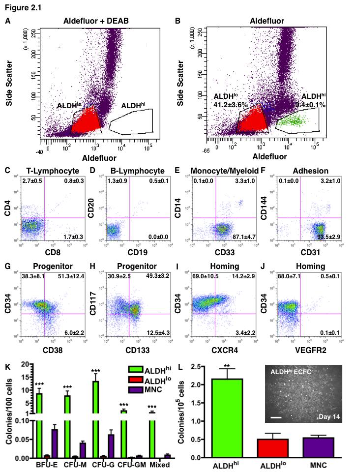
85 63
86 64 Table 2.1 Cell surface marker expression for the hematopoietic (gated CD45+) component of human UCB MNC, ALDH hi cells, and ALDH lo cells. Lineage Markers (%) MNC ALDH hi ALDH lo T-Lymphocyte CD4 47.6±3.2 a 3.5±0.6** b 47.8±2.1 a CD8 24.9±0.9 a 2.5±0.3** b 26.1±3.5 a B-Lymphocyte CD ±1.8 a 0.6±0.1** b 21.1±1.4 a CD ±1.8 a 1.8±0.9** b 21.1±1.7 a Myeloid/ Monocyte CD ±0.9 a 90.4±4.8** b 1.4±0.3** c CD ±1.2 a 3.3±1.1** b 0.6±0.2** b Adhesion CD ±1.2 a 96.7±2.9* b 81.0±3.5 a CD ±2.0 a 3.2±1.0** b 4.9±2.9** b CD34 5.1±1.9 a 89.6±5.9** b 0.3±0.1 a Progenitor CD117/c-kit 15.9±2.8 a 80.2±4.9** b 12.5±5.3 a CD ±0.3 a 61.5±2.3** b 0.2±0.1 a Homing CXCR4 47.3±6.1 a 17.7±2.5* b 37.8±5.8 a KDR/VEGFR2 0.6±0.3 a 0.9±0.3 a 0.3±0.1 a Data represents mean ± SEM (n=4), different letters denote different expression patterns at *p<0.05, **p<0.01 compared to unpurified MNC.
87 65 Table 2.2. Cell surface marker expression for the non-hematopoietic (gated CD45 - ) component of human UCB MNC, ALDH hi cells, and ALDH lo cells. Lineage Markers (%) MNC ALDH hi ALDH lo Adhesion CD ±24.9 a 38.7±16.6 a 20.3±13.2 a CD ±12.5 a 17.8±11.3 a 6.5±3.8 a CD ±9.2 a 65.0±14.8* b 1.3±0.6** c Progenitor CD117/c-kit 22.1±9.8 a 64.9±12.4* b 12.8±3.5 a CD ±1.5 a 22.5±3.9* b 0.4±0.3 a Homing CXCR4 33.9±10.1 a 8.8±3.4* b 7.4±1.1* b KDR/VEGFR2 1.8±0.8 a 1.9±1.0 a 1.4±1.1 a Data represents mean ± SEM (n=4), different letters denote different expression patterns at *p<0.05, **p<0.01 compared to unpurified MNC.
88 UCB ALDH hi cells demonstrated a pro-angiogenic transcription profile Affymetrix microarray identified 1253 transcripts with significantly increased (*p<0.05) expression in 3 UCB ALDH hi cell samples compared to sample-matched ALDH lo cells. These transcripts were first filtered to include unique genes translating to plasma membrane bound (196 hits) or extracellular (108 hits) locations, and subsequently filtered for documented angiogenic biological functions (Tables 2.3 and 2.4). As a control for the selection of cells with high ALDH-activity, ALDH1A1 mrna expression was increased 42-fold in ALDH hi vs ALDH lo cells (p=2.08x10⁻⁵ ). Furthermore, significantly increased CD34, PROM1, and KIT mrna expression in ALDH hi cells (Table 2.3) correlated with increased CD34, CD133, and c-kit/cd117 cell surface protein expression previously observed by FACS (Figure 2.1, Table 2.1). Transcripts associated with early myeloid progenitor differentiation (FLT3, CSF3R, CD33), integrin-mediated cell adhesion (ITGA9, ITGAV, ITGA2B), and vascular cell signaling (TIE1, TIE2, FGFR1, endoglin) were also highly expressed by UCB ALDH hi cells (Table 2.3). Interestingly, transcripts for secreted cytokines associated with vascular functions (ANGPT1, FGF16, VEGFA), immune cell migration (IL-18, IL-1b, IL-8, CSF1, CXCL2), and modulators of EGF and IGF signaling (EREG, AREG, HBEGF, IGFBP7) were also increased in UCB ALDH hi cells (Table 2.4). Collectively, these data suggested that UCB ALDH hi cells possessed a pro-angiogenic transcription profile compared to ALDH lo cells.
89 67 Gene Symbol PROM1 CD34 KIT FLT3 IRAK3 Table 2.3 Angiogenesis-associated transcripts encoding cell surface proteins with increased expression in UCB ALDH hi versus ALDH lo cells. Common Names prominin 1, CD133 transmembrane sialomucin c-kit, CD117 FLT3 receptor tyrosine kinase, CD135 IL-1 receptor associated kinase 3 ITGA9 integrin, alpha 9 colony stimulating CSF3R factor receptor, CD114 TGF-beta-1-binding CD109 protein type 1 transmembrane CD302 C-type lectin receptor EGF-module receptor 2 EMR2 CD312 myeloid cell surface CD33 antigen tyrosine kinase with immunoglobulin-like TIE1 and EGF-like domain 1 Biological Function hematopoiesis, vascular development / remodeling hematopoiesis, vascular development / remodeling HPC survival, maintenance and migration, HPC marker myeloid progenitor proliferation / differentiation stimulates EC proliferation and angiogenesis cell-cell and cell-matrix adhesion granulocyte and neutrophil survival / differentiation cell division, differentiation, motility, adhesion phagocytosis, cell adhesion and migration mucin-like cell adhesion adhesion molecule on monomyelocytic cells regulation of angiogenesis and vasculogenesis ITGAV integrin, alpha V cell-cell, cell-matrix adhesion ENG FGFR1 ITGA2B TMEFF1 TEK/TIE2 endoglin, CD105 fibroblast growth factor receptor 1, CD331 integrin, alpha 2b transmembrane protein with EGF-like and follistatin-like domains tyrosine kinase with immunoglobulin-like and EGF-like domain 2 vascular system development and remodeling, EC migration angiogenesis, embryonic development, cell proliferation, differentiation cell-cell and cell-matrix adhesion unknown function during embryonic development angiogenesis, EC survival, proliferation, migration, adhesion Molecular Function FC vs Alo p-value vs Alo MAPK and Akt signaling x10-6 adherence to BM matrix and cells x10-5 tyrosine-protein kinase signaling x10-6 fms-related tyrosine kinase signaling x10-5 regulates IL-1 receptor signaling x10-6 α9/β1 dimer binds VCAM x10-6 GCSF receptor signaling x10-3 heterodimer with TGFB1 receptor x10-6 carbohydrate binding / adhesion x10-4 G-protein coupled receptor signaling x10-5 tyrosine kinase, binds sialic acid x10-5 modulates TEK / TIE2 binding to angiopoietins x10-3 αv/β3 binds vitronectin x10-3 complexes with TGF-beta, activin, BMP molecules x10-3 tyrosine kinase activity, binds FGF molecules x10-3 α2b/β3 binds fibronectin x10-4 modulates Nodal and BMP signaling x10-3 tyrosine kinase activity, binds angiopoietins x10-3 FC, fold change; BMP, bone morphogenic protein; EC, endothelial cell, EGF, epidermal growth factor; GCSF, granulocyte colony stimulating factor, HPC, hematopoietic progenitor cell; IL-1, interleukin 1; MCSF, macrophage colony stimulating factor; TGF, transforming growth factor; VCAM, vascular cell adhesion molecule
90 68 Table 2.4 Angiogenesis-associated transcripts encoding secreted cytokines with increased expression in UCB ALDH hi versus ALDH lo cells. Gene Symbol Common Name Biological Function Molecular Function FC vs Alo p-value vs Alo ANGPT1 angiopoietin-1 vascular development / remodeling, angiogenesis TIE2 receptor activation x10-5 IL18 interleukin 18 (IFNγinducing molecule) inflammation, EC migration, angiogenesis IL 18 receptor activation x10-6 IL1β interleukin-1 beta inflammation, EC differentiation / proliferation IL-1β receptor activation x10-4 EREG epiregulin EC proliferation, angiogenesis EGF receptor activation x10-4 IL 8 (CXCL8) interleukin 8 chemoattraction of angiogenic and inflammatory cells CXCR1 and CXCR2 activation x10-5 AREG amphiregulin EC proliferation, angiogenesis EGF receptor activation x10-4 IGFBP7 insulin-like growth factor binding protein 7 implicated in tumor angiogenesis binds IGF1 and IGF x10-4 BMP6 bone morphogenic protein 6 EC development, proliferation, angiogenesis BMP receptor activation x10-3 CSF1 colony stimulating factor 1, M-CSF macrophage differentiation, migration and function M-CSF receptor activation x10-4 FGF16 fibroblast growth factor 16 angiogenic factor during development FGFR1 and FGFR2 activation x10-3 FBLN5 fibrulin 5 EC adhesion during vascular development / remodeling vascular ligand for integrins x10-3 CXCL2 chemokine (C-X-C motif) ligand 2 chemoattraction of angiogenic and inflammatory cells CXCR2 activation x10-3 HBEGF heparin-binding EGFlike growth factor endothelial cell proliferation, angiogenesis EGF receptor activation x10-3 VEGFA vascular endothelial growth factor A regulation of angiogenesis and vasculogenesis VEGFR1 and R2 activation x10-3 FC, fold change; EC, endothelial cell
91 UCB ALDH hi cells augment the survival of HUVEC in vitro To assess the paracrine functions of ALDH-purified UCB cells on endothelial cell survival and proliferation in vitro, 40x10 3 HUVEC were seeded into transwell co-cultures for 72 hours using a 5μm pore hanging insert containing 10 5 UCB ALDH hi or ALDH lo cells (or basal media control) suspended above HUVEC. Under optimal conditions containing growth factors (GF + ) and serum, non-contact co-culture with ALDH lo or ALDH hi cells did not impact the 5-fold expansion of HUVEC observed over 72h (Figure 2.2A,E). Conversely, in GF - /serum-free conditions with or without ALDH lo cell supplementation, HUVEC invariably died within 72 hours (Figure 2.2B,E). In contrast, co-culture with ALDH hi cells under GF - /serum-free conditions promoted HUVEC viability for 72 hours (*p<0.05, Figure 2.2B,E). Under GF + /serum-free conditions, ALDH hi cell co-culture promoted a modest expansion (1.7-fold) of HUVEC (67.4±7.1x10 3 cells), compared to >3-fold HUVEC loss with basal media (12.4±2.6x10 3 cells) or ALDH lo cell supplementation (14.4±2.8x10 3 cells, *p<0.05, Figure 2.2C,E). Under GF - /serum + conditions, ALDH lo or ALDH hi co-culture did not significantly alter HUVEC numbers (Figure 2.2D,E). Thus, non-contact co-culture with human ALDH hi cells promoted HUVEC survival under GF - /serum-free conditions. These effects were consistent with the pro-survival stimulus in HUVEC provided by VEGF-A treatment 44, a central pro-angiogenic cytokine with increased mrna-expression in UCB ALDH hi cells (Table 2.4).
92 70 Figure 2.2 UCB ALDH hi cells promote endothelial cell survival under growth factor and serum-free conditions. (A-D) Representative photomicrographs of HUVEC density after 72h co-culture with UCB ALDH lo or ALDH hi cells in hanging transwells with or without growth factor or serum supplementation. Scale bar = 200μm. (E) Non-contact co-culture with human UCB ALDH hi cells promoted the survival of HUVEC under growth factor-free, serum-free conditions (n=5). Data are expressed as mean ± SEM, *p<0.05.
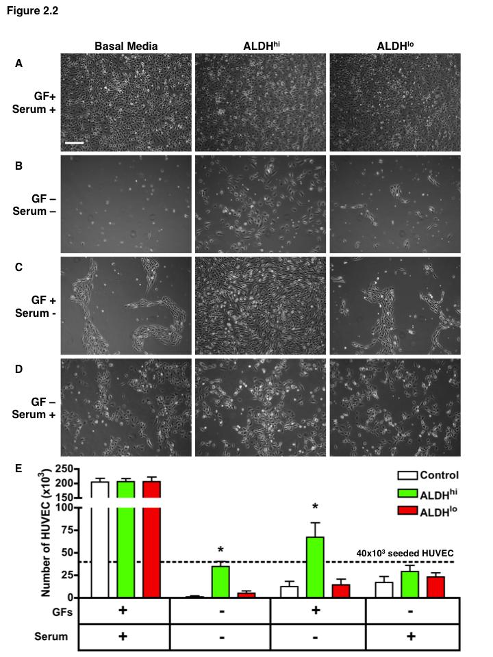
93 71
94 UCB ALDH hi cells augment tube-like cord formation by HUVEC in vitro HUVEC were cultured alone at 50x10 3 per well, or co-cultured with 20x10 3 ALDH hi or ALDH lo cells on growth factor reduced Matrigel. HUVEC established partially completed cord networks within 24 hours (Figure 2.3A). Although co-culture of HUVEC with UCB ALDH lo cells did not significantly alter cord formation, supplementation with ALDH hi cells increased the number of HUVEC tube-like cord formed at 24 hours after plating (Figure 2.3B and C). Although UCB ALDH hi cells established ECFC in liquid culture (Figure 2.1L, inset), UCB ALDH lo or ALDH hi cells seeded alone on growth factor reduced Matrigel did not spontaneously align to form cord networks (Figure 2.3D), and tube-like cord stability of HUVEC was not enhanced by co-culture with UCB ALDH lo or ALDH hi at 72 hours (Figure 2.3E-H). Enumeration of completed branch points at 24 and 48 hours revealed that co-culture with UCB ALDH hi cells augmented cord formation by HUVEC compared to media controls (n=6, *p<0.05, Figure 2.3H). Therefore, co-culture with UCB ALDH hi cells promoted the cord-forming function of HUVEC in vitro.
95 73 Figure 2.3 UCB ALDH hi cells augmented tube-like cord formation by HUVEC. Representative photomicrographs of tubule formation by HUVEC after 24 (A-C) and 72 (E-G) hours co-culture with ALDH lo or ALDH hi cells or media control. Scale bar = 500μm. (D) ALDH hi cells plated without HUVEC did not form cord structures. (H) Coculture with ALDH hi cells augmented the formation of complete tubule networks formed by HUVEC at 24 and 48 hours, but did not augment tubule stability after 72 hours coculture. Data are expressed as mean ± SEM, *p<0.05.
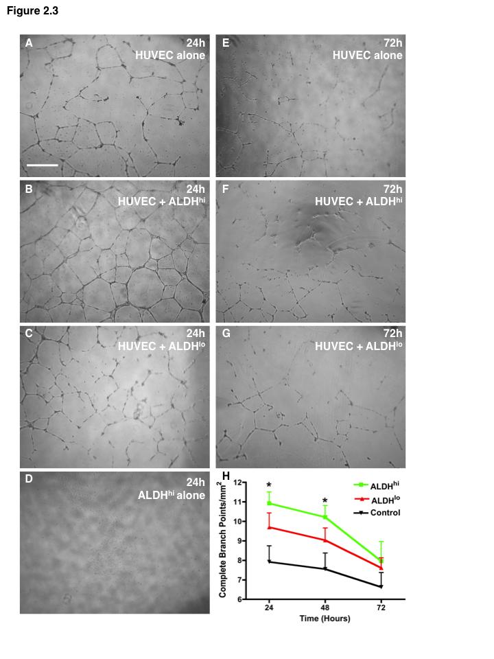
96 74
97 Transplanted UCB ALDH hi cells enhanced the recovery of hindlimb perfusion To determine whether ALDH-purified human UCB cells could support blood vessel regeneration after intravenous transplantation in vivo, we induced acute unilateral hindlimb ischemia in NOD/SCID mice by right femoral artery ligation with complete excision of a 0.5cm section of the femoral artery and vein. Blood flow was assessed by LDPI and the perfusion ratio (PR) in the ischemic versus control limb was decreased >10-fold (PR=0.08±0.02) post-surgery confirming consistent induction of unilateral hindlimb ischemia (Figure 2.4). Within 24 hours of surgery mice were tail vein injected with PBS vehicle control, unsorted UCB MNC, ALDH hi cells, or ALDH lo cells. The representative LDPI shown in Figure 2.4 documents the recovery of limb perfusion after transplantation. PBS-injected control mice showed recovery of PR from 0.07±0.01 after surgery to 0.36±0.05 by day 7 post-surgery without subsequent improvement at later time points (Figure 2.4A,E). This baseline recovery of perfusion was sufficient to prevent excess morbidity or limb loss in PBS transplanted mice. Mice transplanted with 20x10 6 MNC or 10x10 6 ALDH lo cells showed equivalent recovery of limb perfusion similar compared to PBS controls (Figure 2.4B,D-E). In contrast, mice transplanted with fold fewer ALDH hi cells (2x10 5 ) showed increased perfusion by day 14 posttransplantation (PR=0.58±0.07) compared to all other treatments and augmented perfusion was maintained for 28 days post-transplantation (PR=0.64±0.06, **p<0.01, Figure 2.4C,E). Thus, intravenous-transplantation of human UCB ALDH hi cells improved the limb perfusion in mice with acute unilateral hindlimb ischemia.
98 76 Figure 2.4 Transplanted UCB ALDH hi cells augmented perfusion in ischemic hindlimbs. Representative LDPI following right femoral artery ligation and tail vein injection of (A) PBS vehicle control (n=7), (B) 20x10 6 unsorted UCB MNC (n=7), (C) 2x10 5 ALDH hi cells (n=7), or (D) 10x10 6 ALDH lo cells (n=7). Numbers in lower left of each image indicate perfusion ratio of the ischemic versus the non-ischemic hindlimb. (E) Compared to all other treatments, transplantation of UCB ALDH hi cells promoted improved recovery of perfusion in ischemic mouse hindlimbs by day 14 and augmented perfusion was maintained to day 28. Data are expressed as mean ± SEM, **p<0.01.
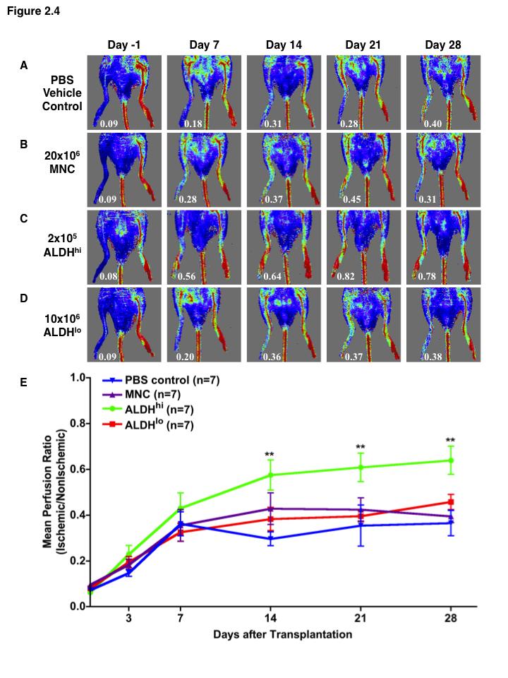
99 77
100 Transplanted UCB ALDH hi cells increased capillary density in the ischemic limb To address the recovery of blood vessels in the ischemic hindlimb, sections from the adductor muscle at the site of artery ligation were stained for both murine CD31 + capillary (Figure 2.5A-C) and vwf + blood vessel (Figure 2.5D-F) density. Compared to the non-ischemic limb at day 28 (65.3±6.1 CD31 + /mm 2, 8.1±1.2 vwf + /mm 2 ), PBSinjected mice showed significant reduction (*p<0.05) of both CD31 + capillaries (53.5±4.8/mm 2, Figure 2.5G) and vwf + blood vessels (5.5±1.4/mm 2, Figure 2.5F) within the ischemic hindlimb. However, mice transplanted with UCB ALDH hi cells (73.7±10.5/mm 2 ) had significantly more CD31 + capillaries compared to mice transplanted with ALDH lo cells (61.1±12.4/mm 2, *p<0.05) or PBS (65.3±6.1/mm 2,**p<0.01, Figure 2.5G). Similarly, mice transplanted with ALDH hi cells (8.8±1.8/mm 2 ) showed higher vwf + blood vessel density than mice injected with ALDH lo cells (5.8±1.2/mm 2, *p<0.05), or MNC (5.8±0.9/mm 2, *p<0.05), or PBS-injected controls (8.1±1.2 vwf + /mm 2, ***p<0.001, Figure 2.5H). Despite only partial recovery of perfusion, CD31 + and vwf + vessel density in the ischemic limbs of ALDH hi cell-treated mice was equivalent to the vessel density of the non-ischemic control limb at day 28 (Figure 2.5G,H). Thus, UCB ALDH hi cell transplantation increased both CD31 + capillary and vwf + blood vessel density in the ischemic hindlimb after femoral artery ligation.
101 79 Figure 2.5. Transplanted UCB ALDH hi cells augmented blood vessel density within ischemic hindlimbs. Representative photomicrographs of adductor muscle sections from the ischemic and non-ischemic limbs of mice injected with PBS or UCB ALDH hi cells stained for (A-C) CD31 + capillaries or (D-F) vwf + blood vessels at day 28 post-transplantation. Scale bar = 100μm. Compared to all other treatments, (G) CD31 + capillary density, and (H) vwf + blood vessel density was increased in the ischemic limb of mice transplanted with human UCB ALDH hi cells. Data are expressed as mean ± SEM, *p<0.05, **p<0.01, ***p<0.001 as determined by two way ANOVA and Bonferroni post-tests to compare multiple means.
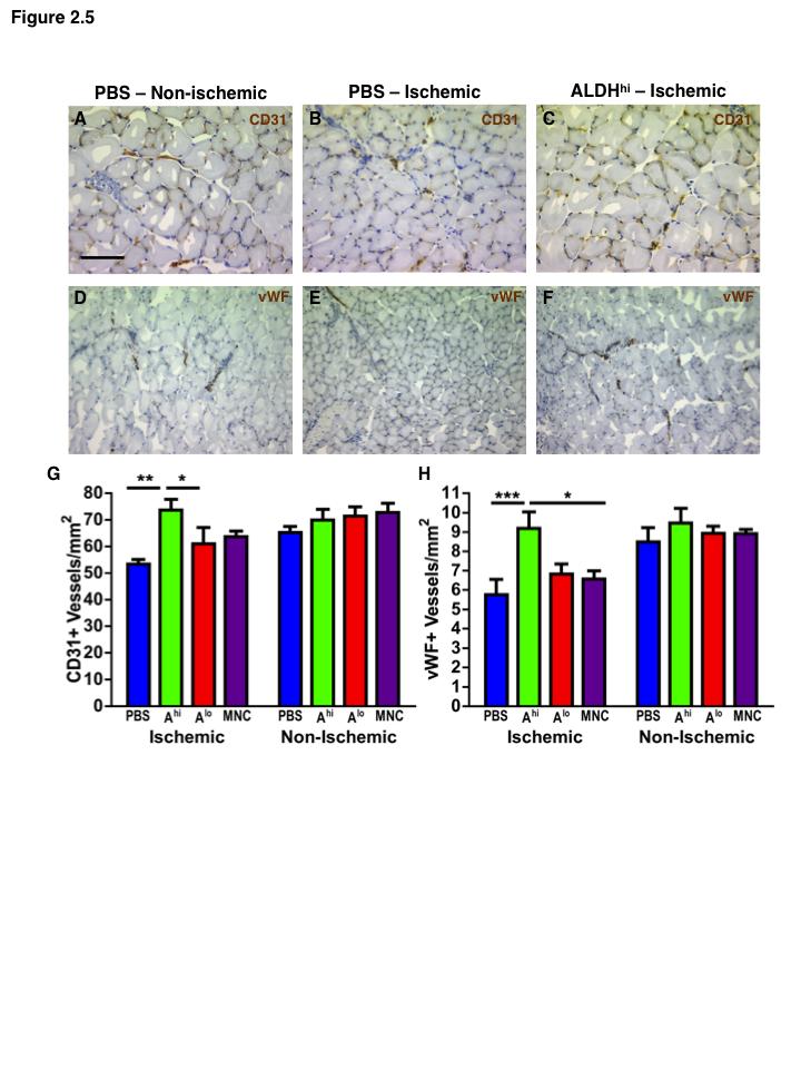
102 80
103 Transplanted UCB ALDH hi cells recruited to the ischemic muscle at low frequency In order to correlate the recovery of perfusion with human cell engraftment, murine BM and adductor muscle were collected at 3, 7, and 28 days after transplantation, and analyzed for the presence of human CD45 + or HLA-A,B,C + cells by flow cytometry. Despite consistent human cell hematopoietic chimerism in the BM of mice transplanted with ALDH hi cells (Figure 2.6A,C) or MNC (Figure 2.6C), human cells were not significantly detected in the adductor muscle of ischemic limbs by flow cytometry (Figure 2.6B,D) at day 28. In addition, mice processed for engraftment at earlier time points showed no evidence of human cells in the ischemic limb at 3 (n=3) or 7 days (n=3) post-transplantation by FACS. Therefore, we repeated engraftment experiments using identical cell doses in the GUSB-deficient NOD/SCID/MPSVII recipient to permit the detection of transplanted human cells at single cell resolution. At 3 and 7 days posttransplantation, the detection of GUSB expressing cells confirmed the recruitment of human cells to the ischemic hindlimbs of mice that received MNC (n=4, Figure 2.6E-F) or ALDH hi cells (n=4, Figure 2.6H-I). However, human cells were not detected in the ischemic limbs of mice that received ALDH lo cells (n=4). Although transplanted cells were never detected in the non-ischemic limb, engrafted MNC and ALDH hi cells were observed in ischemic muscle sections surviving out to 28 days (Figure 2.6G, J). Staining for GUSB-activity combined with murine vwf showed engraftment of single human cells between muscle fibres (Figure 2.6K) and in association with the connective tissue surrounding blood vessels (Figure 2.6L). Also, the majority of GUSB + human cells surviving at day 28 co-stained for CD45 (Figure 2.6M). Despite low frequency engraftment in the ischemic region, early recruitment of ALDH hi progenitor cells to the site of ischemic injury was sufficient to augment blood vessel density and improve limb perfusion.
104 82 Figure 2.6 Transplanted human UCB ALDH hi cells recruited to the ischemic hindlimb. Representative flow cytometry plots illustrating the detection of human cells (CD45 + /HLA-A,B,C + ) cells within (A) the bone marrow and (B) ischemic hindlimbs of mice transplanted with UCB ALDH hi cells. (C,D) Despite engraftment of human cells in the mouse bone marrow, human cells were rarely detected in the ischemic hindlimb by flow cytometry. Representative photomicrographs showing the detection of transplanted human cells (red) in the adductor muscle of the ischemic hindlimb of mice transplanted with (E-G) UCB MNC or (H-J) UCB ALDH hi cells at 3, 7 and 28 days posttransplantation. GUSB + human cells (arrows) were detected in the ischemic limb of mice injected with UCB MNC or ALDH hi cells, but not in mice injected with UCB ALDH lo cells. Co-staining for GUSB-activity with murine vwf at day 28 showed (K) engraftment of single GUSB + cells between muscle fibres, and (L) associated with vwf + blood vessels. (M) GUSB + human cells surviving at day 28 co-stained for CD45. Scale bars = 100μm.
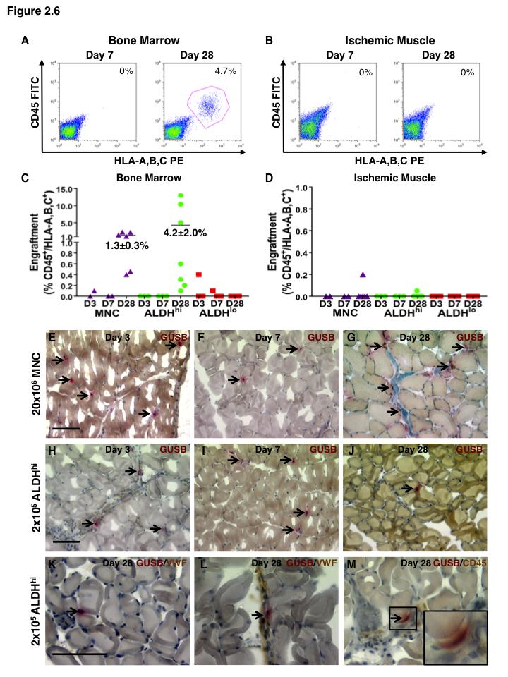
105 83
106 Sublethal irradiation did not improve perfusion in the ischemic hindlimb The requirement for preparative irradiation is an important consideration in the design of clinically applicable cell therapies for ischemic diseases. In an attempt to improve human cell engraftment in the ischemic limb, transplantation experiments were repeated after sublethal irradiation (300cGy) administered as a means to reduce xenorejection by residual innate immunity or NK-cell activity in the NOD/SCID model. Surprisingly, in mice receiving UCB ALDH hi cells, perfusion at early time points (days 3, 7, and 14) was similar for all transplanted groups (Figure 2.7A-E). Although perfusion improved in the ALDH hi cell transplanted cohort at day 21, perfusion was reduced to baseline by day 28 (Figure 2.7E), indicating that only a transient improvement in limb perfusion was observed. Compared to mice transplanted without preparative irradiation (Figure 2.4), irradiated mice receiving an equal dose of UCB ALDH hi cells actually showed delayed recovery of perfusion at day 14 (PR 300cGy = 0.40±0.05 vs PR 0cGy = 0.58±0.07). Nonetheless, at day 28, irradiated mice injected with ALDH hi cells still showed improved vwf + blood vessel density (Figure 2.8) similar to non-irradiated mice. Irradiated mice also showed enhanced recruitment of ALDH hi cells to the BM at day 7 (1.3±0.2%, Figure 2.9A,C), resulting in extensive hematopoietic repopulation by day 28 (44.7±2.0%, Figure 2.9A,C). Unexpectedly, human cells in the ischemic limb were not detected by FACS or GUSB staining at day 7 (Figure 2.9B,D,E), and were only rarely detected at day 28 (Figure 2.9F). Thus, irradiation prior to ALDH hi cells transplantation did not augment engraftment or the recovery of perfusion in the ischemic limb.
107 85 Figure 2.7 Transplanted UCB ALDH hi cells induced transient recovery of perfusion in the ischemic limbs of mice that received preparative irradiation. Representative LDPI following right femoral artery ligation, 300 cgy sublethal preparative irradiation to suppress immune rejection, and tail vein injection with (A) PBS vehicle control (n=6), (B) 20x10 6 unsorted UCB MNC (n=7), (C) 2x10 5 UCB ALDH hi cells (n=8), or (D) 10x10 6 ALDH lo cells (n=8), and monitored for 28 days. Numbers in lower left of each LDPI image indicate perfusion ratio of the ischemic versus the nonischemic hindlimb from ankle to toe. (E) Summary of mean perfusion ratios in all transplanted mice as described above. Compared to all other treatments, transplantation of UCB ALDH hi cells promoted a transient recovery of perfusion in ischemic mouse hindlimbs at day 21 post-transplantation. Data are expressed as mean ± SEM, **p<0.01.
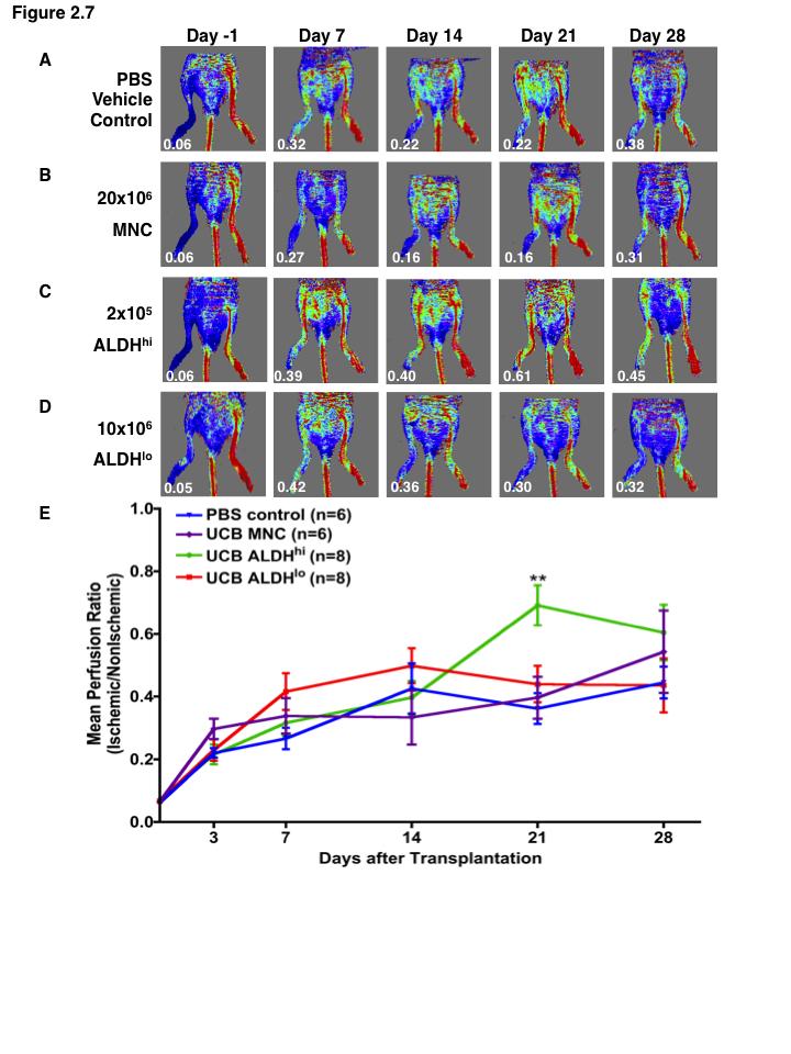
108 86
109 87 Figure 2.8 Transplanted UCB ALDH hi cells augmented blood vessel density in the ischemic hindlimb of irradiated mice. Representative photomicrographs of adductor muscle sections from the non-ischemic and ischemic hindlimbs of mice injected with (A, B) PBS or (C) UCB ALDH hi cells or (D) UCB ALDH lo cells stained for vwf + blood vessels at day 28 post-transplantation. Scale bar = 100µm. (E) Summary of vwf + blood vessel density for each transplanted cohort at 28 days post-transplantation. Compared to all other treatments, blood vessel density was increased in the ischemic limb of mice transplanted with human UCB ALDH hi cells. Data are expressed as mean ± SEM, ***p<0.001.
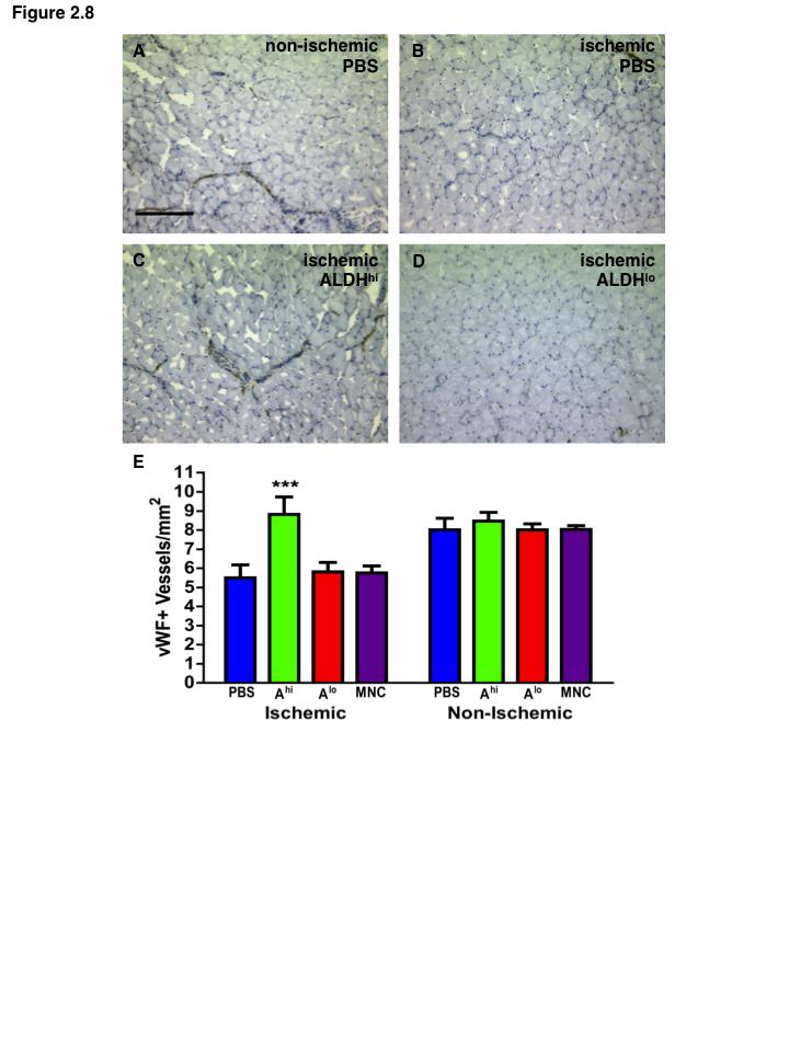
110 88
111 89 Figure 2.9 Transplanted UCB ALDH hi cell showed reduced engraftment in the ischemic limb and increased engraftment in the bone marrow of irradiated mice. (A,B) Representative flow cytometric detection of human cells in (A) bone marrow or (B) ischemic muscle (B) of mice transplanted with UCB ALDH hi cells after sublethal (300cGy) preparative irradiation. (C-D) Summary of human cell (CD45 + /HLA-A,B,C + ) engraftment in (C) the bone marrow or (D) ischemic muscle of all transplanted mice at 7 and 28 days post transplantation. (E, F) Representative photomicrographs showing the detection of transplanted human cells (red) in the adductor muscle of the ischemic hindlimb of mice transplanted with UCB ALDH hi cells at 7 and 28 days posttransplantation. GUSB + human cells (arrows) were detected in the ischemic limb of mice at 28 days post-transplantation, Scale bar = 100μm.
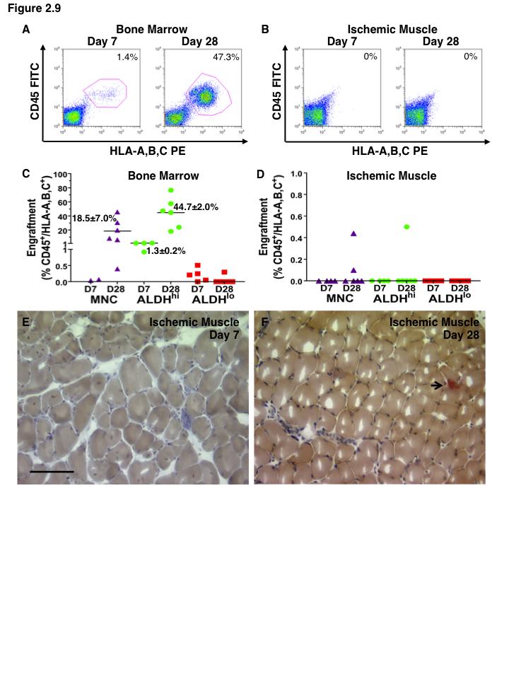
112 90
113 Discussion In the present study we demonstrate that ALDH-expressing cells from human UCB, representing <0.5% of total UCB cells and primarily comprised of primitive myeloid progenitor cells, demonstrated increased expression of mrna transcripts for several angiogenic cytokines, and stimulated the survival and tubule forming functions of EC during co-culture in vitro. Intravenous transplantation of UCB ALDH hi cells also augmented recovery of limb perfusion induced by femoral artery ligation and transection. Although the engraftment of human cells within ischemic muscle was infrequent, UCB ALDH hi cells specifically recruited to the ischemic limb within 3 days of transplantation, and stimulated the recovery of blood vessel and capillary density. Furthermore, ALDH hi cells function within the pancreas to promote islet revascularization after transplantation into streptozotocin-treated mice 45,46. Collectively, these data suggest that UCB ALDH hi progenitor cells represent a readily available population of pro-angiogenic cells for the development of cellular therapies to promote endogenous revascularization. The UCB ALDH hi population was significantly enriched for human hematopoietic progenitor function. Approximately 1 in 4 ALDH hi cells demonstrated multipotent CFU capacity in methylcellulose cultures. UCB ALDH hi cells also showed increased expression of primitive cell surface markers (CD34, c-kit/cd117, and CD133), and >90% of UCB ALDH hi cells expressed the early myeloid marker CD33, indicating a substantial enrichment of early myeloid progenitors. At the same time, UCB ALDH hi cells were depleted of mature T- and B-lymphocytes as well as CD14 expressing monocytes. This is notable as CD14 + monocytes have been specifically implicated in the paracrine support of angiogenesis 47. However, transplantation of UCB ALDH hi cells, containing only 3% CD14 + monocytic cells significantly improved perfusion and blood vessel density in the ischemic limb, whereas transplantation of mice with 100-fold higher doses of unsorted MNC, containing over 17% CD14 + monocytes, failed to promote recovery from hindlimb ischemia. Recent studies have demonstrated that murine common myeloid progenitors preferentially differentiate into pro-angiogenic monocytes
114 92 that support neovessel formation in vivo 48. Although UCB ALDH hi cells may be capable of differentiating into monocytes in vivo, our data indicates that primitive UCB ALDH hi myeloid progenitors play a significant role in support of angiogenesis in vivo. UCB ALDH hi cells also possessed enriched ECFC capacity in vitro. In contrast to the dominant myeloid progenitor composition, ECFC were infrequent at approximately 1 ECFC in 50,000 ALDH hi cells. Although this represents a significant ECFC enrichment from unpurified UCB MNC 9, it is unlikely that the few ECFC within 40x10 3 ALDH hi cells make a major contribution towards the maintenance of HUVEC survival and the induction of tubule forming function that we observed under growth factor and serum starved conditions in vitro. However, paracrine contributions of the non-hematopoietic component of the ALDH hi population should not be overlooked in vivo. Approximately 7% of the UCB ALDH hi population were CD45 -, and these cells demonstrated high coexpression of primitive endothelial precursor markers (CD34 and CD133). Therefore, we propose that UCB ALDH hi cells represents a heterogeneous mixture of primitive hematopoietic and non-hematopoietic progenitors that may act in synergy to generate a supportive microenvironment to promote EC survival and revascularization function. After intravenous transplantation, UCB ALDH hi cells induced stable recovery from acute ischemic injury within 2 weeks of transplantation into mice with unilateral hindlimb ischemia. However, permanent engraftment of ALDH hi cells within the ischemic muscle was below the threshold of detection by flow cytometry. Notably, transplantation of fold greater cell doses of unpurified UCB MNC or ALDH lo cells also showed little engraftment by FACS. Using NOD/SCID/MPSVII mice, a unique cell tracking model capable of detecting GUSB-expressing human cell at the single cell level, we demonstrate that intravenous-injected UCB ALDH hi cells specifically recruited into the ischemic limb at 3 and 7 days after transplantation, and these cells survived in ischemic muscle tissue, albeit at low frequencies, for up to 28 days. Nonetheless, this low frequency engraftment of ALDH hi cells in the ischemic region at early time points was sufficient to augment the revascularization of ischemic muscle indicating a vascular stabilization was mediated by the ALDH hi cells.
115 93 Recent models have proposed a transient role for pro-angiogenic hematopoietic cells in the stabilization of injured vasculature, and in the activation and recruitment of vessel resident endothelial precursors to the sprouting vessel branch 8. In support of this concept we show that sublethal irradiation prior to transplantation of ALDH hi cells reduced recruitment of human cells to the ischemic region, increased engraftment of ALDH hi cells in the murine BM, and delayed the functional recovery of perfusion. Therefore, we propose that early recruitment of ALDH hi cells to the ischemic region is critical for vascular recovery, allowing ALDH hi cells to release pro-angiogenic stimuli that activate an endogenous program for collateral vessel formation resulting in improved limb perfusion. Thus, strategies to improve the efficiency ALDH hi progenitor cell delivery, or to prolong survival at or near the site of ischemic injury, is expected to further augment vascular regeneration via paracrine or contact dependent mechanisms Conclusion and Summary Strategies that purify pro-angiogenic cell subtypes from alternate human sources are required to improve the efficacy of human cell therapy trials 26. Our studies outline several important caveats relevant to the development of cellular therapies to treat ischemic diseases. First, we have identified a mixed population of hematopoietic/myeloid and nonhematopoietic/endothelial ALDH hi progenitor cells from human UCB as a readilyavailable and clinically applicable cell population with potent pro-angiogenic function. Second, the potential use of allogeneic UCB cells will open new avenues towards clinical cell based therapies for ischemic disease, as mounting evidence indicates the potential for progenitor deletion and dysfunction of autologous BM cells in patients with severe diabetes and cardiovascular disease 2, Third, we have shown that neither high-level nor permanent engraftment of human cells were necessary to mediate augmentation of perfusion and improved vessel density in vivo. In an allogeneic context, such as intramuscular delivery of UCB ALDH hi cells in patients with ischemic heart disease or critical limb ischemia 49,50, future clinical transplantation strategies employing UCB may require only short-term immune suppression to initiate potent revascularization. Future studies will increase our understanding of the cellular constituents and paracrine
116 94 pathways that modulate regenerative angiogenesis, and will lead to novel therapies to improve tissue vascularization during ischemic diseases.
117 References 1. Dotsenko O. Stem/Progenitor cells, atherosclerosis and cardiovascular regeneration. Open Cardiovasc Med J. 2010;4: Bozdag-Turan I, Turan RG, Turan CH, et al. Relation between the frequency of CD34 bone marrow derived circulating progenitor cells and the number of diseased coronary arteries in patients with myocardial ischemia and diabetes. Cardiovasc Diabetol. 2011;10: Dimmeler S, Zeiher AM, Schneider MD. Unchain my heart: the scientific foundations of cardiac repair. J Clin Invest. 2005;115: Jin DK, Shido K, Kopp HG, et al. Cytokine-mediated deployment of SDF-1 induces revascularization through recruitment of CXCR4+ hemangiocytes. Nat Med. 2006;12: Rafii S, Lyden D. Therapeutic stem and progenitor cell transplantation for organ vascularization and regeneration. Nat Med. 2003;9: Asahara T, Murohara T, Sullivan A, et al. Isolation of putative progenitor endothelial cells for angiogenesis. Science. 1997;275: Peichev M, Naiyer AJ, Pereira D, et al. Expression of VEGFR-2 and AC133 by circulating human CD34(+) cells identifies a population of functional endothelial precursors. Blood. 2000;95: Richardson MR, Yoder MC. Endothelial progenitor cells: quo vadis? J Mol Cell Cardiol. 2011;50: Yoder MC, Mead LE, Prater D, et al. Redefining endothelial progenitor cells via clonal analysis and hematopoietic stem/progenitor cell principals. Blood. 2007;109: Capoccia BJ, Shepherd RM, Link DC. G-CSF and AMD3100 mobilize monocytes into the blood that stimulate angiogenesis in vivo through a paracrine mechanism. Blood. 2006;108: Estes ML, Mund JA, Mead LE, et al. Application of polychromatic flow cytometry to identify novel subsets of circulating cells with angiogenic potential. Cytometry A. 2010;77: Urbich C, Heeschen C, Aicher A, et al. Relevance of monocytic features for neovascularization capacity of circulating endothelial progenitor cells. Circulation. 2003;108: Grunewald M, Avraham I, Dor Y, et al. VEGF-induced adult neovascularization: recruitment, retention, and role of accessory cells. Cell. 2006;124: Kaplan RN, Riba RD, Zacharoulis S, et al. VEGFR1-positive haematopoietic bone marrow progenitors initiate the pre-metastatic niche. Nature. 2005;438:
118 Kaplan RN, Rafii S, Lyden D. Preparing the "soil": the premetastatic niche. Cancer Res. 2006;66: Rafii S, Lyden D. Cancer. A few to flip the angiogenic switch. Science. 2008;319: Urbich C, Dimmeler S. Endothelial progenitor cells: characterization and role in vascular biology. Circ Res. 2004;95: Vasa M, Fichtlscherer S, Aicher A, et al. Number and migratory activity of circulating endothelial progenitor cells inversely correlate with risk factors for coronary artery disease. Circ Res. 2001;89:E Schatteman GC, Hanlon HD, Jiao C, et al. Blood-derived angioblasts accelerate blood-flow restoration in diabetic mice. J Clin Invest. 2000;106: Tepper OM, Carr J, Allen RJ, Jr., et al. Decreased circulating progenitor cell number and failed mechanisms of stromal cell-derived factor-1alpha mediated bone marrow mobilization impair diabetic tissue repair. Diabetes. 2010;59: Janssens S, Dubois C, Bogaert J, et al. Autologous bone marrow-derived stemcell transfer in patients with ST-segment elevation myocardial infarction: doubleblind, randomised controlled trial. Lancet. 2006;367: Wollert KC, Meyer GP, Lotz J, et al. Intracoronary autologous bone-marrow cell transfer after myocardial infarction: the BOOST randomised controlled clinical trial. Lancet. 2004;364: Schachinger V, Erbs S, et al. Intracoronary bone marrow-derived progenitor cells in acute myocardial infarction. N Engl J Med. 2006;355: Tateishi-Yuyama E, Matsubara H, Murohara T, et al. Therapeutic angiogenesis for patients with limb ischaemia by autologous transplantation of bone-marrow cells: a pilot study and a randomised controlled trial. Lancet. 2002;360: Higashi Y, Kimura M, Hara K, et al. Autologous bone-marrow mononuclear cell implantation improves endothelium-dependent vasodilation in patients with limb ischemia. Circulation. 2004;109: Rosenzweig A. Cardiac cell therapy--mixed results from mixed cells. N Engl J Med. 2006;355: Putman DM, Bell GI, Hess DA. Blood-Derived ALDHhi Cells in Tissue Repair Regenerative Therapy Using Blood-Derived Stem Cells. In: Allan DSS, Strunk D, eds: Humana Press; 2012: Ingram DA, Mead LE, Tanaka H, et al. Identification of a novel hierarchy of endothelial progenitor cells using human peripheral and umbilical cord blood. Blood. 2004;104: Alves G, Wu WW, Wang G, et al. Enhancing peptide identification confidence by combining search methods. J Proteome Res. 2008;7:
119 Werner N, Wassmann S, Ahlers P, et al. Endothelial progenitor cells correlate with endothelial function in patients with coronary artery disease. Basic Res Cardiol. 2007;102: Werner N, Kosiol S, Schiegl T, et al. Circulating endothelial progenitor cells and cardiovascular outcomes. N Engl J Med. 2005;353: Fadini GP, Sartore S, Albiero M, et al. Number and function of endothelial progenitor cells as a marker of severity for diabetic vasculopathy. Arterioscler Thromb Vasc Biol. 2006;26: Jialal I, Devaraj S, Singh U, et al. Decreased number and impaired functionality of endothelial progenitor cells in subjects with metabolic syndrome: implications for increased cardiovascular risk. Atherosclerosis.211: Hess DA, Meyerrose TE, Wirthlin L, et al. Functional characterization of highly purified human hematopoietic repopulating cells isolated according to aldehyde dehydrogenase activity. Blood. 2004;104: Hess DA, Wirthlin L, Craft TP, et al. Selection based on CD133 and high aldehyde dehydrogenase activity isolates long-term reconstituting human hematopoietic stem cells. Blood. 2006;107: Cai J, Cheng A, Luo Y, et al. Membrane properties of rat embryonic multipotent neural stem cells. J Neurochem. 2004;88: Pearce DJ, Taussig D, Simpson C, et al. Characterization of cells with a high aldehyde dehydrogenase activity from cord blood and acute myeloid leukemia samples. Stem Cells. 2005;23: Storms RW, Green PD, Safford KM, et al. Distinct hematopoietic progenitor compartments are delineated by the expression of aldehyde dehydrogenase and CD34. Blood. 2005;106: Corti S, Locatelli F, Papadimitriou D, et al. Transplanted ALDHhiSSClo neural stem cells generate motor neurons and delay disease progression of nmd mice, an animal model of SMARD1. Hum Mol Genet. 2006;15: Capoccia BJ, Robson DL, Levac KD, et al. Revascularization of ischemic limbs after transplantation of human bone marrow cells with high aldehyde dehydrogenase activity. Blood. 2009;113: Hess DA, Craft TP, Wirthlin L, et al. Widespread nonhematopoietic tissue distribution by transplanted human progenitor cells with high aldehyde dehydrogenase activity. Stem Cells. 2008;26: Hofling AA, Vogler C, Creer MH, et al. Engraftment of human CD34+ cells leads to widespread distribution of donor-derived cells and correction of tissue pathology in a novel murine xenotransplantation model of lysosomal storage disease. Blood. 2003;101: Bhatia M, Bonnet D, Murdoch B, et al. A newly discovered class of human hematopoietic cells with SCID-repopulating activity. Nat Med. 1998;4:
120 Gerber HP, McMurtrey A, Kowalski J, et al. Vascular endothelial growth factor regulates endothelial cell survival through the phosphatidylinositol 3'-kinase/Akt signal transduction pathway. Requirement for Flk-1/KDR activation. J Biol Chem. 1998;273: Bell GI, Meschino MT, Hughes-Large JM, et al. Combinatorial Human Progenitor Cell Transplantation Optimizes Islet Regeneration Through Secretion of Paracrine Factors. Stem Cells Dev. 46. Bell GI, Putman DM, Hughes-Large JM, et al. Intrapancreatic delivery of human umbilical cord blood aldehyde dehydrogenase-producing cells promotes islet regeneration. Diabetologia. 47. Awad O, Dedkov EI, et al. Differential healing activities of CD34 + and CD14 + endothelial cell progenitors. Arterioscler Thromb Vasc Biol. 2006;26: Wara AK, Croce K, Foo S, et al. Bone marrow-derived CMPs and GMPs represent highly functional proangiogenic cells: implications for ischemic cardiovascular disease. Blood.118: Balber AE. Concise review: aldehyde dehydrogenase bright stem and progenitor cell populations from normal tissues: characteristics, activities, and emerging uses in regenerative medicine. Stem Cells.29: Perin EC, Silva G, et al. A randomized, controlled study of autologous therapy with bone marrow-derived aldehyde dehydrogenase bright cells in patients with critical limb ischemia. Catheter Cardiovasc Interv.78:
121 99 Chapter 3 3 Ex Vivo Expanded Hematopoietic Progenitor Cells Promote Endothelial Cell Survival and Ischemic Limb Revascularization Due to the relative rarity of ALDH hi cells from UCB or BM, I aimed in Chapter 3 to determine if it was possible to expand UCB-derived ALDH hi cells ex vivo to generate more cells for therapeutic applications without losing beneficial vascular regenerative effect. Therefore the objectives of this chapter 3 were two-fold: To optimize the ex vivo expansion of UCB ALDH hi hematopoietic cells under clinically relevant serum-free conditions and to characterize vascular regenerative functions of the expanded progenitor cells in vitro. To investigate the paracrine mechanisms by which expanded hematopoietic progenitor cells derived from UCB ALDH hi contribute to revascularization after direct intramuscular injection into the ischemic limb. The studies presented in this chapter used HMVEC as the EC source as minor adjustment compared to the HUVEC used in Chapter 2 to more closely model the phenotype of the EC thought be involved in angiogenesis and vascular regeneration. Vascular regeneration tends to affect the capillary microcirculation more so than a large vessel venous EC like HUVEC. The work presented in this chapter build directly upon the findings presented in Chapter 2. Specifically, it demonstrates that UCB ALDH hi cells can be efficiently expanded ex vivo without loss of vascular regenerative phenotype. Moreover, it introduces further refinement of the in vivo and in vitro models of vascular regeneration from those used in Chapter 2. The in vitro model used in this chapter introduces further mechanistic characterizing of the effects of HPC on cultured EC in terms of cell survival and death as measured by flow cytometry. Building on the in vivo murine hindlimb ischemia model from the previous chapter, here I present techniques to measure limb usage in addition to blood perfusion data. Quantitation of limb usage in addition to simple measurement of perfusion indicates that mice regain functional usage of the limb in addition to recovery of simple blood flow, strengthening the model. Furthermore, the
122 100 transplantation method was changed to intramuscular injection to more closely model the transplantation strategy from most PVD and CLI trials as well as to decrease delays in therapeutic benefit due to delayed and inefficient recruitment of cells transplanted intravenously. 3.1 Introduction Ischemia, caused by insufficient blood flow to a given tissue, represents a widespread burden on human health. Cardiovascular disease (CVD) is the leading cause of death in North America, and accounts for roughly one third of all deaths and related healthcare costs for patients with CVD and stroke is now estimated to be in excess of $300 billion in the US alone 1,2. Since Asahara et al. first described a role for circulating bone marrowderived cells in vascular regeneration, cell therapy has emerged as an important strategy to treat ischemic diseases 3-7. Thus, the development of novel cellular therapies for the management of acute cardiovascular diseases such as myocardial infarction and stroke, as well as chronic ischemic disease such as coronary and peripheral arterial disease, remain the focus of intense preclinical research. Early clinical trials building upon the preclinical promise shown by bone marrow and umbilical cord blood-derived endothelial and hematopoietic progenitor cells showed that transplantation in humans was well-tolerated and safe 6-8. Unfortunately, many of these therapies showed mixed results in improving cardiovascular disease outcomes, leading researchers to refocus on better understanding of the role of circulating progenitor cells in vascular repair 9. As such, several cell types have been implicated in vascular regeneration including multiple cell types from the hematopoietic lineage Recent cell-based approaches for vascular regeneration have focused on better functional characterization of purified cellular subpopulations and the mechanisms by which transplanted progenitor cells mediate vascular regeneration. Several groups have demonstrated that cells of the myeloid lineage recruit to areas of ischemia and support vessel regeneration though paracrine signaling to vessel-resident endothelial cells Our lab has demonstrated that transplantation of primitive hematopoietic progenitor cells with high aldehyde dehydrogenase (ALDH) activity, derived from human bone marrow
123 101 or umbilical cord blood, promote recovery from acute ischemic injury in immune deficient mice with hindlimb ischemia more effectively than unpurified, more heterogeneous populations (Chapter 2) 18,19. However, implementation of a cell-based therapy using UCB or BM-derived ALDH hi cells is hindered by the rarity of the ALDH hi cell population as it accounts for less than 1% of total MNC in BM or UCB samples. However, interest in developing strategies to increase the number of hematopoietic stem and progenitor cells for transplantation into patients with hematological disorders has led to identification of clinically-applicable culture strategies for hematopoietic progenitor expansion using defined, xeno-free conditions supplemented with stem cell factor (SCF), thrombopoietin (TPO) and fms-related tyrosine kinase 3 ligand (FLT3L) In this study we characterize serum-free ex vivo expansion of UCB-derived ALDH hi hematopoietic progenitor cells as a transplantable cell population for vascular regenerative cell therapies. We demonstrate that intramuscular transplantation of expanded ALDH hi cells can accelerate the recovery of perfusion and limb use after femoral artery ligation-induced ischemic injury. Even after a 20-fold increase in the total number of myeloid cell progeny available for transplantation, expanded HPC prevented capillary loss as effectively as freshly isolated UCB ALDH hi cells. In addition, the secretion of pro-angiogenic and pro-inflammatory effectors from expanded ALDH hi cells correlated with improved survival and tube formation by human microvascular endothelial cells (HMVEC) during co-culture in vitro. We propose that ex vivo-expanded myeloid progeny derived from the ALDH hi subset of UCB represent a readily available, clinically applicable population for future development of cell therapies to prevent limb loss in patients with severe peripheral artery disease. 3.2 Methods Purification of human umbilical cord blood cells based on aldehyde dehydrogenase activity Human umbilical cord blood samples were collected with informed consent immediately after scheduled, full-term Cesarean sections by venipuncture at the London Health Sciences Birthing Centre. The Human Research Ethics Committee at the University of
124 102 Western Ontario approved all studies. Cord blood samples were labeled with RosetteSep human cord blood progenitor cell enrichment tetrameric antibody complexes to prospectively deplete cells expressing lineage specific (Lin-) markers CD2, CD3, CD14, CD16, CD19, CD24, CD56, CD66b and glycophorin. After hypaque-ficoll centrifugation (Stem Cell Technologies, Vancouver, Canada) Lin MNC were further depleted of erythrocytes by ammonium chloride lysis and assayed for ALDH activity using Aldefluor reagent (Stem Cell Technologies) as previously described (Appendix 1, Chapter 2) 18,19,23,24. Fluorescence activated cell sorting was performed to isolate cells with low side scatter and high (ALDH hi ) versus low (ALDH lo ) ALDH activity. Before use in experiments, sorted cell population of >98% purity were washed in phosphate-buffered saline (PBS) to allow the efflux of Aldefluor substrate via ATP-binding cassettetransporters, leaving the cells unmodified for subsequent functional characterization and transplantation experiments Ex vivo expansion of hematopoietic progenitors from UCB ALDH hi cells After fluorescence activated cell sorting, UCB ALDH hi cells were plated on recombinant human fibronectin coated (1 hour coating with mg/ml fibronectin in PBS followed by sterile water rinse) dishes in X-vivo 15 (Lonza, Basel, Switzerland) supplemented with stem cell factor (SCF, 10ng/mL), Fms-related tyrosine kinase 3 ligand (FLT3LG, 10ng/mL), and thrombopoietin (TPO, 10ng/mL). All growth factors were from Life Technologies, Burlington, ON. Growth media was changed every 3 days and conditioned media was collected after 3 and 6 days of expansion. Ex vivo expanded hematopoietic progenitors (HPC) were harvested and enumerated by Trypan blue viability at day Assessment of ALDH-activity and cell surface marker expression after ex vivo expansion After 6 days ex vivo expansion, HPC were first assayed for the retention of high ALDH activity using Aldefluor reagent by flow cytometry to compare fluorescence levels to DEAB-inhibited control samples. Freshly isolated UCB ALDH hi cells and day 6 ex vivoexpanded HPC progeny were labeled with anti-human antibodies for lineage-specific
125 103 markers for T lymphocytes (CD3, Biolegend, San Diego, CA), macrophages (CD11b, BD Biosciences), monocytes (CD14, BD Bioscience), B lymphocytes (CD19, BD Bioscience), myeloid cells (CD33, BD Bioscience), and with antibodies for primitive progenitor-specific markers CD34 (Biolegend), CD38 (BD Biosciences), CD133 (Miltenyi Biotech, Auburn CA) and CD117 (BD Bioscience). Viable, 7-aminoactinomycin D (7AAD, BD Bioscience)-excluding cells were assessed for ALDH-activity and cell surface marker expression using an LSRII flow cytometer at the London Regional Flow Cytometry Facility and analyzed using FlowJo software (Treestar, Ashland, OR) Quantitation of hematopoietic colony forming capacity in vitro Freshly isolated UCB ALDH hi or 6-day ex vivo-expanded HPC were cultured in semisolid methylcellulose media (Methocult H4434, SCT) at limiting dilution. Hematopoietic colony formation at 14 days was enumerated by manual counting under bright field light microscopy, based on cell morphology identifying burst-forming units of erythrocytes (BFU-E), colony-forming units of granulocytes (CFU-G), macrophages (CFU-M), granulocytes and macrophages (CFU-GM), or multilineage mixed colonies (CFU-Mixed) containing cells of all 3 myeloid cell types listed above Microarray assessment of gene transcription after ex vivo expansion mrna was isolated from 6 day culture expanded HPC or freshly-isolated (uncultured) UCB ALDH hi cells using the mrneasy Mini kits (n=3, Qiagen, Mississauga, ON). Microarray analysis was performed in triplicate using human array chips (Affymetrix, Santa Carla, CA) at the London Regional Genomics Centre (London, ON). Genes differentially expressed by uncultured UCB ALDH hi cells versus ex vivo-expanded HPC were analyzed using the Partek Genomic Suite and angiogenesis associated-transcripts coding for secreted proteins were identified (Partek, Inc., St. Louis, MO).
126 HMVEC survival when exposed to HPC-secreted factors For endothelial cell growth and survival assays, 4x10 4 HMVEC (adult, dermal source) were cultured in endothelial basal media (EBM2-MV, Lonza) with or without growth factors (EGF, VEGF-A, FGF, IGF-1) and serum (5%) supplementation. To assess whether co-culture with expanded HPC could support HMVEC survival under serumstarved, growth factor depleted conditions, 8x10 4 HPC were seeded into porous (1µm) hanging transwells suspended above the HMVEC. The number of trypan blue excluding HMVEC present after 72 hours co-culture was enumerated by blinded hemocytometer counts. To assess HPC-conditioned media could support endothelial cell survival, 3x10 3 HMVEC were plated in endothelial basal media with or without growth factors, or in serum-free HPC-conditioned media collected after 3 or 6 days of expansion. The number of HMVEC present after 72 hours of culture was quantified using the CyQUANT cell proliferation assay (Life Technologies) performed as described by the manufacturers protocol. Briefly, total lysed-cell DNA content was quantified by a fluorescence plate reader (SpectraMax M5, Molecular Devices, Sunnyvale, CA) and cell number was interpolated by a standard curve. To quantify HMVEC apoptosis rates during co-culture or after exposure to HPC-conditioned media, HMVEC were also harvested after 24 hours and labelled with viability dye 7AAD and co-stained with anti-human annexin V and assayed using flow cytometry. Annexin V is expressed on the external cell membrane early during the apoptotic cascade and identified apoptotic cells and 7AAD staining was used to identify live versus dead cells. The frequency of live (7AAD /annexin V ), apoptotic (7AAD /annexin V + ), and dead HMVEC (7AAD + ) was quantified using FlowJo analysis software HMVEC tubule network formation when exposed to HPCsecreted factors To assess endothelial cell tubule formation, 1.4x10 4 HMVEC were cultured on growth factor-reduced Matrigel (BD Biosciences) for up to 48 hours. HMVEC Matrigel cultures were bathed with either endothelial basal media (EBM) without growth factors or serum, complete EGM2-MV (EBM + growth factors, +5% serum), or serum-free HPC expansion media, or serum-free HPC-conditioned media collected after 3 or 6 days of
127 105 expansion. To assess whether direct cell contact could augment endothelial cell function, HMVEC were also co-cultured on growth factor reduced Matrigel in direct co-culture with 1x10 5 ex vivo-expanded HPC in EBM. The number of complete branch points in the tubule networks formed by HMVEC was enumerated on an inverted microscope by blinded manual counts of complete branch points using four fields of view in triplicate wells at 6, 24 and 48 hours Identification of secreted angiogenic proteins during HPC- HMVEC co-culture Conditioned EBM (without serum or growth factors) was harvested after 72h of culture of 4x10 4 HMVEC or 8x10 4 HPC to assess cytokines secreted by each cell type under basal culture conditions. To identify angiogenic proteins differentially secreted when cross-talk between EC and HPC was permitted, HMVEC and HPC were co-cultured together to generate EBM media conditioned by both cell types in direct co-culture simultaneously. Angiogenesis-associated proteins were detected in EC-, HPC-, and EC/HPC co-culture-conditioned media samples using a human angiogenesis antibody array C1000 kit and chemiluminescence imaging of the membranes as per manufacturer instructions (Human Angiogenesis Antibody Array C1000, RayBiotech, Norcross, GA, USA). Mean signal intensities for each protein detected was normalized to signal intensity of negative and positive control spots to adjust for differences in exposures between arrays and between samples as outlined in manufacturer guidelines. Relative fold-changes in protein levels in conditioned media were calculated by densitometry comparing array signal density between 3 different biological samples using Image J software Murine femoral artery ligation and intramuscular transplantation of expanded HPC To induce unilateral hindlimb ischemia, surgical ligation and complete resection of the right femoral artery and vein was performed on anesthetised NOD/SCID (Jackson Laboratory, Bar Harbor, ME) or NOD/SCID/MPSVII mice as previously described in detail in Putman et al. in Appendix 1 and elsewhere (Chapter 2, Appendix 1) 19,24. Within 24 hours of surgery, mice were transplanted by intramuscular injection at three sites into
128 106 the right (surgical/ischemic) adductor muscle with 50μL saline as vehicle control, 20x10 6 unsorted UCB MNC, 1x10 5 UCB ALDH hi cells, 1x10 5 ALDH lo cells, or 5x10 5 ex vivoexpanded HPC. Total cell doses injected were selected based on frequency of ALDH hi cells in unpurified UCB MNC and based on the frequency of cells that retained high ALDH-activity after 6 days of culture Quantification of hindlimb perfusion using laser Doppler perfusion imaging Anesthetized mice were stabilized at 37 C for 5 minutes on a heat pad before blood flow in the lower hindlimbs (ankle to toe) was quantified using laser Doppler perfusion imaging (LDPI, Moor Instruments, Devon, UK). The perfusion ratio of the ischemic/surgical limb compared to the uninjured/control limb was assessed after surgery to confirm induction of acute ischemic injury with a perfusion ratio (PR<0.1) <10% compared to the normal limb. Subsequent LDPI measurements were taken in each mouse at 3, 7,14, 21, and 28 days after transplantation to track recovery of perfusion temporally Gait analysis to assess recovery of limb use after injury Noldus CatWalk software and equipment (CatWalk 7.1, Noldus, Wageningen, Netherlands) was used to quantify usage of the hindlimb after femoral artery ligationinduced injury. Baseline limb usage was determined by relative print intensity in the right versus left hindlimb, before and after surgery. PBS or expanded HPC-transplanted mice mice were reassessed for limb usage at 3, 7, 14, 21, and 28 days after transplantation by running on the CatWalk system. Relative usage of the injured right hindlimb was quantified by normalizing print intensity of the right hindlimb compared to the contralateral uninjured leg at each time point Assessment of hindlimb muscle vascularization Adductor muscles from transplanted mice were were harvested, embedded in optimum cutting temperature medium (Tissue Tek, Sakura Finetek, Tokyo, Japan), frozen, and cryosectioned. Muscle sections were fixed in 10% formalin (Sigma), and blocked with mouse-on-mouse reagent (Vector Labs, Burlingame, CA). Adductor muscle blood vessel
129 107 density in ischemic and nonischemic limbs was quantified at 7 and 28 days after transplantation by quantifying mouse CD31 + blood vessels using rat anti-mouse CD31 (1:100; BD Biosciences) and peroxidase-labeled anti-rat secondary antibodies (Vector Labs) visualized using 3,3' diaminobenzidine substrate (Vector), and counterstained with hematoxylin. Blood vessel density was counted in a blinded fashion from nine photomicrographic fields per sample using light microscopy Statistics Analysis of significance was performed by one-way ANOVA with Tukey s multiple comparison tests for progenitor frequency assays, EC survival/proliferation assays, cytokine array screens, and vessel density histology. Two-way repeated measures ANOVA with Bonferroni post-hoc tests were used for LDPI and Matrigel tube forming time courses. Catwalk limb usage data were analysed using an unpaired Student s t-test. All statistical analyses were performed using Graphpad Prism software. 3.3 Results Ex vivo expansion of UCB ALDH hi cells Lineage-depleted (Lin ) human UCB MNC with low side scatter and high (ALDH hi, 2.4±0.4% of Lin cells) or low (ALDH lo, 16.2±3.6% of Lin cells) aldehyde dehydrogenase activity were selected based on diamniobenzaldehyde (DEAB) inhibition gating controls (n=18, Figure 3.1). Next, the FACS-purified UCB ALDH hi cells were plated on fibronectin-coated dishes in serum-free X-vivo-15 media supplemented with 10ng/ml of each recombinant human TPO, SCF, and FLT3L. After 6 days in culture, there was a 20.0±5.6 fold increase in total cell number. 13.2±2.4% of expanded cells (Table 3.1, n=4) maintained high ALDH activity amounting to a 2.6-fold expansion of total ALDH hi cells over 6 days ex vivo expansion. After 6 days culture under expansion conditions, resulting hematopoietic cells were assessed for changes in cell surface marker expression compared to freshly isolated UCB ALDH hi cells. Expanded progeny showed a decreased frequency of cells expressing the hematopoietic progenitor marker CD34 (29.5±2.7%) compared to freshly isolated
130 108 ALDH hi cells (92.5±2.7%, Table 3.1 n=4). Similarly, expanded cells showed a diminished frequency of cells expressing prominin 1 (CD133) from 73.0±1.2% to 10.2±1.8%, and the receptor tyrosine kinase for SCF (c-kit/cd117) from 33.5±8.1% to 2.9±0.6% (Table 3.1, n=4). However, taking into account the 20-fold expansion in total cell number, the total number of CD34 + cells was increased 6.4-fold, the total number of CD133 + cells was increased 2.8-fold, and the total number of CD117 + cells was increased 1.7-fold after 6 days expansion. Culture expanded cells maintained high expression of the early myeloid progenitor marker Siglec-3 (CD33, 96.2±0.07%) compared to parental UCB ALDH hi cells (96.1±0.8%, Table 3.1, n=4). Finally, after 6 days ex vivo culture there was no increase in the expression of mature lineage-specific markers for monocytes (0.3±0.1% CD14 +, Table 3.1, n=4), macrophages (0.4±0.2% Mac-1/CD11b + ), B cells (0.0±0.0% CD19 +, Table 3.1 n=4), or T cells (0.1±0.1% CD3 +, Table 3.1 n=4). Collectively, these data indicate that ex vivo culture of UCB ALDH hi cells results in the expansion of the total number of primitive cells that retained early myeloid phenotype without increasing cell surface marker expression associated with mature monocyte or macrophage lineages.
131 109 Figure 3.1. Ex vivo expansion of hematopoietic progenitor cells from lineage depleted UCB ALDH hi cells. Following ficoll-paque centrifugation and bead-labeled antibody negative selection of mature lineage markers (A) human UCB mononuclear cells were selected according to size (forward scatter) and granularity (side scatter). Inhibition of ALDH activity by DEAB prevents accumulation of Aldefluor fluorescence. Cells with low side scatter selected based on low (ALDH lo ) or high (ALDH hi ) Aldefluor fluorescence (n=18). (B) Schematic overview of ex vivo expansion of hematopoietic progenitor cells (HPC). (C) Representative FACS plots of measures of retention of high ALDH activity in ex vivoexpanded HPC (n=4).
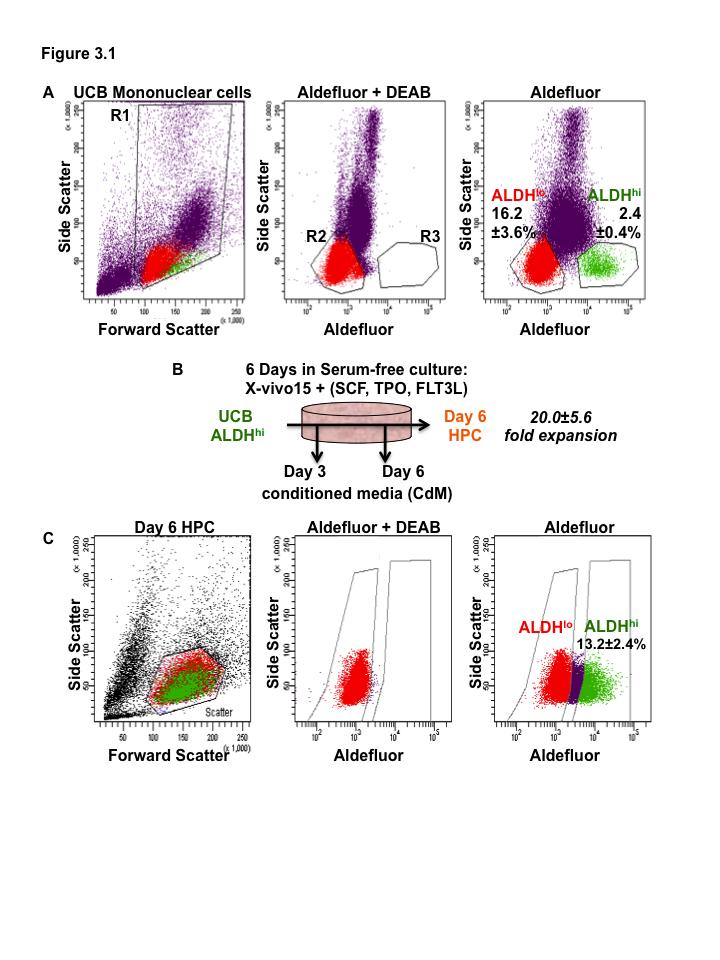
132 110
133 111 Table 3.1. Cell surface marker phenotype changes after ex vivo expansion of HPC. Lineage Marker Hematopoietic Progenitor Myeloid Monocyte B cell T cell UCB ALDH hi cells (%) ex vivo expanded HPC (%) ALDH ±2.4 CD ± ±4.4 CD ± ±7.5 CD ± ±1.8 CD ± ±0.6 CD ± ±0.7 CD14 2.8± ±0.1 CD19 0.8± ±0.0 CD3 3.0± ±0.1
134 Ex vivo expansion of ALDH hi UCB cells increases the total number of hematopoietic progenitor cells. Fresh UCB ALDH hi cells and 6 day expanded progeny were plated at limiting dilution in semi solid methylcellulose media with hematopoietic growth factors to enumerate clonogenic hematopoietic colony formation before and after 6 days ex vivo expansion. Notably, the ex vivo-expanded HPC retained significant multipotent myeloid colony forming capacity (Figure 3.2A). In freshly isolated UCB ALDH cells, approximately 1 in 3 cells showed hematopoietic colony forming function in methylcellulose. After ex vivo culture, HPC progeny displayed a reduced frequency of colony forming units at approximately 1 in 8 cells. At 20.0±5.6-fold total cell expansion this amounts to a 7.5- fold increase in total hematopoietic colony forming cells after 6 days ex vivo culture. Ex vivo-expanded HPC also showed a significant decrease in the overall frequency of erythroid blast forming units (BFU-E, macrophage (CFU-M) and granulocyte (CFU-G) colony forming units (Figure 3.2A, n=4, p<0.05). Furthermore, ex vivo expansion caused a shift in myeloid lineage specification away from granulocytic differentiation towards erythroid differentiation (Figure 3.2B,C). Taken together, these data illustrate that ex vivo culture of UCB ALDH hi cells effectively increases the total number of hematopoietic progenitor cells (HPC) available for further application.
135 113 Figure 3.2. Ex vivo expanded HPC retain myeloid multipotency. (A) Following 6 days of serum free culture expanded HPC retain diminished progenitor cell marker phenotype. (C) HPC plated in semi-solid methylcellulose media show myeloid multipotency at diminished levels compared to (B) freshly isolated ALDH hi cells. (*p<0.05, n=5).
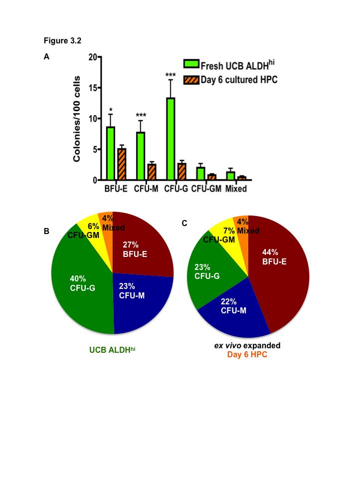
136 114
137 Expanded HPC retain a vascular regenerative transcription profile Affymetrix microarray identified multiple transcripts with significantly changed expression levels (>1.5 fold increase, p<0.01) in 3 ex vivo-expanded HPC samples compared to 3 parental, uncultured UCB ALDH hi cell samples. Confirming cell surface phenotype as measured by flow cytometry, ex vivo-expanded HPC expressed significantly less KIT (CD117, p=8.31x10⁻⁶ ) and PROM1 (CD133, p= 6.88x10⁻⁴ ) compared to UCB ALDH hi cells. Interestingly, despite significant reduction in CD34 expression at the cell surface there was no significant reduction in CD34 transcript levels in HPC compared to UCB ALDH hi cells. The raw data was next filtered to focus on transcripts coding for secreted proteins with documented biological functions that potentially modulate endothelial cell survival and pro-angiogenic processes that were significantly upregulated after ex vivo expansion (n=3, Table 3.2). Ex vivo-expanded HPC showed upregulation of several transcripts which code for secreted growth factors known to modulate vascular regeneration including epidermal growth factor (EGF), vascular endothelial growth factor A and B (VEGF-A, VEGF-B), and angiopoietin 1 (ANGPT1, Table 3.2). Furthermore, HPC expressed significantly higher levels of transcripts for proteins that modulate vascular growth factor signaling including ribonuclease/angiogenin inhibitor-1 (RNH1, angiogenin regulator) and insulin-like growth factor binding protein 3 (IGFBP3, insulin-like growth factor modulator, Table 3.2). Ex vivo expansion of HPC from UCB ALDH hi cells also increased expression of transcripts associated with matrix modifying proteins, including tissue inhibitor of metalloproteinases 1 and 3 (TIMP-1, TIMP-3, Table 3.2). Collectively, these data suggest that ex vivo-expanded HPC retain a vascular regenerative transcription profile.
138 116 Table 3.2. Transcripts for secreted proteins associated with vascular regeneration upregulated in ex vivo-expanded HPC Gene Symbol Common Name Biological Function Molecular Function Fold- Change p-value vs. UCB vs. UCB ALDH hi ALDH hi promotes endothelial cell EGF epidermal growth factor proliferation, survival, differentiation, and migration EGF receptor activation x10-4 TIMP3 tissue inhibitor of metalloproteinases prevents matrix remodeling, 3 stabilizing preexisting vasculature inhibition of MMP activity x10-5 ribonuclease/ modulates cellular RNH1 angiogenin inhibitor 1 promotes cell survival under stress conditions, decreases apoptosis localization, regulatory activity of angiogenin x10-7 VEGFA vascular primary regulator and activator of endothelial growth angiogenesis, autocrine signaling factor A promotes vascular homeostasis VEGFR2 receptor activation x10-3 ANGPT1 angiopoietin 1 stabilizes the blood vasculature by suppressing EC responses during vascular stress TIE2 receptor activation x10-3 TIMP1 tissue inhibitor of MMP-independent functions include metalloproteinases inhibition of apoptosis, induction of 1 angiogenesis and cell proliferation inhibition of MMP activity; stimulating the Akt survival pathway x10-4 VEGFB vascular critical for blood vessel survival under endothelial growth pathological conditions, non-essential VEGFR1 receptor factor B role in vessel formation activation x10-4 IGFBP3 insulin-like growth factor binding protein 3 inhibits endothelial cell apoptosis, modulates IGF signaling x10-4
139 Expanded HPC support endothelial cell tubule formation in vitro. To assess whether these expanded HPC supported EC tubule forming function via paracrine actions, human microvascular endothelial cells (HMVEC) were plated on a thin gel of growth factor reduced matrigel bathed in growth factor+/serum+ endothelial growth medium (EGM2-MV, Figure 3.3A), or growth factor-/serum endothelial basal media (EBM, Figure 3.3B), to serve as positive and negative controls for tubule formation respectively. In addition, HMVEC were exposed to basal HPC expansion media (X-vivo 15 + cytokines) or day 3 or day 6 HPC conditioned media (Figure 3.3D). Finally, HMVEC were co-cultured directly with expanded HPC in EBM (Figure 3.3C). Interestingly, HPC-conditioned media showed no effect on tubule formation compared to basal media controls (n=8). However, after 6 hours of direct co-culture with ex vivoexpanded HPC, HMVEC formed significantly more branch points compared to HMVEC grown in EBM or X-vivo 15 vehicle controls (p<0.01, Figure 3.3D, n=4). Furthermore, direct co-culture with HPC supported tubule formation equivalent to HMVEC grown in positive control conditions containing EGM fully supplemented with growth factors and serum. Importantly, HPC plated alone on growth factor reduced matrigel did not result in tubule formation. Together, this data shows that direct co-culture with HPC promotes EC tubule formation under growth factor-free and serum-free conditions.
140 118 Figure 3.3. Ex vivo-expanded HPC support tubule formation by HMVEC in serumfree, growth factor reduced conditions. EC were seeded onto growth factor-reduced Matrigel supplemented with endothelial basal media with (A) 5% serum and EC growth factors, (B) basal EC media, (C) 5x10 3 expanded HPC in direct co-culture, xvivo15 basal, or day 3 or day 6 HPC conditioned media. Photomicrographs were taken at 6h (A-C), 24h, and 48h and branch points were enumerated by blinded counts (Scale bar = 500 μm). (D) Summary of complete branch points counts per well. (**p<0.01).

141 119
142 Expanded HPC support HMVEC survival under growth factorfree, serum-free conditions HMVEC were grown on plastic in EGM2 media fully supplemented with serum and growth factors (EGM2-MV, Figure 3.4A), or in basal endothelial cell media (EBM) without serum or growth factors (Figure 3.4B) or in non-contact transwells containing ex vivo-expanded HPC in EBM (Figure 3.4C). Under fully supplemented conditions (EGM2 + serum + cytokines) HMVEC demonstrated a 4-fold increase in cell number in 72 hours, whereas culture in serum and growth factor depleted basal conditions resulted in a 2-fold reduction in cell number. HPC co-culture prevented HMVEC cell loss after 72 hours under basal growth factor-free, serum-free conditions (Figure 3.4D, n=5-8, p<0.05). To further address the paracrine support of endothelial cell survival under growth factorfree, serum-free conditions, HMVEC were plated in HPC conditioned media and cells were enumerated after 72 hours using the CyQuant system. Similar to the transwell coculture results, HMVEC grown in day 3 or day 6 conditioned media showed the prevention of cell loss associated under growth factor-free, and serum-free conditions (Figure 3.4E, n=6-7, p<0.05). Due to the pro-survival responses elicited in HMVEC when exposed to HPC secreted factors, HMVEC were also harvested after 24 hours culture in EGM2-MV, EBM, or in non-contact transwell co-culture with HPC and assessed for apoptosis using 7- aminoactinomycin D (7AAD) combined with annexin V-staining measured by flow cytometry (Figure 3.4F, n=4-6). HPC co-culture significantly reduced the frequency of dead cells (% 7AAD+ cells) compared to EBM-cultured controls (Figure 3.4G, n=4-6, p<0.05). However, HPC co-culture only modestly reduced the frequency of early apoptotic cells (% 7AAD-/Annexin V+). In contrast, HPC conditioned media did not alter total cell death or apoptosis rates compared to EBM or X-vivo 15 vehicle controls (Figure 3.4G). Collectively, these data indicate that HPC co-culture supported the survival of EC in serum-free, growth factor-free conditions.
143 121 Figure 3.4. Ex vivo-expanded UCB HPC and conditioned media support the survival of endothelial cells under serum-starved conditions. (A-C) 4x10 4 HMVEC were cultured for 72 hours in basal media (EBM2) with or without growth factors (EGF, VEGF, FGF, IGF-1) and serum (5%) supplementation. Into each culture a hanging 1μm pore transwell was suspended above the EC and filled with 8x10 4 day 6 expanded HPC or basal media (Scale bar = 200μm). (D) Non-contact co-culture with D6 expanded HPC promoted the survival of EC under growth factor-free, serumstarved conditions (*p<0.05, n=5-8, in duplicate). (E) 3x10 3 EC were cultured in HPC expansion basal media (Xvivo 15) with or without EC growth factors (EGF, VEGF, FGF, IGF-1) and serum (5%) supplementation. Day 3 or Day 6 conditioned medium (X vivo 15 base) collected during HPC expansion supported survival of EC under serum and growth factor-free conditions (n=6-7 in triplicate, *p<0.05). (F) Representative plots showing flow cytometric detection of Annexin V/7AAD survival status of endothelial cells 24 hours after culture. (G) Summary statistics for cell survival status at 24 hours. (different letter denotes *p<0.05).
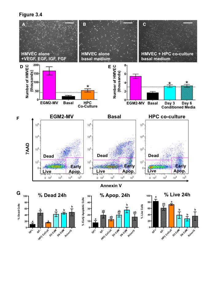
144 122
145 HMVEC co-culture with HPC increased the secretion of prosurvival signals HMVEC and HPC were grown in basal endothelial cell media (EBM) alone, or in direct co-culture with both cell types in basal EBM. After 72 hours, the conditioned media from each culture condition was collected and screened for secreted protein content using antibody array. When grown in basal endothelial media without serum or growth factors both HMVEC and ex vivo-expanded HPC secreted growth factors, chemokines, matrix modifying proteins and secreted receptor proteins associated with endothelial cell function during angiogenesis (Figures 3.5, 3.6, n=3). Notably, co-culture of HPC with EC significantly increased levels of several growth factors associated with vascular regeneration including epidermal growth factor (EGF, Figure 3.5, n=3, p<0.05) and angiopoietin-2 (Figure 3.6, n=3, p<0.05). Moreover, HPC co-culture increased the amount of angiopoietin-2 secreted into the media significantly more than the additive signals of HMVEC or HPC cultured alone, indicating a potential cellular crosstalk regulating increased angiopoietin 2 secretion (n=3, p<0.05). Many chemokines which have been implicated in the recruitment of inflammatory cell types to areas of ischemia were also significantly increased during HPC co-culture including GROα,β,γ/CXCL1,2,3, IL8/CXCL8, and RANTES/CCL5 (Figure 3.5, n=3, p<0.05). Several matrix modifying proteins were significantly upregulated during HPC co-culture including tissue inhibitor of metalloproteinases 1 and 2 (TIMP-1, TIMP-2, Figure 3.5, n=3, p<0.05) and matrix metalloproteinase-9 (MMP-9, Figure 3.6, n=3, p<0.05). TIMP-2 was significantly upregulated in co-culture compared to the additive signals of HMVEC and HPC alone (n=3, p<0.05). Overall, HPC co-culture with HMVEC in unsupplemented media increased the paracrine and autocrine secretion of several angiogenesis-associated growth factors, chemokines and matrix modifying proteins.
146 124 Figure 3.5. HMVEC co-culture with HPC increases secretion of pro-survival signals: Part 1. (A) Chemiluminescent imaging of RayBio Membrane-Based Antibody Human Angiogenesis Array C1000 was used to screen and compare expression levels of angiogenesis-associated cytokines, growth factors, proteases, and soluble receptors secreted in basal endothelial media after 72 in culture. (B) HMVEC grown in basal unsupplemented media co-cultured with ex vivo-expanded HPC are exposed to higher levels of pro-survival signals (n=3, *p<0.05).
147 125
148 126 Figure 3.6. HMVEC co-culture with HPC increases secretion of pro-survival signals: Part 2. (A) Chemiluminescent imaging of RayBio Membrane-Based Antibody Human Angiogenesis Array C1000 was used to screen and compare expression levels of angiogenesis-associated cytokines, growth factors, proteases, and soluble receptors secreted in basal endothelial media after 72 in culture. (B) HMVEC grown in basal unsupplemented media co-cultured with ex vivo-expanded HPC are exposed to higher levels of pro-survival signals (n=3, *p<0.05).
149 127
150 Expanded HPC enhance recovery of limb perfusion after transplantation into ischemic limbs Previously in Chapter 2, I demonstrated that freshly isolated UCB ALDH hi cells support recovery of perfusion and revascularization in the ischemic hindlimb of mice after intravenous transplantation into mice with unilateral hindlimb ischemia (Chapter 2) 19. To confirm these findings with UCB ALDH hi cells and to assess whether expanded HPC could impact the recovery of perfusion in vivo, freshly isolated UCB ALDH hi cells and ex vivo-expanded HPC were transplanted intramuscularly into immune deficient NOD/SCID mice with acute unilateral hindlimb ischemia after femoral artery ligation surgery. Hindlimb perfusion was assessed by laser Doppler perfusion imaging (LDPI) and the perfusion ratio of the ischemic limb versus the contralateral control limb was determined weekly for up to 28 days. After femoral artery ligation surgery, the perfusion ratio (PR) of the treatment leg was less than 10% of the control leg in all transplanted mice (PR 0.1). Mice treated with intramuscular phosphate buffered saline (PBS, Figure 3.7A) vehicle control established the baseline recovery of perfusion (PR=0.49±0.03 at 28 days after treatment). Mice with ischemic adductor muscle transplanted intramuscularly with freshly isolated 20x10 6 UCB MNC or 1x10 5 purified UCB ALDH lo cells showed no significant changes in recovery of perfusion compared to PBS controls (Figure 3.7D). In contrast, mice that were treated by intramuscular transplantation of 1x10 5 purified UCB ALDH hi cells (Figure 3.7B, Day 7 PR=0.45±0.09) or 5x10 5 ex vivo-expanded HPC (Figure 3.7C, Day 7 PR=0.40±0.02) showed significantly accelerated recovery of perfusion of the ischemic limb compared to PBS vehicle controls (Figure 3.7A, Day 7 PR=0.25±0.02) within 7 days after transplant (Figure 3.7D). At a frequency of 13.2% of expanded HPC retaining high ALDH activity, mice transplanted with 5x10 5 HPC receive an equivalent dose of 6.6x10 4 ALDH hi cells, a lower effective dose of ALDH hi cells. Importantly, improved perfusion at day 7 translated to a significant increase in perfusion at later time points (days 14-28) compared to PBS injected controls. Collectively, these data confirm that transplantation of UCB ALDH hi cells promote recovery or perfusion in the ischemic hindlimb of mice after femoral artery ligation. Furthermore, the data indicates that ex vivo expansion of HPC from UCB ALDH hi vascular regenerative function. cells does not diminish
151 129 To further assess recovery from acute ischemic injury, limb use by gait analysis of transplanted mice was assessed using Noldus Catwalk 7.1 software. Mice treated by intramuscular injection of ex vivo-expanded HPC showed significantly increased usage of the ischemic limb at 7 days after transplant compared to PBS treated control mice (Figure 3.7E). These findings demonstrate that the recovery of perfusion in mice transplanted with UCB ALDH hi cells or ex vivo-expanded HPC is accompanied by augmented recovery of limb usage in the injured limb.
152 130 Figure 3.7. Direct intramuscular injection of UCB ALDH hi cells augment perfusion of ischemic limbs. (A-C) Representative LDPI following right femoral artery ligation and tail vein injection of PBS vehicle control, 20x10 6 unsorted UCB MNC, 1x10 5 ALDH hi cells, 1x10 5 ALDH lo cells, or 5x10 5 ex vivo-expanded HPC monitored for 28 days (n=7-15). Numbers in bottom left of each LDPI image indicate perfusion ratio of the ischemic versus the nonischemic hindlimb from ankle to toe. (D) Summary of mean perfusion ratio ± SEM as above. Transplantation of UCB ALDH hi cells promoted robust, maintained recovery of perfusion in ischemic mouse hindlimbs by day 7 post-transplantation as compared to saline injected controls (*p<0.05,**p<0.01). (E) Noldus catwalk paw print intensity at day 7 post transplantation showed that mice transplanted with HPC regained use of their ischemic leg faster than saline vehicle controls (n=6).
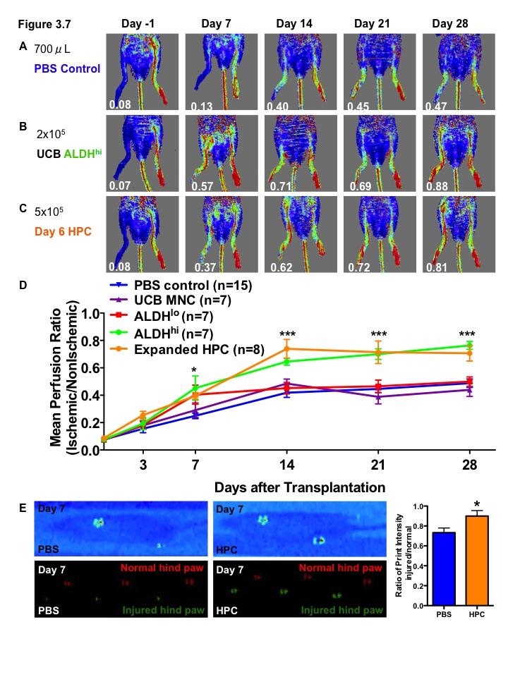
153 131
154 Intramuscular transplantation of HPC prevents loss of endogenous blood vessels after acute ischemic injury Ischemic and control adductor muscle were cryosectioned and stained for CD31 expression to enumerate the blood vessels present at 7 and 28 days after injury. Within 7 days of femoral artery ligation, saline-treated control mice showed a significant loss of the CD31 + positive vessels in the ischemic limb (61.9±2.1 CD31 + vessels/mm 2 ) compared to the contralateral control limb (69.9±0.8 CD31 + vessels/mm 2, p<0.05, n=7, Figure 3.8C). This loss of vascularization in the ischemic limb (62.5±1.6 CD31 + vessels/mm 2, Figure 3.8D) of saline treated mice was consistent at 28 days after induction of ischemic injury compared to normal limb (69.4±0.6 CD31 + vessels/mm 2, Figure 3.8D). Mice treated by intramuscular injection of UCB MNC or ALDH lo cells showed no difference in ischemic limb vascularization compared to PBS controls (Figure 3.7). Mice transplanted with ex vivo-expanded HPC showed prevention of CD31 + cell loss at 7 (72.3±0.6 CD31 + vessels/mm 2, Figure 3.8C) and 28 days after treatment (68.1±1.8 CD31 + vessels/mm 2, Figure 3.8D). Purified ALDH hi cells from fresh UCB similarly showed a reduction in the loss of vascularization in the ischemic muscle at 28 days after treatment (71.8±2.3 CD31 + vessels/mm 2, Figure 3.8D). These findings suggested that UCB ALDH hi cells and ex vivoexpanded HPC prevent the loss of CD31 + vessels in the ischemic hindlimb of mice after ischemic injury.
155 133 Figure 3.8. Intramuscular transplantation of ex vivo-expanded cord blood hematopoietic progenitor cells prevents loss of vascularization of ischemic hindlimb muscle after acute ischemic injury. (A, B) Representative photomicrographs of CD31 staining in ischemic adductor muscle sections taken 28 days after right femoral artery ligation and intramuscular injection of PBS, ALDH hi cells, or HPC. (C, D) Summary of mean vessel density ± SEM in the ischemic or non-ischemic limb at (C) 7 days, or (D) 28 days after transplantation. Mice that were transplanted with fresh UCB ALDH hi cells or ex vivo-expanded HPC demonstrated increased vascularization of the surgical limb. (*p<0.05).
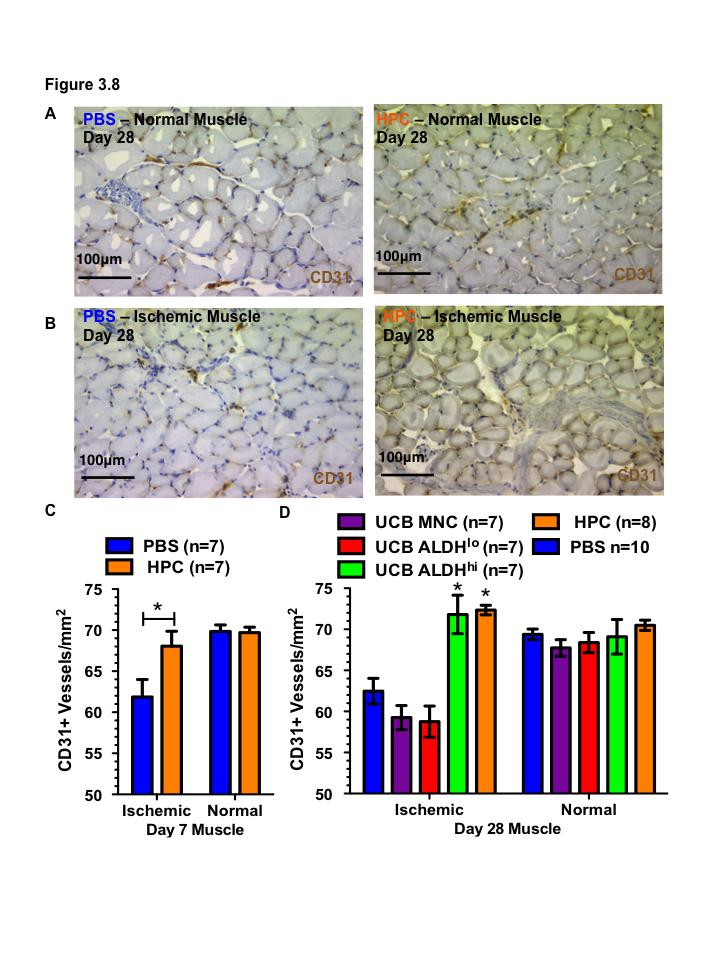
156 134
157 Discussion In this study I demonstrate that human UCB-derived cells with high ALDH activity can be readily expanded in serum free conditions without diminishing vascular regenerative function after direct intramuscular transplantation in vivo. After 6 days ex vivo culture in clinically relevant serum free conditions, 20-fold expansion of total hematopoietic cell number was obtained with substantial expansion of total myeloid progenitor cell number. Our findings show that ex vivo-expanded HPC maintained an angiogenic vascular regenerative phenotype by mrna transcriptional profile. Furthermore, we demonstrate the HPC responded to EC cell signals in culture and increase secretion of angiogenic growth factors, matrix-modifying proteins and chemokines that recruit support cells to the site of injury, inflammation and hypoxia. This vascular regenerative phenotype of HPC was confirmed by demonstration that HPC supported endothelial cell survival and tubule formation in growth factor-free, serum-free co-culture culture conditions. Importantly, transplantation of ex vivo-expanded HPC into immune-deficient mice with femoral artery ligation induced hindlimb ischemia promoted recovery of perfusion and prevented loss of vascularization in the hindlimb similar to previously characterized UCB ALDH hi cells (Chapter 2) 19. Thus, we propose that ex vivo expansion of UCB ALDH hi cells represents a clinically applicable means of effectively increasing the number myeloid progenitor cells for cardiovascular disease therapy without reducing vascular regenerative function. After 6 days of serum free expansion culture UCB ALDH hi cells expanded 20-fold in total cell number. Of these expanded HPC, only 13.2±2.4% retained high ALDH activity, amounting to a total 2.6 fold expansion of ALDH hi cells over 6 days ex vivo expansion. Based on other progenitor cell surface marker expression our 6-day expansion protocol also produced a 6.4-fold expansion of CD34 + cells, a 2.8-fold expansion of CD133 + cells and a 1.7-fold expansion in the number of total CD117 + cells, despite a decrease in the relative frequency of cells expressing progenitor markers. The general decrease in progenitor marker expression frequency despite total progenitor cell number is consistent with other published hematopoietic cell expansion protocols 20,25,26. Furthermore, it is
158 136 possible that the number of CD117 expressing cells is higher than detected by flow cytometry as after prolonged culture in high concentrations of SCF, the ligand for CD117/c-Kit, leads to receptor internalization and recycling as is the case with many receptor tyrosine kinases 27. Relative decreases in hematopoietic progenitor marker expression were confirmed by microarray results showing significantly lower expression of both CD117/c-Kit (KIT) and CD133 (PROM1) transcripts. Thus, we demonstrate here that ex vivo expansion of UCB ALDH hi cells effectively increases the number of angiogenic myeloid ALDH hi /CD34 + /CD133 + /CD117 + progenitors cells available for cell therapy applications. It has recently been shown that decreasing autocrine inhibitory signaling during HPC expansion by use of a batch-fed system, it is possible to significantly increase the expansion of hematopoietic stem and progenitor cells 28,29. It is plausible that by applying new, more efficient methods of ex vivo HPC expansion, and by the use of novel molecules to prevent progenitor differentiation, it will be possible to further increase the number of hematopoietic cells that retain vascular regenerative function for cell therapy applications outside of transplantation for haematological disorders. This is an important area of development for applications employing UCB-derived cells in an allogeneic setting, because available cell number currently prohibits clinical application. We also demonstrated that co-culture of HPC with endothelial cells in growth factor-free, serum-free, basal endothelial cell media led to increased survival of endothelial cells as measured by cell counts and by viability dye measures in vitro. Furthermore, intramuscular transplantation of HPC into the ischemic hindlimb of mice augmented recovery of perfusion and prevented loss of capillary density within the ischemic muscle tissue. As such, HPC conditioned media contained secreted EGF and co-culture with EC increased the concentrations of EGF significantly compared to EC cultured alone. Moreover, EGF transcript levels were increased 6.5-fold in ex vivo-expanded HPC compared to fresh UCB ALDH hi cells. The observed pro-survival effect on endothelial cells in both in vitro and in vivo contexts is consistent with potential paracrine stimulatory effects by secreted EGF on endothelial cells. Endothelial cells express epidermal growth
159 137 factor receptor (EGFR) and EGFR activation has been shown to activate the PI3K/Akt pathway thereby regulating endothelial cell survival 33. Furthermore, we show that ex vivo-expanded HPC retain a vascular regenerative, angiogenic transcriptional and secretory profile beyond EGF expression. After culture, HPC showed upregulation of important regulators of endothelial cell function including angiopoietin 1 (ANGPT1) and vascular endothelial growth factors A and B (VEGF-A, VEGF-B). During homeostasis in vivo EC-EC contacts are stabilized and cell survival is promoted by perivascular cells who secrete angiopoietin 1 and activate thetie2 receptor on endothelial cells 34,35. Vascular endothelial growth factor A (VEGF-A) is the primary regulator of the angiogenic cascade by which endothelial cells become activated to proliferate and migrate in response to hypoxia or injury 36. Furthermore, VEGF-A signaling has been shown to be necessary for EC survival signaling in homeostatic quiescence and in hypoxic conditions 36,37. Additionally, VEGF-B signaling has been demonstrated to be critical for blood vessel survival under pathological conditions promoting not only EC survival but increasing survival of mural smooth muscle cells or pericytes 38. ANG2 is expressed by EC and its expression is increased under hypoxic conditions 34,39. ANG2 is stored in endothelial Weibel Palade bodies which are secreted in response to inflammation and mediate endothelial cell activation 34,40,41. However, in the absence of VEGF, angiopoietin 2 induces destabilization and regression of the vasculature. During hypoxia, which promoted both ANG2 and VEGF secretion, neovascularization is induced by a VEGF mediated pro-angiogenic program referred to as the angiogenic switch 34,39. HPC co-culture increased secreted ANG2 levels in conditioned media significantly indicating a potential role for HPC in increasing activation of endothelial cells during inflammation and hypoxia. Taken together, these data indicate that ex vivo-expanded HPC maintain the transcriptional prolife and secretory functions necessary to support vascular stabilization after ischemic injury pushing the EC cell program from regression to survival and stabilization. HPC co-culture with HMVEC also increased the secretion of TIMP-1 and TIMP-2 in vitro. Moreover, TIMP1 transcript was upregulated in HPC after ex vivo expansion from UCB ALDH hi cells. Increased TIMP expression been shown in hematopoietic progenitor
160 138 cells and myeloid cells previously, and play a role in modifying the extracellular matrix in response to vascular injury or hypoxia 42. Broadly, TIMPs inhibit MMP-mediated remodelling of the extracellular matrix and basement membrane, an important step in both vascular regression and angiogenesis. TIMPs promote the stability of blood vessels by preventing breakdown of the supportive extracellular matrix and promoting basement membrane deposition 36,43. Beyond the normal activity of TIMPs regulating MMP activity, TIMP-1 has been shown to have MMP-independent functions which include inhibition of apoptosis, induction of angiogenesis and cell proliferation 44. In particular, TIMP-1 has specifically been shown to prevent TNF-α mediated apoptosis in EC though the Akt pathway 45. Both the MMP-independent and MMP-mediated effects of TIMP-1 on EC and the vasculature promote endothelial cell survival by supressing cell death are consistent with our in vitro and in vivo findings showing less cell death in HPC-EC coculture conditions. Ex vivo HPC expansion led to increases in transcripts associated with modulating vascular growth factor signaling ribonuclease/angiogenin inhibitor 1 (RNH1) and insulinlike growth factor binding protein 3 (IGFBP3). RNH1 has been shown to promote cell survival in stress conditions by decreasing apoptosis and modulating the cellular localization and activity of angiogenin which is promoter of angiogenesis 46. IGFBP3 has been demonstrated to inhibit endothelial cell apoptosis by an IGF-independent mechanism 47. Thus, angiogenin and IGFBP3 may represent important regulators EC function and the effects of these two regulators may also contribute to the pro-survival mechanism consistent with our findings. Co-culture of HPC with EC increased secreted levels of several chemokines including members of the CXC and CC families, GRO/CXCL1-3, IL-8/CXCL8, and RANTES/CCL5. GRO and CXCL8 are known to play a role in chemokine-mediated regulation of angiogenesis and regulate recruitment of neutrophils, macrophages and lymphocytes to the site of injury Interestingly, MMP-9 was also secreted by ex vivoexpanded HPC. MMP-9 is thought to be important in activating the angiogenic switch, playing a role in increasing VEGF bioavailability by freeing it from deposits within the extracellular matrix 53. MMP-9 has been further implicated in vascular regeneration by
161 139 playing a role in promoting vasculogenesis 54. Notably, MMP-9 has been implicated in the mobilization of circulating angiogenic cells that also modulate a pro-angiogenic niche in areas of hypoxia. Collectively, the cumulative effects of these molecular mediators all act to increase the potential recruitment of circulating cells from the host, which can help mediate a vascular regenerative program at the site of injury. These proposed mechanisms could play a plausible role in vivo in vascular regeneration seen after transplantation of HPC into ischemic tissues. Our lab has previously described a subset of hematopoietic progenitor cells with from human BM high ALDH activity that play a role in paracrine support of vascular regeneration (Chapter 2) 18,24,55. Perin et al. have recently reported phase I/II clinical trials using autologous BM-derived ALDH hi cells for the treatment of critical limb ischemia secondary to peripheral artery disease and ischemic heart failure 56,57. Both studies demonstrated the safety of ALDH hi cells in patients with cardiovascular disease. Importantly, in patients with critical limb ischemia, intramuscular injection of autologous BM ALDH hi promoted improved Rutherford category scores and reduced resting pain. Furthermore patients treated by ALDH hi cell transplant showed modest but significantly improved ankle to brachial index scores indicating improved perfusion of the lower limb. Finally, patients who were treated with BM ALDH hi cells reported significantly improved quality of life after treatment 56. These data demonstrate that ALDH hi cells are a viable clinical cell population for the treatment of ischemic disease, and our data further suggests that this population may be expanded using clinically applicable conditions to increase the number of pro-vascular progenitor cells available for transplantation. Notably however, there is emerging evidence that application of autologous bone marrow progenitor cells in patients with diabetes or cardiovascular disease might be problematic. There exists convincing evidence for decreased frequency and reduced regenerative function for circulating and bone marrow progenitor cells in patients with chronic diabetes, cardiovascular disease and increasing age Unfortunately, the patients who would most benefit from vascular regenerative therapy are generally of increasing age and overwhelming affected by diabetes and cardiovascular disease 1, making the development of allogeneic transplantation strategies desirable. As such, human umbilical
162 140 cord blood represents an allogeneic source is readily available and may possess distinct advantages over autologous bone marrow as source of cells for vascular regenerative therapy. Most notably, umbilical cord blood is early in ontogeny and free of chronic disease and age related dysfunctions. We have also shown here that ex vivo culture of UCB derived ALDH hi cells permits expansion of the number of cells available for therapy. However, it is possible that similar ex vivo expansion strategies could be applied to autologous bone marrow transplants in patients as an alternative in order bypass the need for immune suppression in the allogeneic setting. Nonetheless, we propose that UCB ALDH hi cells represent a readily available, and expandable HPC population with robust vascular regenerative properties. Therefore, expanded UCB-derived ALDH hi cells represent an excellent candidate population for the development of allogeneic cell therapy for ischemic disease. Our current work demonstrates that is possible to increase the number of hematopoietic progenitor cells available from UCB ALDH hi cells for applications in vascular regeneration without losing regenerative function. We propose that purification of BM or UCB based on high aldehyde dehydrogenase activity followed by ex vivo expansion to increase the progenitor pool poses a promising clinical approach to developing cell therapy for vascular regeneration and the treatment of ischemic disease.
163 References 1. Lackland DT, Roccella EJ, Deutsch AF, et al. Factors influencing the decline in stroke mortality: a statement from the American Heart Association/American Stroke Association. Stroke. 2014;45(1): Dotsenko O. Stem/Progenitor cells, atherosclerosis and cardiovascular regeneration. Open Cardiovasc Med J. 2010;4: Asahara T, Murohara T, Sullivan A, et al. Isolation of putative progenitor endothelial cells for angiogenesis. Science. 1997;275(5302): Dimmeler S, Zeiher AM, Schneider MD. Unchain my heart: the scientific foundations of cardiac repair. J. Clin. Invest. 2005;115(3): Rafii S, Lyden D. Therapeutic stem and progenitor cell transplantation for organ vascularization and regeneration. Nat Med. 2003;9(6): Gupta NK, Armstrong EJ, Parikh SA. The Current State of Stem Cell Therapy for Peripheral Artery Disease. Curr Cardiol Rep. 2014;16(2): Michler RE. Stem cell therapy for heart failure. Cardiology in Review. 2014;22(3): Botham CM, Bennett WL, Cooke JP. Clinical Trials of Adult Stem Cell Therapy for Peripheral Artery Disease. Methodist DeBakey Cardiovascular Journal. 2013;9(4): Rosenzweig A. Cardiac cell therapy--mixed results from mixed cells. N Engl J Med. 2006;355(12): Wara AK, Croce K, Foo S, et al. Bone marrow-derived CMPs and GMPs represent highly functional proangiogenic cells: implications for ischemic cardiovascular disease. Blood. 2011;118(24): Okuno Y, Nakamura-Ishizu A, Kishi K, Suda T, Kubota Y. Bone marrow-derived cells serve as proangiogenic macrophages but not endothelial cells in wound healing. Blood. 2011;117(19): Ohtani K, Vlachojannis GJ, Koyanagi M, et al. Epigenetic regulation of endothelial lineage committed genes in pro-angiogenic hematopoietic and endothelial progenitor cells. Circ. Res. 2011;109(11): Rohde E, Schallmoser K, Reinisch A, et al. Pro-angiogenic induction of myeloid cells for therapeutic angiogenesis can induce mitogen-activated protein kinase p38-dependent foam cell formation. Cytotherapy. 2011;13(4): Yoder MC, Mead LE, Prater D, et al. Redefining endothelial progenitor cells via clonal analysis and hematopoietic stem/progenitor cell principals. Blood. 2007;109(5): Urbich C, Heeschen C, Aicher A, et al. Relevance of monocytic features for neovascularization capacity of circulating endothelial progenitor cells. Circulation. 2003;108(20):
164 Grunewald M, Avraham I, Dor Y, et al. VEGF-induced adult neovascularization: recruitment, retention, and role of accessory cells. Cell. 2006;124(1): Estes ML, Mund JA, Mead LE, et al. Application of polychromatic flow cytometry to identify novel subsets of circulating cells with angiogenic potential. Cytometry A. 2010;77(9): Capoccia BJ, Robson DL, Levac KD, et al. Revascularization of ischemic limbs after transplantation of human bone marrow cells with high aldehyde dehydrogenase activity. Blood. 2009;113(21): Putman DM, Liu KY, Broughton HC, Bell GI, Hess DA. Umbilical cord bloodderived aldehyde dehydrogenase-expressing progenitor cells promote recovery from acute ischemic injury. Stem Cells. 2012;30(10): Flores-Guzman P, Fernandez-Sanchez V, Mayani H. Concise Review: Ex Vivo Expansion of Cord Blood-Derived Hematopoietic Stem and Progenitor Cells: Basic Principles, Experimental Approaches, and Impact in Regenerative Medicine. Stem Cells Translational Medicine. 2013;2(11): Gilmore GL, DePasquale DK, Lister J, Shadduck RK. Ex vivo expansion of human umbilical cord blood and peripheral blood CD34(+) hematopoietic stem cells. Exp. Hematol. 2000;28(11): Piacibello W, Sanavio F, Garetto L, et al. Extensive amplification and selfrenewal of human primitive hematopoietic stem cells from cord blood. Blood. 1997;89(8): Storms RW, Trujillo AP, Springer JB, et al. Isolation of primitive human hematopoietic progenitors on the basis of aldehyde dehydrogenase activity. Proc. Natl. Acad. Sci. U.S.A. 1999;96(16): Putman DM, Hess DA. Isolation of human umbilical cord blood aldehyde dehydrogenase-expressing progenitor cells that modulate vascular regenerative functions in vitro and in vivo. Curr Protoc Stem Cell Biol. 2013;Chapter 2:Unit 2A Fernández-Sánchez V, Pelayo R, Flores-Guzmán P, et al. In vitro effects of stromal cells expressing different levels of Jagged-1 and Delta-1 on the growth of primitive and intermediate CD34(+) cell subsets from human cord blood. Blood Cells Mol. Dis. 2011;47(4): Dahlberg A, Delaney C, Bernstein ID. Ex vivo expansion of human hematopoietic stem and progenitor cells. Blood. 2011;117(23): Orinska Z, Foger N, Huber M, et al. I787 provides signals for c-kit receptor internalization and functionality that control mast cell survival and development. Blood. 2010;116(15): Csaszar E, Kirouac DC, Yu M, et al. Rapid Expansion of Human Hematopoietic Stem Cells by Automated Control of Inhibitory Feedback Signaling. Cell Stem Cell. 2012;10(2):
165 Csaszar E, Chen K, Caldwell J, Chan W, Zandstra PW. Real-time monitoring and control of soluble signaling factors enables enhanced progenitor cell outputs from human cord blood stem cell cultures. Biotechnol. Bioeng. 2013;111(6): Stanic B, Pandey D, Fulton DJ, Miller FJ. Increased Epidermal Growth Factor- Like Ligands Are Associated With Elevated Vascular Nicotinamide Adenine Dinucleotide Phosphate Oxidase in a Primate Model of Atherosclerosis. Arteriosclerosis, Thrombosis, and Vascular Biology. 2012;32(10): Kato M, Inazu T, Kawai Y, et al. Amphiregulin is a potent mitogen for the vascular smooth muscle cell line, A7r5. Biochem. Biophys. Res. Commun. 2003;301(4): Dreux AC, Lamb DJ, Modjtahedi H, Ferns GAA. The epidermal growth factor receptors and their family of ligands: Their putative role in atherogenesis. Atherosclerosis. 2006;186(1): Makki N, Thiel K, Miller F. The Epidermal Growth Factor Receptor and Its Ligands in Cardiovascular Disease. IJMS. 2013;14(10): Eklund L, Saharinen P. Angiopoietin signaling in the vasculature. Experimental Cell Research. 2013;319(9): Jeansson M, Gawlik A, Anderson G, et al. Angiopoietin-1 is essential in mouse vasculature during development and in response to injury. J. Clin. Invest. 2011;121(6): Carmeliet P, Jain RK. Molecular mechanisms and clinical applications of angiogenesis. Nature. 2011;473(7347): Wietecha MS, Cerny WL, DiPietro LA. Mechanisms of vessel regression: toward an understanding of the resolution of angiogenesis. Curr. Top. Microbiol. Immunol. 2013;367: Zhang F, Tang Z, Hou X, et al. VEGF-B is dispensable for blood vessel growth but critical for their survival, and VEGF-B targeting inhibits pathological angiogenesis. Proceedings of the National Academy of Sciences. 2009;106(15): Holash J. Vessel Cooption, Regression, and Growth in Tumors Mediated by Angiopoietins and VEGF. Science. 1999;284(5422): Fiedler U, Scharpfenecker M, Koidl S, et al. The Tie-2 ligand angiopoietin-2 is stored in and rapidly released upon stimulation from endothelial cell Weibel- Palade bodies. Blood. 2004;103(11): Fiedler U, Augustin HG. Angiopoietins: a link between angiogenesis and inflammation. Trends Immunol. 2006;27(12): Kessenbrock K, Plaks V, Werb Z. Matrix Metalloproteinases: Regulators of the Tumor Microenvironment. Cell. 2010;141(1):52 67.
166 Benjamin MM, Khalil RA. Matrix Metalloproteinase Inhibitors as Investigative Tools in the Pathogenesis and Management of Vascular Disease. Experientia Supplementum. 2012;103(Chapter 7): Grunnet M, Mau-Sørensen M, Brünner N. Tissue inhibitor of metalloproteinase 1 (TIMP-1) as a biomarker in gastric cancer: a review. Scand J Gastroenterol. 2013;48(8): Boulday G, Fitau J, Coupel S, Soulillou J-P, Charreau B. Exogenous tissue inhibitor of metalloproteinase-1 promotes endothelial cell survival through activation of the phosphatidylinositol 3-kinase/Akt pathway. Ann. N. Y. Acad. Sci. 2004;1030: Pizzo E, Sarcinelli C, Sheng J, et al. Ribonuclease/angiogenin inhibitor 1 regulates stress-induced subcellular localization of angiogenin to control growth and survival. J. Cell. Sci. 2013;126(Pt 18): Zhang Q, Jiang Y, Miller MJ, et al. IGFBP-3 and TNF- Regulate Retinal Endothelial Cell Apoptosis. Investigative Ophthalmology & Visual Science. 2013;54(8): Zaja-Milatovic S, Richmond A. CXC chemokines and their receptors: a case for a significant biological role in cutaneous wound healing. Histol. Histopathol. 2008;23(11): Kopp H-G, Ramos CA, Rafii S. Contribution of endothelial progenitors and proangiogenic hematopoietic cells to vascularization of tumor and ischemic tissue. Curr Opin Hematol. 2006;13(3): Suffee N, Richard B, Hlawaty H, et al. Angiogenic properties of the chemokine RANTES/CCL5. Biochem. Soc. Trans. 2011;39(6): Crola Da Silva C, Lamerant-Fayel N, Paprocka M, et al. Selective human endothelial cell activation by chemokines as a guide to cell homing. Immunology. 2009;126(3): Schober A. Chemokines in Vascular Dysfunction and Remodeling. Arteriosclerosis, Thrombosis, and Vascular Biology. 2008;28(11): Bergers G, Coussens LM. Extrinsic regulators of epithelial tumor progression: metalloproteinases. Current Opinion in Genetics & Development. 2000;10(1): Ahn G-O, Brown JM. Matrix metalloproteinase-9 is required for tumor vasculogenesis but not for angiogenesis: role of bone marrow-derived myelomonocytic cells. Cancer Cell. 2008;13(3): Putman DM, Bell GI, Hess DA. Blood-derived ALDHhi cells in tissue repair. 2012; Perin EC, Silva G, Gahremanpour A, et al. A randomized, controlled study of autologous therapy with bone marrow-derived aldehyde dehydrogenase bright cells in patients with critical limb ischemia. Catheter Cardiovasc Interv. 2011;78(7):
167 Perin EC, Silva GV, Zheng Y, et al. Randomized, double-blind pilot study of transendocardial injection of autologous aldehyde dehydrogenase-bright stem cells in patients with ischemic heart failure. Am. Heart J. 2012;163(3): e Vasa M, Fichtlscherer S, Aicher A, et al. Number and migratory activity of circulating endothelial progenitor cells inversely correlate with risk factors for coronary artery disease. Circ. Res. 2001;89(1):E Schatteman GC, Hanlon HD, Jiao C, Dodds SG, Christy BA. Blood-derived angioblasts accelerate blood-flow restoration in diabetic mice. J. Clin. Invest. 2000;106(4): Tepper OM, Carr J, Allen RJJ, et al. Decreased circulating progenitor cell number and failed mechanisms of stromal cell-derived factor-1alpha mediated bone marrow mobilization impair diabetic tissue repair. Diabetes. 2010;59(8): Berezin AE, Kremzer AA. Circulating Endothelial Progenitor Cells as Markers for Severity of Ischemic Chronic Heart Failure. Journal of Cardiac Failure
168 146 Chapter 4 4 High Aldehyde Dehydrogenase Activity Identifies a Subpopulation of Multipotent Stromal Cells with Increased Vascular Regenerative Function BM MSC are a population of progenitor cells that expand readily in ex vivo cultures with promising potential for vascular regenerative cellular therapies. However, our lab has demonstrated that different MSC lines from different donors showed variable efficacy to promote regeneration. Furthermore, the Hess lab had previously shown that re-selection of MSC that retained high ALDH activity after culture increased islet regenerative capacity. Therefore the objective of the studies presented in this chapter was as follows: To characterize the differentiative and vascular regenerative functions of MSC subpopulations selected after expansion for low versus high ALDH activity. The studies presented in Chapter 4 use some of the general findings from Chapters 2 and 3 and other work by the Hess laboratory, specifically that selection of cells with high ALDH activity isolates a population of cells with increased vascular regenerative potential, as the basis for an approach to prospectively identify a subset of MSC that might be superior candidates for future development of cellular therapy for CVD. It uses the in vitro models established in Chapters 2 and 3 to establish proof of concept for proposed future in vivo studies with populations of MSC purified by ALDH activity level.
169 Introduction Despite significant progress and advances to improve clinical management, cardiovascular disease (CVD) remains the leading cause of premature death worldwide and represents a huge burden on the health care systems in North America 1-5. Ischemia is characterized by insufficient blood flow to a given tissue or organ and includes both acute conditions like myocardial infarction and stroke and chronic ischemic conditions like atherosclerotic arterial disease. Direct and indirect costs of medical management and treatment of CVD is a growing burden on global health care systems, estimated to now cost in excess of $310 billion annually in the United States alone 3,4,6-8. Multipotent stromal cells (MSC) also referred to as mesenchymal stromal stem cells can be isolated from bone marrow (BM) and other tissues. MSC are defined by their ex vivo culture method; they must be plastic adherent, express the markers CD73 (ecto 5 nucleotidase), CD90 (Thy-1), and CD105 (endoglin) after culture and must not express hematopoietic markers like CD45 or CD14 1,2,7-10. Additionally, MSC must demonstrate definitive multipotency, by ability to differentiate into osteocytes, adipocytes and chondrocytes 1-5. Broadly, MSC are considered as good candidates for allogeneic cell therapy applications because of their low immunogenicity; they do not express MHC class II (HLA-DR) nor co-stimulatory molecules like CD40, CD80 and CD86 3,4,6-8. Furthermore, MSC have been shown to demonstrate multi-tissue engraftment and little proliferative capacity after transplantation, reducing risk of transplantation related malignancies 1,2,7-10. In general, MSC contributions towards regenerative therapy is primarily through the modulation of the tissue microenvironment as opposed to cell replacement 6,8,11. Indeed MSC have been demonstrated to readily support many repair processes including vascular regeneration 1,8-10. MSC have also been shown to augment cardiomyocyte 1,10-14 and EC 1,10,15,16 survival, and secrete VEGF-A, FGF-2, ANG-1, and MCP-1 1,10,12-14,17. MSC have also been recently identified as a component of vascular pericytes that express CD146 in multiple human organs in situ and help stabilize newly formed vessels when co-transplanted with ECs in vivo 15,16, MSC have furthermore been shown to play an important role in
170 148 immunomodulation presenting interesting potential paracrine applications in autoimmune disease and inflammatory conditions 17,18, Taken together these unique properties underscore the considerable promise of MSC from human BM or UCB as a potential source for vascular regenerative cell therapies. BM MSC represent a readily available population of progenitor cells that expand efficiently in ex vivo cultures with promising potential for vascular regenerative cellular therapies. However, an outstanding limitation to the successful implementation of MSCmediated regeneration in various clinical therapeutic settings is major inter-donor variability in MSC function. The Hess laboratory have previously shown that transplanted BM MSC populations show substantial donor variation in ability to support regeneration of pancreatic islets in a streptozotocin-induced beta cell deletion model of diabetes 19,20. Other groups have demonstrated donor variation in MSC in terms of osteogenic differentiative capacity 21,23,24. Furthermore, MSC have been shown to undergo replicative senescence under differentiative growth conditions and subsequent reduction in regenerative function over prolonged culture in several systems 19,25. This decline in MSC function over extended ex vivo culture also demonstrates notable inter-donor variability 24. To improve cell therapy using MSC, it is desirable to identify prospective markers to identify subpopulation with increased regenerative capacity. Our laboratory has previously demonstrated that aldehyde dehydrogenase (ALDH), a cytosolic enzyme involved in retinoic acid synthesis and in cellular self-protection against oxidative stress is a useful marker of primitive progenitor cells associated with increased vascular regenerative function (Chapter 2) 26,27. Indeed the Hess laboratory have shown that selection of BM mononuclear cells with high ALDH activity isolates an enriched starting population of mixed hematopoietic, endothelial and mesenchymal progenitor cells that supported robust recovery of perfusion and increased vascularization after femoral artery ligation-induced ischemic injury in an immunodeficient mouse model 26. Furthermore, the Hess laboratory has demonstrated the MSC derived from BM cells with high ALDH activity support increased regeneration in the diabetic pancreas 19. Thus, we hypothesized that MSC subpopulations with increased ALDH activity will improve vascular regenerative function via the paracrine support of endothelial cell
171 149 activation and neo-vessel forming function. In this chapter, we present preliminary characterization of the differentiation and vascular regenerative properties of MSC subpopulations with high versus low ALDH activity sorted after expansion. 4.2 Methods BM MSC derivation and culture BM aspirates were acquired with informed consent during routine pre-screening for BM donation by Dr. Anargyros Xenocostas at the London Health Sciences Centre in London, Ontario, Canada. The mononuclear cell fraction was isolated using ficoll centrifugation. BM mononuclear cells were then depleted of residual red blood cells by lysis in ammonium chloride. Mononuclear cells were plated at 2.7x10 5 cells/cm 2 on tissue culture treated plastic dishes in fully-supplemented Amniomax C-100 media (Gibco, Grand Island, NY). Culture media was changed 48 hours after plating to allow adherent stromal cells to adhere to plastic. Thereafter, growth media was changed 3 times per week for maintenance and cell expansion. Adherent MSC colonies were detectable by light microscopy within 2 weeks of plating. MSC were harvested by trypsin and subsequently passaged at 4.7x10 3 cells/cm 2 for expansion culture. After passage 2-3, MSC were harvested using trypsin and suspended in FBS + 10% DMSO for cryopreservation. Thawed MSC were seeded at 4.7x10 3 cells/cm 2 in Amniomax to reestablish MSC culture and subsequently passaged prior to reaching 80% confluence until sufficient cell numbers were accrued for experiments Isolation of MSC subpopulations based on ALDH activity To assay BM MSC for relative ALDH activity, growth media was aspirated, cells were washed with PBS and cells were collected after trypsin-mediated detatchment. After cell counting, MSC were assayed for ALDH activity using Aldefluor reagent at 10x10 6 cells/ml as per manufacturer instructions (Stemcell Technologies, Vancouver, BC). Fluorescence activated cell sorting (FACS) was performed to isolate cells with high (ALDH hi ) or low (ALDH lo ) ALDH activity to >99% purity. ALDH lo gating was set by using DEAB inhibition of ALDH activity in control samples. Sorted cells were subsequently washed in PBS to allow efflux of Aldefluor substrate via reactivation of
172 150 inhibited ABC-transporters prior to plating at 2.7x10 5 cells/cm 2 on tissue culture treated plastic dishes in fully supplemented Amniomax C-100 media as described above Cell surface marker phenotype analysis BM MSC were harvested with trypsin, collected in PBS+5%FBS and assayed for ALDH activity level by labeling with Aldefluor as above. Subsequently, cells were stained with anti-human antibodies for MSC marker expression CD73, CD90, CD105. To confirm that MSC populations are negative for hematopoietic markers MSC were also labelled with antibodies raised against monocyte marker CD14, and pan-leukocyte marker CD45. Finally, to further characterise MSC sub populations Aldefluor labelled MSC were labelled with antibodies for pericyte marker CD146 and LNGFR (CD271, Miltenyi Biotech, Auburn CA). All other antibodies were obtained from BD Biosciences (Missisauga, ON). Viable, 7-amino-actinomycin D (7AAD, BD Bioscience)-excluding cells were assessed for ALDH-activity and cell surface marker expression using an LSRII flow cytometer at the London Regional Flow Cytometry Facility and analyzed using FlowJo software (Treestar, Ashland, OR). All gating was performed based on fluorescence minus-one controls (FMO.) in vitro adipogenesis assay To test differentiation of MSC into the adipose lineage, active ALDH hi or ALDH lo MSC cultures were harvested using trypsin and reseeded in 12 well plates at 1x10 4 cells/cm 2 and grown for 2-4 days to near confluency in Amniomax media. Amniomax media was aspirated and cells were washed with PBS before replacement with adipogenesis differentiation media (Gibco). Adipogenesis media was changed every 3-4 days for 14 days. After 14 days of adipocyte induction, cultures were fixed in formalin and subjected to Oil Red O staining to label intracellular lipid stores. Cells were counterstained with hematoxylin to visualise cell nuclei. Relative induction of adipogenesis was quantified by percentage red pixels of total in photomicrographs measured using ImageJ software.
173 in vitro osteogenesis assay To assess differentiation of MSC into osteocytes, active ALDH hi or ALDH lo MSC cultures were harvested using trypsin and reseeded in 12 well plates at 5x10 3 cells/cm 2 and grown for 2-4 days to near confluency in Amniomax. Amniomax media was aspirated and cells were washed with PBS before replacement with osteogenesis differentiation media (Gibco). Osteogenesis media was changed every 3-4 days for 21 days. After 21 days of osteocyte induction, cultures were fixed in formalin and processed for Alizarin Red S staining to detect calcification of osteocytes in vitro chondrogenesis assay To assess differentiation of MSC into chondrocytes, active ALDH hi or ALDH lo MSC cultures were harvested using trypsin, counted, pelleted and resuspended at 1.6x10 7 cells/ml of Amniomax. To create MSC micromass cultures 5μL aliquots of the concentrated cell solution were carefully seeded in multiwall plates. After establishment and 2 hours of incubation in high humidity conditions at 37 C, chondrogenesis differentiation media (Gibco) was added to the micromass cultures. Chondrogenesis media was changed every 2-3 days for 21 days. After 21 days of chondrocyte induction micromasses were collected and embedded in optimum cutting temperature medium and frozen. Cryosections were prepared at 8μm and fixed in formalin prior to Alcian Blue staining to detect proteoglycans produced by chondrocytes. Cryosections were counterstained with Nuclear Fast Red to identify nuclei in chondrocyte micromasses Generation of BM ALDH lo and ALDH hi MSC conditioned media After FACS isolation, ALDH hi or ALDH lo MSC were seeded at 4.7x10 3 cells/cm 2 and grown in Amniomax for 3 days at 37 C to reestablish culture. After 3 days the Amniomax media was aspirated and replaced with serum-free, growth factor-free, endothelial basal media (EBM, Lonza, Basel, Switzerland). After an additional 48 hours in EBM culture without supplement, ALDH hi MSC- or ALDH lo MSC-conditioned media was collected and filtered at 0.2μm and frozen for later use.
174 HMVEC Culture Human dermal microvascular endothelial cells (HMVEC) were grown in endothelial basal medium-2 (EBM2, Lonza) supplemented with single-quots to make endothelial growth medium (EGM2-MV, Lonza) containing fetal bovine serum (5% FBS), epidermal growth factor (EGF), vascular endothelial growth factor (VEGF), basic fibroblast growth factor (FGF-B), insulin-like growth factor-1 (IGF-1), and ascorbic acid. Cells were seeded at 4.7x10 3 cells/cm 2. For maintenance culture, EGM2-MV was changed every 2-3 days when required CyQUANT endothelial cell proliferation assays HMVEC were seeded at 3x10 3 cells/well in 96 well plates using 100μL of fetal bovine serum and growth factor supplemented endothelial growth media (EGM2-MV, GF+/Serum+, Lonza), basal endothelial media with or without growth factors and serum, (GF+/Serum-, GF-/serum+, or GF-/serum-), or in ALDH hi MSC- or ALDH lo MSCconditioned basal endothelial media. Cultures were maintained for 72 hours at 37 C before each well carefully inverted and blotted to remove media from each well. The whole 96-well microplate was then frozen at -70 C for later use. To quantitate increased DNA content representative of increased cell division, adherent cells were lysed by addition of lysis buffer with CyQUANT GR dye in each well for 2-5 minutes at room temperature, and protected from light as per manufacturer instructions (Molecular Probes, Eugene, OR, USA). Fluorescent signal in each well was assessed by fluorescence microplate reader at 480nm excitation and 520nm emission maxima. Cell number in each well was interpolated from a standard curve generated with known number of HMVEC per well Fluorescent cell labeling for co-culture studies HMVEC were labeled using CellTracker Green CMFDA (Molecular Probes), at a concentration of 2μM in PBS+ PBS+5%FBS. ALDH hi or ALDH lo MSC tagged using CellTracker Red CMPTX (Molecular Probes) at a concentration of 3μM in PBS+5%FBS. Growth medium was aspirated from cell culture flasks and washed with PBS. After washing, cells were incubated in appropriate dye solution for min, at 37 C. The
175 153 dye solution was then aspirated and replaced with normal growth medium, followed by incubation at 37 C for at least 30 min prior to harvest for co-culture experiments HMVEC and MSC co-culture tubule formation assays on Geltrex basement membrane extract Geltrex reduced growth factor basement membrane extract (Gibco) was coated as a thin gel on a 24-well plate at a volume of 100μL per well, followed by incubation at 37 C for 1-2 hours to allow the gel to set. The appropriate conditioned medium from ALDH hi versus ALDH lo MSC, or from EGM-2 or EBM controls was then added at a volume of 400μL per well. EBM was added to negative control and experimental wells and EGM2- MV was added to positive control wells. CellTracker Green CMFDA tagged HMVEC, with previously established cells under EGM2-MV conditions, were harvested for secondary culture and 4.5x10 4 cells were added to each well. CellTracker Red CMPTXdyed MSC were harvested and labeled with Aldefluor and sorted into ALDH lo ALDH hi MSC as described above. CMPTX-tagged ALDH hi and ALDH lo MSC populations and were added to experimental wells, at 9x10 3 ALDH hi MSC or ALDH lo MSC per well. Endothelial cell tubule formation was assessed at 6 hours and 24 hours after plating by phase contrast and fluorescent micrographs. Tubule branch points were enumerated using 4 different fields of view by a blinded observer. All co-culture assays were performed in duplicate Murine femoral artery ligation and intramuscular transplantation of expanded MSC To assess the effects of MSC in vascular regeneration in vivo we induced acute unilateral hindlimb ischemia by surgical ligation and complete resection of the right femoral artery and vein on anesthetised NOD/SCID (Jackson Laboratory, Bar Harbor, ME) mice as previously described (Chapter 2, Appendix 1) 27,28. Within 24 hours of surgery, mice were transplanted by intramuscular injection at three sites into the right (surgical/ischemic) adductor muscle with 50μL saline as vehicle control or 5x10 5 BM MSC. and
176 Quantification of hindlimb perfusion using laser Doppler perfusion imaging Anesthetized mice were stabilized at 37 C for 5 minutes on a heating pad before blood flow in the lower hindlimbs (ankle to toe) was quantified using laser Doppler perfusion imaging (LDPI, Moor Instruments, Devon, UK). The perfusion ratio of the ischemic/surgical limb compared to the uninjured/control limb was assessed after surgery to confirm induction of acute ischemic injury with a perfusion ratio (PR<0.1) of <10% compared to the normal limb. Subsequent LDPI measurements were taken in each mouse at 3, 7,14, 21,and 28 days after transplantation to track recovery of perfusion temporally Statistics Data and results are reported as mean values ± SEM. Statistical comparisons were made using a Student s t-test for adipogenesis induction and one-way ANOVA test followed by Tukey s Post-Hoc tests for all other assays. 4.3 Results BM MSC phenotype by ALDH activity level Human BM-derived MSC cultures were established and assayed for ALDH activity using Aldefluor and FACS. Sorted MSC subpopulations with high (37.4±3.2% ALDH hi, Figure 4.1) and low (45±4.2% ALDH lo, Figure 4.1) ALDH activity were isolated based upon DEAB-inhibited controls. Both ALDH hi and ALDH lo MSC displayed robust expression of characteristic stromal markers CD73, CD90, CD105 (Figure 4.2, Table 4.1) but did not express hematopoietic marker CD45 (Figure 4.2D, Table 4.1) or monocyte marker CD14 as detected by flow cytometry (Table 4.1). Furthermore, both ALDH hi and ALDH lo MSC highly expressed pericyte marker CD146 and LNGFR/CD271 (Figure 4.2E, Table 4.1). Taken together, these data indicated that both subpopulations BM MSC express MSC cell surface markers, fulfilling an important determinant in the minimal criteria for MSC as set by the International Society for Cellular Therapy 2.
177 155 Figure 4.1. Bone marrow MSC purified according to high and low levels of ALDH activity using Fluorescence Activated Cell Sorting. (A) DEAB inhibits ALDH activity and establishes a negative gate to mark the ALDH lo MSC subset. ALDH lo and ALDH hi MSC subsets sorted by FACS. (B) Representative photomicrographs of MSC FACS sorted in to (B) ALDH hi and (C) ALDH lo populations and plated in Amniomax media and grown by plastic adherence (n=14).
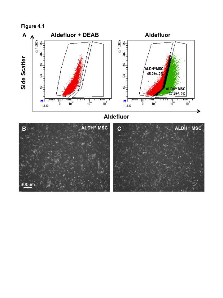
178 156
179 157 Figure 4.2. Bone marrow MSC with high and low levels of ALDH activity display MSC markers. Bone Marrow subpopulations with high and low ALDH activity were isolated by FACS and analyzed for cell surface marker expression associated with MSC phenotype. Both ALDH hi and ALDH lo MSC showed high levels of (A) CD73, (B) CD90, and (C) CD105 expression. Both populations were negative for hematopoietic marker (D) CD45. Both ALDH populations expressed pericyte marker (E) CD146. All gates were set by fluorescence-minus-one controls (FMO).
180 158
181 159 Table 4.1. Bone marrow MSC with high and low levels of ALDH activity display MSC markers. Lineage Marker ALDH hi MSC (+%) ALDH lo MSC (+%) Hematopoietic CD45 0.2± ±0.3 CD14 0.3± ±1.8 MSC CD ± ±5.0 CD ± ±12.1 CD ± ±15.0 Pericyte CD ± ±13.4 Other CD ± ±8.5 (n=3-6)
182 Both BM MSC ALDH hi and ALDH lo subpopulations demonstrate multipotent stromal cell differentiation Human BM-derived MSC were separated into subpopulations by level of ALDH activity by FACS and were subjected to in vitro differentiation conditions to characterize mesenchymal multipotency. Both ALDH hi and ALDH lo MSC formed Oil Red O positivestaining adipocytes after 14 days culture in adipogenesis induction media (Figure 4.3). Notably, ALDH hi MSC (Figure 4.3A,C) demonstrated significantly increased frequency of adipocyte induction compared to ALDH lo MSC (Figure 4.3B,D) as measured by percent Oil Red O positive staining by culture area (Figure 4.3E, p<0.05, n=4). Both ALDH hi (Figure 4.4A,C) and ALDH lo MSC (Figure 4.4A,C) also demonstrated induction of osteocyte formation as assayed by Alizarin Red S detection of calcium deposition after 21 days in osteogenesis media for 21 days (Figure 4.4). Furthermore, both ALDH hi (Figure 4.5A) and ALDH lo MSC (Figure 4.5B) demonstrated induction of chondrocyte differentiation in micromass culture in chondrogenesis media for 21 as measured by detection of proteoglycan deposition by chondrocytes using Alcian Blue staining in cryosections (Figure 4.5). Taken together, these data indicate that subpopulations of BM MSC with both high and low levels of ALDH activity demonstrate mesenchymal multipotency consistent with the defining minimal criteria 2.
183 161 Figure 4.3. Bone marrow-derived ALDH hi MSC show increased adipogenesis. (A,C) ALDH lo MSC and (B,D) ALDH hi MSC were grown for 14 days in adipogenesis differentiation media prior to Oil Red O staining. Representative (A,B) bright field and (C,D) phase contrast photomicrographs of cell cultures stained with Oil Red O and hematoxylin. (E) ALDH hi MSC show greater induction of adipocyte formation as measured by % red pixels indicating positive Oil Red O staining after 14 days in adipogenesis medium compared to ALDH lo MSC (n=4, p<0.05).
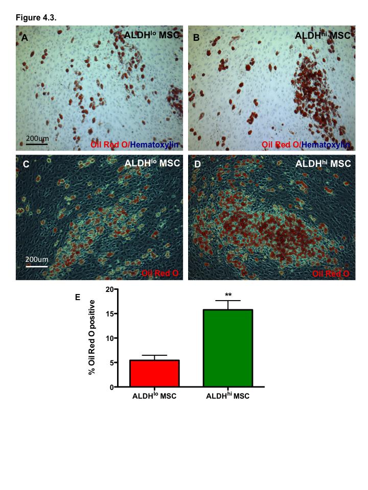
184 162
185 163 Figure 4.4. Bone marrow-derived ALDH hi and ALDH lo MSC differentiate into osteocytes. (A,C) ALDH lo MSC and (B,D) ALDH hi MSC were grown for 21 days in osteogenesis differentiation media prior to Alizarin Red S staining. Representative (A,B) bright field and (C,D) phase contrast photomicrographs of cell cultures stained with Alizarin Red S. Both ALDH hi and ALDH lo MSC induction of osteocyte formation as measured by % red pixels indicating calcium deposition in osteocytes (n=4).
186 164
187 165 Figure 4.5 ALDH activity level does not affect chondrocyte differentiation by MSC. Both bone marrow-derived (A) ALDH lo and (B) ALDH hi MSC showed robust formation of chondrocytes after 21 days micromass culture in chondrogenesis medium. Induction of chondrogenesis in micromass cryosections was confirmed by Alcian blue staining indicating proteoglycan deposition. Nuclear Fast Red counterstaining labels nuclei.
188 166
189 ALDH hi MSC demonstrated increased support of endothelial cell proliferation To test differences in paracrine support of support of endothelial cell proliferation ALDH-purified ALDH hi or ALDH lo MSC subpopulations were cultured in GF-free serum-free endothelial basal media to generate MSC-conditioned media used for HMVEC growth studies. Dermal-origin HMVEC were cultured on plastic in basal endothelial media with or without GFs (EGF, VEGF-A, FGF, IGF-1) and 5%FBS, or in ALDH hi MSC-conditioned or ALDH lo MSC-conditioned media generated without serum or growth factor supplementation. Conversely, HMVEC grown without either or both serum and growth factors showed no proliferation over 72hours. HMVEC cultured in GF+/FBS+ endothelial growth media demonstrated approximately 3.5 fold increase in cell number over 72 hours as measured by CyQuant quantification. Notably, ALDH hi MSC-conditioned media significantly increased proliferation of HMVEC compared to ALDH lo MSC-conditioned media and basal endothelial media (p<0.01, n=5). Interestingly, there was notable variation in HMVEC proliferative responses between BM donor source. Pairwise comparisons of ALDH hi vs. ALDH lo MSC-conditioned media within all other donor samples showed significant increases in EC proliferation in ALDH hi treated cultures (p<0.05, n=5). Intriguingly, BM45 MSC conditioned media was most effective at promoting proliferation in both ALDH hi and ALDH lo subpopulations further illustrating inter-donor variability in the induction of HMVEC growth. Taken together these data demonstrate the purification of BM MSC based on high ALDH activity isolated a subpopulation of MSC with increased paracrine secretory support of EC expansion.
190 168 Figure 4.6. ALDH hi MSC conditioned media supports proliferation of endothelial cells. Representative photo-micrographs of human micro-vascular endothelial cells (HMVEC) grown in (A) growth factor+/serum+ EGM2 media, (B) basal growth factor-free/serumfree, (C) ALDH hi MSC-conditioned media or (D) ALDH lo MSC-conditioned media. (E) Compared to ALDH lo MSC-conditioned basal media, and basal media, ALDH Hi MSC conditioned media increased HMVEC proliferation. (F) Induction of HMVEC proliferation varies according to the individual donor bone marrow source from which the MSC were derived. (E) Different letters denote significant differences in mean (p<0.01, n=5).
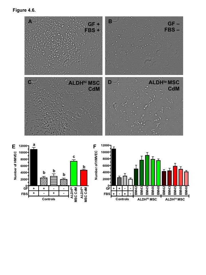
191 169
192 ALDH hi and ALDH lo MSC augmented HMVEC tubule stability To assess the potential for MSC-mediated support of endothelial cell tube formation, CellTracker Green CMFDA fluorescent-tagged HMVEC were plated on growth factor reduced Geltrex in basal endothelial media without serum or growth factor supplementation. ALDH sorted MSC subpopulations were fluorescently labelled with CellTracker Red CMPTX and co-cultured with EC at a ratio of 1 MSC:5 EC. Within 6 hours of co-culture, red-labelled MSC were detected in association with newly formed endothelial cell tubules, consistent with perivascular wrapping function of MSC (Figure 4.7G,H). Direct co-culture of HMVECs with either ALDH lo or ALDH hi MSC did not significantly increase tubule branch point formation by HMVECs at 6 hours relative to negative control basal endothelial cell media (Fig. 4.7A-H,Q, n=3). Moreover, the number of branch points formed by HMVEC in MSC co-cultures was reduced compared to the number of branch points formed by HMVEC alone in growth factor- and serumsupplemented media (Figure 4.7Q). However, after 24 hours of co-culture, only ALDH hi MSC supported significantly increased endothelial cell tubule formation compared to basal conditions (Figure 4.7R). Moreover, ALDH hi MSC co-culture promoted tube formation at equivalent levels to HMVEC cultured alone in growth factor and serum supplemented endothelial growth media. ALDH lo MSC co-culture did not significantly alter HMVEC tubule formation compared to controls or ALDH hi MSC co-culture (Figure 4.7R, n=3). These data demonstrated that purification of MSC with high ALDH activity isolates a population of cells with increased support of endothelial cell tubule stabilization.
193 171 Figure 4.7. Co-culture with bone marrow-derived ALDH hi and ALDH lo MSC augments HMVEC tubule formation. Representative photomicrographs of tubule and branch point formation by HMVECs after (A-H) 6 hours or (I-P) 24 hours culture in basal media (EBM, A,E,I,M), positive control media (EGM2-MV, B,F,J,N), EBM with ALDH lo MSC co-culture (C,G,K,O), or EBM with ALDH hi MSC co-culture (D,H,L,P). Scale bar = 300μm. (Q) Co-culture with ALDH hi and ALDH lo MSC significantly augmented tubule branch point formation relative to basal media at 6 hours. (R) ALDH hi MSC-conditioned media supported maintenance of significantly more branch points compared to ALDH lo at 24 hours. Data are expressed as mean ± SEM. (Q,R) Different letters denote significant difference, p<0.05, n=3.
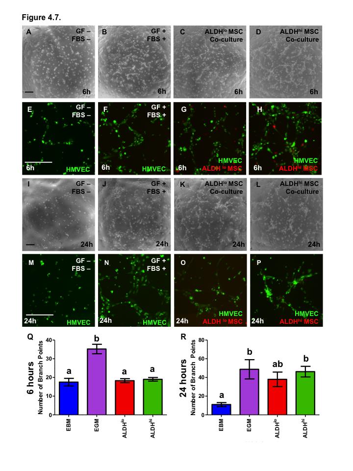
194 172
195 Intramuscular injection of BM MSC promotes accelerated recovery of perfusion after acute ischemic injury. To establish proof of concept in vivo and to assess the functional role of transplanted MSC in vascular regeneration, NOD/SCID mice were subjected to unilateral femoral artery ligation and transplanted by intramuscular injection of BM MSC (non-sorted) into the injured leg adductor muscle. Mice were monitored for recovery of perfusion using laser Doppler perfusion imaging for 28 days. Seven days after treatment of PBS vehicleinjected control mice had recovered a perfusion ratio (PR, ischemic limb/normal limb) of 0.25±0.03 (Figure 4.8A, n=7). However, mice treated with heterogeneous BM MSC showed significantly augmented rate of recovery of perfusion (PR=0.52±0.07) within 7 days compared to PBS-treated mice (Figure 4.8A, p<0.01, n=6). Although perfusion was increased in MSC transplanted mice at days 7 and 14, significantly improved perfusion was not maintained at later time points (Days 21 and 28). Two different BM MSC donor sources were used. At day 7 post-treatment mice injected with BM20 MSC (Figure 4.8B, PR=0.67±0.06, n=3) showed significantly augmented recovery of perfusion compared to both PBS (Figure 4.8B, PR=0.25±0.03, n=7, p<0.01) and BM18 MSC (Figure 4.8B, PR=0.37±0.03, n=3, p<0.01) treated mice. Notably, mice treated with BM18 MSC showed no significant differences in recovery of perfusion compared to PBS controls. Taken together, these data indicate that BM MSC support the recovery of perfusion in the ischemic hindlimb of immune deficient mice after acute injury. However, inter-donor variability in the induction of recovered perfusion by different MSC lines was evident. This in vivo model will form the basis for future investigations to determine the in vivo vascular regenerative potential of ALDH lo or ALDH hi MSC sub-populations and to further identify other phenotypes of BM MSC samples associated with increased regenerative function.
196 174 Figure 4.8. Bone marrow MSC show differential support of vascular regeneration by donor source. LDPI was used to quantify perfusion in the hind limb following right femoral artery ligation and intramuscular injection PBS vehicle control (n=7), or 5x10 5 BM MSC (n=6) monitored for 28 days. (A) Perfusion ratio of the ischemic versus the non-ischemic hindlimb from ankle to toe in all transplanted mice as described above. Compared to saline controls, transplantation of BM MSC promotes recovery of perfusion in ischemic mouse hindlimbs by day 7 post-transplantation. (B) Data presented in (A) sub-divided by MSC donor, MSC from BM20 showed increased support of perfusion recovery compared to donor BM18 MSC. Data are expressed as mean ± SEM, *p<0.01 v PBS, #p<0.01 v BM18 MSC.
197 175
198 Discussion In this chapter, we demonstrate that BM MSC can be sub-fractionated into populations based upon the level of ALDH enzyme activity. Both ALDH hi and ALDH lo MSC display the characteristic cell surface marker phenotype typical of mesenchymal stromal cells devoid of hematopoietic cell contamination. Moreover, both ALDH hi and ALDH lo subpopulations display robust mesenchymal multipotency, demonstrating differentiation capacity to adipocytes, osteocytes and chondrocytes under lineage specific differentiation conditions. We show that the MSC subpopulation with high ALDH activity after culture expansion demonstrates increased paracrine support of endothelial cell proliferation in vitro, under serum-free, growth factor free conditions. Furthermore, we demonstrated that ALDH hi MSC also augmented endothelial cell tubule formation, stabilizing tube-like structures possibly through perivascular wrapping. We propose that high ALDH activity provides a clinically relevant, FACS-selectable, and prospective marker to identify MSC subpopulations with increased vessel supportive functions for the development of improved cell therapies for ischemic disease. The Hess laboratory and others have previously identified cells high levels of ALDH activity as a marker which selects hematopoietic (Chapter 2) 26,27,29-35, endothelial (Chapter 2) 26,27,36-38, neural 39,40, and mesenchymal 19,26 cells with primitive, stem/progenitor cell phenotype and function. Our lab has previously demonstrated that initial selection of ALDH hi mixed progenitor cells from BM 26 or UCB (Chapter 2) 27 demonstrate increased support of endothelial cell function in vitro and enhanced vascular regeneration in vivo. The Hess laboratory have shown that while MSC-like cells can be derived from the ALDH lo BM cells, these cells do not demonstrate full multi-lineage differentiation, and are restricted primarily to the adipocytes lineage 26. In contrast, MSC grown from ALDH hi BM mononuclear cells represent true MSC, which expand efficiently in ex vivo culture and demonstrate full multipotecy forming fat, bone and cartilage 26. Furthermore, the frequency of mesenchymal colony forming cells is significantly higher in the ALDH hi population of human BM compared to ALDH lo BM 26. Together these findings led us to hypothesize that high ALDH activity might be a good prospective marker to further purify an ALDH hi MSC subset with increased capacity for vascular regeneration after
199 177 expansion ex vivo. Importantly, both the ALDH lo and ALDH hi subsets of expanded MSC demonstrated extensive expression of CD146, a cell surface marker previously associated with perivascular wrapping function for MSC isolated from multiple human tissues in situ 16. The findings that BM ALDH hi cell promote recovery of hindlimb ischemia in mice was used as preclinical justification for initiating clinical trials in patients with critical limb ischemia, the most severe form of peripheral arterial disease (PAD)(NCT ) 41. Intramuscular transplantation of freshly isolated (uncultured) autologous BM ALDH hi cells at multiple sites in the ischemic limb was compared directly compared to unpurified autologous BM MNC in PAD patients with a Rutherford score of 4 (resting pain without significant ulceration) or 5 (resting pain with unresolved ulceration or necrosis) 41. Results of this pioneering study demonstrated safety and patients that received autologous BM ALDH hi cells showed improved Rutherford category and reduced resting pain. Furthermore patients in the ALDH hi treatment group showed improved ankle to brachial index scores more rapidly than the MNC group indicating improved perfusion of the lower limb. However, patients in both cohorts reported significantly improved quality of life indices. 41 More recently, a phase 2 placebo-controlled randomized clinical study has been initiated to investigate transplantation of BM ALDH hi cells to improve symptoms of intermittent claudication (NCT ). The Hess laboratory have previously shown that the BM ALDH hi progenitor cells are primarily (>90%) comprised of hematopoietic precursor cells that efficiently form myeloid colonies in vitro, and are also highly enriched in CD45 - progenitor cells that generate multipotent fibroblast colonies (1 CFU-F in 1500 BM ALDH hi cells) that expand efficiently and adherent to plastic. Furthermore, MSC have been widely investigated in clinical trials for cardiovascular disease. As a readily ex vivo expandable population of cells from BM, MSC have been proposed as excellent candidates for cell therapy due to their robust paracrine activities and perivascular wrapping functions. Indeed in diabetic rat models, BM MSC have been demonstrated to promote vascular repair and wound healing Moreover, a randomized controlled clinical trial was undertaken to compare BM MNC and BM MSC therapy in diabetic patients with critical limb ischemia 45. This study
200 178 showed that both BM MNC and MSC were safely administered by intramuscular administration and both BM MNC or BM MSC promoted improvements in perfusion, tissue oxygenation, and pain free walking time at 24 weeks post treatment 45. Notably, greater cell numbers could be generated during the ex vivo expansion of BM MSC compared to the fresh BM MNC, indicating the appeal of ex vivo expansion to increase cell numbers for transplantation. However, a number of preclinical models have shown that BM MNC and MSC from patients with diabetes or cardiovascular disease show diminished function for vascular regeneration For this reason, cell therapy approaches using BM MSC combined with ex vivo expansion are appealing as ex vivo expansion and targeted intra-muscular transplantation may restore MSC vascular regenerative function, or alternatively, you could potentially source BM from healthy donors for use in an allogeneic context. In addition, the establishment of national registries to cryopreserve and HLA-phenotype UCB samples may provide a renewable source of vascular regenerative MSC from early ontogeny and devoid of chronic cardiovascular disease co-morbidities. Moving forward towards essential experiments that will establish proof-of-concept, I propose that ALDH activity presents a useful functional marker to identify MSC subpopulations with increased vascular regenerative potential. Furthermore, selection of ALDH hi MSC might also be helpful in screening cell lines to identify sample with increased or diminished vascular regenerative potential. Taken together I propose that BM-derived MSC with high ALDH activity represent a promising candidate for further development of cell therapy for cardiovascular disease broadly and for critical limb ischemia and peripheral arterial disease specifically.
201 References 1. Wu Y, Chen L, Scott PG, Tredget EE. Mesenchymal Stem Cells Enhance Wound Healing Through Differentiation and Angiogenesis. Stem Cells. 2007;25(10): Horwitz EM, Le Blanc K, Dominici M, et al. Clarification of the nomenclature for MSC: The International Society for Cellular Therapy position statement. Cytotherapy. 2005;7(5): Pittenger MF, Mackay AM, Beck SC, et al. Multilineage Potential of Adult Human Mesenchymal Stem Cells. Science. 1999;284(5411): Lackland DT, Roccella EJ, Deutsch AF, et al. Factors influencing the decline in stroke mortality: a statement from the American Heart Association/American Stroke Association. Stroke. 2014;45(1): Wielgosz A, Canada PHAO. Tracking Heart Disease and Stroke in Canada. Ottawa, Ontario: Public Health Agency of Canada; Bronckaers A, Hilkens P, Martens W, et al. Mesenchymal stem/stromal cells as a pharmacological and therapeutic approach to accelerate angiogenesis. Pharmacology & Therapeutics Le Blanc K, Tammik C, Rosendahl K, Zetterberg E, Ringdén O. HLA expression and immunologic propertiesof differentiated and undifferentiated mesenchymal stem cells. Experimental Hematology. 2003;31(10): Caplan AI, Correa D. The MSC: An Injury Drugstore. Cell Stem Cell. 2011;9(1): Prockop DJ, Oh JY. Medical therapies with adult stem/progenitor cells (MSCs): A backward journey from dramatic results in vivo to the cellular and molecular explanations. J. Cell. Biochem. 2012;113(5): Hung SC, Pochampally RR, Chen SC, Hsu SC, Prockop DJ. Angiogenic effects of human multipotent stromal cell conditioned medium activate the PI3K-Akt pathway in hypoxic endothelial cells to inhibit apoptosis, increase survival, and stimulate angiogenesis. Stem Cells. 2007;25(9): Mirotsou M, Zhang Z, Deb A, et al. Secreted frizzled related protein 2 (Sfrp2) is the key Akt-mesenchymal stem cell-released paracrine factor mediating myocardial survival and repair. Proc. Natl. Acad. Sci. U.S.A. 2007;104(5): Kinnaird T, Stabile E, Burnett MS, et al. Local delivery of marrow-derived stromal cells augments collateral perfusion through paracrine mechanisms. Circulation. 2004;109(12): Chen L, Tredget EE, Wu PYG, Wu Y. Paracrine Factors of Mesenchymal Stem Cells Recruit Macrophages and Endothelial Lineage Cells and Enhance Wound Healing. PLoS ONE. 2008;3(4):e1886.
202 Boomsma RA, Geenen DL. Mesenchymal Stem Cells Secrete Multiple Cytokines That Promote Angiogenesis and Have Contrasting Effects on Chemotaxis and Apoptosis. PLoS ONE. 2012;7(4):e Au P, Tam J, Fukumura D, Jain RK. Bone marrow-derived mesenchymal stem cells facilitate engineering of long-lasting functional vasculature. Blood. 2008;111(9): Crisan M, Yap S, Casteilla L, et al. A perivascular origin for mesenchymal stem cells in multiple human organs. Cell Stem Cell. 2008;3(3): Ben-Ami E, Berrih-Aknin S, Miller A. Mesenchymal stem cells as an immunomodulatory therapeutic strategy for autoimmune diseases. Autoimmun Rev. 2011;10(7): Carmeliet P, Jain RK. Molecular mechanisms and clinical applications of angiogenesis. Nature. 2011;473(7347): Bell GI, Broughton HC, Levac KD, et al. Transplanted Human Bone Marrow Progenitor Subtypes Stimulate Endogenous Islet Regeneration and Revascularization. Stem Cells and Development. 2012;21(1): Bell GI, Meschino MT, Hughes-Large JM, et al. Combinatorial human progenitor cell transplantation optimizes islet regeneration through secretion of paracrine factors. Stem Cells and Development. 2012;21(11): Siddappa R, Licht R, van Blitterswijk C, de Boer J. Donor variation and loss of multipotency during in vitro expansion of human mesenchymal stem cells for bone tissue engineering. J. Orthop. Res. 2007;25(8): Wietecha MS, Cerny WL, DiPietro LA. Mechanisms of vessel regression: toward an understanding of the resolution of angiogenesis. Curr. Top. Microbiol. Immunol. 2013;367: Mentink A, Hulsman M, Groen N, et al. Predicting the therapeutic efficacy of MSC in bone tissue engineering using the molecular marker CADM1. Biomaterials. 2013;34(19): Bieback K, Hecker A, Schlechter T, et al. Replicative aging and differentiation potential of human adipose tissue-derived mesenchymal stromal cells expanded in pooled human or fetal bovine serum. Cytotherapy. 2012;14(5): Tokalov SV, Grüner S, Schindler S, et al. Age-Related Changes in the Frequency of Mesenchymal Stem Cells in the Bone Marrow of Rats. Stem Cells and Development. 2007;16(3): Capoccia BJ, Robson DL, Levac KD, et al. Revascularization of ischemic limbs after transplantation of human bone marrow cells with high aldehyde dehydrogenase activity. Blood. 2009;113(21): Putman DM, Liu KY, Broughton HC, Bell GI, Hess DA. Umbilical cord bloodderived aldehyde dehydrogenase-expressing progenitor cells promote recovery from acute ischemic injury. Stem Cells. 2012;30(10):
203 Putman DM, Hess DA. Isolation of human umbilical cord blood aldehyde dehydrogenase-expressing progenitor cells that modulate vascular regenerative functions in vitro and in vivo. Curr Protoc Stem Cell Biol. 2013;Chapter 2:Unit 2A Storms RW, Trujillo AP, Springer JB, et al. Isolation of primitive human hematopoietic progenitors on the basis of aldehyde dehydrogenase activity. Proc. Natl. Acad. Sci. U.S.A. 1999;96(16): Fallon P, Gentry T, Balber AE, et al. Mobilized peripheral blood SSCloALDHbr cells have the phenotypic and functional properties of primitive haematopoietic cells and their number correlates with engraftment following autologous transplantation. Br. J. Haematol. 2003;122(1): Hess DA, Meyerrose TE, Wirthlin L, et al. Functional characterization of highly purified human hematopoietic repopulating cells isolated according to aldehyde dehydrogenase activity. Blood. 2004;104(6): Lioznov MV, Freiberger P, Kroger N, Zander AR, Fehse B. Aldehyde dehydrogenase activity as a marker for the quality of hematopoietic stem cell transplants. Bone Marrow Transplant. 2005;35(9): Storms RW, Green PD, Safford KM, et al. Distinct hematopoietic progenitor compartments are delineated by the expression of aldehyde dehydrogenase and CD34. Blood. 2005;106(1): Hess DA, Wirthlin L, Craft TP, et al. Selection based on CD133 and high aldehyde dehydrogenase activity isolates long-term reconstituting human hematopoietic stem cells. Blood. 2006;107(5): Gentry T, Deibert E, Foster SJ, et al. Isolation of early hematopoietic cells, including megakaryocyte progenitors, in the ALDH-bright cell population of cryopreserved, banked UC blood. Cytotherapy. 2007;9(6): Gentry T, Foster S, Winstead L, et al. Simultaneous isolation of human BM hematopoietic, endothelial and mesenchymal progenitor cells by flow sorting based on aldehyde dehydrogenase activity: implications for cell therapy. Cytotherapy. 2007;9(3): Nagano M, Yamashita T, Hamada H, et al. Identification of functional endothelial progenitor cells suitable for the treatment of ischemic tissue using human umbilical cord blood. Blood. 2007;110(1): Povsic TJ, Zavodni KL, Kelly FL, et al. Circulating progenitor cells can be reliably identified on the basis of aldehyde dehydrogenase activity. J. Am. Coll. Cardiol. 2007;50(23): Corti S, Locatelli F, Papadimitriou D, et al. Transplanted ALDHhiSSClo neural stem cells generate motor neurons and delay disease progression of nmd mice, an animal model of SMARD1. Hum Mol Genet. 2006;15(2):
204 Corti S, Locatelli F, Papadimitriou D, et al. Identification of a primitive brainderived neural stem cell population based on aldehyde dehydrogenase activity. Stem Cells. 2006;24(4): Perin EC, Silva G, Gahremanpour A, et al. A randomized, controlled study of autologous therapy with bone marrow-derived aldehyde dehydrogenase bright cells in patients with critical limb ischemia. Catheter Cardiovasc Interv. 2011;78(7): Kwon DS, Gao X, Liu YB, et al. Treatment with bone marrow-derived stromal cells accelerates wound healing in diabetic rats. Int Wound J. 2008;5(3): Shibata T, Naruse K, Kamiya H, et al. Transplantation of bone marrow-derived mesenchymal stem cells improves diabetic polyneuropathy in rats. Diabetes. 2008;57(11): Iwase T, Nagaya N, Fujii T, et al. Comparison of angiogenic potency between mesenchymal stem cells and mononuclear cells in a rat model of hindlimb ischemia. Cardiovasc. Res. 2005;66(3): Lu D, Chen B, Liang Z, et al. Comparison of bone marrow mesenchymal stem cells with bone marrow-derived mononuclear cells for treatment of diabetic critical limb ischemia and foot ulcer: a double-blind, randomized, controlled trial. Diabetes Res. Clin. Pract. 2011;92(1): El-Ftesi S, Chang EI, Longaker MT, Gurtner GC. Aging and diabetes impair the neovascular potential of adipose-derived stromal cells. Plastic and Reconstructive Surgery. 2009;123(2): Tamarat R, Silvestre J-S, Le Ricousse-Roussanne S, et al. Impairment in ischemia-induced neovascularization in diabetes: bone marrow mononuclear cell dysfunction and therapeutic potential of placenta growth factor treatment. Am. J. Pathol. 2004;164(2): Tepper OM, Carr J, Allen RJJ, et al. Decreased circulating progenitor cell number and failed mechanisms of stromal cell-derived factor-1alpha mediated bone marrow mobilization impair diabetic tissue repair. Diabetes. 2010;59(8): Dimmeler S, Leri A. Aging and disease as modifiers of efficacy of cell therapy. Circ. Res. 2008;102(11):
205 183 Chapter 5 5 Summary and Discussion 5.1 Summary of Major Findings The overall objective of this thesis was to expand our understanding of the role of human progenitor cells in vascular regeneration as a means to improve the outcome of potential cell therapy applications in ischemic cardiovascular disease. Towards this end, I have characterized a population of umbilical cord blood derived progenitors purified by selection of cells with high ALDH activity, a mixed stem and progenitor cell population with enhanced regenerative functions. Using complementary in vitro and in vivo models, I have demonstrated the vascular regenerative effects of UCB ALDH hi cells. Furthermore, I have demonstrated that it is feasible to increase the number of these useful, proangiogenic hematopoietic cells for vascular regenerative therapy by clinically applicable, serum-free ex vivo expansion without diminishing vascular regenerative activity. Building upon the abundant pre-clinical promise of MSC for vascular regenerative therapies and the concept of high ALDH activity as a marker of cells with increased progenitor cell and vascular regenerative phenotypes, I further demonstrated that MSC with high ALDH activity demonstrate the support of endothelial cell function in vitro, further reinforcing the use of ALDH as a prospective marker of multiple progenitor cell lineages with enhanced vascular regenerative functions. Taken together, my studies have demonstrated the viability of high ALDH activity as a useful marker for the isolation and purification of blood and BM-derived cell types with increased efficacy in treatment of ischemic disease (Figure 5.1).


206 Figure 5.1 Summary of thesis findings. 184
207 Chapter 2 UCB ALDH hi cells induce vascular regeneration The aims of my studies in chapter 2 were to characterize the capacity of UCB ALDH hi cells to support vascular function in vitro in models of endothelial cell co-culture and in vivo in a murine model of acute unilateral hind limb ischemia. Our lab had previously demonstrated that selection of BM cells with high ALDH activity isolates a subpopulation of cells that reconstitute hematopoiesis 1,2, show widespread tissue distribution 3, and recruit to areas of ischemia after intravenous transplantation 4. Furthermore, the Hess lab have demonstrated that BM ALDH hi cells augment revascularization of the ischemic limb in transplanted mice after acute ischemic injury 4. Numerous studies have demonstrated that patients affected by CVD and/or diabetes have decreased numbers of circulating pro-angiogenic cells, and furthermore these few circulating have diminished efficacy in supporting vascular regeneration For this reason my studies focused on the investigation of UCB as an alternate source of ALDH hi pro-angiogenic progenitor cells for allogeneic transplantation approaches to augment the diminished efficacy and frequency of circulating cells for vascular regeneration in patients with CVD where autologous transplantation may be of limited effectiveness. At the onset of these studies, I hypothesized that transplantation of human UCB ALDH hi HPC would promote vascular regeneration of ischemic limbs in immune-deficient mice with unilateral hind limb ischemia via paracrine mechanisms. I demonstrated that selection of UCB ALDH hi cells isolates a population with increased myeloid HPC phenotype by cell surface marker expression and by colony forming cell capacity in vitro. Furthermore, my studies demonstrated that isolation of cells with high ALDH activity from UCB enriches and selects for rare CD45 ECFC within the more heterogenous MNC population. I showed that the UCB ALDH hi population demonstrated a pro-angiogenic transcription profile compared to UCB cells with low ALDH activity. Indeed, transcription of several important vascular regeneration-associated cytokines including ANG1 and VEGF-A were expressed at significantly higher levels in ALDH hi cells than in UCB ALDH lo cells. In co-culture with EC, UCB ALDH hi cells promoted the survival of EC under growth factor-free, serum-free conditions. Moreover, in co-culture with EC on growth factor-reduced matrigel, UCB ALDH hi cells supported augmented EC tubule
208 186 formation. Importantly, intravenous transplantation of UCB ALDH hi cells in an immunedeficient mouse model of acute unilateral hind limb ischemia significantly augmented recovery of perfusion and increased vascularization in the ischemic limb compared to vehicle-transplanted controls. Notably, the value of purification of the ALDH hi progenitor cell subset was shown by increased recovery of perfusion and vascularization compared to transplantation of heterogeneous UCB MNC containing an equivalent dose of ALDH hi cells. After intravenous transplantation of UCB ALDH hi cells mice showed low levels of human cell engraftment specifically in the ischemic muscle. Preparative irradiation to reduce human cell rejection did not enhance recovery of perfusion by enhancing engraftment or hematopoietic reconstitution. Indeed, irradiated mice showed increased recruitment of human cells to the BM consistent with previously demonstrated hematopoietic reconstituting capacity 1,2. Transplantation of UCB ALDH hi cells into irradiated mice showed delayed, less robust recovery of perfusion or vascularization in the ischemic hindlimb compared to non-irradiated mice consistent with decreased early recruitment of transplanted ALDH hi cells to the ischemic muscle. Indeed, our lab has previously shown that tail vein injected BM-derived ALDH hi cells recruit specifically to the ischemic hind limb within 6-24 hours after femoral artery ligation surgery indicating the chemotactic nature of ALDH hi cells in response to ischemic damage stimuli 4. We concluded that although increased engraftment and survival of ALDH hi cells in the ischemic hindlimb was predicted to improve revascularization, permanent or long-term residence in the ischemic limb was not required to achieve potent recovery of small capillary function. My work in chapter 2 demonstrated that purification of ALDH hi cells from UCB is a viable, readily available and less invasive alterative source of ALDH hi cells to BM. Due to the early ontogeny of UCB, it will not have any CVD or age-associated morbidities that might impair the most effective applications of cell therapy. The evidence that purified ALDH hi cells were more effective than transplantation of heterogeneous MNC containing as many ALDH hi cells indicates the importance of prospective selection strategies to isolate cell types with increase vascular regenerative function while depleting cells with either no benefit or potentially inhibitory effects. Overall, these
209 187 studies indicated that UCB ALDH hi cells are a promising candidate for future development of cell therapies for the treatment of CVD and more specifically for PAD and CLI Chapter 3 ex vivo expanded UCB HPC retain pro-angiogenic capacity Building directly upon the findings of chapter 2, my second aim was to address a primary shortcoming of the use of blood derived ALDH hi cells. Namely, selection of ALDH hi cells for use in a human setting may be hindered by the number of cells available for transplant because the ALDH hi population is very rare accounting for less than 0.5% of total MNC in UCB and less than 1% in total BM MNC 4,12. The first goal in chapter 3 was to develop an ex vivo expansion protocol which could increase the number of cells available for therapy without untoward differentiation that may decrease the effectiveness of the cells for vascular regeneration. Thus my aim for the studies presented in chapter 3 was to optimize the ex vivo expansion of UCB ALDH hi hematopoietic cells under clinically relevant serum-free conditions and to characterize vascular regenerative functions of the expanded progenitor cells in vitro. In addition, I wanted to investigate the paracrine mechanisms by which expanded hematopoietic progenitor cells derived from UCB ALDH hi contribute to revascularization after direct intramuscular injection into the ischemic limb. The hypothesis for these studies was that ex vivo expansion of UCB ALDH hi cells would increase the number of pro-angiogenic hematopoietic progenitor cells for application in vascular regeneration without decreasing EC-supportive functions. I demonstrated that ex vivo expansion of UCB ALDH hi cells in clinically relevant, serumfree media with recombinant human TPO, FLT3L, and SCF for 6 days imparted a 20-fold expansion of early myeloid cells retaining hematopoietic progenitor phenotype without increasing the number of mature monocytes or macrophages. Moreover, after ex vivo expansion from UCB ALDH hi cells, the resultant progeny retained hematopoietic progenitor colony forming function in vitro and maintained a pro-angiogenic transcriptional profile similar to freshly isolated UCB ALDH hi parental cells. In fact, the expanded HPC population showed upregulation of several important vascular regeneration-associated transcripts including significant upregulation of EGF, VEGF-A
210 188 and ANG1. In direct co-culture with HMVEC, ex vivo-expanded HPC augmented tubule formation in serum-free growth factor reduced conditions. Furthermore, HPCconditioned media and co-culture of HPC with HMVEC promoted increased survival of HMVEC in growth factor-free serum-starved conditions. I demonstrated that during coculture with HMVEC, in basal unsupplemented media, HPC increased secretion of the vascular regenerative growth factors EGF and ANG2. Furthermore HPC co-culture increased secretion of chemokines that have been shown to play a role in modulating recruitment of other support cells that are implicated in vascular regeneration in response to injury or hypoxia including GRO/CXCL1-3, IL-8/CXCL8, and RANTES/CCL5. Finally, HPC co-culture also increased secretion levels of matrix modifying proteins, MMP-9, TIMP-1 and TIMP-2. These pro-angiogenic and matrix-modifying proteins have been implicated in the induction of angiogenic sprouting in vivo and in vascular stabilization and extracellular matrix remodeling respectively. Using our well established immune-deficient mouse model of acute hindlimb ischemia, I demonstrated that intramuscular injection of ex vivo-expanded HPC augmented recovery of perfusion and increases vascularization of the ischemic muscle. Furthermore, I showed that this improved vascular repair after femoral artery ligation in mice treated with ex vivo expanded UCB HPC also resulted in improved recovery of limb usage compared to vehicle controls. This indicates the usefulness of cell therapy for vascular regeneration reaches beyond improvements in blood flow to ischemic tissue and includes accelerated limb usage during recovery. Adequate perfusion and limb usage in ischemic tissues is an important factor in moderating quality of life improvements in patients afflicted with peripheral artery disease and critical limb ischemia. Taken together, my work in Chapter 3 demonstrated that it is possible to increase the number of pro-angiogenic hematopoietic cells using clinically relevant expansion conditions. Importantly, I demonstrated that this expansion of cell number does not decrease the efficacy for vascular regeneration compared to the parental UCB ALDH hi cell population. Collectively, the findings from Chapters 2 and 3 demonstrated that UCB is a readily available, expandable source of progenitor cells with vascular regenerative potential, presenting a promising candidate cell population for development of cell therapies for ischemic PAD.
211 Chapter 4 MSC with high ALDH activity Multipotent stromal cells can be isolated from bone marrow (BM) and many other tissues. Broadly, MSC are considered good candidates for allogeneic cell therapy applications because of their established safety after infusion, low immunogenicity and potential immunomodulatory effects. Moreover, MSC are considered to be excellent cellular sources of regenerative cytokines for many tissue repair processes and specifically they have been shown to secrete pro-angiogenic factors Implementation of MSC therapy has been somewhat hindered by variability in MSC function between donor source and by lack of prospective markers for selection of samples with the optimal regenerative potential for a given application. Our laboratory has previously demonstrated that selection of high ALDH activity isolates an increased frequency of MSC from BM MNC 4 and as MSC differentiate in prolonged ex vivo culture, ALDH activity and regenerative capacity is reduced. Furthermore, my earlier studies from Chapters 2 and 3 have shown that high ALDH activity identifies hematopoietic cells with increased vascular regenerative potential. For these reasons I hypothesized that selection of MSC subpopulations with high ALDH activity after expansion culture would identify a population of MSC with enhanced vascular regenerative functions. In Chapter 4, I demonstrated that after expansion ex vivo, both ALDH hi and ALDH lo MSC subpopulations expressed the cell surface markers characteristic of MSC, CD73, CD90, and CD105, and were devoid of contaminating hematopoietic cells and monocytes. Moreover, I showed that both MSC sub-populations expressed the pericyte marker CD and the prospective MSC marker low affinity nerve growth factor receptor (LNGFR) also called CD Furthermore, I demonstrated that both ALDH hi and ALDH lo subsets of expanded MSC demonstrate mesenchymal multipotency, showing growth factor induced differentiation into bone, fat and cartilage. Thus, both ALDH hi and ALDH lo MSC met the strict definition of MSC set forth by the International Society for Cellular Therapy 20. Adapting the in vitro co-culture systems I developed in Chapters 2 and 3, I demonstrated that ALDH hi MSC showed greater support of endothelial cell proliferation in basal conditions compared to ALDH lo MSC. Moreover, using fluorescent tagging and co-
212 190 culture with HMVEC, I demonstrated that ALDH hi MSC demonstrated increased vessel integration/association and provided structural support for the stabilization of EC tubule formation in vitro compared to ALDH lo MSC. Based on my findings in Chapter 4, I propose that BM-derived MSC with high ALDH activity represent an excellent candidate for further development of cellular therapies for cardiovascular disease. Moreover, after intramuscular injection of unpurified, culture expanded MSC into immune deficient mice with unilateral hindlimb ischemia I showed evidence for inter-donor sample variability in the capacity of BM MSC to promote vascular regeneration. These findings confirmed the need for further study to identify additional prospective markers to identify MSC with effective vascular regenerative function. 5.2 Clinical Applications The findings presented in this thesis provide substantial pre-clinical justification for further investigations of potential application of purified UCB ALDH hi cells, ex vivoexpanded HPC derived from UCB ALDH hi cells, and MSC subpopulations with high ALDH activity. Indeed, Phase I/II clinical trials headed by Perin and colleagues have demonstrated the safety and efficacy of autologous BM ALDH hi cells by intramyocardial injection in patients with heart failure and by intramuscular injection in patients with critical limb ischemia 21,22. My work demonstrates the potential application of UCBderived ALDH hi cells in allogeneic transplant strategies both by intravenous and intramuscular injection. Specifically in patients with severe PAD, transplantation of BMderived ALDH hi cell has been demonstrated to support the recovery of perfusion and improved disease outcome, decreasing Rutherford score and resting pain with concomitant increases in self reported quality of life 21. These promising findings demonstrate the potential for application of ALDH hi cells broadly for CVD and specifically for patients with PAD and CLI. Purification by high ALDH activity does not represent the only subpopulation of BM MNC that has been investigated at the clinical level; autologous BM CD34 + have been previously assessed in patients with moderate to severe CLI 23. These studies also demonstrated safety and suggested a promising trend towards reduced amputation and
213 191 improved clinical indices 23. However, I postulate that ALDH may represent a superior marker to isolate a subpopulations of cells as it isolates not only the hematopoietic progenitor cell types as would CD34 selection, but also isolates increased frequency of MSC and ECFC 4,12. However, to date the majority of cell therapy trials for CVD have investigated heterogenous BM MNC populations. Despite the mixed results in BM MNC therapy for acute myocardial infarction, meta-analyses have shown small benefits in left ventricle ejection fraction 24. Furthermore, the clinical trials investigating application of BM MNC for PAD and CLI have shown some promise despite the small sample size of trials completed As larger trials are completed, higher-powered analyses will be needed clarify disease outcome improvements, and our preclinical data suggests that prospective purification of pro-angiogenic progenitors and concomitant removal of potentially interfering inflammatory cells could potentially help improve trial outcomes. Interestingly, it has been demonstrated that MSC derived from a type 2 model diabetic (db/db) mice showed decreased frequency, recruitment and efficacy in the support of wound healing in diabetic mice compared to wild type controls 28. Furthermore, MSC isolated from rats with STZ-induced diabetes showed diminished differentiation, paracrine cytokine secretion, and anti-apoptotic activity compared to healthy controls 29. However, studies have shown that using ex vivo growth factor (IGF and FGF2) activation of diabetic-derived MSC has been shown to abrogate disease associated dysfunction suggesting possible rejuvenation strategies to augment autologous MSC for CVD patients may be possible 30. Moreover, pretreatment of MSC from type 2 (db/db) diabetic mice with EGF has been shown to abrogate diabetes-related dysfunction and to support recovery of perfusion in mouse model of hindlimb ischemia 31. These finding point towards the potential benefit of a combined cell therapy approach using HPC cotransplantation with HLA-matched MSC. Indeed, our pre-clinical characterization of proangiogenic effectors showed that HPC secrete EGF and EGF-family cytokines that are expected improve MSC-mediated vascular regenerative effects in vivo. Using a similar approach, clinical trials using co-administration of expanded BM MSC with BM MNC in patients with moderate to severe PAD showed safety and suggested improvements in both hemodynamic and clinical indices 32.
214 192 Broadly, most cell therapies investigated for ischemic disease at the clinical level encompass autologous approaches, despite accumulating evidence of disease associated progenitor cell dysfunction in the patients with CVD 8,9,28,29,33,34. Thus, the preclinical work presented in this thesis may represent important findings regarding possible alternative allogeneic sources (namely UCB) of purified angiogenic supportive progenitor cell types untouched by chronic CVD co-morbidities. In addition, despite demonstrating safety in multiple applications, arguably many of the clinical trials completed to date have been underpowered to properly assess efficacy in treatment of CVD 25,26,35,36. While many have shown promising trends improving disease outcomes, further study is required to properly identify the best candidate and delivery techniques for future cell therapy for CVD 37. To date, there have been few significant clinical trials investigating application of allogeneic BM MSC or UCB cells for PAD 27. Notwithstanding, the findings presented in this thesis and by other research groups demonstrate the abundant promise shown in preclinical experiments for possible allogeneic cell therapy with BM MSC 14,38 or UCBderived cells 12,39-42 for PAD and CVD broadly. As such, I propose that purified UCB ALDH hi cells, their ex vivo-expanded HPC progeny, and the ALDH hi subset of expanded BM MSC all represent promising candidates for future cell therapy applications Angiogenic Hematopoietic Cells It is important to note that UCB ALDH hi cells represent a heterogeneous progenitor cell population, that contained enriched frequency of both hematopoietic and endothelial colony forming cells 4,12. Throughout this thesis I have taken great care to delineate the contributions of supportive hematopoietic progenitors from the potential contributions of non-hematopoietic endothelial progenitor cells or EPC. Indeed, confusion in the field still exists because many populations that have been termed EPC are in fact hematopoietic cell types that do not give rise to mature EC. Yoder et al. have established the phenotype of the true, non-hematopoietic progenitor cells which they termed ECFC, defined by cobblestone morphology, late outgrowth after plating from blood and no expression of hematopoietic markers CD45 or CD14 6. Others have called ECFC the late blood outgrowth EPC in contrast to the early outgrowth cells which should be more properly called circulating angiogenic cells (CAC) or colony forming unit-hill cells (CFU-
215 193 Hill) 43,44. The population of cells that Asahara et al. termed EPC in the seminal 1997 manuscript 45, and early outgrowth CAC in fact more closely resemble heterogeneous populations of VEGF-activated monoctyes Nonetheless, culture-expanded CAC populations derived primarily from CD14 + monocytes have been shown to be an important circulating angiogenic cell type 40,50,51. UCB ALDH hi cells and ex vivo-expanded HPC do not contain notable CD14 + monocytes, an important distinction between UCB ALDH hi cells and CAC broadly. In addition our ALDH hi cell-derived HPC are generated using traditional hematopoietic expansion conditions whereas CAC are expanded using angiogenic growth factors. Both CAC and CFU-Hill cells have been shown to play a role in paracrine support of angiogenesis and vascular regeneration 44,47,48. More generally, ALDH hi cells are different from CAC or CFU-Hill cells in that ALDH hi selection depletes mature macrophages and lymphocytes, which are known to be constituent cells types of those other angiogenic cell populations. However, both CFU-Hill and CAC include hematopoietic progenitor cells, which would have high ALDH activity, indicating some overlap in these pro-angiogenic but non-vessel integrated cellular populations. Since selection of high ALDH activity isolates cells with progenitor cell function, it remains possible that ALDH hi cells or expanded HPC differentiate into mature effectors like monocytes or macrophages in vivo which may contribute to improved revascularization and perfusion after transplantation. However, whether the vascular regenerative effects of UCB ALDH hi cells or expanded HPC is mediated directly by the transplanted cells or by differentiated progeny after transplantation remains to be elucidated after future studies. 5.3 Future Directions Our screening approach to identify secreted soluble factors that might modulate vascular regeneration was limited by availability of panel candidate factors. To identify a more global profile of blood-derived progenitor cell paracrine factors that modulate vascular regeneration, I propose the development of a proteomic approach to identification of the complete secretome of UCB ALDH hi cells, expanded HPC or ALDH hi MSC. Other groups have done similar proteomic screening to identify the secretome of human embryonic stem cells 52, EPC 46,53, and to identify stem cell-mediated cytokine deposition in extracellular matrix 54,55. Identification of a global secretory profile of angiogenic
216 194 hematopoietic cells including UCB ALDH hi cells and ex vivo-expanded HPC will help determine the mechanisms by which blood cell-mediated vascular regeneration can occur and will help direct future development of rational cell, peptide or drug therapies for CVD. Beyond identification of the paracrine and juxtacrine factors that regulate angiogenic blood cell-mediated vascular interactions in ischemic disease, I propose that future cell therapy clinical trails should only be undertaken on cell types with better characterized molecular mechanisms of action to prevent unnecessary expenditures on suboptimal cellular populations. For this reason, gain and loss-of-function studies on critical paracrine mediators should be performed on candidate cellular populations to develop a more in depth understanding of cell-mediated vascular regeneration after acute and under chronic ischemic conditions. Better understanding of the molecular mediators regulating effective regeneration could also direct possible future applications of autologous cell therapies with ex vivo or correction of CVD-associated angiogenic cell dysfunction. A limitation of my mouse model for investigating potential cell therapy for CAD or PAD is that femoral artery ligation induces an acute ischemic injury and does not recapitulate the atherosclerotic chronic disease condition. The most common mouse models used to assess atherosclerosis are the apolipoprotein E-deficient (ApoE -/- ) and low density lipoprotein receptor knockout (LDLR -/- ) The ApoE knockout model is characterized by spontaneous development of atheromatous lesions, the development of which is accelerated on a Western diet 57. More recently, investigations have combined the ApoE knockout mice model of atheroscelosis with the acute injury model of femoral artery ligation 59. Moreover, one group demonstrated that transplantation of CD34 + /M-cadherin + BM-derived progenitor cells promoted increased arteriogenesis in the ischemic hindlimb of ApoE -/- mice with surgically-induced unilateral hindlimb ischemia. These findings indicate that a population of hematopoietic progenitor cells promotes vascular regeneration in a chronic atheromatous model of hindlimb ischemia. I propose investigating application of UCB ALDH hi cells, HPC, and ALDH hi MSC in similar models of chronic ischemic disease to further confirm my findings.
217 195 Recently, progress has been made in advancing the effectiveness of ex vivo expansion of hematopoietic stem and progenitors cells. A notable example of these novel factors are the aryl hydrocarbon receptor antagonists which have been shown to dramatically increase the expansion of hematopoietic stem cells 60. Moreover, the Sauvageau lab has recently identified many new factors involved in HSC regulation 61. Beyond new small molecule and protein mediators of HSC/HPC expansion, novel ex vivo culture techniques have augmented our ability to increase the number of progenitor cells available after expansion. The Zandstra group demonstrated recently that using a batch-fed culture system which reduces accumulation of autocrine inhibitory factors that dampen expansion significantly increases expansion rates of HPC 62,63. Use of these novel expansion strategies will augment our ability in the future to increase the number of cells available for cell therapy in disease conditions beyond simple HSC transplantation for hematological disorders. As the key cellular mediators of progenitor cell vascular regeneration are better characterized and the molecular mechanisms are elucidated it will become increasingly feasible to consider ex vivo bio-engineering approaches to develop de novo blood vessels for clinical application. As new scaffolds are identified and characterized such as decellularized blood vessel explants 64, our increased understanding of the important cell types in vascular regeneration will help advance possible autologous seeding with BM or blood-derived cells before scaffold implantation. Moreover, it is important to recognize that sufficient vascularization is necessary for long-term successful engraftment of many tissue transplant strategies broadly 65. Thus, it will be increasingly important to understand possible autologous or allogeneic cell sources that may augment vascularization of tissue grafts. Indeed, in the practice of reconstructive plastic surgery it is well recognized that proper vascularization is a limiting factor in the implementation of many promising tissue engineering applications This demonstrates the importance of a better understanding of the cellular mediators of vascular regeneration beyond vascular disease as a key component of regenerative medicine broadly. The work presented in my thesis has advanced our understanding of the particular subpopulations of blood derived cells that help mediate a vascular regenerative niche and demonstrated the effectiveness of UCB
218 196 ALDH hi cells, ex vivo-expanded HPC and ALDH hi MSC for regenerative medicine applications. 5.4 Conclusions Overall, the findings presented in this thesis demonstrate that UCB cells with high ALDH activity are a readily available population of pro-angiogenic cells which can be expanded in ex vivo culture and present a promising source for cell therapy applications in treatment of CVD. Moreover, I demonstrated that high ALDH activity identifies subpopulations of progenitor cells with increase vascular regenerative functions from UCB HPC and also from BM MSC. My work has advanced the development of further preclinical characterization of candidate cell populations and biomarkers for the future development of allogeneic and autologous cell therapy.
219 References 1. Hess, D. A. et al. Functional characterization of highly purified human hematopoietic repopulating cells isolated according to aldehyde dehydrogenase activity. Blood 104, (2004). 2. Hess, D. A. et al. Selection based on CD133 and high aldehyde dehydrogenase activity isolates long-term reconstituting human hematopoietic stem cells. Blood 107, (2006). 3. Hess, D. A. et al. Widespread nonhematopoietic tissue distribution by transplanted human progenitor cells with high aldehyde dehydrogenase activity. Stem Cells 26, (2008). 4. Capoccia, B. J. et al. Revascularization of ischemic limbs after transplantation of human bone marrow cells with high aldehyde dehydrogenase activity. Blood 113, (2009). 5. Bozdag-Turan, I. et al. Relation between the frequency of CD34 bone marrow derived circulating progenitor cells and the number of diseased coronary arteries in patients with myocardial ischemia and diabetes. Cardiovasc Diabetol 10, 107 (2011). 6. Yoder, M. C. et al. Redefining endothelial progenitor cells via clonal analysis and hematopoietic stem/progenitor cell principals. Blood 109, (2007). 7. Urbich, C. & Dimmeler, S. Endothelial progenitor cells: characterization and role in vascular biology. Circ. Res. 95, (2004). 8. Cohen, K. S. et al. Circulating CD34+ progenitor cell frequency is associated with clinical and genetic factors. Blood 121, e50 e56 (2013). 9. Vasa, M. et al. Number and migratory activity of circulating endothelial progenitor cells inversely correlate with risk factors for coronary artery disease. Circ. Res. 89, E1 7 (2001). 10. Schatteman, G. C., Hanlon, H. D., Jiao, C., Dodds, S. G. & Christy, B. A. Bloodderived angioblasts accelerate blood-flow restoration in diabetic mice. J. Clin. Invest. 106, (2000). 11. Tepper, O. M. et al. Decreased circulating progenitor cell number and failed mechanisms of stromal cell-derived factor-1alpha mediated bone marrow mobilization impair diabetic tissue repair. Diabetes 59, (2010). 12. Putman, D. M., Liu, K. Y., Broughton, H. C., Bell, G. I. & Hess, D. A. Umbilical cord blood-derived aldehyde dehydrogenase-expressing progenitor cells promote recovery from acute ischemic injury. Stem Cells 30, (2012). 13. Wu, Y., Chen, L., Scott, P. G. & Tredget, E. E. Mesenchymal Stem Cells Enhance Wound Healing Through Differentiation and Angiogenesis. Stem Cells 25, (2007). 14. Hung, S. C., Pochampally, R. R., Chen, S. C., Hsu, S. C. & Prockop, D. J. Angiogenic effects of human multipotent stromal cell conditioned medium
220 198 activate the PI3K-Akt pathway in hypoxic endothelial cells to inhibit apoptosis, increase survival, and stimulate angiogenesis. Stem Cells 25, (2007). 15. Kinnaird, T. et al. Local delivery of marrow-derived stromal cells augments collateral perfusion through paracrine mechanisms. Circulation 109, (2004). 16. Chen, L., Tredget, E. E., Wu, P. Y. G. & Wu, Y. Paracrine Factors of Mesenchymal Stem Cells Recruit Macrophages and Endothelial Lineage Cells and Enhance Wound Healing. PLoS ONE 3, e1886 (2008). 17. Boomsma, R. A. & Geenen, D. L. Mesenchymal Stem Cells Secrete Multiple Cytokines That Promote Angiogenesis and Have Contrasting Effects on Chemotaxis and Apoptosis. PLoS ONE 7, e35685 (2012). 18. Crisan, M. et al. A perivascular origin for mesenchymal stem cells in multiple human organs. Cell Stem Cell 3, (2008). 19. Jones, E. A. et al. Isolation and characterization of bone marrow multipotential mesenchymal progenitor cells. Arthritis Rheum. 46, (2002). 20. Horwitz, E. M. et al. Clarification of the nomenclature for MSC: The International Society for Cellular Therapy position statement. Cytotherapy 7, (2005). 21. Perin, E. C. et al. A randomized, controlled study of autologous therapy with bone marrow-derived aldehyde dehydrogenase bright cells in patients with critical limb ischemia. Catheter Cardiovasc Interv 78, (2011). 22. Perin, E. C. et al. Randomized, double-blind pilot study of transendocardial injection of autologous aldehyde dehydrogenase-bright stem cells in patients with ischemic heart failure. Am. Heart J. 163, e1 (2012). 23. Losordo, D. W. et al. A randomized, controlled pilot study of autologous CD34+ cell therapy for critical limb ischemia. Circ Cardiovasc Interv 5, (2012). 24. Simari, R. D. et al. Bone Marrow Mononuclear Cell Therapy for Acute Myocardial Infarction: A Perspective From the Cardiovascular Cell Therapy Research Network. Circ. Res. 114, (2014). 25. Botham, C. M., Bennett, W. L. & Cooke, J. P. Clinical Trials of Adult Stem Cell Therapy for Peripheral Artery Disease. Methodist DeBakey Cardiovascular Journal 9, (2013). 26. Raval, Z. & Losordo, D. W. Cell therapy of peripheral arterial disease: from experimental findings to clinical trials. Circ. Res. 112, (2013). 27. Gupta, N. K., Armstrong, E. J. & Parikh, S. A. The Current State of Stem Cell Therapy for Peripheral Artery Disease. Curr Cardiol Rep 16, 447 (2014). 28. Shin, L. & Peterson, D. A. Impaired therapeutic capacity of autologous stem cells in a model of type 2 diabetes. Stem Cells Translational Medicine 1, (2012).
221 Jin, P. et al. Streptozotocin-induced diabetic rat-derived bone marrow mesenchymal stem cells have impaired abilities in proliferation, paracrine, antiapoptosis, and myogenic differentiation. Transplantation Proceedings 42, (2010). 30. Khan, M., Akhtar, S., Mohsin, S., N Khan, S. & Riazuddin, S. Growth factor preconditioning increases the function of diabetes-impaired mesenchymal stem cells. Stem Cells and Development 20, (2011). 31. Amin, A. H. et al. Modified multipotent stromal cells with epidermal growth factor restore vasculogenesis and blood flow in ischemic hind-limb of type II diabetic mice. Lab Invest 90, (2010). 32. Lasala, G. P., Silva, J. A., Gardner, P. A. & Minguell, J. J. Combination stem cell therapy for the treatment of severe limb ischemia: safety and efficacy analysis. Angiology 61, (2010). 33. Fadini, G. P. et al. Diabetes impairs progenitor cell mobilisation after hindlimb ischaemia-reperfusion injury in rats. Diabetologia 49, (2006). 34. Jialal, I., Devaraj, S., Singh, U. & Huet, B. A. Decreased number and impaired functionality of endothelial progenitor cells in subjects with metabolic syndrome: implications for increased cardiovascular risk. Atherosclerosis 211, (2010). 35. Sanganalmath, S. K. & Bolli, R. Cell therapy for heart failure: a comprehensive overview of experimental and clinical studies, current challenges, and future directions. Circ. Res. 113, (2013). 36. Lawall, H., Bramlage, P. & Amann, B. Treatment of peripheral arterial disease using stem and progenitor cell therapy. J. Vasc. Surg. 53, (2011). 37. Rosenzweig, A. Cardiac cell therapy--mixed results from mixed cells. N Engl J Med 355, (2006). 38. Gupta, P. K. et al. A double blind randomized placebo controlled phase I/II study assessing the safety and efficacy of allogeneic bone marrow derived mesenchymal stem cell in critical limb ischemia. J Transl Med 11, 143 (2013). 39. Murohara, T. et al. Transplanted cord blood-derived endothelial precursor cells augment postnatal neovascularization. J. Clin. Invest. 105, (2000). 40. Urbich, C. et al. Relevance of monocytic features for neovascularization capacity of circulating endothelial progenitor cells. Circulation 108, (2003). 41. Lavergne, M. et al. Cord blood-circulating endothelial progenitors for treatment of vascular diseases. Cell Prolif. 44 Suppl 1, (2011). 42. Greco, N. & Laughlin, M. J. Umbilical cord blood stem cells for myocardial repair and regeneration. Methods Mol. Biol. 660, (2010). 43. Basile, D. P. & Yoder, M. C. Circulating and tissue resident endothelial progenitor cells. J. Cell. Physiol. 229, (2014).
222 Hill, J. M. et al. Circulating endothelial progenitor cells, vascular function, and cardiovascular risk. N Engl J Med 348, (2003). 45. Asahara, T. et al. Isolation of putative progenitor endothelial cells for angiogenesis. Science 275, (1997). 46. Medina, R. J. et al. Molecular analysis of endothelial progenitor cell (EPC) subtypes reveals two distinct cell populations with different identities. BMC Med Genomics 3, 18 (2010). 47. Rohde, E. et al. Pro-angiogenic induction of myeloid cells for therapeutic angiogenesis can induce mitogen-activated protein kinase p38-dependent foam cell formation. Cytotherapy 13, (2011). 48. Rehman, J., Li, J., Orschell, C. M. & March, K. L. Peripheral blood endothelial progenitor cells are derived from monocyte/macrophages and secrete angiogenic growth factors. Circulation 107, (2003). 49. Fadini, G. P., Losordo, D. & Dimmeler, S. Critical reevaluation of endothelial progenitor cell phenotypes for therapeutic and diagnostic use. Circ. Res. 110, (2012). 50. Dalton, H. J. et al. Monocyte Subpopulations in Angiogenesis. Cancer Res. 74, (2014). 51. Arras, M. et al. Monocyte activation in angiogenesis and collateral growth in the rabbit hindlimb. J. Clin. Invest. 101, (1998). 52. Hughes, C. S., Nuhn, A. A., Postovit, L. M. & Lajoie, G. A. Proteomics of human embryonic stem cells. Proteomics 11, (2011). 53. Brea, D. et al. Proteomic analysis shows differential protein expression in endothelial progenitor cells between healthy subjects and ischemic stroke patients. Neurol. Res. 33, (2011). 54. Hughes, C. et al. Mass spectrometry-based proteomic analysis of the matrix microenvironment in pluripotent stem cell culture. Mol. Cell Proteomics 11, (2012). 55. Hughes, C. S., Radan, L., Betts, D., Postovit, L. M. & Lajoie, G. A. Proteomic analysis of extracellular matrices used in stem cell culture. Proteomics 11, (2011). 56. Ishibashi, S. et al. Hypercholesterolemia in low density lipoprotein receptor knockout mice and its reversal by adenovirus-mediated gene delivery. J. Clin. Invest. 92, (1993). 57. Plump, A. S. et al. Severe hypercholesterolemia and atherosclerosis in apolipoprotein E-deficient mice created by homologous recombination in ES cells. Cell 71, (1992). 58. Zhang, S., Reddick, R., Piedrahita, J. & Maeda, N. Spontaneous hypercholesterolemia and arterial lesions in mice lacking apolipoprotein E. Science 258, (1992).
223 Peck, M. A. et al. A Functional Murine Model of Hindlimb Demand Ischemia. Annals of Vascular Surgery 24, (2010). 60. Boitano, A. E. et al. Aryl hydrocarbon receptor antagonists promote the expansion of human hematopoietic stem cells. Science 329, (2010). 61. Deneault, E. et al. A Functional Screen to Identify Novel Effectors of Hematopoietic Stem Cell Activity. Cell 137, (2009). 62. Csaszar, E. et al. Rapid Expansion of Human Hematopoietic Stem Cells by Automated Control of Inhibitory Feedback Signaling. Cell Stem Cell 10, (2012). 63. Csaszar, E., Chen, K., Caldwell, J., Chan, W. & Zandstra, P. W. Real-time monitoring and control of soluble signaling factors enables enhanced progenitor cell outputs from human cord blood stem cell cultures. Biotechnol. Bioeng. 111, (2013). 64. Bertanha, M. et al. Morphofunctional characterization of decellularized vena cava as tissue engineering scaffolds. Experimental Cell Research (2014). doi: /j.yexcr Zieber, L., Or, S., Ruvinov, E. & Cohen, S. Microfabrication of channel arrays promotes vessel-like network formation in cardiac cell construct and vascularization in vivo. Biofabrication 6, (2014). 66. Weber, B., Emmert, M. Y. & Schoenauer, R. Tissue engineering on matrix: future of autologous tissue replacement. Seminars in (2011). 67. Glotzbach, J. P., Levi, B., Wong, V. W., Longaker, M. T. & Gurtner, G. C. The Basic Science of Vascular Biology: Implications for the Practicing Surgeon. Plastic and Reconstructive Surgery 126, (2010). 68. Brennan, M. P. et al. Tissue-engineered Vascular Grafts Demonstrate Evidence of Growth and Development When Implanted in a Juvenile Animal Model. Transactions of the... Meeting of the American Surgical Association 126, (2008).
224 202 Appendices Appendix 1. Isolation of Human Umbilical Cord Blood Aldehyde Dehydrogenase Expressing Progenitor Cells That Modulate Vascular Regenerative Functions in vitro and in vivo iii David M. Putman and David A. Hess iii This appendix has been previously published and is reproduced here with permission. Putman DM, Hess DA. Isolation of Human Umbilical Cord Blood Aldehyde Dehydrogenase Expressing Progenitor Cells that Modulate Vascular Regenerative Functions In Vitro and In Vivo. Current Protocols in Stem Cell Biology :2A A.10.19
225 203 ABSTRACT This unit describes the isolation and application of human umbilical cord blood progenitors cells to modulate vascular regenerative functions using in vitro co-culture systems and in vivo transplantation models. Using aldehyde dehydrogenase as a marker of stem cell function, blood-derived progenitors can be efficiently purified form human umbilical cord blood using flow cytometry. We describe in vitro approaches to measure cell-mediated effects on the survival, proliferation and tube forming function of endothelial cells using growth rate assays and Matrigel tube forming assays. Additionally, we provide a detailed protocol for inducing acute unilateral hindlimb ischemia in immune-deficient mice to assess progenitor cell modulated effects on vascular regeneration by tracking the recovery of blood flow using non-invasive laser Doppler perfusion imaging. Collectively, we present combined in vitro and in vivo transplantation strategies for the pre-clinical assessment of human progenitor cell-based therapies to treat ischemic disease. Keywords: aldehyde dehydrogenase stem cells umbilical cord blood critical limb ischemia angiogenesis
226 204 INTRODUCTION This section describes the isolation and application of pro-angiogenic progenitor cells from human umbilical cord blood (UCB) for the development of cellular therapies to treat ischemic vascular diseases. Angiogenesis, defined as the sprouting of new blood vessels from pre-existing vessels, represents a central process for tissue repair, and allows the delivery of circulating pro-angiogenic progenitor cells from multiple lineages to form a vascular regenerative microenvironment (Rafii and Lyden, 2003). Due to recent widespread initiatives to HLA-phenotype and cryopreserve human UCB in North America and Europe, UCB now represents a readily available source of progenitor cells for cell therapy applications. Umbilical cord blood represents a rich and renewable source of hematotopoietic (HPC) and endothelial (EPC) progenitor cells that demonstrate a variety of pro-angiogenic functions. Early in ontogeny and untouched by chronic vascular disease-related pathologies, human UCB contains expandable precursor populations of endothelial cells (EC) that represent the building blocks of neovessels (Asahara et al., 1999; Asahara et al., 1997) and early myeloid progenitor cells that provide paracrine support of EC survival and vessel forming functions (Putman et al., 2012). This unit focuses on the prospective isolation and functional characterization of progenitor cells from human UCB. First, we describe fluorescence activated cell sorting (FACS) of lineage depleted UCB mononuclear cells using high aldehyde dehydrogenase (ALDH hi ) activity, a conserved stem cell function that enriches for HPC and EPC frequencies. Second, we outline co-culture strategies to determine the paracrine communication between signal-sending progenitor lineages that support signal-receiving EC survival, proliferation and tube-like structure formation in vitro. Third, we describe in detail, human progenitor cell transplantation methodologies into immunodeficient (NOD/SCID) models of critical limb ischemia induced by unilateral femoral artery ligation and transection. These models have been optimized to permit the survival and function of delivered human progenitor cells in areas of ischemia, and allow the temporal quantification of blood flow recovery in live mice using laser Doppler perfusion imaging,
227 205 coupled with detailed characterization of human cell engraftment correlated with the induction of capillary formation in vivo. Collectively, these technologies represent valuable platforms to dissect the molecular cross-talk between vascular and hematopoietic progenitor subsets during regenerative angiogenesis. These vascular regenerative processes may also be manipulated to promote the repair of ischemic tissues including the damaged myocardium after infarct, and the recovery of vascular function following stroke. The protocols presented below are detailed descriptions of those previously published by our group (Cappocia et al., 2009, Putman et al., 2012). NOTE: All cell isolation and culture procedures are to be performed in a Class II biological safety cabinet. NOTE: Aseptic technique is required for handling of all the solutions and equipment in contact with living human cells.
228 206 BASIC PROTOCOL 1 ISOLATION OF UMBILICAL CORD BLOOD PROGENITOR CELLS WITH HIGH ALDEHYDE DEHYDROGENASE ACTIVITY Using high aldehyde dehydrogenase (ALDH hi ) activity, a cytosolic enzyme involved in retinoic acid metabolism and cellular self-protection from oxidative damage (Hess et al., 2004; Hess et al., 2006; Cai et al., 2004; Pearce et al., 2005; Storms et al., 2005; Corti et al., 2006), and progenitor-associated cell surface markers (CD34, CD133), we have developed fluorescence activated cell sorting (FACS) strategies to enrich for progenitor cells from hematopoietic, endothelial, mesenchymal lineages (Capoccia et al., 2009). This isolation is non-toxic and relies on the principle that ALDH-activity is downregulated during progenitor cell differentiation (Storms et al., 1999). Thus, high ALDH-activity efficiently isolates highly-purified and viable progenitor subtypes for characterization of pro-angiogenic functions using traditional molecular approaches (cell surface marker analyses, Affymetrix microarray, cytokine production by ELISA or western blot; not discussed in this unit). Furthermore, these starting cell populations can be used to establish highly proliferative hematopoietic and endothelial progenitor cultures amenable to co-culture analyses and transplantation into immunodeficient mouse models of limb ischemia described in detail below. Materials 50mL heparinized syringe (250U) with 16-gauge needle 15mL or 50mL conical tubes liquid pipettor and sterile 25mL, 10mL, and 5mL pipettes 1-mL and 200-µL pipettors and sterile filtered tips RosetteSep human UCB progenitor enrichment cocktail (Stem Cell Technologies (SCT), cat. no ) peripheral buffered saline (PBS) + 2% fetal bovine serum (FBS HyClone) Ficoll-Hypaque gradient (density g/l, SCT) Sterile glass pipettes EasySep Red Cell Lysis Buffer (SCT, cat. no ) Beckman GPR centrifuge with horizontal rotor
229 207 hemacytometer and inverted light microscope (Zeiss), or automated cell counter Isolation of UCB Lineage Depleted Mononuclear Cells 1. Using a 50mL heparinized syringe, draw 50mL of human UCB by venipuncture from the umbilical vein of clamped placental and umbilical cord obtained immediately after live birth. UCB can be stored at room temperature for up to 24 hours prior to cell processing without a significant loss of viability. 2. To deplete mature T-lymphocytes, B-lymphocytes, monocytes, NK-cells and red blood cells (optional), transfer UCB sample from syringe into 2-50mL conical tubes and add RosetteSep Human UCB Progenitor Enrichment cocktail at 10µL/mL and incubate for 20 min at room temperature. RosetteSep is comprised of tetrameric antibody complexes recognizing CD2, CD3, CD14, CD16, CD19, CD24, CD56, CD66b and glycophorin A. Unwanted mature cells are pelleted with red blood cells after Ficoll-Hypaque centrifugation (see below). 3. Add an equal volume of room temperature PBS and mix well. 4. Slowly layer 10mL UCB/PBS mixture on the surface of 5mL of Ficoll-Hypaque solution at room temperature. For large volume samples 30mL UCB/PBS mixture can be layered on the surface of 15mL Ficoll Hypaque solution. To maintain the Ficoll-Hypaque / blood interface, hold the receiving tube at a 45º angle and allow the UCB mixture to carefully flow down the side of the tube to settle on the top of the Ficoll-Hypaque without mixing. 5. Centrifuge for min at 450 x g, at 20ºC, with no brake. Human UCB progenitor cells are enriched in the cloudy mononuclear cell layer, above the pelleted RBC and Ficoll-Hypaque and below the clear plasma layer.
230 Using a sterile pipette, carefully remove the mononuclear cell layer by gently swirling the pipette tip as cells are drawn, and transfer mononuclear cell layer to a fresh 15mL or 50mL conical tube. 7. Wash cells by adding excess PBS + 2% FBS (>3 times the volume of the mononuclear cell layer) and centrifuge for 7 min at 240 x g, at 20ºC, with the brake on. After centrifugation, aspirate the supernatant, and resuspend the cell pellet in PBS + 2% FBS, repeat this step 2 times. The washing step described above is required to reduce platelet contamination and cellular debris from the mononuclear cell layer and wash remaining Ficoll- Hypaque solution from the sample. 8. If visible red cell contamination occurs in the washed mononuclear cell sample (pink tinge in soluion), lyse residual RBC by adding 25mL of 1X red cell lysis buffer solution. Allow to stand for 5 min at room temperature, add 25mL of PBS + 2% FBS, and centrifuge for 7 min at 240 x g. After centrifugation, aspirate the supernatant and resuspend the cell pellet in PBS + 2% FBS, repeat this step 2 times. 1-2 rounds of red cell lysis may be required. Some UCB samples may contain a significant amount of nucleated red blood cells that resist lysis. However, after 2 rounds of lysis and washing the red cell:leukocyte ratio should be <2:1, and manual hemacytometer counts will allow for an accurate quantification of lineage depleted mononuclear cell numbers. 9. After the final wash, resuspend the purified mononuclear cells in 1mL of Aldefluor assay buffer. Count viable cell numbers by trypan blue exclusion using a hemocytometer or an automated cell counter. A typical 50mL UCB sample contains x 10 6 undepleted mononuclear cells. After RosetteSep (as above) the total mononuclear cell number is reduced to 5-50x10 6 lineage depleted cells allowing for a more efficient isolation of ALDH-expressing cells by FACS as described below.
231 209 SUPPORT PROTOCOL 1 CELL SEPARATION BASED ON ALDEHYDE DEHYDROGENASE ACTIVITY The Aldefluor reagent kit is used to identify UCB progenitor cells that exhibit high activity levels of the intracellular enzyme ALDH. Activated Aldefluor reagent is an aminoacetaldehyde moiety conjugated to a Bodipy fluorochrome (BAAA). BAAA is lipid soluble and freely diffuses into intact and viable mononuclear cells. In the presence of ALDH, BAAA is converted into Bodipy-aminoacetate (BAA - ) anion, and trapped within the cell due to its negative charge, allowing the accumulation of fluorescent product proportional to the amount of active ALDH within the cell. BAA - can be cleared from the cell acting as a substrate for ATP-binding cassette (ABC) transporters. During the Aldefluor reaction, active efflux of BAA - is minimized by an efflux inhibitor contained within the Aldefluor assay buffer. After cell sorting, this pharmacological inhibition can be removed by washing in PBS. Aldeflour reagent is not cytotoxic. Thus, viable progenitor cells with high ALDH activity can be purified from highly heterogeneous cell populations without affecting progenitor cell functions in subsequent assays. A specific inhibitor of ALDH, diethylaminobenzaldehyde (DEAB), is used to control for background fluorescence. The Aldefluor separation protocol detailed below has been optimized for human umbilical cord blood, but is also applicable to the isolation of ALDH hi cells from human bone marrow. Materials Aldefluor kit (Stem Cell Technologies, cat. no ) containing: Dry Aldefluor reagent, 50 μg DEAB inhibitor, 1.5 mm, in 95% ethanol, 1 ml 2N HCl, 1.5 ml DMSO, 1.5 ml Aldefluor Assay Buffer, 4 x 25 ml High-speed fluorescence activated cell sorter, FACS Aria III (Beckton Dickenson) 5mL conical tubes with 500µL PBS+5%FBS (for sorted cell collection) NOTE: Store kit reagents at 4 8ºC as per manufactures instructions. Do not freeze.
232 210 NOTE: The following protocols have been optimized for lineage depleted UCB samples. The protocol has been slightly modified from the manufacturers instructions to conserve reagents. Aldefluor Activation 1. Assemble all kit reagents and warm to room temperature before use. 2. Add 25µL of DMSO to the vial with dry Aldefluor reagent, mix well with pipettor, and let stand at room temperature for 1 minute. The dry Aldefluor reagent is an orange red powder that changes to a bright yellow green upon addition of DMSO. 3. Add 25µL of 2N HCl to the Aldefluor vial, mix well with pipettor, and incubate the mixture for 15 minutes at room temperature. 4. Add 350µL of Aldefluor buffer and mix well. The final concentration of the solubilized Aldefluor reagent is 300µM. 5. Divide the Aldefluor reagent into aliquots and store at -20ºC until needed for the Aldefluor assay. Aldefluor Assay 1. Adjust the cell sample to 5-10x10 6 cells/ml in Aldefluor assay buffer. 2. Add 5µL of DEAB inhibitor to the DEAB control tube. Recap the tube and the DEAB vial immediately. DEAB is provided in 95% ethanol and will evaporate if not capped after use. 3. Add 5µL of Aldefluor per ml of cell suspension. Mix immediately and add 0.5mL of the cell and Aldefluor mixture to the DEAB control tube and recap each tube. Incubate each tube for 30 min at 37 C (do not exceed 60 min).
233 211 The ALDH enzymatic reaction begins immediately upon the addition of the activated substrate to the cell suspension. Because the DEAB tube is important in establishing the sort gates for the ALDH lo versus the ALDH hi populations, it is important that the aliquot of Aldefluor reacted cells is added to the DEAB control tube without delay. 4. After incubation, centrifuge tubes at 450 x g for 7 min and remove supernatant. Resuspend each tube in 0.5mL of Aldefluor assay buffer 5. Count viable cell numbers using trypan blue exclusion using a hemocytometer or an automated cell counter. For optimal sorting concentration adjust Aldefluor treated cell suspension to 30-40x10 6 cells/ml in Aldefluor assay buffer and store on ice or at 4 C until cell sorting. If cell sorting using ALDH activity in combination with cell surface markers (CD34, CD133, etc) is to be performed, aliquot 0.5 ml of Aldefuor treated cells to individual tubes and add 2-5µL of PE- or APC conjugated antibody to cell suspension. To prevent efflux of Aldefluor product it is important to perform antibody incubation (20-30 min) in Aldefluor assay buffer. Whenever possible keep the cells on ice or at 4 C to minimize fluorescent product efflux. Cell Sorting Based on ALDH activity 1. Generate a side scatter (SSC) versus forward scatter (FSC) dot plot. In setup mode, place the DEAB control sample on the flow cytometer and adjust the voltages to center the nucleated cell populations to the to the center of the SSC versus FSC plot. Create region (R1) that includes the nucleated cell population and excludes anucleated erythrocytes and cell debris based on the UCB FSC versus SSC profile (Fig. 1A). Granular cells show high SSC and FSC. Nucleated cells (monocytes, macrophages, T-lymphocytes, B-lymphocytes, and progenitor cells) show low
234 212 SSC with intermediate FSC. Platelets, erythrocytes and cell debris show low SSC and FSC (Fig. 1A). 2. Create a SSC versus Aldefluor fluorescence dot plot (Fig. 1B) gated on R1. In set-up mode, adjust the Aldefluor fluorescence photomultiplier tube voltage so that the stained cells in the DEAB control tube are centered in the third log decade on the dot plot (Fig. 1B). Create a region R2 that includes DEABinhibited cells with low SSC and low Aldefluor fluorescence. Switch the flow cytometer to data acquisition mode and collect 100,000 events in the R1 region. Even in the DEAB-inhibited sample, all live nucleated cells will demonstrate Aldefluor fluorescence. Thus, the DEAB analyses (Aldefluor fluorescence in the presence of DEAB inhibitor) is a necessary control to distinguish background cellular fluorescence (ALDH lo cells) versus cells with high ALDHactivity (ALDH hi cells) using the test sample. 3. In set-up mode, place the test sample on the flow cytometer. Without adjusting the photomultiplier voltage, create a region (R3) that includes low SSC cells with bright Aldefluor fluorescence (ALDH hi cells). Switch the flow cytometer to data acquisition mode and collect 100,000 events in the R1 region. 4. In sorting mode with the sample chiller on, load ALDH lo cell and ALDH hi cell collection tubes each containing 500µL Aldefluor assay buffer, and collect the ALDH lo SSC lo cells (R1 and R2) and ALDH hi SSC hi cells (R1 and R3) at 10,000-20,000 events per second (Fig. 1C). 5. After cell sorting, perform a purity check on the collected ALDH lo cell versus ALDH hi cell tubes. Cell populations should be >99% pure. 6. Centrifuge sorted cell tubes at 240 x g for 7 min and aspirate supernatant. Wash cells with 5mL PBS + FBS to remove ABC-transporter inhibition and allow the efflux activated Aldefluor from viable cells.
235 213 Sorted cell populations are undamaged by cell sorting and ready for use in the in vitro co-culture or in vivo transplantation experiments described below. In addition, UCB ALDH hi cells can be used to establish highly-proliferative cultures of hematopoietic progenitor cells (Fig. 1D) in X-Vivo 15 basal media + 10ng/mL SCF, TPO, Flt-3 ligand (Lonza, cat. no Q) on fibronectin coated plates, or endothelial progenitor cells (Fig. 1E) in EGM-2 media + EGF, IGF, FGF, VEGF (Lonza, cat. no. CC-3162) on fibronectin-coated plates.
236 214 Appendix 1. Figure 1. Flow cytometric isolation of human UCB mixed progenitor cells using high aldehyde dehydrogenase activity. (A-C) Representative flow cytometry plots showing the selection of ALDH lo versus ALDH hi mononuclear cells after lineage depletion. (D) Representative photomicrograph of human hematopoietic progenitors from ALDH hi UCB in culture. (E) Representative photomicrograph of endothelial progenitor cells in plastic adherent culture.
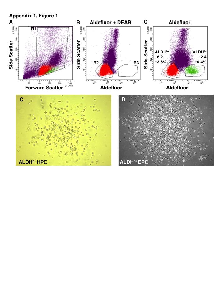
237 215
238 216 BASIC PROTOCOL 2 CO-CULTURE OF UCB ALDH hi CELLS TO ASSESS PARACRINE EFFECTS ON ENDOTHELIAL CELL SURVIVAL, PROLIFERATION, AND FUNCTION This protocol was adapted to characterize paracrine and juxtacrine support provided UCB progenitor cells on mature endothelial cell survival, proliferation and tubule formation in vitro (Putman et al., 2012). However, these co-culture systems could easily be adapted to test the potential vascular regenerative paracrine functions of any cell type of interest. NOTE: For this protocol human microvascular endothelial cells (HMVEC) are selected to model capillary or small vessel pro-angiogenic responses. Conversely, human umbilical vein endothelial cells (HUVEC) are used to model pro-angiogenic responses by larger vessel-resident endothelial cells. Materials Endothelial cells (HUVEC or HMVEC expanded in adherent culture, ATCC) EBM-2 Basal Medium (Lonza, cat. no. CC-3156) EGM-2 MV SingleQuot Kit & Growth Factors (Lonza, cat. no. CC-4147) Contains a cocktail of growth factors (EGF, VEGF, FGF, IGF-1) and fetal bovine serum (5% FBS) supplements to be added to basal media. Tissue culture treated dishes/plates (Corning; 75cm 2 flasks for expansion, cat. no , 12 well plate 3512, 24-well plate 3524 for co-culture assays) Phosphate Buffered Saline (PBS, prepared ahead) Trypsin Solution (Gibco, TrypLE Express (1X), no Phenol Red, cat. no ) Phosphate Buffered Saline + 5% fetal bovine serum (PBS+FBS, prepared ahead) ALDH-activity purified UCB cells (ALDH hi and ALDH lo from Basic Protocol 1) 50mL conical centrifuge tubes Hanging polycarbonate transwell inserts (3µm pore, Corning, Cat. no. 3402) Matrigel Basement Membrane Matrix - Growth Factor Reduced (BD Biosciences, cat. no ) Pipettors and 1000µL, 200µL, 20µL sterile tips Beckman GPR centrifuge with horizontal rotor hemacytometer and inverted light microscope or automated cell counter
239 217 Cell Culture Incubator at 37 C, 5% CO 2 Endothelial Cell Harvest 1. Aspirate growth media (EGM2-MV) and gently rinse culture dish with 10mL pure PBS (no FBS). 2. Add 1mL Trypsin solution to culture dish and swirl to cover surface. Incubate at room temperature or 37 C for 2-5 min to dissociate adherent HMVEC. 3. If cells do not lift easily from the growth surface you can force cell into suspension by hitting the base of the flask against the palm of your hand. 4. Once cells have lifted from flask (check visually on inverted light microscope) add 10mL PBS + 5% FBS solution and mix by repeated pipetting. Check flask to ensure no cells are left behind uncollected. 5. Collect cells into a 50mL conical tube and centrifuge at 240 x g for 7 minutes. 6. Aspirate supernatant and resuspend cells in 1 ml of EBM2 (Basal EC Media). 7. Count cells using trypan blue exclusion using a hemocytometer and light microscope or automated cell counter. 8. Distribute cells into a 50mL tube for each media condition to be used. Adjust the ratio of cell suspension added to each tube depending on number of wells needed for assay: I. GF+ Serum+ (EGM2-MV; fully supplemented) Positive control conditions for optimal EC survival and growth. II. GF- Serum- (EBM2; unsupplemented basal media) Negative control conditions for baseline EC survival. III. GF+ Serum- (EBM-2 + growth factors) Suboptimal conditions for EC growth without serum. IV. GF- Serum+ (EBM-2 + 5% FBS) Suboptimal conditions for EC growth without growth factors.
240 Spin all tubes at 240 x g for 7 minutes. 10. Aspirate supernatants and resuspend cells in appropriate media at 10 6 cells/ml Endothelial Cell Growth Rate Assay 1. Seed 40x10 3 HMVEC per well in tissue culture-treated 12-well plate. Cell densities have been optimized for a 12 well plate format and should be adjusted accordingly for culture in 6 or 24 well plates. 2. Under each growth condition, add 10 5 ALDH hi or ALDH lo cells into porous 3µm hanging transwells suspended above the HMVEC in the 12 well plate (Fig. 2A). 3. Incubate cell culture dishes at 37 C for 72 hours. 4. At 24, 48, and 72 hours, observe cells under stereomicroscope and record photomicrographs (Fig. 2B). 5. Enumerate trypan blue excluding HMVEC by blinded hemocytometer counts or automated cell counting to determine the total number and frequency of viable versus dead cells. Matrigel Tubule Formation Assay 1. Thaw matrigel overnight in ice bucket in fridge. 2. Plate 200μL into each well of a 24 well plate. Carefully, pipet Matrigel to evenly cover the base of the well. It is important to prechill your plate and pipet tips in the fridge to prevent the Matrigel from solidifying prematurely. Keep plate on an ice block, and Matrigel in ice bucket. Shake the dish if needed to evenly coat well. 3. Incubate plate at 37 C for at least 30 minutes to allow matrigel layer to set before plating cells.
241 Add appropriate media to each well up to a total volume 400μL. EGM2-MV; positive control EBM2; negative control and basal media for cell supplementation 5. Seed 50x10 3 HMVEC to each Matrigel-coated well. 6. Add 20x10 3 ALDH hi or ALDH lo cells. 7. Observe under stereomicroscope and obtain photomicrographs of 3 fields of view per well of matrigel at 24, 48 and 72 hours (Fig. 2C). 8. Enumerate number of branch points per field of view and quantify average tube length using image analysis software such as ImageJ.
242 220 SUPPORT PROTOCOL 2 PREPARING CONDITIONED MEDIA TO ASSESS PARACRINE SUPPORT OF ENDOTHELIAL CELL FUNCTION If desired, basic protocol 2 can be adapted for use with cell-conditioned basal media, prepared in advance, in place of direct co-culture in hanging transwell inserts (Fig. 2A). These assays demonstrate the added benefit of assessing only paracrine effects without the possibility of cell migration through the transwell. 1. Seed standardized number of ALDH lo or ALDH hi cells into appropriate sized culture dish containing EBM-2, basal media without serum or growth factors. For optimal cell density, seed x10 3 cells in a T75-flask (10-15mL), x10 3 cells in a T25 flask (3-5mL), or 50x10 3 cells in a 6 well plate (1mL). 2. Incubate culture for hours at 37ºC. 3. Using a pipet collect conditioned media into a 50mL conical centrifuge tube and spin at 450 x g for 5-7 minutes. 4. Collect supernatant and aliquot conditioned media into polystyrene tubes. 5. Store conditioned media at -30ºC until use.
243 221 Appendix 1. Figure 2. Co-culture assays to assess cell-mediated effect on survival proliferation tube forming capacity of endothelial cells (A) Schematic overview of hanging transwell co-culture and cell conditioned media assays to assess endothelial cell proliferation. (B) Representative photomicrograph of human endothelial cells cultured on plastic. (C) Representative photomicrograph of human endothelial cells growing on a thin layer of matrigel to assess modulation of tubelike structure formation.
244 222
245 223 BASIC PROTOCOL 3 MURINE FEMORAL ARTERY LIGATION AND EXCISION AND QUANTIFICATION OF VASCULAR REGENERATION IN VIVO This protocol outlines a surgical procedure for inducing unilateral acute hindlimb ischemia in immunodeficient mice. This protocol includes a double ligation with complete resection of the fermoral artery and vein. This procedure induces more severe ischemia than single ligation or non-resecting models. We have used immune deficient NOD/SCID mice (Jackson Laboratory, Bar Harbor, ME) to allow the survival and function of xenotransplanted human progenitor cells with reduced rejection due to complete knockout of T- and B-lymphocyte function (SCID mutation) and reduced innate immunity (NOD mutaton). However, this protocol can be applied to any immunodeficient mouse strain (NUDE, NOD/SCID IL-2R g null, NOD/SCID/MPSVIII mice) noting that there are strain specific differences in the magnitude and rate of recovery of perfusion following human cell transplantation. NOTE: The procedures described for the transplantation of human progenitor cells into highly immunodeficient mouse models of limb ischemia requires a dedicated barrier facility with an aseptic surgical and monitoring suite and sterile housing using hepafiltered ventilated racks. Materials Ketamine hydrochloride (100mg/mL, 60-70mg/kg) Xylazine (20mg/mL, 2-4 mg/kg) Eye lubricant (Allergan Refresh Lacrilube) Surgical scissors and forceps Sterile cotton tipped applicator 5-0 Silk Sutures Nexaband Liquid Topical Tissue Adhesive (Abbott Animal Health) Microsugical inverted microscope (Nikon SMZ2B) Microinjection Syringes Mobile Laboratory Animal Anesthesia System (VetEquip, cat. no )
246 224 Electric hair clippers (Wahl) Hair removal cream (Nair) Bacteriostatic soap Isopropyl alcohol Betadine solution Sterile cotton tipped applicators Cautery tool (Alcon Surgical Optemp II) Bupreorphine (0.1 mg/kg, subcutaneous) Heat Pad Laser Doppler imager (Moor LDI2) OCT tissue embedding medium Tissue moulds Liquid nitrogen Dry ice Cryostat microtome Mouse anti-human HLA-A,B,C antibody (1:500, Beckton Dickenson, cat. no ) Rat anti-mouse CD31 antibody (1:100 Beckton Dickenson, cat. no ) vwf antibody (1:200 Millipore, cat. no. AB7356) DAPI reagent Immunofluorescent Microscope (Zeiss AxioScope or similar) Anesthesia 3 Induce initial anesthesia by intraperitoneal injection of ketamine hydrochloride (60-70mg/kg) and xylazine (2-4mg/kg) solution. For ease of use and to reduce the number of injections needed prepare a working solution of ketamine and xylazine in saline or sterile water to use to intraperitoneal injection. 4 Place mouse into anesthesia induction chamber and fill chamber with 2-5% isoflurane in 100% O 2 at a flow rate of 0.8L/min.
247 225 5 Once mouse is unresponsive to external stimuli by foot pinch, place mouse on surgical surface in supine position and immobilize the right hindlimb by taping the foot to the surgical surface. 6 Maintain anesthesia for the duration of the surgical procedure using a nose cone with 1-2% isoflurane in 100% O 2 at a flow rate of 0.8L/minute. Surgery 1. Apply bepanthene ophthalmic ointment to the eyes to prevent drying during surgery. 2. Shave the hair from the lower limb and inguinal region using an electric shaver for bulk hair removal. Completely remove residual hair from lower limb by applying Nair hair removal cream briefly (1 min). 3. Wipe away excess hair removal cream and clean the skin and prepare surgical field with three rounds of bacteriostatic soap wash followed by isopropyl alcohol swab and betadine scrub. From here forward the protocol is most easily completed using a dissecting microscope. 4. Carefully make a 10-15mm longitudinal incision in the skin following the femoral vessel from the inguinal crease distally down the midpoint of the thigh using surgical scissors and forceps. 5. Use moistened sterile cotton tipped applicator and surgical scissors to gently clear subcutaneous fat away to expose the femoral artery and vein and associated neurovascular bundle (Fig. 3A). Locate the superficial epigastric artery branch point as a landmark.
248 Using the cautery tool, cauterize transversely the subcutaneous fat and the superficial epigastric artery. This will expose the muscle and neurovascular bundle underneath. 7. Carefully separate the femoral nerve from the femoral artery and vein starting near the groin and moving distally towards the knee using forceps and scissors. Avoid damaging the femoral nerve while separating it from the vessels. 8. Thread silk suture under the femoral artery and vein and gently slide it proximally toward the groin to maximize the length of the femoral artery and vein to be excised. Ligate the artery and vein using double surgical knots. 9. Thread silk suture under the femoral artery and vein and gently slide it proximally to the bifurcation of the popliteal and saphenous arteries that descend into the calf. Ligate the artery and vein using double surgical knots. 10. Using the cautery tool, cauterize the collateral vessels that emerge from the femoral artery between the ligation sites. 11. Carefully resect and excise the femoral artery and vein bundle between ligation sites with surgical scissors. Typically, it is possible to excise a 5mm segment of the femoral artery and vein between ligation sites. Carefully check the ligation site for full occlusion and for any signs of bleeding. 12. Close skin incision using silk sutures and/or Nexaband tissue glue. 13. For post-operative pain management, administer buprenorphine (0.1mg/kg) by subcutaneous injection. 14. Transfer mouse to recovery cage with direct access to water and food and monitor the mice for min ensuring they emerge from anesthesia and retain use of the surgical limb.
249 227 It is expected that the mice will show reduced mobility and use of the ischemic limb after surgery. Mice will demonstrate a notable limp for hours postoperatively. Gradually, mice will recover full mobility in the surgical limb. Be sure to monitor the incision sites daily for up to 7 days and look for signs of bleeding or excessive grooming that may result in the re-opening of the incision. If signs of incision damage occur, move mouse to a separate cage without littermates to allow healing. 15. Allow mice to recover for 24 hours before administration of the compound or cell type of interest to assess effects on the recovery of blood flow by laser Doppler perfusion imaging of the mouse hindquarters as described below. Useful administration techniques include intravenous tail vein injection to deliver cells or compounds to the general circulation to study the recruitment of injected cells to the ischemic region. Alternatively, direct intramuscular injection into the ischemic adductor or gastrocnemius muscle can be used to bypass the inefficiencies of cellular recruitment to the ischemic region. Laser Doppler Perfusion Imaging (LDPI) Analyses NOTE: LDPI timepoints and a representative example of the recovery of limb perfusion after intravenous injection of 2x10 5 UCB ALDH hi cells is shown in Fig. 3B. 1. Induce anesthesia as described in the surgery section above. Depending on the strain of mouse used the injection of xylazine and ketamine to induce anesthesia may not be necessary and induction using inhaled anesthesia may be sufficient. 2. Completely remove any residual hair from lower limb by applying hair removal cream briefly. Excess hair re-growth will interfere with the accuracy of the laser Doppler measurements. Typically, residual hair removal will be required every 1-2 weeks depending on mouse strain and fur color.
250 Transfer mouse to 37ºC draped heating plate for 5 minutes to stabilize body temperature. It is important to ensure stable body temperature in the mouse before LDPI scanning as decreased body will significantly alter blood flow in the hindlimbs. 4. Maintain anesthesia with 1-2% isoflurane in 100% oxygen at 0.8L/min. Carefully maintain anesthetic depth to prevent scan artifact from the mouse moving if too lightly anesthetized or from falsely low measurement of blood flow if too deeply anesthetized. 5. Place mouse in supine position under laser Doppler perfusion imager and extend hindlimbs. Follow manufacturer instructions for proper calibration of laser and measurement of superficial blood flow using consistent settings between subsequent measures at later time points. 6. Using the LDPI software acquire the perfusion image data. Adjust the background threshold to display LDPI flux measurements on the mouse hindquarters from the waist to the tips of the toes. 7. To quantify relative blood flow in the ischemic versus the control limb set a region of interest on each leg between the toes and heel (Fig. 3A), and measure the units of flux in the ischemic limb versus the non-ischemic limb. 8. To ensure consistent induction of acute unilateral hindlimb ischemia by the surgical procedures described above. Relative perfusion in the ischemic limb of NOD/SCID mice should be <10% of the perfusion flow in the control limb. 9. Repeat measurement of blood flow using the identical protocol at each time point to track the recovery of perfusion over a 28 day time course. We recommend performing LDPI at post-transplantation day 0, 3, 7, 14, 21, and 28 (Fig. 3B) in 8-10 mice per cohort to allow for statistical comparison between the recovery of perfusion in control (PBS injected) versus cellular transplanted treatments. Collection of Muscle Samples for Histological Analysis 1. At the predetermined end point of your experiment, euthanize the mice by cervical dislocation under anesthesia or by CO 2 inhalation.
251 229 Depending on the purpose of the experiment we routinely euthanize mice from each transplantation cohort at day 7; to address the recruitment of human cells to the ischemic region and to generate a snapshot of active vascular regenerative processes, at day 14 to address the survival of human cells in the ischemic region at the midpoint of the regenerative process, and at day 28 to document the overall extent of vascular recovery. At each endpoint extensive histochemical analyses can be performed (see below) at the site of surgery in the quadriceps muscle and distally in the gastrocnemius muscle. We routinely monitor human cell engraftment and survival, the recovery of capillary density and small neovessel formation, the induction of EC proliferation and prevention of apoptosis in the ischemic region, in order to fully document the cellular mechanisms of vascular recovery. 2. Remove skin and fasciae from the legs and lower limbs of the mouse. Using dissection scissors remove the superficial thigh muscles by making an incision beginning laterally across the knee and carefully peeling and cutting back the muscle from along the femur up towards the groin. 3. Embed the muscle in OCT compound in tissue mould blocks. Align embedded tissue so that the lateral cut across the knee or ankle is along the base of the block. 4. Dissect the gastrocnemius (calf) muscle by making incisions along the tendons inserting at the ankle and cut proximally along the lower leg bones towards the knee. 5. Embed the calf muscle in OCT compound in tissue mould blocks. Align embedded tissue so that the lateral cut across the knee is along the base of the block. Careful placement of each muscle sample in the tissue block allows efficient cross-sectional cuts through the muscle fibers during cryosectioning 6. Quickly freeze muscle sections suspended in OCT using liquid nitrogen flash freeze or by placing trays on a dry ice block. 7. Frozen tissue can be stored at -20ºC for up to 7 days. For long-term storage, transfer to a -80ºC freezer. 8. Cut 10-15μm sections using a cryostat microtome at -14ºC to -18C. Store dry sections at -20ºC. Transfer dry frozen sections to -80ºC freezer for long-term storage.
252 230 Human Cell Engraftment and Blood Vessel Density in Hindlimb Muscle Sections 1. Following standard immunohistochemistry or immunfluorescence protocols ( detect transplanted cell recruitment/engraftment using HLA-A,B,C expression to identify human cells. 2. Similary, immunostain for mouse CD31 expression to visualize murine capillary beds using immunofluorescent microscopy counterstained with DAPI or light microscopy counterstained with hematoxylin to mark nuclei (Fig. 3C). 3. Similarly, immunostain for mouse vwf expression to visualize larger murine arterioles using immunofluorescent microscopy counterstained with DAPI or light microscopy counterstained with hematoxylin to mark nuclei (Fig. 3D). This labels endothelial cells of larger higher shear stress vessels such as arterioles. Whereas CD31 staining can be inconsistent in larger vessels vwf reliably identifies larger vessels in muscle sections (Pusztaszeri et al. 2006). 4. Enumerate HLA-A,B,C + human cells, CD31 + capillary structures, and vwf + arterioles by blinded manual counts of 3 fields of view in each of 3 sections for each muscle sample.
253 231 Appendix 1. Figure 3. Transplantation model of acute unilateral hindlimb ischemia. (A) Diagram of the vasculature of the mouse hindlimb from the inguinal crease to the bifurcation of the femoral artery into the saphenous and popliteal arteries which descend into the calf and lower limb. Ligation sites for femoral artery ligation surgery are indicated. Representative laser Doppler perfusion images of mouse hindquarters before and after ligation and transection of femoral artery and vein. Regions of interest are selected from ankle to toe to quantify relative perfusion in the surgical limb. (B) Outline and suggested timing for tracking recovery of perfusion in the ischemic limb after surgical induction of acute ischemia. Number in lower left quadrant of each laser Doppler perfusion image is the ratio of perfusion in the ischemic limb to the perfusion in the control limb. (C,D) Representative photomicrographs of ischemic adductor muscle sections stained for (C) CD31and (D) vwf.
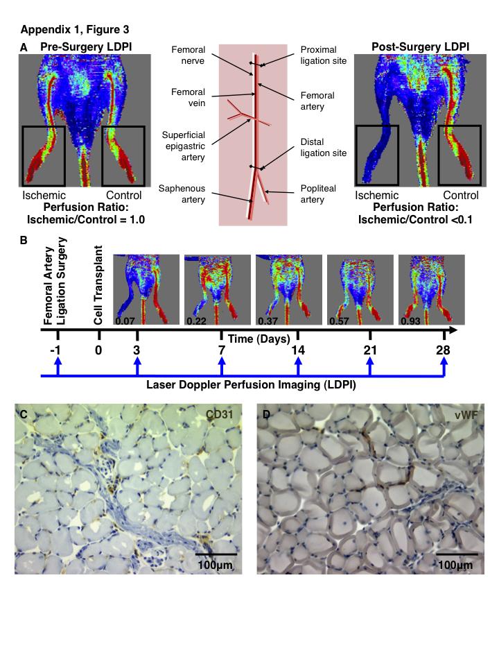
254 232
Growth factor delivery
 Growth factor delivery S. Swaminathan Director Centre for Nanotechnology & Advanced Biomaterials School of Chemical & Biotechnology SASTRA University Thanjavur 613 401 Tamil Nadu Joint Initiative of IITs
ISCT Telegraft Column: Mesenchymal Stromal Cell (MSC) Product Characterization and Potency Assay Development
 Product Characterization and Potency Assay Development") ISCT Telegraft Column: Mesenchymal Stromal Cell (MSC) Product Characterization and Potency Assay Development University of Wisconsin-Madison, Production Assistance for Cellular Therapies (PACT) Over the
ISCT Telegraft Column: Mesenchymal Stromal Cell (MSC) Product Characterization and Potency Assay Development University of Wisconsin-Madison, Production Assistance for Cellular Therapies (PACT) Over the
At the conclusion of this lesson you should be able to:
 Learning Objectives At the conclusion of this lesson you should be able to: Understand the key terms and definitions regarding stem cells Differentiate between the adult and embryonic stem cells Differentiate
Regulation of Hematopoietic Stem Cells and their Bone Marrow Niches by the Coagulation System*.
 Regulation of Hematopoietic Stem Cells and their Bone Marrow Niches by the Coagulation System*. * Via PAR-1 & CXCR4 upregulation, SDF-1 secretion and EPCR shedding. By Tsvee Lapidot, Ph.D. Weizmann Institute
NPTEL Biotechnology Tissue Engineering. Stem cells
 Stem cells S. Swaminathan Director Centre for Nanotechnology & Advanced Biomaterials School of Chemical & Biotechnology SASTRA University Thanjavur 613 401 Tamil Nadu Joint Initiative of IITs and IISc
Des cellules-souches dans le poumon : pourquoi faire?
 Des cellules-souches dans le poumon : pourquoi faire? Karl-Heinz Krause Dept. of Pathology and Immunology, Medical Faculty Dept. of Genetic and Laboratory Medicine, University Hospitals Geneva, Switzerland
About ATCC. Established partner to global researchers and scientists
 Discovering ATCC Primary Immunology Cells - Model Systems to Study the Immune and Cardiovascular Systems James Clinton, Ph.D. Scientist, ATCC July 14, 2016 About ATCC Founded in 1925, ATCC is a non-profit
Fundamental properties of Stem Cells
 Stem cells Learning Goals: Define what a stem cell is and describe its general properties, using hematopoietic stem cells as an example. Describe to a non-scientist the current progress of human stem cell
Preclinical development for SSc indications NO COPY. a preclinical portfolio in a perfect world. Jörg Distler
 Preclinical development for SSc indications a preclinical portfolio in a perfect world Jörg Distler Department of Internal Medicine 3 and Institute for Clinical Immunology University of Erlangen-Nuremberg
Contribution and Mobilization of Mesenchymal Stem Cells in a mouse model of carbon tetrachloride-induced liver fibrosis
 Contribution and Mobilization of Mesenchymal Stem Cells in a mouse model of carbon tetrachloride-induced liver fibrosis Yan Liu 1,*, Zhipeng Han 1,*, Yingying Jing 1,*, Xue Yang 1, Shanshan Zhang 1, Chen
BNG 331 Cell-Tissue Material Interactions. Wound Healing I
 BNG 331 Cell-Tissue Material Interactions Wound Healing I Course update LBL 4 Friday; paper posted Need a volunteer to switch groups! Much better job on LBL 3 figure summaries BNG spring seminar 3 this
Stem cells and tissue engineering
 Stem cells and tissue engineering S. Swaminathan Director Centre for Nanotechnology & Advanced Biomaterials School of Chemical & Biotechnology SASTRA University Thanjavur 613 401 Tamil Nadu Joint Initiative
Endothelium conference
 Endothelium conference Epithelium FUNCTIONS: COVER ORGANS, LINE VISCERA AND BLOOD VESSELS, SECRETORY CELLS OF GLANDS DISTINGUISHING FEATURES AND DISTRIBUTION: ALWAYS SIT ON A BASEMENT MEMBRANE, BUT COME
Figure S1. Phenotypic characterization of transfected ECFC. (a) ECFC were transfected using a lentivirus with a vector encoding for either human EPO
 ECFC were transfected using a lentivirus with a vector encoding for either human EPO") Figure S1. Phenotypic characterization of transfected ECFC. (a) ECFC were transfected using a lentivirus with a vector encoding for either human EPO (epoecfc) or LacZ (laczecfc) under control of a cytomegalovirus
Figure S1. Phenotypic characterization of transfected ECFC. (a) ECFC were transfected using a lentivirus with a vector encoding for either human EPO (epoecfc) or LacZ (laczecfc) under control of a cytomegalovirus
SmartPReP 2: The Gold Standard
 The Optimal Platelet Rich Plasma Composition SmartPReP 2: The Gold Standard Harvest Technologies is the leader in developing point-of-care cellular platforms to isolate and concentrate autologous growth
BUILT TO CONCENTRATE. Magellan is an autologous concentration system that delivers concentrated platelets and cells at the point of care.
 BUILT TO CONCENTRATE Magellan is an autologous concentration system that delivers concentrated platelets and cells at the point of care. DELIVER PERSONALIZED MEDICINE Every patient has a unique biology
Strategies for Assessment of Immunotoxicology in Preclinical Drug Development
 Strategies for Assessment of Immunotoxicology in Preclinical Drug Development Rebecca Brunette, PhD Scientist, Analytical Biology SNBL USA Preclinical Immunotoxicology The study of evaluating adverse effects
Supplemental Information Inventory
 Cell Stem Cell, Volume 6 Supplemental Information Distinct Hematopoietic Stem Cell Subtypes Are Differentially Regulated by TGF-β1 Grant A. Challen, Nathan C. Boles, Stuart M. Chambers, and Margaret A.
Regenerative Medicine and Stem Cell Therapies
 Regenerative Medicine and Stem Cell Therapies Regenerative Medicine Major component of successful regenerated / tissue engineered organs Scaffolds A critical element is the binding of the repopulating
UNIT CELL PROCESSES UNDERLYING TISSUE ENGINEERING AND REGENERATIVE MEDICINE
 Massachusetts Institute of Technology Harvard Medical School Brigham and Women s Hospital VA Boston Healthcare System 2.79J/3.96J/20.441/HST522J UNIT CELL PROCESSES UNDERLYING TISSUE ENGINEERING AND REGENERATIVE
Double Syringe System Autologous Conditioned Plasma
 TM Double Syringe System Autologous Conditioned Plasma Autologous Conditioned Plasma Introduction Autologous blood products have created a growing interest for use in a number of orthopaedic therapies.
Stem Cel s Key Words:
 Stem Cells Key Words: Embryonic stem cells, Adult stem cells, ips cells, self-renewal, differentiation, pluripotent, multipotent, Inner cell mass, Nuclear transfer (Therapeutic cloning), Feeder cells,
Mesenchymal Stem Cell Characterization. Peiman Hematti, M.D. Department of Medicine
 Mesenchymal Stem Cell Characterization Peiman Hematti, M.D. Department of Medicine Terminology has evolved over the years: Colony forming units-fibroblasts (CFU-E) Fibroblast colony forming cells (FCFC)
UvA-DARE (Digital Academic Repository) Factor XI as target for antithrombotic therapy van Montfoort, M.L. Link to publication
 Factor XI as target for antithrombotic therapy van Montfoort, M.L. Link to publication") UvA-DARE (Digital Academic Repository) Factor XI as target for antithrombotic therapy van Montfoort, M.L. Link to publication Citation for published version (APA): van Montfoort, M. L. (2014). Factor XI
UvA-DARE (Digital Academic Repository) Factor XI as target for antithrombotic therapy van Montfoort, M.L. Link to publication Citation for published version (APA): van Montfoort, M. L. (2014). Factor XI
cardiovascular cell Solutions
 The Essentials of Life Science Research Globally Delivered cardiovascular cell Solutions We love what we do. And now we do it even better! As part of your portfolio for success, ATCC now provides a range
Time allowed: 2 hours Answer ALL questions in Section A, ALL PARTS of the question in Section B and ONE question from Section C.
 UNIVERSITY OF EAST ANGLIA School of Biological Sciences Main Series UG Examination 2017-18 CELL BIOLOGY BIO-5005B Time allowed: 2 hours Answer ALL questions in Section A, ALL PARTS of the question in Section
Outcomes in Mesenchymal Stem Cell Manufacturing. Athena Russell, MT(AAB) Human Cellular Therapy Laboratory Mayo Clinic Jacksonville, FL
 Human Cellular Therapy Laboratory Mayo Clinic Jacksonville, FL") Outcomes in Mesenchymal Stem Cell Manufacturing Athena Russell, MT(AAB) Human Cellular Therapy Laboratory Mayo Clinic Jacksonville, FL Background HCTL established in 1992 to support BMT programs of Mayo
Outcomes in Mesenchymal Stem Cell Manufacturing Athena Russell, MT(AAB) Human Cellular Therapy Laboratory Mayo Clinic Jacksonville, FL Background HCTL established in 1992 to support BMT programs of Mayo
REGENERATIVE MEDICINE
 REGENERATIVE MEDICINE Umbilical Cord Blood-Derived Aldehyde Dehydrogenase-Expressing Progenitor Cells Promote Recovery from Acute Ischemic Injury DAVID M. PUTMAN, KEVIN Y. LIU, HEATHER C. BROUGHTON, GILLIAN
Regenerative Medicine. Speaker Disclosures. What is regenerative medicine? 6/22/2016
 Regenerative Medicine Halland Chen, MD Speaker Disclosures I disclose that I am not a consultant or have any financial interests. 2 What is regenerative medicine? Regeneration of human cells, tissues,
THE SCIENCE OF PLATELET RICH PLASMA
 THE SCIENCE OF PLATELET RICH PLASMA Cheri A. Ong, MD FACS ONG INSTITUTE FOR PLASTIC SURGERY & HEALTH WHAT IS PRP BLOOD PLASMA 55% of the body s today body volume 45% blood cells:- Red blood cells, White
Tissue Engineering. Mesenchymal Stem Cells for. Tissue Engineering
 Tissue Engineering Mesenchymal Stem Cells for Tissue Engineering Reference: Culture of Cells for Tissue Engineering (Culture of Specialized Cells), Chapter 2 Shu-Ping Lin, Ph.D. Date: 03.20.2012 Institute
Stem cells in Development
 ANAT 2341 Embryology Lab 10 8 Oct 2009 Therapeutic Use of Stem Cells Practical Hurdles & Ethical Issues Stem cells in Development Blastocyst Cord blood Antonio Lee PhD Neuromuscular & Regenerative Medicine
Stem cells in Development
 ANAT 2341 Embryology Lab 10 8 Oct 2009 Therapeutic Use of Stem Cells Practical Hurdles & Ethical Issues Stem cells in Development Blastocyst Cord blood Antonio Lee PhD Neuromuscular & Regenerative Medicine
PCCS Growth Media, Cell Tagging, Cell Separation Final Assignment. Igneris Rosado-Erazo. Panama College of Cell Science
 Running Head: Growth Media, Cell Tagging, Cell Separation PCCS Growth Media, Cell Tagging, Cell Separation Final Assignment Igneris Rosado-Erazo Panama College of Cell Science In partial fulfillment of
Introduction to Cell/ Biomaterial Engineering
 Introduction to Cell/ Biomaterial Engineering Module 3, Lecture 1! 20.109 Spring 2011! Topics for Lecture 1 Introduction to tissue engineering! motivation! basic principles + examples! Introduction to
hpsc Growth Medium DXF Dr. Lorna Whyte
 hpsc Growth Medium DXF Dr. Lorna Whyte 27.06.2014 Training from Heidelberg Overview Background: Stem Cells Introduction: Human Pluripotent Stem Cells (hpsc) vs. Adult Stem Cells Promise of PSC Research
Stem Cells, Regenerative Medicine and cgmp (GTP)
") Stem Cells, Regenerative Medicine and cgmp (GTP) Encompass Stem cell based therapies activities Collection source Purification Isolation from other cell types if needed Manipulation Minimal vs Moderate
Stem Cells, Regenerative Medicine and cgmp (GTP) Encompass Stem cell based therapies activities Collection source Purification Isolation from other cell types if needed Manipulation Minimal vs Moderate
Adult Stem Cells for Chronic Pain. Dr. John Hughes, DO January 24 th, 2018
 Adult Stem Cells for Chronic Pain Dr. John Hughes, DO January 24 th, 2018 Dr. John Hughes, DO Doctor of Osteopathy From Georgia Arizona College of Osteopathic Medicine - 2007 Aspen Integrative Medicine
Intradermal delivery of plasmids encoding angiogenic growth factors by electroporation promotes wound healing and neovascularization
 University of South Florida Scholar Commons Graduate Theses and Dissertations Graduate School 2009 Intradermal delivery of plasmids encoding angiogenic growth factors by electroporation promotes wound
Cytomics in Action: Cytokine Network Cytometry
 Cytomics in Action: Cytokine Network Cytometry Jonni S. Moore, Ph.D. Director, Clinical and Research Flow Cytometry and PathBioResource Associate Professor of Pathology & Laboratory Medicine University
BEH.462/3.962J Molecular Principles of Biomaterials Spring 2003
 Lecture 17: Drug targeting Last time: Today: Intracellular drug delivery Drug targeting Reading: T.J. Wickham, Ligand-directed targeting of genes to the site of disease, Nat. Med. 9(1) 135-139 (2003) Drug
Introduction to Cell and Biomaterial Engineering! Module 3, Lecture 1!! Spring 2014!
 Introduction to Cell and Biomaterial Engineering! Module 3, Lecture 1!! 20.109 Spring 2014! Topics for Lecture 1! Introduction to tissue engineering! motivation! basic principles! examples! Introduction
Lesson 7A Specialized Cells, Stem Cells & Cellular Differentiation
 Lesson 7A Specialized Cells, Stem Cells & Cellular Differentiation Learning Goals I can explain the concept of cell differentiation and cell specialization. I can explain how the cell structure relates
Mesenchymal Stromal/Stem Cells
 Mesenchymal Stromal/Stem Cells Peiman Hematti, M.D. Department of Medicine Financial Disclosure: Consulting Fees from Celgene Cellular Therapeutics in 2011 & 2012 1 Caplan used the term Mesenchymal Stem
3D Endothelial Pericyte Coculturing Kit Product Description Kit Components
 3D Endothelial Pericyte Coculturing Kit (3D-EPC) Cat #8728 Product Description Blood vessels are responsible for transporting blood throughout the body as part of the circulatory system. Their main components
Sign up to receive ATOTW weekly -
 BLOOD PHYSIOLOGY PART 2 ANAESTHESIA TUTORIAL OF THE WEEK 231 11 TH JULY 2011 Dr Karen Hayes Royal Devon & Exeter Correspondence to: kmhayes@hotmail.co.uk QUESTIONS Before continuing, try to answer the
The niche/microenvironment shapes the fate of researchers and stem cells
 The niche/microenvironment shapes the fate of researchers and stem cells Beate Heissig, MD Division of Stem Cell Dynamics Center of Stem Cell Biology and Regenerative Medicine The Institute of Medical
Future of Stem Cell Engineering. Jaeseung Jeong, Ph.D Department of Bio and Brain Engineering KAIST
 Future of Stem Cell Engineering i Jaeseung Jeong, Ph.D Department of Bio and Brain Engineering KAIST Keywords of Stem Cell Engineering g Embryo and Fetus (Foetus) Adult stem cells and embryonic stem cells
Supplementary Information. Conversion of vascular endothelial cells into multipotent stem-like cells
 Supplementary Information Conversion of vascular endothelial cells into multipotent stem-like cells Damian Medici 1, Eileen M. Shore 2,3,4, Vitali Y. Lounev 2,4, Frederick S. Kaplan 2,4,5, Raghu Kalluri
Lyset BOOST YOUR CELL CULTURE TODAY FOR THE EXPERIMENTS OF TOMORROW
 Lyset BOOST YOUR CELL CULTURE TODAY FOR THE EXPERIMENTS OF TOMORROW Lyset, the human platelet derived supplement for cell culture Among the different alternatives to animal serum, platelet derived preparations
Molecular Medicine. Stem cell therapy Gene therapy. Immunotherapy Other therapies Vaccines. Medical genomics
 Molecular Medicine Molecular Medicine Stem cell therapy Gene therapy Tumor therapy Immunotherapy Other therapies Vaccines Genetic diagnostics Medical genomics Medication Diagnostics medicine: old desire
PRIME-XV Hematopoietic Cell Basal XSFM
 PRIME-XV Hematopoietic Cell Basal XSFM Xeno-free, serum-free basal medium for human hematopoietic progenitor cell culture Optimized to support vigorous expansion of hematopoietic progenitor cells while
Structure of IgG and IgM
 Structure of IgG and IgM Fig. 5-1 A,B Crystal Structure of Secreted IgG Fig. 5-1 C Structure of an Ig Domain Fig. 5-2 Proteolytic Fragments of IgG (1) Fig. 5-3A Proteolytic Fragments of IgG (2) Fig. 5-3B
Supplementary Figure 1. Reconstitution of human-acquired lymphoid system in
 Supplementary Figure 1. Reconstitution of human-acquired lymphoid system in mouse NOD/SCID/Jak3 null mice were transplanted with human CD34 + hematopoietic stem cells. (Top) Four weeks after the transplantation
Module 1 Objectives Regenerative Cell Therapy. Module 1. Why Use Regenerative Cell Therapy? What is Regenerative Medicine? Regenerative Therapy Goals
 Module 1 Principles of Regenerative Medicine Module 1 Objectives Regenerative Cell Therapy Learn why to use regenerative cells Learn what they are Learn the relevant mechanisms of action Learn why adipose
In Vitro Angiogenesis Assay Kit
 In Vitro Angiogenesis Assay Kit Catalog Number KA1323 100 assays Version: 02 Intended for research use only www.abnova.com Table of Contents Introduction... 3 Background... 3 Principle of the Assay...
ALL THE ESSENTIALS OF HEALING, NATURALLY
 ALL THE ESSENTIALS OF HEALING, NATURALLY Harvest Technologies Developing Cellular platforms that may help optimize conditions for healing orthopedic sites Unlocking the Biologic Potential What is the Optimal
Cell-based therapies in tissue engineering and regenerative medicine (TE/RM)
") Cell-based therapies in tissue engineering and regenerative medicine (TE/RM) Pamela Gehron Robey, Ph.D. Craniofacial and Skeletal Diseases Branch National Institutes of Dental and Craniofacial Research
Cell-based therapies in tissue engineering and regenerative medicine (TE/RM) Pamela Gehron Robey, Ph.D. Craniofacial and Skeletal Diseases Branch National Institutes of Dental and Craniofacial Research
International Foundations of Medicine Basic Science Exam Blueprint
 International Foundations of Medicine Basic Science Exam Blueprint General Principles 28% 30% Biochemistry and molecular biology gene expression: DNA structure, replication, and exchange gene expression:
The Effects of Hydrostatic Pressure on Early Endothelial Tubulogenic Processes
 University of Kentucky UKnowledge Theses and Dissertations--Biomedical Engineering Biomedical Engineering 2013 The Effects of Hydrostatic Pressure on Early Endothelial Tubulogenic Processes Ryan M. Underwood
Regenerative medicine approaches to the treatment of cardiovascular disease
 Regenerative medicine approaches to the treatment of cardiovascular disease Author: Noah Weisleder, Ph.D. Abstract: Ischemic heart disease related myocardial infarction (MI) remains the leading cause of
بسم اهلل الرمحن الرحيم
 بسم اهلل الرمحن الرحيم Definition Multiple myeloma is a neoplastic plasma cell dyscrasia (PCD) that generally produced a monoclonal immunoglobulin protein, characterized by a clinical pentad: (a) anemia;
Physiology Unit 3 HEMATOLOGY
 Physiology Unit 3 HEMATOLOGY In Physiology Today Hematocrit Percentage of blood volume that is erythrocytes 45% in men 42% in women Average blood volume in is 5 L Plasma Large amounts of organic/inorganic
The European Stroke Network
 The European Stroke Network A Unique Alliance for Combating Stroke Prof. Dr. Stephen Meairs Department of Neurology University Medicine Mannheim University of Heidelberg, Germany European Stroke Network
Assuring Multipotency of human Mesenchymal Stem Cells (hmsc)
") Assuring Multipotency of human Mesenchymal Stem Cells (hmsc) Introduction Over the past decade, stem cell research has provided new avenues for deeper investigation into tissue repair and aging processes,
Assuring Multipotency of human Mesenchymal Stem Cells (hmsc) Introduction Over the past decade, stem cell research has provided new avenues for deeper investigation into tissue repair and aging processes,
PRIME-XV Cell Therapy Products by
 PRIME-XV Cell Therapy Products by Product List 2017 TRINOVA BIOCHEM now distributes the PRIME-XV Cell Therapy line by Irvine Scientific in Germany, Austria and Switzerland. Irvine Scientific is a worldwide
YOUR OWN LIFE
 YOUR OWN LIFE Stromal Tissue (ST), a Regenerative Source There is a worldwide consensus that the isolation and collection of regenerative Mesenchymal Stem Cells (MSC s) from differentiated body tissues
initial single-cell analysis, with a pragmatic focus on surface markers with the highest potential for
 Supplementary Figure 1: Summary of the exclusionary approach to surface marker selection for initial single-cell analysis, with a pragmatic focus on surface markers with the highest potential for protein
Transdifferentiated Human Vascular Smooth Muscle Cells are a New Potential Cell Source for Endothelial Regeneration
 Transdifferentiated Human Vascular Smooth Muscle Cells are a New Potential Cell Source for Endothelial Regeneration Hong, X., Margariti, A., Le Bras, A., Jacquet, L., Kong, W., Hu, Y., & Xu, Q. (2017).
Hematopoietic Progenitor Cell Product Characterization
 Hematopoietic Progenitor Cell Product Characterization Carolyn A. Taylor, Ph.D. Professor of Medicine Director of BMT Program Cell Processing Laboratory Product Testing and Characterization Goals Required
PTH Effects Bone Healing and Vasculogenesis in Calvaria Bone Allograft Model
 PTH Effects Bone Healing and Vasculogenesis in Calvaria Bone Allograft Model Dmitriy Sheyn, PhD 1, Doron Cohn-Yakubovich, BSc 2, Ilan Kallai, MSc 2, Susan Su, MD 1, Xiaoyu Da, MSc 1, Gadi Pelled, DMD,
How You Can Use Stem Cells
 How You Can Use Stem Cells Dr. Ralph T Bright Macquarie Cosmetic Medicine 21B Bathurst Street Liverpool NSW 2170 www.cosmeticmedicine.com.au (02) 9824 3044 Stem Cell Transfer Many of the people in this
NEXT GENERATION ECM-BASED ALLOGRAFT TECHNOLOGY:
 NEXT GENERATION ECM-BASED ALLOGRAFT TECHNOLOGY: Potent biological scaffolds strategically control stem cell fate and function, allowing our allografts to harness the regenerative potential of patient s
ASX Small to Mid Caps Conference Singapore. May 27 th, 2010
 ASX Small to Mid Caps Conference Singapore May 27 th, 2010 Mesoblast Capital Overview Fund Raisings $m IPO @ 50 cents 21.0 Equity Placements: Jul-06 17.4 Dec-07 13.4 Apr-09 10.8 May-10 37.0 Option funds
CELL THERAPY FOR THE TREATMENT OF DAMAGED MYOCARDIUM
 CELL THERAPY FOR THE TREATMENT OF DAMAGED MYOCARDIUM Non-Discrimination Statement and Multi-Language Interpreter Services information are located at the end of this document. Coverage for services, procedures,
All THE E SSEn TIAl S OF H EAl I ng, natu RAlly. P RIvATE PRACTICE
 All THE E SSEn TIAl S OF H EAl I ng, natu RAlly P RIvATE PRACTICE Harvest Technologies Developing Cellular platforms that may help optimize conditions for healing orthopedic sites Unlocking the Biologic
ARTHREX ACP. Features and Benefits: Introduction
 ARTHREX ACP Introduction Autologous blood products have created a growing interest for use in a number of orthopaedic therapies. The healing effects of plasma are supported by growth factors released by
Regulation of advanced blood cell therapies
 Regulation of advanced blood cell therapies www.pei.de Clinical trials using cell-based products Substantially manipulated cells and cells for non-homologous use Quality, safety and non-clinical aspects
PRODUCT REPORT. Premiere Regenerative Medicine.
 PRODUCT REPORT Premiere Regenerative Medicine 01 KEY TERMS Regenerative medicine; Cord blood; Growth factors; Mesenchymal stem cells (MSCs); Vascular enothelial growth factor (VEGF); Fibroblast basic growth
Stem Cells Canadian Perspective
 Stem Cells Canadian Perspective John Laffey Critical Illness and Injury Research Centre, Keenan Research Centre, St Michael s Hospital. Departments of Anesthesia, Physiology and Interdepartmental Division
ANAT 2341 Embryology Lecture 18 Stem Cells
 ANAT 2341 Embryology Lecture 18 Stem Cells 29 September 2010 Dr Antonio Lee Neuromuscular & Regenera
TISSUE ENGINEERING AND REGENERATION: TECHNOLOGIES AND GLOBAL MARKETS
 TISSUE ENGINEERING AND REGENERATION: TECHNOLOGIES AND GLOBAL MARKETS HLC101B August 2014 Yojana Jeevane Project Analyst ISBN: 1-56965-894-3 BCC Research 49 Walnut Park, Building 2 Wellesley, MA 02481 USA
Leading the world in novel adult stem cell therapies Half-Year Financial Results
 Leading the world in novel adult stem cell therapies 2013 Half-Year Financial Results CAUTIONARY NOTE REGARDING FORWARD-LOOKING STATEMENTS This presentation, including any comments made during or following
AIP1 functions as an endogenous inhibitor of VEGFR2-mediated signaling and inflammatory angiogenesis
 SUPPLEMENTAL MATERIALS functions as an endogenous inhibitor of VEGFR2-mediated signaling and inflammatory angiogenesis Haifeng Zhang 1*, Yun He 1*, Shengchuan Dai 1*, Zhe Xu 2*, Yan Luo 2, Ting Wan 2,
Characteristics. Capable of dividing and renewing themselves for long periods of time (proliferation and renewal)
") STEM CELLS (p 2-8) Overview The body is made up of about 200 different kinds of specialised cells, such as muscle cells, nerve cells, fat cells and skin cells. Cells with the capacity to give rise to the
STEM CELLS (p 2-8) Overview The body is made up of about 200 different kinds of specialised cells, such as muscle cells, nerve cells, fat cells and skin cells. Cells with the capacity to give rise to the
ANAT 3231 Cell Biology Lecture 21 Stem Cells
 ANAT 3231 Cell Biology Lecture 21 Stem Cells Outline What are Stem Cells? Totipotency - Pluripotency - Multipotency What are different sources of Stem Cells? Embryonic vs Adult Pros and Cons for each type
Cell membrane repair in acute lung injury: from bench to bedside. Disclosure. Cell membrane repair is an elemental physiological process
 Cell membrane repair in acute lung injury: from bench to bedside Disclosure Jianjie Ma, Ph.D. Karl P. Klassen Chair of Thoracic Surgery Department of Surgery Davis Heart and Lung Research Institute The
Accelerating skin wound healing by M-CSF through generating SSEA-1 and -3 stem cells. in the injured sites
 Accelerating skin wound healing by M-CSF through generating SSEA-1 and -3 stem cells in the injured sites Yunyuan Li, Reza Baradar Jalili, Aziz Ghahary Department of Surgery, University of British Columbia,
Adv Pathophysiology Unit 4 Page 1 of 10. Learning Objectives for this file:
 Adv Pathophysiology Unit 4 Page 1 of 10 Learning Objectives for this file: Topics include structure & function of: 1. Coagulation process platelets, clotting factors 2. Coagulation cascade formation of
Kieran McGourty, PhD. Short BIO. Long BIO
 Kieran McGourty, PhD Lecturer in Biochemistry and Cell Biology, Department of Chemical Sciences University of Limerick, Ireland Short BIO Kieran did his PhD and his Post-Doctoral scientific training in
Matrices sourcebook. Your guide to Gibco extracellular matrices products
 Matrices sourcebook Your guide to Gibco extracellular matrices products Introduction Gibco products, including extracellular matrices, 3D scaffolds, and attachment proteins, are essential tools for providing
Andrew Finnerty General Manager - CCMI
 Andrew Finnerty General Manager - CCMI Manufacturing Human Mesenchymal Stem Cells for Clinical Trials Quality Considerations Biopharma and Pharma RQA Regional Forum Bioclin Laboratories, Athlone 13 May
Anika Therapeutics, Inc.
 Anika Therapeutics, Inc. Jefferies Global Healthcare Conference June 4, 2015 Safe Harbor Statement The statements made in this presentation that are not statements of historical fact are forward looking
Mid-term review. Recitation5 03/31/2014. Stem Cell Biology and Function W4193 1
 Mid-term review Recitation5 03/31/2014 Stem Cell Biology and Function W4193 1 What to expect in the midterm2 It will be all so far but mainly whatever was NOT covered by the previous mid-term- not a huge
Hypoxic Preconditioning Strategy for Stem Cell Transplantation Therapy after Ischemic Stroke
 The 13 th International Conference on Dose-Response Preconditioning: Adaptive Responses in Biology and Medicine; UMass Amherst Hypoxic Preconditioning Strategy for Stem Cell Transplantation Therapy after
Promises and Challenges
 Vladimir Mironov & Richard Visconti Regenerative Medicine: Promises and Challenges What is Regenerative Medicine? Regeneration is a well known biological term and phenomenon. According to the popular Webster
Introduction to Cell- Biomaterial Engineering!
 Introduction to Cell- Biomaterial Engineering! Module 3, Lecture 1! 20.109 Spring 2010! Topics for Lecture 1!! Introduction to tissue engineerin! motivation"! basic principles + examples"! Introduction
ARM Cell Potency Project & Developing a Bioassay for an Allogeneic Cell Therapy Product
 ARM Cell Potency Project & Developing a Bioassay for an Allogeneic Cell Therapy Product Dr. Gil Van Bokkelen CASSS BioAssays 2013 March 4 th, 2013 ARM - Potency Project Overview 2011-2012 Project goal:
Erectile and voiding dysfunction: will stem cells solve it all?
 Erectile and voiding dysfunction: will stem cells solve it all? Maarten Albersen MD PhD Dept. of Development and Regeneration Leuven University, Belgium Disclosures No conflicts of interest. ED stem cell
THE THERAPEUTIC REALITIES OF STEM CELLS
 Ferghana Partners Group is a Life Sciences investment banking house with offices in London and New York. We specialise in creating and executing effective strategic transactions for pharmaceutical, chemical,