Histopathological techniques The adoption of routine fixation and paraffin wax embedding.
|
|
|
- Florence Barnett
- 6 years ago
- Views:
Transcription
1 CIHRT Exhibit P-3359 Page 1 Routine tissue preparation in modern diagnostic histopathology. Bryan R. Hewlett ART, MLT. Histopathological techniques The adoption of routine fixation and paraffin wax embedding Baker - Alcohol preservation of Hydra 1851 Clarke - Alcohol/acetic as fixative 1879 Fredericq - Alcohol/turpentine/wax (evaporated) 1881 Giesbrecht - Alcohol/turpentine/molten wax 1893 Blum - Formaldehyde as fixative 1899 Hardy - Studied variety of fixative/wax processes 1905 Brasil - Formaldehyde/alcohol/acetic fixative Medical schools include microtechnique FFPE becomes prevalent technique 1 * Bracegirdle Fixation Fixation is the single MOST important preparative histological technique. Poor fixation CANNOT be remedied at any later stage. The influence of processing Dependent on quality of fixation 3 4 1
2 CIHRT Exhibit P-3359 Page 2 Processing of tissues for histological analysis Dehydration Intermediate solvent (Clearing) Infiltration with support media Dehydration effects Following optimal fixation in NBF, ethanol removes some lipids and a few proteins not immobilized by cross-linking. This can produce a small amount of tissue dependent shrinkage (2-15%). Some hardening also occurs. Ethanol fixation produces tissue dependent shrinkage of 35-40% and much more hardening!!!! 5 6 Intermediate solvent effects Xylene is a true clearing agent i.e. it raises the R.I. of tissue. It also removes some lipids, causes some shrinkage and also some hardening. Paraffin wax effects Removes lipids and causes some hardening. The heated wax causes the majority of tissue shrinkage (may total 30-40%). May be reduced by minimizing the heat shock on transfer from xylene to molten wax. (Time may be shortened by agitation and negative pressure) 7 8 2


3 CIHRT Exhibit P-3359 Page 3 Effects of fixation/processing Loss of constituents Shrinkage Hardening Change in optical properties Inactivation of most enzymes Change in acidophilic/basophilic properties Destruction or masking of antigen epitopes Change in morphology ALL OF THESE EFFECTS ARE MINIMIZED FOLLOWING OPTIMAL FORMALDEHYDE FIXATION!! 9 The nature of fixatives Non-Coagulant fixatives Formaldehyde Protein secondary structure intact. Only modifies tertiary and quaternary structures, (Methylene bridge cross-links) mostly (90%) retrievable, with little loss (<1%) of protein. Coagulant Fixatives Alcohol Protein primary structure intact. Alters secondary and tertiary structures, (Hydrophilic/phobic inversion) often irretrievably, with loss of up to 40% of protein. 12 3


4 CIHRT Exhibit P-3359 Page 4 Non-coagulant fixative Fast penetration Slow fixation Little loss of constituents (< 1%) Little shrinkage Soft fixative Many effects are reversible Ileum NBF fixed section Ileum Alcohol fixed section 13 Most realistic overall morphology, allows widest range of histochemistry 14 Alcohol fixation Coagulant fixative Medium penetration (K = 1.0) Fast fixation (fixes as it penetrates) Loss of constituents ( 40%) Causes shrinkage Hard fixative Not readily reversible Fixation Reality #1 provides the most realistic overall morphology and becomes the standard fixative for the majority of routine diagnostic histopathologists! Great for nuclear morphology and staining of nucleoproteins, restricts range of histochemistry


5 CIHRT Exhibit P-3359 Page 5 Routine tissue preparation Development of tissue preparative techniques The current state of the art Fixation re-visited Effects on staining Effects on QA Formaldehyde allows the widest range of histochemical stains 17 What we need to do to improve 18 automated processing 1950 s processor (optional 24 hour and 7 day timers)


6 CIHRT Exhibit P-3359 Page 6 Fixation Reality #2 Following formaldehyde fixation; automated processing techniques provide an advantage in speed and ease of use, with no loss of morphology! 21 Modern closed processor (flexible timer and pressure/heat control) 22 Fixation Reality #3 Formaldehyde fixed automated processing Today s Routine becomes integrated with automated processing, providing a further advantage in speed and ease of use. The change in morphology is deemed acceptable and becomes Routine!


7 CIHRT Exhibit P-3359 Page 7 Technical Quality Standards Few universal standards are applied to routine histological techniques. Standards that exist are usually of a local, subjective nature, such as; our pathologist likes it this way we ve always done it this way it looks alright to me Technical Quality Standards Variations in one histological technique are often introduced as a local response to a real or perceived problem. The root cause of the problem may actually lie in another histological technique. These variations are empirically derived and are spread anecdotally. 25 This causes a wide range of reported results. 26 Small GI biopsy Routinely NBF-fixed and processed overnight. Feels Gritty on sectioning 27 THE ASSERTION: The tissue is; over-fixed, over-dehydrated over-processed PROPOSED SOLUTION: Change the pertinent processing times! 28 7


8 CIHRT Exhibit P-3359 Page 8 THE REALITY: The section was cut too rapidly, resulting in Knife edge vibrations or chattering. THE SOLUTION: Cut the section slowly, The chatters disappear! 29 The same small GI biopsy Routinely NBF-fixed and processed overnight. Sectioning performed at a slower rate, no longer feels gritty 30 Routine tissue preparation Development of tissue preparative techniques The current state of the art Fixation myths Formaldehyde fixes at a rate of 1.0 mm/hour. Fixation re-visited Effects on staining Effects on QA What we need to do to improve
9 CIHRT Exhibit P-3359 Page 9 How long will it take to fix? What is the fixative penetration rate? Tissue slice = 5 mm thick Core biopsy =1.5 mm thick Medawar, (1941) established that fixatives obey the diffusion laws. That is, the depth penetrated is proportional to the square root of time. Each fixative has a unique coefficient of diffusibility, designated K. Penetration rate may be determined from the formula; d = K x t 5 hours? 1.5 hours? Where d = depth in mm K = the Medawar coefficient t = the square root of fixation time in hours What is the coefficient of diffusibility (K)? Tellyesnicszky (1926) used thick tissue and long times. K = 0.78 Medawar (1941) used Plasma clots. K = 5.5 Baker(1958) used Gelatin/Albumen models. K = 3.6 (Helander s (1994) data indicates K must be AT LEAST 2.0 and probably closer to 3.5) Size = 5 cm Penetration rate Time = 1 hour K = 3.6 (Baker) Penetration = 3.6 mm Size = 5 mm Fully penetrated
10 CIHRT Exhibit P-3359 Page 10 How long will it take to fix? Penetration time at K = hour = 3.6 mm 4 hours = 7.2 mm (1.8 mm/hr) 16 hours = 14.4 mm (0.9 mm/hr) 64 hours = 28.8 mm (0.45 mm/hr) 256 hours = 57.6 mm (0.225 mm/hr) Fixation myths Formaldehyde fixes at a rate of 1.0 mm/hour. Small pieces of tissue fix faster than larger pieces. (to double the depth takes 4x the time) How long will it take to fix? Penetration time at K = 3.6 Tissue slice = 5 mm thick Core biopsy =1.5 mm thick 30 minutes <5 minutes K= hours 1 hour 1.0 cm thick tissue slice requires - 36 hours How long will it take to fix? The formaldehyde paradox. Histologists have known for more than 70 years, that fixation of tissue in formaldehyde demonstrates a bizarre effect. Namely, that formaldehyde is one of the fastest fixing agents to penetrate tissue but one of the slowest to fix. The paradox was explained (Burnett) in However, many histologists remain unaware of the implications
11 CIHRT Exhibit P-3359 Page 11 How long will it take to fix? How long will it take to fix? The chemical reaction. The reaction rate (The Clock reaction). 1) Formaldehyde covalently binds to reactive side chains on proteins at random to form unstable addition complexes. 2) Once a sufficient number of addition complexes are formed, they may slowly cross-link to each other by formation of methylene bridges. 3) Progressive formation of cross-links promotes gel formation and confers stability on the tissue. 4) These reactions are readily reversible. 41 1) In aqueous solution, formaldehyde is hydrated and mainly exists as methylene glycol. (< 1 part in 100,000 exists as free formaldehyde ) 2) Methylene glycol penetrates the tissue rapidly but does not fix. 3) Binding of the little available free aldehyde starts the clock reaction and allows slow decomposition of glycol to aldehyde over several hours C labeled formalin binding time 14 C labeled formalin binding time 100% Fox, et al 1985 Helander, 1994 Fox, et.al μ m section = 24 hours Helander, x 4 x 4 mm tissue cube = 25 hours BINDING 0% hours 6 days TIME
12 CIHRT Exhibit P-3359 Page 12 For initial stabilization of fixation to occur binding time is crucial, NOT penetration time. How long will it take to fix? The reaction rate. = 24 hours minimum for a 1.5 mm thick core biopsy. Tissue slice = 5 mm thick Core biopsy =1.5 mm thick = 24 hours minimum for a 5 mm thick tissue slice. Binding time + penetration time = Reaction rate! 24.5 hours! 24.1 hours! Fixation myths Formaldehyde fixes at a rate of 1.0 mm/hour. Small pieces of tissue fix faster than larger pieces. The optimal fixation time for formaldehyde is 24 hours. How long will it take to fix? The reaction rate. The minimum binding/stabilization time does not, unfortunately, denote complete fixation time. Progressive cross-linking continues over time. Complete fixation is thought to take at least 7 days. However, the stabilization at 24 hours is sufficient to allow reproducible results
13 CIHRT Exhibit P-3359 Page 13 Fixation Reality #4 Acceptable morphology on routine H&E stain does NOT correlate with acceptable staining by other methods! Especially with Immunohistochemistry! Why would anyone use Immunohistochemistry? IHC provides a final visual label on microscopic entities of interest, many of which cannot be identified by other histochemical techniques. IHC can answer the questions; What is it? Where is it? How much is present? Immunohistochemistry Immunohistochemistry Many different protocols in use. Sensitivity and Specificity varies with small changes in protocol. Wide range of reported results. Can provide answers to additional questions, such as; (Prognosis) What is the likely future course of the patient s disease? (Predictive) How will the disease respond to therapy?
14 CIHRT Exhibit P-3359 Page 14 Immunohistochemistry Immunohistochemistry The current and future use of IHC as a stand alone diagnostic, prognostic and predictive tool, demands reliable and reproducible results. These results may determine patient treatment! Current estimates indicate that IHC is required in up to 25% of malignancies. This will increase as proteomic studies produce more targeted therapeutic agents. Standardized IHC kits for antigens of interest and automated IHC instruments help, but variability of results is still problematic Quality in histotechnology Many histopathology laboratories use both quality control (QC) and quality assessment (QA) activities for the various individual steps involved in completing the daily workload. Unfortunately, little attention is given to the overall integration of these daily activities, to assess total quality (TQ). Quality control (QC) QC activities are prospective. i.e. they look forward at what will happen if all the steps in the process are followed. QC defines a product s quality and imparts to it the credibility needed for it s intended purpose. QC activities are the result of advanced planning and are applied to everything that contributes to the final product (on-line controls)
 QA activities are retrospective. i.e. they look back at what has happened, with a view to measuring the degree to which the desired outcomes are successful.")
 TQ, or the total test approach is a more holistic look at all the steps, in all the various processes used, from")


15 CIHRT Exhibit P-3359 Page 15 Quality assessment (QA) QA activities are retrospective. i.e. they look back at what has happened, with a view to measuring the degree to which the desired outcomes are successful. QA provides opportunities to subsequently modify the processes contributing to the final product (off-line controls). 57 Total quality (TQ) TQ, or the total test approach is a more holistic look at all the steps, in all the various processes used, from obtaining the sample until the final reporting of results. TQ involves integrating all of the QC and QA findings and understanding how changes to any of the various processes will affect the final outcome. The latter may involve experimentation to provide provenance for any proposed modification of the processes and the consequent QC and QA activity. 58 Immunohistochemistry IHC is technically complex. No aspect of this complexity can be ignored. QC of the IHC procedure alone is insufficient. QA and particularly EQA helps to provide valuable additional information for TQ. cytokeratins 59 DAB Routine IHC Vimentin DAB 60 15
16 CIHRT Exhibit P-3359 Page 16 Number of Labs PATH-0402 Overall Vimentin scores Immunohistochemistry The Total Quality Approach Looks at global factors that may influence IHC performance, from the collection of the sample to final interpretation. Both off-line and on-line controls are utilized to give total test performance Score 62 The Total Quality Approach Standardize factors that can be standardized. Understand the effect of those factors that cannot be standardized. Optimize the IHC procedure to accommodate all factors. Immunohistochemistry Immunohistochemical techniques rely upon the steric interaction (best fit) between the antibody paratope and the matching epitope of its target antigen. Recognition critically depends upon the epitope remaining unaltered and available to react. Validate the results!


17 CIHRT Exhibit P-3359 Page 17 Factors effecting the IHC Detection threshold Biological variation Sample collection Fixation/processing Section thickness Section pre-treatment Antibody-type/clone Antibody-dilution Sensitivity of detection reagents Histochemical reaction 68 17


18 CIHRT Exhibit P-3359 Page 18 The impact, of the nature & duration of fixation and processing, on IHC detection threshold is ENORMOUS! The routine histological section: Is it really formaldehyde fixed? (90% of IHC staining problems) 69 Ileum NBF fixed section H&E 70 Reality #5 No universal standard exists so what, exactly, IS a routinely formaldehyde-fixed, paraffin section? Your routine or mine? 71 Routine A Bowel polyp H & E Routine B 72 18


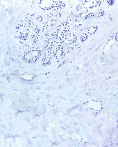
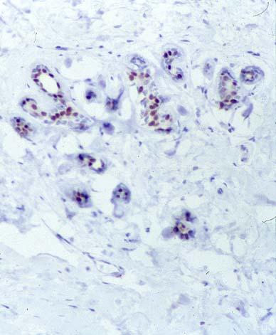
19 CIHRT Exhibit P-3359 Page 19 Routine A Breast Ca. IHC ER Routine B 73 Routine A Breast Ca. IHC HER2 Routine B 74 Routine A Seminoma IHC CD117 Routine B 75 Routine A = 6 hr E M Routine B = 24 hr 76 19
20 CIHRT Exhibit P-3359 Page 20 Reality #6 Routine fixation/processing consists of allowing tissues to fix for variable periods of time. The actual fixation time being dictated by the start time of the processing machine! Forms methylene bridges at reactive side chains Fast penetration (3.6 mm in 1 st hr) Slow fixation ( hrs) many effects reversible (<24 hrs) No shrinkage Soft fixative Mildly cross-links proteins Most versatile C labeled formalin Binding(24 hr) Reversal time at 25C 0% Helander, 1994 Non-Coagulant fixatives Formaldehyde The nature of fixatives Coagulant Fixatives Alcohol *during processing 50% 90% 100% hours 6 days TIME Protein secondary structure intact. Only modifies tertiary and Quaternary structures, (Methylene bridge cross-links) mostly (90%) retrievable, with little loss (<1%) of protein. Protein primary structure intact but alters secondary and tertiary structures, (Hydrophilic/phobic inversion) often irretrievably, with loss of up to 40% of protein


21 CIHRT Exhibit P-3359 Page 21 The routine histological section reality #7: Most are NOT really formaldehyde fixed! They are variably fixed by a combination of formaldehyde and alcohol. NBF = 24hr Alcohol = 24hr Ileum Adjacent blocks 3µm sections Batched H&E The shorter the time in NBF the more like Alcohol fixation Ileum Routinely NBF fixed H & E 81 Routine - NBF = 8 hr 82 Routine formaldehyde fixation/processing; Results in; variable morphology and cell content variable shrinkage and hardening variable masking/destruction of epitopes variable porosity variable basophilic/acidophilic relations variable intensity of stains variable success/failure of staining techniques! 83 Alcohol-fixed IHC - Chromogranin NBF-fixed 84 21


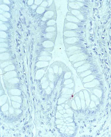
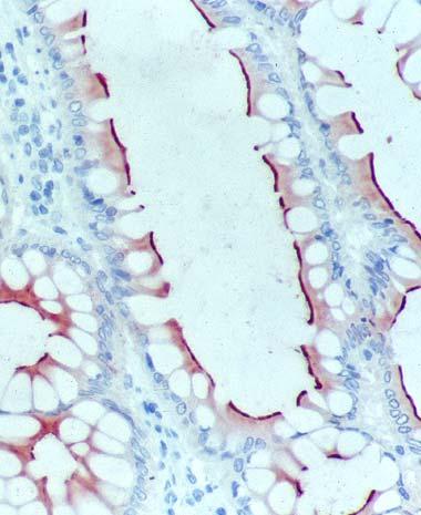
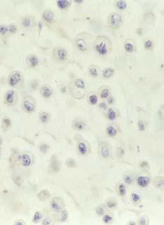
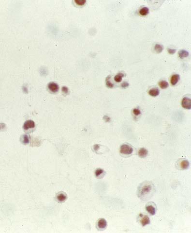
22 CIHRT Exhibit P-3359 Page 22 Alcohol-fixed IHC - NSE NBF-fixed 85 Alcohol-fixed IHC - CEA NBF-fixed 86 Alcohol-fixed MCF-7 cells IHC - ER NBF-fixed 87 Alcohol-fixed Breast IHC ER NBF-fixed 88 22


 (ALL")
 Referred-in positivity rate = 8% ( + 20%")
 Lymph node, Breast Ca.")
23 CIHRT Exhibit P-3359 Page 23 Membrane protein stripping Edge of clot Alcohol-fixed SKBR3 cells IHC HER2 NBF-fixed 89 SKBR3 cells NBF-fixed for 4 hours IHC HER2 90 Impact of routine tissue preparation on HER2 IHC positivity rates (2002). In-house positivity rate = 22% (+ 5% equivocal) (ALL fixed for minimum of 24 hours, including core biopsies) Referred-in positivity rate = 8% ( + 20% equivocal) ( routinely fixed) Over-all positivity rate = 13% (Accepted range = 18 22%) Lymph node, Breast Ca. NBF-fixed for approx. 8 hours IHC HER


 for the")


24 CIHRT Exhibit P-3359 Page 24 Myths Formalin fixation for more than 24 hours is Overfixation and will destroy immunoreactivity. Reality #9 There is NO such thing as Overfixation in formalin. Progressive cross-linking does occur over time. This may lead to masking of antigens. This does not occur within any reasonable time frame (5-7 days) for the majority of antigens of clinical interest. NBF, 24hr. fixation CD5 NBF, 7day fixation Fixation Fixation is the single MOST important preparative histological technique. We have tested ER, HER2 and CD117 for up to 90 days of fixation time, finding NO significant loss in immunoreactivity after 24 hours. Poor fixation CANNOT be remedied at any later stage
25 CIHRT Exhibit P-3359 Page 25 Routine tissue preparation Reality #10 Development of tissue preparative techniques The current state of the art Standardize fixation! Fixation re-visited Effects on staining Effects on QA Standardize fixation times for all tissues requiring prognostic/predictive markers! (24 hour minimum in NBF) What we need to do to improve 97 Is this the time to duck? 98 Strategies Standardize Fixation by; 1) Cut thin (3-4mm) blocks. 2) Use a 20:1 fixative tissue ratio. 3) Adopt a routine minimum 24hr fixation time. OR 4) Fix only Special tissues/blocks for 24hrs. OR 5) Stay with variable routine fixation and HOPE! Consequences 1) Improved fixation and processing. 2) Improved maintenance of ph and fixation. 3) Extension of current turn-around time (TAT). OR 4) Only Special tissues/blocks TAT affected. OR 5) No standardization! Standardized fixation improves IHC performance!

 July 2003. Goldstein et. al.")


. CAP/ASCO.")
26 CIHRT Exhibit P-3359 Page 26 Recommended fixation times Estrogen Receptor protein. OAP/CAP- SenGupta et. al. Fixation in buffered formaldehyde for hours. AJCP, 120(1) July Goldstein et. al. NBF fixed 8 hours NBF fixed 24 hours Minimum formalin fixation time for reliable IHC ER results is 6-8 hours regardless of the type or size of specimen. MCF-7 cells ER Breast Cancer. ER = Positive (range of expression) Standardized IHC protocol 103 Recommended fixation times HER2 protein. HercepTest. Tissues from the biopsy should be blocked into a thickness of 3 or 4 mm and fixed for hours in neutral buffered formalin (NBF). CAP/ASCO. Fixation in NBF for 6-48 hours. QMP-LS. Fixation in phosphate buffered formaldehyde for a minimum of hours


27 CIHRT Exhibit P-3359 Page 27 NBF fixed 8 hours NBF fixed 24 hours SKBR3 cells 1+ 0 HER2 105 Range of IHC staining for HER2 seen in breast cancer Standardized IHC protocol 106 Myths The IHC control tissues should be fixed and processed in a similar manner to the test sample. CLSI guidelines for IHC Single Tonsil, NBF strategic time set H&E 7 day, 3 day, 24hr, 16hr Reality #11 Yes they should, but unless you have adopted a standard fixation time for ALL test samples, this is virtually impossible! In practice, a strategic fixation time set on a given control tissue will satisfy this requirement. 2 hr, 4hr, 8hr, 12hr


28 CIHRT Exhibit P-3359 Page 28 Myths 4hr. fixation Every antibody should have AR pre-treatment. Reality #15 Each antigen-antibody interaction is unique. The necessity for any form of pre-treatment is dictated by the response to fixation of individual antigen epitopes. 24hr. fixation CD23 7day fixation Pre-treatment May un-mask or improve the accessibility of epitopes. May damage other epitopes. May enhance unwanted cross-reactions. May degrade morphology. All are dependent on fixation/processing! Pre-treatment There are NO standards for pre-treatment! For proteolytic agents, standardize the time, temperature and concentration for use. (activity rating in units/mg solid) For HIER, standardize the time at temperature, the buffer concentration and ph. 111 These can ONLY be truly standardized if the fixation is!

29 CIHRT Exhibit P-3359 Page 29 Use The Total Quality Approach Standardize factors that can be standardized. Understand the effect of those factors that cannot be standardized. Any smoothly functioning technology has the appearance of magic Optimize the IHC procedure to accommodate all factors. Validate the results! Arthur C. Clark
TheraLin. Universal Tissue Fixative Enabling Molecular Pathology
 TheraLin Universal Tissue Fixative Enabling Molecular Pathology TheraLin Universal Tissue Fixative Enabling Molecular Pathology Contents Page # TheraLin Universal Tissue Fixative 3 Introduction 5 Easy
Preparation of tissues for study
 Preparation of tissues for study HISTOLOGY : It is the branch of science which deals with the microscopic study of normal tissue HISTOPATHOLOGY : It is the branch of science which deals with the microscopic
General Comments. Misinformation in Immunohistochemistry. Common Misinformation s in Immunohistochemistry 4/13/2017
 Common Misinformation s in Immunohistochemistry Tri State Meeting May 3, 2017 Steven Westra Reagent Product Specialist Leica Biosystems Misinformation in Immunohistochemistry Flood of New Markers Diagnostic
CIHRT Exhibit P-1764 Page 1 IMMUNOHISTOCHEMISTRY ACCURATE LOCALIZATION OF TISSUE OR CELLULAR CONSTITUENTS WITH ANTIBODIES
 CIHRT Exhibit P-1764 Page 1 IMMUNOHISTOCHEMISTRY ACCURATE LOCALIZATION OF TISSUE OR CELLULAR CONSTITUENTS WITH ANTIBODIES CIHRT Exhibit P-1764 Page 2 FUNCTIONAL ROLE OF ANTIBODIES Identify the tissue of
Comparing the Quality of Fixation for Gel-based Formalin (Formagel) versus Traditional Liquid-Based Formalin for Immunohistochemistry
 versus Traditional Liquid-Based Formalin for Immunohistochemistry") Comparing the Quality of Fixation for Gel-based Formalin (Formagel) versus Traditional Liquid-Based Formalin for Immunohistochemistry Brian H. Le, M.D., Reading Hospital Reviewed by Michael R. LaFrinere,
Comparing the Quality of Fixation for Gel-based Formalin (Formagel) versus Traditional Liquid-Based Formalin for Immunohistochemistry Brian H. Le, M.D., Reading Hospital Reviewed by Michael R. LaFrinere,
Methodology for Immunohistochemistry. Learning Objectives:
 Proteomics Methodology for Immunohistochemistry Methodology for Immunohistochemistry A staining process for identifying the proteins location in cells, tissues by using antigen-antibody property. Immuno
Overview of Immunohistochemistry
 Overview of Immunohistochemistry Immunohistochemistry (IHC) combines anatomical, immunological and biochemical techniques to identify discrete tissue components by the interaction of target antigens with
THE BASICS OF IMMUNOHISTOCHEMISTRY
 THE BASICS OF IMMUNOHISTOCHEMISTRY Introduction Immunohistochemistry (IHC) identifies specific tissue components by means of a specific antigen/antibody reaction tagged with a visible label. IHC makes
KCC Path-Core Page 1 of 5
 Instructions for Sample preparation for Paraffin embedding PLEASE NOTE: There is no one-size-fits-all method of tissue preparation for all experimental designs. Before harvesting tissue, you need to assess
Overview of Immunohistochemistry. (with a focus on wax-embedded sections)
") Overview of Immunohistochemistry (with a focus on wax-embedded sections) Overview of Immunohistochemistry (with a focus on wax-embedded sections) Overview of Immunohistochemistry IHC is like cooking. There
Overview of Immunohistochemistry (with a focus on wax-embedded sections) Overview of Immunohistochemistry (with a focus on wax-embedded sections) Overview of Immunohistochemistry IHC is like cooking. There
PREPARATION OF HISTOLOGICAL SPECIMENS
 PREPARATION OF HISTOLOGICAL SPECIMENS Histo-techniques Preparation of tissue for microscopic examination Series of processes Ultimate aim to make tissue visible as it is Pathology Vs Anatomy Steps vary
Preparation of thin slices for light microscopy
 Preparation of thin slices for light microscopy Optical light microscopy course 23.10.2012 Kirsi Rilla Shortly: Histological sample preparation for microscopy 1. Fixation: To fix the tissue components
Technical Note. Tissue Section Imaging. Published August The most recent version of this Technical Note is posted at licor.com/bio/support.
 Technical Note Tissue Section Imaging Published August 2017. The most recent version of this Technical Note is posted at licor.com/bio/support. Page 2 - Tissue Section Imaging Table of Contents Page I.
Laboratory Accreditation Test Validation: A Brave New World for Anatomic Pathology
 Laboratory Accreditation Test Validation: A Brave New World for Anatomic Pathology Francis E. Sharkey, MD, FCAP University of Texas Health Science Center, San Antonio, TX Richard W. Brown, MD, FCAP Memorial
Product Datasheet and Instructions for Use
 Product Code: MP-109-CM01 (0.1ml conc) MP-109-CM05 (0.5ml conc) MP-109-CM1 (1ml conc) MP-109-PM6 (6ml RTU) Product Description: Androgen Receptor Concentrated and Prediluted Monoclonal Antibody Control
TOTAL TISSUE DIAGNOSTICS PATHOS DELTA. World s first hybrid tissue processor (microwave + conventional) A fully automatic system to fit your workflow
 A fully automatic system to fit your workflow") TOTAL TISSUE TM DIAGNOSTICS MILESTONE H E L P I N G P A T I E N T S From surgical specimen to diagnosis PATHOS DELTA World s first hybrid tissue processor (microwave + conventional) A fully automatic system
TOTAL TISSUE TM DIAGNOSTICS MILESTONE H E L P I N G P A T I E N T S From surgical specimen to diagnosis PATHOS DELTA World s first hybrid tissue processor (microwave + conventional) A fully automatic system
PRACTICAL -3 SELECTION OF THE TISSUE BLOCK
 PRACTICAL -3 SELECTION OF THE TISSUE BLOCK Ms. Khadija Al-Zahrani Before the specimens come to the laboratory: 1. Immediate fixation & the suitable fixative. 2. Enough amount of fixative. 3. Suitable container.
Frozen tissue section
 IHC Protocol - Frozen Tissue Author : Dan Souw Immunohistochemistry on Frozen tissues IHC Protocol - Frozen Tissue: An introduction This is the second post in a series on immunohistochemistry (IHC). The
Risk Mitigation in Breast Predictive Factor Testing. Elizabeth H Hammond MD
 Risk Mitigation in Breast Predictive Factor Testing Elizabeth H Hammond MD Risk Defintion Risk is the opportunity for error, and risk of an inaccurate test result for the patient. Goal is the right test
Web Based Promotion! 10% Off any initial product order. Mention promo code 1204!
 Web Based Promotion! 10% Off any initial product order. Mention promo code 1204! Volume 2, Number 2 1998 FROM PATIENT TO EMBEDDING CENTER IN TWO HOURS OR LESS The single biggest factor in health care today
Immunohistochemistry guide
 Immunohistochemistry guide overview immunohistochemistry Overview Immunohistochemistry is a laboratory technique utilized for the visual detection of antigens in tissue. When working with cells this technique
Product Datasheet and Instructions for Use
 Product Datasheet and Instructions for Use Product Code: MP-323-CM01 (0.1ml conc) MP-323-CM05 (0.5ml conc) Product Description: CD24 Concentrated Monoclonal Antibody Control Number: 901-323-052510 ISO
GenomeMe. GeneAbTM Her2/Neu. Clone: IHC002 Source: Mouse Monoclonal Positive Control: Breast Carcinoma
 GeneAbTM Her2/Neu Clone: IHC002 Source: Mouse Monoclonal Positive Control: Breast Carcinoma 1. Intended Use This antibody is intended for in vitro diagnostic (IVD) use. The Her2/Neu (IHC002) antibody is
GenomeMe. GeneAb TM BRAF V600E. Clone: IHC600 Source: Mouse Monoclonal Positive Control: Colorectal Adenocarcinoma
 GeneAb TM BRAF V600E Clone: IHC600 Source: Mouse Monoclonal Positive Control: Colorectal Adenocarcinoma 1. Intended Use This antibody is intended for in vitro diagnostic (IVD) use. The BRAF V600E (IHC600)
SOP# version e1.0 Material Handling and Documentation Preservation of Tissue: Paraffin Embedding
 CTRNet Standard Operating Procedure SOP Number: 8.3.005 Version e1.0 Supersedes: SR 001.001 Effective Date 09 Jan 08 Subject: Preservation of Tissue: Paraffin Embedding Category Material Handling and Documentation
College of American Pathologists Laboratory Accreditation Program. AP for Histotechs and Secretaries. Learning Objectives
 College of American Pathologists AP for Histotechs and Secretaries Francis E. Sharkey, M.D., FCAP University of Texas Health Science Center - San Antonio February 20th, 2008 Copyright 2008 College of American
Product Datasheet and Instructions for Use
 Product Datasheet and Instructions for Use Product Code: MP-138-CM01 (0.1ml conc) MP-138-CM05 (0.5ml conc) MP-138-CM1 (1ml conc) Product Description: Biotinylated Bromodeoxyuridine (BrdU) Concentrated
ab TripleStain IHC Kit: M&M&R on human tissue (DAB, Red/AP & DAB/Ni)
") ab183287 TripleStain IHC Kit: M&M&R on human tissue (DAB, Red/AP & DAB/Ni) Instructions for Use For the detection of Rabbit and Mouse Primary antibodies on Human tissue or cell samples. This product is
ab183287 TripleStain IHC Kit: M&M&R on human tissue (DAB, Red/AP & DAB/Ni) Instructions for Use For the detection of Rabbit and Mouse Primary antibodies on Human tissue or cell samples. This product is
Tissue Tackle AEC Mouse Immunohistochemistry System Cat # HCS26
 Tissue Tackle AEC Mouse Immunohistochemistry System Cat # HCS26 Table of Contents Page Intended Use... 1 Background... 2 Principle of the Assay... 2 Materials Provided... 3 Materials Required But Not Provided...
Which hydrogel preparation for immunostaining protocol should I use?
 Protocol: Preparation of TissueSpec hydrogels for immunostaining This protocol may be used prior to immunostaining cells, organoids, or patient-derived xenografts cultured in TissueSpec matrix hydrogels.
GenomeMe. GeneAb TM Collagen Type IV. Clone: IHC549 Source: Mouse Monoclonal Positive Control: Lung, Muscle
 GeneAb TM Collagen Type IV Clone: IHC549 Source: Mouse Monoclonal Positive Control: Lung, Muscle 1. Intended Use This antibody is intended for in vitro diagnostic (IVD) use. The Collagen Type IV (IHC549)
2013 Continuing Compliance Master Series Checklist Updates for Anatomic Pathology. Gerald A. Hoeltge, MD, FCAP July 17,
 2013 Continuing Compliance Master Series Checklist Updates for Anatomic Pathology Gerald A. Hoeltge, MD, FCAP July 17, 2013 www.cap.org Today s Presenter Gerald A. Hoeltge, MD FCAP Chair, Checklists Committee
Supporting Protocols
 Supporting Protocols This protocol may be used prior to immunostaining cells, organoids, or patient-derived xenografts cultured in TissueSpec ECM Hydrogels. Introduction Cells and organoids may form complex
ab EXPOSE Rabbit Specific HRP/DAB Detection IHC Kit
 Version 3 Last updated 3 November 2017 ab80437 - EXPOSE Rabbit Specific HRP/DAB Detection IHC Kit For the detection of a specific antibody bound to an antigen in tissue sections. This product is for research
ab EXPOSE Mouse and Rabbit Specific HRP/DAB Detection IHC Kit
 Version 7 Last updated 17 January 2018 ab80436 - EXPOSE Mouse and Rabbit Specific HRP/DAB Detection IHC Kit For the detection of a specific antibody bound to an antigen in tissue sections. This product
ab Human on human IHC kit (HRP/DAB)
") Version 1 Last updated 13 September 2016 ab214749 Human on human IHC kit (HRP/DAB) For staining human primary antibodies on human tissues without background staining This product is for research use only
Version 1 Last updated 13 September 2016 ab214749 Human on human IHC kit (HRP/DAB) For staining human primary antibodies on human tissues without background staining This product is for research use only
ab TripleStain IHC Kit: R&R&M on human tissue (DAB, AP/Red & Green/HRP)
") ab183288 TripleStain IHC Kit: R&R&M on human tissue (DAB, AP/Red & Green/HRP) Instructions for Use For the detection of Rabbit and Mouse Primary antibodies on Human Tissue. This product is for research
ab183288 TripleStain IHC Kit: R&R&M on human tissue (DAB, AP/Red & Green/HRP) Instructions for Use For the detection of Rabbit and Mouse Primary antibodies on Human Tissue. This product is for research
A Disruptive Approach.
 1 Taking Control of Fixation for Improved IHC. A Disruptive Approach. 2 ASCO/CAP Recommendations for HER2 Results: Approx. 20% of current HER2 testing may be inaccurate What can be done? ASCO/CAP Guideline
ab64254 Liquid Fast-Red Substrate Kit (75X)
") Version 1 Last updated 6 June 2018 ab64254 Liquid Fast-Red Substrate Kit (75X) For the immunohistochemical staining. This product is for research use only and is not intended for diagnostic use. Table
Version 1 Last updated 6 June 2018 ab64254 Liquid Fast-Red Substrate Kit (75X) For the immunohistochemical staining. This product is for research use only and is not intended for diagnostic use. Table
Fixation and Processing
 Fixation and Processing Freida L. Carson Fixation Fixation is the single most influential factor in the long sequence of steps between procurement of the specimen and coverslipping the stained slide; nearly
ab Human on human IHC kit (AP/Permanent
 Version 1 Last updated 13 September 2016 ab214753 Human on human IHC kit (AP/Permanent Red) For staining human primary antibodies on human tissues without background staining This product is for research
ab Mouse and Rabbit Specific HRP/DAB (ABC) Detection IHC Kit
 Detection IHC Kit") ab64264 - Mouse and Rabbit Specific HRP/DAB (ABC) Detection IHC Kit Instructions for Use For the detection of a specific antibody bound to an antigen in tissue sections. This product is for research use
ab64264 - Mouse and Rabbit Specific HRP/DAB (ABC) Detection IHC Kit Instructions for Use For the detection of a specific antibody bound to an antigen in tissue sections. This product is for research use
COPYRIGHTED MATERIAL. Tissue Preparation and Microscopy. General Concepts. Chemical Fixation CHAPTER 1
 CHAPTER 1 Tissue Preparation and Microscopy General Concepts I. Biological tissues must undergo a series of treatments to be observed with light and electron microscopes. The process begins by stabilization
Keratin 19 (KRT19) Immunohistochemistry Kit
 Immunohistochemistry Kit") Keratin 19 (KRT19) Immunohistochemistry Kit For Immunohistochemical Staining of Keratin 19 (KRT19) in human FFPE Tissue RUK-KKR01-20 For Research Use Only Riverside Biosciences Inc. 2327 S 5th Ave, North
Keratin 19 (KRT19) Immunohistochemistry Kit For Immunohistochemical Staining of Keratin 19 (KRT19) in human FFPE Tissue RUK-KKR01-20 For Research Use Only Riverside Biosciences Inc. 2327 S 5th Ave, North
1. Paraffin section slides can be stored at room temperature for a long time.
 Immunohistochemistry (IHC) Protocols Immunohistochemistry (IHC) Protocol of Paraffin Section 1. Fix dissected tissues with 10% formalin for no less than 48 hours at room temperature. Inadequately fixation
ebioscience BrdU Kit for IHC/ICC Colorimetric Catalog Number: RUO: For Research Use Only. Not for use in diagnostic procedures.
 Page 1 of 1 ebioscience BrdU Kit for IHC/ICC Colorimetric Catalog Number: 8800-6599 RUO: For Research Use Only. Not for use in diagnostic procedures. Product Information Contents: ebioscience BrdU Kit
Central Laboratory Services. Anatomic Pathology Histology Services
 Central Laboratory Services Anatomic Pathology Histology Services Oncology and Beyond... There is no question that innovations in technology have the potential to significantly impact a number of clinical
Adenomatous Polyposis Coli (APC) Immunohistochemistry Kit
 Immunohistochemistry Kit") Adenomatous Polyposis Coli (APC) Immunohistochemistry Kit For Immunohistochemical Staining of Adenomatous Polyposis Coli (APC) in human FFPE Tissue RUK-KAP01-20 For Research Use Only Riverside Biosciences
Adenomatous Polyposis Coli (APC) Immunohistochemistry Kit For Immunohistochemical Staining of Adenomatous Polyposis Coli (APC) in human FFPE Tissue RUK-KAP01-20 For Research Use Only Riverside Biosciences
Best IHC Staining Practices
 Best IHC Staining Practices Featuring Cell Marque Tissue & Cellular Diagnostics The life science business of Merck KGaA, Darmstadt, Germany operates as MilliporeSigma in the U.S. and Canada. Immunohistochemistry
ab TripleStain IHC Kit: M&M&R on Human tissue (DAB, AP/Red & HRP/Green)
") ab183286 TripleStain IHC Kit: M&M&R on Human tissue (DAB, AP/Red & HRP/Green) Instructions for Use For the detection of Rabbit and Mouse Primary antibodies on Human Tissue. This product is for research
ab183286 TripleStain IHC Kit: M&M&R on Human tissue (DAB, AP/Red & HRP/Green) Instructions for Use For the detection of Rabbit and Mouse Primary antibodies on Human Tissue. This product is for research
ab DoubleStain IHC Kit: R&Rt on Human/Mouse Tissue (Green/HRP & AP/Red)
") ab183285 DoubleStain IHC Kit: R&Rt on Human/Mouse Tissue (Green/HRP & AP/Red) Instructions for Use For the detection of Rat and Rabbit Primary antibodies on Human/Mouse Tissue. This product is for research
ab183285 DoubleStain IHC Kit: R&Rt on Human/Mouse Tissue (Green/HRP & AP/Red) Instructions for Use For the detection of Rat and Rabbit Primary antibodies on Human/Mouse Tissue. This product is for research
Product Datasheet and Instructions for Use
 Product Code: MP-066-CM01 (0.1ml conc) MP-066-CM05 (0.5ml conc) MP-066-CM1 (1ml conc) MP-066-PM6 (6ml RTU) Product Description: Neurofilament Concentrated and Prediluted Monoclonal Antibody Control Number:
ab DoubleStain IHC Kit: M&R on human tissue (DAB & AP/Red)
") ab210059 DoubleStain IHC Kit: M&R on human tissue (DAB & AP/Red) Instructions for use: For the detection of mouse and rabbit primary antibodies on human tissue. This product is for research use only and
ab210059 DoubleStain IHC Kit: M&R on human tissue (DAB & AP/Red) Instructions for use: For the detection of mouse and rabbit primary antibodies on human tissue. This product is for research use only and
1, Run 2. May Colonic mucosa. 2. Tonsil, and 1. Participation: The background. antibody clone/vendor. overall score.
 General module. Cycle 1, Run 2 The slides to be stained for CD3 comprised : 1. Colonic mucosa, 2. Tonsil, and 3. PTCL All tissuess sent were fixed in 10% neutral buffered formalin. www.qcmark.org 1 3 2
Cytokeratin 19 (CK19)
") Assessment Run 50 207 Cytokeratin 9 (CK9) Material The slide to be stained for CK9 comprised:. Thyroid gland 2. Esophagus 3. Colon 4. Thyroid papillary carcinoma 5. Breast carcinoma 6. Breast neuroendocrine
Assessment Run 50 207 Cytokeratin 9 (CK9) Material The slide to be stained for CK9 comprised:. Thyroid gland 2. Esophagus 3. Colon 4. Thyroid papillary carcinoma 5. Breast carcinoma 6. Breast neuroendocrine
Sectioning of Paraffin and OCT Embedded Tissue. Signature
 Title SOP Code Effective Date Sectioning of Paraffin and OCT Embedded Tissue SOP117_01 01-Sep-2012 Site Approvals Name and Title (typed or printed) Signature Date dd/mon/yyyy 1.0 PURPOSE This Standard
ab Mouse and Rabbit Specific HRP/AEC IHC Detection Kit - Micropolymer
 Version 4 Last updated 21 June 2018 ab236467 Mouse and Rabbit Specific HRP/AEC IHC Detection Kit - Micropolymer For the detection of a specific antibody bound to an antigen in tissue sections. This product
Retriever For Antigen Unmasking. Examples of Staining. IHC Antigen Retrievers. Follow us on...
 embedded tissue of human duodenum Antibody PC-10 against proliferation marker PCNA (nuclear) was used E-cadherin (Membranous, extracellular domain) embedded tissue of human cervix were deparaffinied and
Contents. 11 The Use of Epitope Tags in Histochemistry References... 98
 Contents 1 Antibodies for Immunohistochemistry... 1 1.1 Structure of Antibodies... 2 1.2 Polyclonal Antibodies... 4 1.3 Mouse Monoclonal Antibodies... 4 1.4 Rabbit Monoclonal Antibodies... 5 1.5 Protein
Anti-Ig HRP Detection Kits
 BD Pharmingen Anti-Ig HRP Detection Kits Instruction Manual Detection Kit Cat. No. Anti-Mouse Ig HRP 551011 Anti-Rat Ig HRP 551013 Anti-Hamster Ig HRP 551012 2014 Becton, Dickinson and Company. All rights
FineFIX. The New Formalin-free Fixative for Optimal Morphology and Molecular Analysis MILESTONE
 FineFIX The New Formalin-free Fixative for Optimal Morphology and Molecular Analysis MILESTONE C O M P A S S I O N A T E T E C H N O L O G I E S F O R S A M E - D A Y D I A G N O S I S MILESTONE A Company
celldatasci.com/dnastorm DNAstorm DNA Isolation Kit for FFPE Tissue Samples 50 extractions (CD502)
") celldatasci.com/dnastorm info@celldatasci.com DNAstorm DNA Isolation Kit for FFPE Tissue Samples 50 extractions (CD502) Support: Email: support@celldatasci.com Phone: 650.285.2376 (option 2) Toll-free:
celldatasci.com/dnastorm info@celldatasci.com DNAstorm DNA Isolation Kit for FFPE Tissue Samples 50 extractions (CD502) Support: Email: support@celldatasci.com Phone: 650.285.2376 (option 2) Toll-free:
NEW STAINS IN TISSUE DIAGNOSIS
 NEW STAINS IN TISSUE DIAGNOSIS CHARLES F. GESCHICKTER, M.D. (Prom the Surgical Pathological Lahoratory of the Johns Hopkins Hospital ami Univel'sily, Baltimore, Maryland) The use of stains has long been
A Pragmatic Revolution for Histology Laboratories. Start with the end in mind.
 A Pragmatic Revolution for Histology Laboratories Start with the end in mind. 1 Improve diagnostic quality by introducing standardization, automation, and documentation of the pre-analytical step. 2 Improve
Boost turnaround time for faster diagnosis
 Boost turnaround time for faster diagnosis Tissue-Tek Xpress x120 Rapid Tissue Processor True rapid continuous tissue processing The Tissue-Tek Xpress x120 is a standardized, turnkey solution combining
THE NEW FORMALIN-FREE FIXATIVE FOR OPTIMAL MORPHOLOGY AND MOLECULAR ANALYSIS
 FineFIX THE NEW FORMALIN-FREE FIXATIVE FOR OPTIMAL MORPHOLOGY AND MOLECULAR ANALYSIS M I L E S T O N E The advantage FineFIX - The advantages of form THE SEARCH FOR THE IDEAL FIXATIVE IS ON The use of
Survivin (BIRC5) Immunohistochemistry Kit
 Immunohistochemistry Kit") Survivin (BIRC5) Immunohistochemistry Kit For Immunohistochemical Staining of Survivin (BIRC5) in human FFPE Tissue RUK-KBI01-20 For Research Use Only Riverside Biosciences Inc. 2327 S 5th Ave, North Riverside,
Survivin (BIRC5) Immunohistochemistry Kit For Immunohistochemical Staining of Survivin (BIRC5) in human FFPE Tissue RUK-KBI01-20 For Research Use Only Riverside Biosciences Inc. 2327 S 5th Ave, North Riverside,
Solvent Free Environmentally Friendly IHC
 Solvent Free Environmentally Friendly IHC Furler Chase¹, Wall Carolyn¹, Henry Marianne¹, Henry Jim ¹ and Heras Alfonso¹ Bio SB Inc., www.biosb.com 69 Santa Felicia Dr., Santa Barbara, CA, 93117 INTRODUCTION
External QA in Immuhohistochemistry
 Cadiz 13 th April 2018 External QA in Immuhohistochemistry Nordic Immunohistochemical Quality Control Jan Klos, MD Department of Pathology Stavanger University Hospital Norway Conflict of interests NONE
ab Mouse and Rabbit AP/Fast-Red (ABC) Detection IHC Kit
 Detection IHC Kit") ab128967 - Mouse and Rabbit AP/Fast-Red (ABC) Detection IHC Kit Instructions for Use For the detection of a specific antibody bound to an antigen in tissue sections. This product is for research use only
ab128967 - Mouse and Rabbit AP/Fast-Red (ABC) Detection IHC Kit Instructions for Use For the detection of a specific antibody bound to an antigen in tissue sections. This product is for research use only
2-step or indirect immunofluorescence 1. Substrate on which cells are plated: plastic vs. glass; coating vs. non
 Variables in standard immunostaining protocol 2-step or indirect immunofluorescence 1. Substrate on which cells are plated: plastic vs. glass; coating vs. non 2. Plating density: sparse vs. confluent 3.
BrdU IHC Kit. For the detection and localization of bromodeoxyuridine incorporated into newly synthesized DNA of actively proliferating cells
 K-ASSAY BrdU IHC Kit For the detection and localization of bromodeoxyuridine incorporated into newly synthesized DNA of actively proliferating cells Cat. No. KT-077 For Research Use Only. Not for Use in
Optimized Immunohistochemistry Workflow Facilitated by New Dako Autostainer Link 48 Software
 Optimized Immunohistochemistry Workflow Facilitated by New Dako Autostainer Link 48 Software * Corresponding Author E-mail: ronald.stead@holburn.com This e-mail address can be published Elizabeth C. Colley,
Product Datasheet and Instructions for Use
 Product Code: MP-357-CMK01 (conc 0.1ml) MP-357-CMK05 (conc 0.5ml) MP-357-CMK1 (conc 1ml) MP-357-PM6 (RTU 6ml) Product Description: Factor XIIIa Concentrated and Prediluted Monoclonal Antibody Control Number:
SOP# version e1.0 Material Handling and Documentation TMA's from Paraffin Embedded Tissue Blocks
 CTRNet Standard Operating Procedure SOP Number: 8.3.010 Version e1.0 Supersedes: SR 001.001 Effective Date 09 Jan 08 Subject: TMA's from Paraffin Embedded Tissue Blocks Category Material Handling and Documentation
Technical Overview Cross-linking fixatives: What they are, what they do, and why we use them
 Technical Overview Cross-linking fixatives: What they are, what they do, and why we use them Focus on: Formaldehyde, Glutaraldehyde, and Osmium tetroxide M. Kuwajima/Kristen Harris Lab EM processing and
GenomeMe. GeneAb TM Nerve Growth Factor Receptor (NGFR) Clone: IHC637 Source: Mouse Monoclonal Positive Control: Breast
 Clone: IHC637 Source: Mouse Monoclonal Positive Control: Breast") GeneAb TM Nerve Growth Factor Receptor (NGFR) Clone: IHC637 Source: Mouse Monoclonal Positive Control: Breast GeneAb TM Nerve Growth Factor Receptor (NGFR) (IHC637) on Cervix Product Information IHC637-100
GeneAb TM Nerve Growth Factor Receptor (NGFR) Clone: IHC637 Source: Mouse Monoclonal Positive Control: Breast GeneAb TM Nerve Growth Factor Receptor (NGFR) (IHC637) on Cervix Product Information IHC637-100
TECHNOLOGIES & SERVICES FOR THERAPEUTIC ANTIBODY DEVELOPMENT
 Specialist in tissue analysis by Histology, Immunohistochemistry and In Situ Hybridization TECHNOLOGIES & SERVICES FOR THERAPEUTIC ANTIBODY DEVELOPMENT CONTENTS Histalim: who we are Our areas of expertise
Manufactured by. Zyagen Barnes Canyon Road San Diego, CA 92121, USA
 Alkaline Phosphatase Immunohistochemistry Detection kits For detection of mouse, rabbit, goat, rat, sheep, chicken, guinea pig, and human primary antibodies Size: 500 Tests Catalog #: AK-011, Mouse Kit
Evaluation of Tissue Adhesion and Staining Performance of BOND Adhesive Microscope Slides: A Comparative Study on the BOND-III and BOND-MAX Platforms
 Evaluation of Tissue Adhesion and Staining Performance of BOND Adhesive Microscope Slides: A Comparative Study on the BOND-III and BOND-MAX Platforms By Brandon Pohl, Sue Kapoor, Stanley E. Hansen, Travis
Materials and Methods Materials Required for Fixing, Embedding and Sectioning
 Page 1 Introduction Immunofluorescence uses the recognition of cellular targets by fluorescent dyes or antigenspecific antibodies coupled to fluorophores. Depending on the antibody or dye used, proteins,
BrdU Immunohistochemistry Kit Instruction Manual
 BrdU Immunohistochemistry Kit Instruction Manual Features Easy to use system Reagents titered for success Proven protocol Ordering Information Catalog Number X1545K Size 50 Slides Format Immunohistochemistry
Pinpoint Slide DNA Isolation System Catalog No. D3001
 INSTRUCTIONS Pinpoint Slide DNA Isolation System Catalog No. D3001 Highlights Easily isolates genomic DNA in any targeted microscopic tissue area on a slide. The simple procedure combines Pinpoint tissue
Introduction to Histology
 Introduction to Histology Histology The term "Histology" is derived from the Greek word for a tissue "Histos", and "-logos" = the study of Histology : Is the study of tissues and how they are arranged
IHC staining protocol. Paraffin, frozen and free-floating sections
 IHC staining protocol Paraffin, frozen and free-floating sections IHC staining protocol Contents Paraffin and frozen sections Immunostaining free-floating sections Signal amplification Paraffin and frozen
TissueSAFE plus. Biospecimen vacuum system MILESTONE
 TissueSAFE plus Biospecimen vacuum system MILESTONE H E L P I N G P A T I E N T S Problem: formalin in surgery suites. The International Agency for Research on Cancer (IARC 1 ) has classified formaldehyde
NordiQC. Seminal results during 15 years.. Søren Nielsen Scheme Manager NordiQC Aalborg University Hospital, Denmark
 Seminal results during 15 years.. Søren Nielsen Scheme Manager NordiQC Aalborg University Hospital, Denmark Now and the After 55 Years 2 IHC Quality Problem 9 nordic labs 2001 Run 2 CK-LMW 3 The biomarker
Introduction to histology and its methods of study
 Introduction to histology and its methods of study Li shulei lishulei@tom.com Department of Histology & Embryology 1 What is histology Definition Cell: smallest units functions in the human body Tissue
Supporting Information
 Supporting Information Chakrabarty et al. 10.1073/pnas.1018001108 SI Materials and Methods Cell Lines. All cell lines were purchased from the American Type Culture Collection. Media and FBS were purchased
RNAstorm FFPE. RNA Isolation Kit for FFPE Tissue Samples. Kit Manual. 20 extractions (CD201) 50 extractions (CD501) rev. 1.3
 50 extractions (CD501) rev. 1.3") https://celldatasci.com info@celldatasci.com RNAstorm FFPE RNA Isolation Kit for FFPE Tissue Samples Kit Manual 20 extractions (CD201) 50 extractions (CD501) rev. 1.3 Support: Email: support@celldatasci.com
https://celldatasci.com info@celldatasci.com RNAstorm FFPE RNA Isolation Kit for FFPE Tissue Samples Kit Manual 20 extractions (CD201) 50 extractions (CD501) rev. 1.3 Support: Email: support@celldatasci.com
celldatasci.com/rnastorm RNAstorm RNA Isolation Kit for FFPE Tissue Samples Sample Kit (20 extractions)
") celldatasci.com/rnastorm info@celldatasci.com RNAstorm RNA Isolation Kit for FFPE Tissue Samples Sample Kit (20 extractions) Support: Email: support@celldatasci.com Phone: 650.285.2376 (option 2) Toll-free:
celldatasci.com/rnastorm info@celldatasci.com RNAstorm RNA Isolation Kit for FFPE Tissue Samples Sample Kit (20 extractions) Support: Email: support@celldatasci.com Phone: 650.285.2376 (option 2) Toll-free:
Updated PRODUCT INSERT. Burlington, MA USA Hypoxyprobe F6 Kit
 Updated 2015 1 PRODUCT INSERT Hypoxyprobe, Inc. 121 Middlesex Turnpike Burlington, MA 01803 USA www.hypoxyprobe.com Hypoxyprobe F6 Kit Kit Contents: Solid CCI-103F (Hypoxyprobe -F6) Diluted Rabbit anti-cci-103f
CLEARING AGENT KEY BENEFITS: The Next Generation of Environmentally Safe Histology Products.
 CLEARING AGENT l Embalming Historically, toluene and xylene have been used as clearing agents throughout the histology lab in the processes of tissue and slide preparation to remove alcohol and other dehydrants
Anti-Piscirickettsia salmonis monoclonal antibody. Product no: P05
 Anti-Piscirickettsia salmonis monoclonal antibody Product no: P05 Product Description The monoclonal antibody (Mab) against Piscirickettsia salmonis is specific for this bacterium. The specificity of the
KOS The microwave multifunctional tissue processor you were waiting for. MILESTONE
 KOS The microwave multifunctional tissue processor you were waiting for. Tissue processing Decalcification Special stains Fixation Gross hardening Antigen retrieval In a fraction of the time MILESTONE
HISTOS 3/5 RA P ID MI CR O WAVE HIS TO P R O C ESSOR FOR SPE C IMENS UP TO 5 MM IN THI C K N ESS
 HISTOS 3/5 RA P ID MI CR O WAVE HIS TO P R O C ESSOR FOR SPE C IMENS UP TO 5 MM IN THI C K N ESS DUAL PROCESSING TECHNOLOGY FOR CONTINUOUS ACCESS DOUBLE PRODUCTIVITY UP TO 220 CASSETTES IN 90 M I L E S
Ariol. Clinical IHC and FISH Breast Marker Analysis Quality. Efficiency. Confidence.
 Ariol Clinical IHC and FISH Breast Marker Analysis Quality. Efficiency. Confidence. 3 Behind every breast slide in your laboratory is a woman waiting for life changing answers With over 1.25 million women
ultraview Universal Alkaline Phosphatase Red Detection Kit
 ultraview Universal Alkaline Phosphatase Red Detection Kit 760-501 05269814001 250 Red Precipitate AP Multimer Enhancer Naphthol Fast Red INTENDED USE Ventana Medical Systems' (Ventana) ultraview Universal
Assessment Run CD31
 Assessment Run 38 203 CD3 Material The slide to be stained for CD3 comprised:. Appendix, 2. Tonsil, 3. Liver, 4. Angiosarcoma All tissues were fixed in 0% neutral buffered formalin. Criteria for assessing
ab BrdU Immunohistochemistry Kit
 ab125306 - BrdU Immunohistochemistry Kit Instructions for Use For the detection and localization of bromodeoxyuridine incorporated into newly synthesized DNA of actively proliferating cells. This product