Regina R. Reimann
|
|
|
- Ashley Gilbert
- 6 years ago
- Views:
Transcription
1 Interdisciplinary Technical Journal Club: Special series on Laboratory Animal Science recognized by the Veterinary Office of the Canton of Zurich In vivo imaging - In accordance with 3R policies Regina R. Reimann Institute of Neuropathology
: https://www.nc3rs.org.uk")
2 The Three R s Small-animal models a bridge between basic science and clinic application Human animal research (Russell, 1957):
 : Time course and the anatomic hot-spots of the disease (model)")
3 Reduction: Longitudinal in vivo studies Reduction of variance : each mouse is it s own control Monitoring of disease progression in the individual mouse Enabling a systematic collection of tissue (in further studies) : Time course and the anatomic hot-spots of the disease (model) are known
4 Refinement by the use of in vivo imaging It constitutes a way of assessing biological structures and function in vivo by non invasive means, allowing the collection of quantitative information, both in health and disease states (Zanzonico, 2011) Acquisition of an impressive amount of unique often multi-modular information (without interfering with the biological process under study) Real time studying of disease in a quantitative way Macroscopic level and molecular level Repeatedly and non-invasively monitor disease progression or response to treatment Translational aspect : nearly identical settings than used in clinic

5 In vivo imaging : main modalities Electromagnetic radiation: Partical radiation : Hildebrandt,


6 X-ray computed tomography Spiral CT scanner: X-Ray tube: Micro CT: Application: - Assessment of skeletal and lung abnormalities - Heart function - Tumor growth and angiogenesis Advantages: - High spatial resolution with a relative low time required for scanning Drawbacks: - Radiation burden - Volume of contrast agent (animal research)


7 Magnetic Resonance Imaging Micro-MRI: - Require stronger magnets (at least 4.7 T), specific receiver, stronger gradient sets - 23 % of all small-animal imaging Application: Multiple due to the big variety of MRI techniques Advantages: - Non-ionizing, use of tissue properties - Excelent contrast and spatial resolution - Variety of MRI techniques (signal weighting, contrast agents, DWI, fmri) Drawbacks: - Very expensive - Longer aquisition time

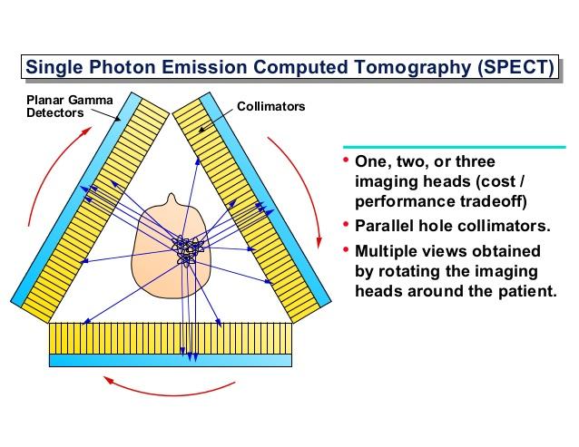
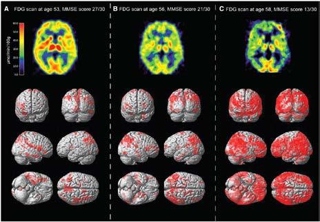
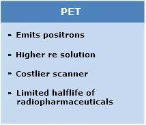
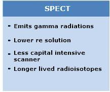
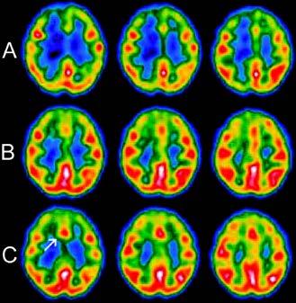
8 Positron emission tomography Single-photon emission computed tomography Application: - SPECT: cardiology, neurology (brain perfusion, neurotransmission) - PET: metabolism, angiogenesis, hypoxia, amyloid imaging (11C-PiB PET)

9 Optical imaging Modalities: - Bioluminescence (luciferin substrate) - Fluorescence and near-infrared fluorescence (NIR) - Diffuse optical tomography (DOT) Application: - Molecular imaging of reporters - Enzymatic imaging, tumor angiogenesis Advantages: - High sensitivity - Low costs - Relatively high throughput - Short acquisition time Drawbacks: - Low resolution - Limited depth

10 Photoacousting imaging: Listen to absorption Is the combination of optic imaging and US. a, Pulsed light of time-shared multiple wavelengths illuminates the tissue of interest b, In the tissue there are absorbing elements. In response to the fast absorption of light by this elements acoustic responses are generated. They can then be detected with acoustic detectors. Advantage: Combination of high optical contrast and submillimeter ultrasound resolution V. Ntziachristos, Nature America 2010

11 Contrast agent versus molecular imaging probe Anatomical imaging: = Contrast agent CT: Iodine-based, barium MRI: Gd, Mn, SPIO Molecular imaging: = imaging probes PET: 18 F, 11 C, 13 N, 68 Ga SPECT: 99m Tc, 123 I, 111 In OI: Organic fluophores, inorganic semiconductor nanoparticles
12


13 How are these images made? Nice pictures... Are they biomedical useful? And from the view of a small animal? In vivo ultrafast ultrasound localization microscopy in the rat brain Velocity map s separating vessels in two populations with opposite blood flow rate (reproduced from Errico in Nature methods 2016) Ultrafast ultrasound localization microscopy for deep super-resolution vascular imaging Claudia Errico, Juliette Pierre, Sophie Pezet, Yann Desailly, Zsolt Lenkei, Olivier Couture*, Mickael Tanter* Nature, November Unprecedented spatiotemporal resolution - Deep penetration & super-resolution - Images are acquired in 150s! - In-vivo, Minimal-invasive : Catheterized jugular vein for the administration of the contrast agent (conventional microbubbles also used in clinical applications), thinnedskull imaging window
 Piezoelectric elements https://en.wikipedia.")
 http://www.ndk.")


14 Basic principle of Ultrasound Sonar imaging, Langevin (20th century) Piezoelectric elements Acoustic waves : wavelength (λ), frequence (f), amplitude (A) Basic for the contrast in US: Tanter and Fink, Array of multiple piezoelectric elements - Line-per-line - Transmission of a slightly defocused US beam and parallel processing of 4 US beam in the receive mode (25 fps) reflection scattering divergence interference absorption
 waves - Tilted plane waves with")
 : functional imaging (tissue motion, brain activity via")
15 Ultrafast Ultrasound Imaging Leaving sonar principles: Plane wave insonifying Tanter and Fink, 2015 Bercoff, Transmission of plane (or unfocused) waves - Tilted plane waves with different angles - Full region of interest, using all array elements - Higher amplitudes - Short acquisition (hundreds of microseconds) : functional imaging (tissue motion, brain activity via blood flow)
16 Ultrasound Contrast agent Primarily designed to detect small blood vessels Biocolloid : Colloid particles made from biocompatible materials Smaller than the wavelength of diagnostic ultrasound ( μm) - Gas spheres (perfluocarbon) - Gas core with a low density Basic for resonation - A clinical US system is in principle capable of detecting the signature from a single microbubble (resolution ) - Shell : in clinical use phospholipid (older albumin, protein shelled) - A clean microbubble is inherently unstable Molecular Imaging, Weisleder et al, 2010 Solid and liquid nanoparticles: - Less echogenic than gas bubbles (incompressible) - Stable ; Advantageous pharmacokinetic properties - Enhanced permeability and retention principle
 Solution: 500 frames per second averaging In 150 s Cox and Beard, 2015 about: Claudia Errico, ( ), Olivier Couture*, Mickael Tanter*, 2015 Pinpointing the location of the")
17 The PALM approach applied to ultrasound The problem to solve: Conventional ultrasound contrast imaging is limited by the classical wave diffraction theory and corresponds roughly to the ultrasonic wavelength (200 μm 1 mm) Solution: 500 frames per second averaging In 150 s Cox and Beard, 2015 about: Claudia Errico, ( ), Olivier Couture*, Mickael Tanter*, 2015 Pinpointing the location of the few, well-separated microbubbles that degraded between each image ( blinking of bubbels ) Viessmann et al. 2013: Achievement of the necessary separation by sufficient dilute of microbubbles. However hour-long imaging acquisition.
 c, three independent microbubbles")
 http://www.nature.com/nature/journal/v527/n7579/fig_tab/nature16066_sv1.html?")
18 The principle of ultrafast ultrasound localization microscopy a, Average stack of 250 beamformed images b, Frames separated by 44 ms and filters (to remove the tissue signal) c, three independent microbubbles blinking over several miliseconds Red cross: Exact position of centroid (deconvolution with point-spread function) Claudia Errico, 2015

 Claudia")
19 Spatial resolution and bubble velocity maps a, Microbubble density maps with a spatial resolution of λ/10 Resolution in depth and lateral direction : 8 μm x 10 μm b, Same area in a conventional power Doppler image c, Interpolated profiles the lines marked in a d, dynamic tracing of bubbles separates vessels in two populations e, f, Velocity lines associated with d (n = number of bubbles) Claudia Errico, 2015


20 Thinned skull window versus intact skull a, uulm preformed through shinned skull (8 μm x 10 μm) b, corresponding velocity map ab/nature16066_sv2.html c, uulm performed through the intact skull : attenuation of the ultrasound wave in the presence of bone (12.5 μm x 10 μm) d, corresponding velocity map Claudia Errico, 2015
21 Nice pictures... Are they biomedical useful? Possible fields: - Normal and diseased blood-vessel function, - identification of microvessel-related disorders, - angiogenesis in neoplasms, - vascular dementia etc - functional imaging in neuroscience (combining this technique with functional ultrasound) Conventional clinical ultrasound: Resolution inversely correlates to penetration (f) In ultrafast ultrasound localization microscopy: Resolution is related to: - SNR, - BW of backscattered echoes, -N of array elements = high resolution deep into organs could be reached = clinical application s (liver, kidney, breast) Human brain? By the use of longer wavelength maybe the challenge of the thick human skull can be circumvented
22 And from the view of a small animal? Replacement : Realistic non-invasive human application Reduction : Longitudinal (functional) studies Refinement : Fast acquisition, minimal invasive
23 Are this biccoloids of further use? Molecular Imaging, Weisleder et al, 2010
 - NP platform which integrates a variety of imaging and")
24 Theranostics: Agents for diagnosis and Therapy Theranostic: The combination of diagnostic and therapeutic entities into one drug delivery vehicle for simultaneous diagnosis and treatment of disease A large variety of inorganic and organic based nanoparticles Nanoporphyrin: - All-in-one porphyrin-based organic nanoconstruct (nanoporphyrin NP) - NP platform which integrates a variety of imaging and therapeutic functions Yuanpei Li et al, 2014

25 From micro to nano bacteriochlorophyll-lipid In situ conversion of porphyrin microbubbles to nanoparticles for multimodality imaging Elizabeth Huynh, Ben Y. C. Leung, Brandon L. Helfield, Mojdeh Shakiba, Julie-Anne Gandier, Cheng S. Jin, Emma R. Master, Brian C. Wilson, David E. Goertz and Gang Zheng* Nature Nanotechnology, March Drug delivery by using ultrasound to implode microbubbles into nanoparticles - Bacteriochlorophyll-lipid (BChl-lipid) shell; porphyrins confer photoacoustic and fluorescent properties - In-vivo anatomical guidance by the microbubble US contrast - Nanodroplets are better able to penetrate fenestrated or compromised vessel - Interaction of ultrasound with drug-loaded microbubbles causing non-lethal transient pores in blood vessels
 Expanding and Contraction E-G) Collaps Basis for a strong and unique echo, microbubble resonate at frequencies typical used in US imaging Effect of pulse driving pressure on bubble radial")
26 The echogenity of microbubbles Microbubble passage of an acoustic wave A) Bubble with an initial radius of 1.5 μm B-E) Expanding and Contraction E-G) Collaps Basis for a strong and unique echo, microbubble resonate at frequencies typical used in US imaging Effect of pulse driving pressure on bubble radial oscillation A and C) Driving (acoustic) pressure; A = 50 kpa and C = 500 kpa B and D) Expansion ratio in response to either A or C B represents a linear dynamic D represents a nonlinear dynamic Molecular Imaging, Weisleder et al, 2010
27 Pulse-inversion imaging : Contrast between bubbles and tissue A and B) Transmitted pulses (A: 50kPa, B: 100 kpa); B is inverted (180 ) C and D) Corresponding echoes from 1-micron bubble and tissue Bubble echoes: With the higher pressure non-linear dynamic E) Summation of 2 times echo in C plus echo in D The linear echoes from tissue are cancelled while nonlinear echoes from the bubble are acquired Molecular Imaging, Weisleder et al, 2010

 Microbubbles generate both linear and")
28 Basic properties of BChl-lipid microbubbles a) Acoustic attenuation measurement of pmbs, Ressonance attenuation peak at 4.5 MHz b) Linear and nonlinear ultrasound properties of pmbs Tissue-mimicking flow phantom (agar and graphite) Microbubbles generate both linear and non-linear ultrasound signals Elizabeth Huynh, 2015

29 Conversion ultrasound Conversion ultrasound : 1 MHz, high-duty-cycle (50 %), ultrasound (2 W cm -2 ) f, light microscopy image of microbubbles before conversion ultrasound g, Electron microscopy of porphyrin nanoparticles after ten ultrasound pulses c, Size distribution of microbubbles before and after conversion ultrasound d, Concentration of microbubbles and nanoparticles before and after conversion ultrasound e, Size distribution of nanoparticles before and after conversion ultrasound Elizabeth Huynh, 2015

 ten pulses b-d, Quantified signals (b: ultrasound, c: photoacoustic, d: fluorescence")
30 Multimodal imaging of Microbubbles (pmbs) and resulting nanoparticles (pnps) following ultrasound-induced conversion a, Acrylamide gel phantom imaged with ultrasound, photoacoustic and fluorescence 1) PBS 2) without conversion US 3) one pulse of conversion US 4) three pulses 5) ten pulses b-d, Quantified signals (b: ultrasound, c: photoacoustic, d: fluorescence Elizabeth Huynh, 2015
 Non con.")
31 Conversion of pmbs to pnps in vivo in mice Mouse model: Subcutaneous inoculation of 2x106 KB cells (HeLa derivative) into the right flank of athymic nude mice. Experiments were performed when tumors reached a surface of 5-7 diameter. a, Cross section of the tumor (linear and non-linear imaging) Non con. US: After injection, the pmbs circulate into the tumor, reaching a peak in circulation at 20 s and could be observed continuously in the circulation beyond 40 s With con. US: A decrease of non-linear signal after the 20 s time point b, ROI analysis without conversion ultrasound (top) and with conversion ultrasound (bottom) Elizabeth Huynh, 2015

32 Successful delivery of pnps into the tumor xenograft c, Retention of pnps in the tumor xenograft enabled by conversion from pmbs to pnps d, Normalized photoacoustic signal over time in the tumor, indicative for a successful delivery Elizabeth Huynh, 2015
33 Nice pictures... Are they biomedical useful? Not adressed: - Penetration of the conversion ultrasound? (1 MHz = high penetration) - Limitations for the drug delivery? (size, chemical properties etc) Possible fields: - Translational medicine (drug delivery) - The use of nanoporphyrin offer an expansion to other imaging modalities
34 And from the view of a small animal? Replacement : (further investigation needed, before application in humans are possible) Reduction : Longitudinal studies are possible (Reaches the drug the target tissue?) Refinement : Multimodality imaging (multiple information) Drawbasks: Setup experimental, photoacoustic longitudinal studies performed over 2 h


35 Thank you for your attention! - Questions? - Get your forms signed!
BME101 Introduction to Biomedical Engineering Medical Imaging Özlem BİRGÜL Ankara University Department of Biomedical Engineering
 BME101 Introduction to Biomedical Engineering Medical Imaging Özlem BİRGÜL Ankara University Department of Biomedical Engineering Outline What is Medical Imaging? History of Medical Imaging X-Ray Imaging
1st Faculty of Medicine, Charles University in Prague Center for Advanced Preclinical Imaging (CAPI)
") ADVANTAGES Optical Imaging OI Optical Imaging is based on the detection of weak light by a highly sensitive and high resolution CCD camera DISADVANTAGES High sensitivity Limited penetration depth Easy
ADVANTAGES Optical Imaging OI Optical Imaging is based on the detection of weak light by a highly sensitive and high resolution CCD camera DISADVANTAGES High sensitivity Limited penetration depth Easy
Computer Assisted Surgery Basics of medical imaging
 Computer Assisted Surgery Basics of medical imaging Prof. Leo Joskowicz School of Engineering and Computer Science The Hebrew University of Jerusalem, ISRAEL Medical Image Processing Basics of medical
Translational Multimodality Optical Imaging
 Translational Multimodality Optical Imaging Fred S. Azar Xavier Intes Editors 0 ARTECH H O U S E BOSTON LONDON artechhouse.com Contents Foreword Preface xv xvii CHAPTER1 Introduction to Clinical Optical
Novel Kidney Imaging. Objectives. Imaging surveillance in VHL. Disclosures. From nephrology perspective 10/11/2018
 Disclosures Novel Kidney Imaging Contract with Lantheus Medical Imaging (Definity) Contrast-enhanced ultrasound Emily Chang, MD Nephrology October 4, 2018 13 th International VHL Symposium 10/11/2018 2
Principles of translational medicine: imaging, biomarker imaging, theranostics
 Principles of translational medicine: imaging, biomarker imaging, theranostics Compiled by: Endre Mikus PhD, CEO Budapest, 21/9/2015 Imaging and imaging biomarkers An imaging biomarker is an anatomic,
Photoacoustic imaging of vascular networks in transgenic mice
 Photoacoustic imaging of vascular networks in transgenic mice J.G. Laufer 1, J.O. Cleary 1,2, E.Z. Zhang 1, M.F. Lythgoe 2, P.C. Beard 1 1. Department of Medical Physics and Bioengineering, University
PREPARED FOR: U.S. Army Medical Research and Materiel Command Fort Detrick, Maryland
 AD Award Number: W81XWH-07-1-0231 TITLE: Spectroscopic Photoacoustic Tomography of Prostate Cancer PRINCIPAL INVESTIGATOR: Xueding Wang CONTRACTING ORGANIZATION: University Of Michigan Ann Arbor, MI 48109-1274
Basic principles of quantification using optical techniques
 Contents Basic principles of quantification using optical techniques Adrian Taruttis Helmholtz Zentrum München Chair for Biological Imaging Technische Universität München Light/ tissue interactions Planar
Miniature fibre optic probe for minimally invasive photoacoustic sensing
 Miniature fibre optic probe for minimally invasive photoacoustic sensing Sunish J. Mathews*, Edward Z. Zhang, Adrien E. Desjardins and Paul C. Beard Department of Medical Physics and Biomedical Engineering,
Photoacoustic Imaging in Biomedicine Critical Review by Saurabh Vyas Group 9: Interventional Photoacoustic Ultrasound CIS II: 600.
 Photoacoustic Imaging in Biomedicine Critical Review by Saurabh Vyas Group 9: Interventional Photoacoustic Ultrasound CIS II: 600.446, Spring 2011 Introduction Photoacoustic imaging (PA Imaging) is the
K-Space analysis for plane-wave ultrasound imaging. Georg Schmitz
 K-Space analysis for plane-wave ultrasound imaging Georg Schmitz Abstract: Ultrasound imaging can be considered as an inverse scattering problem in which the unknown distribution of the material parameters
PREPARED FOR: U.S. Army Medical Research and Materiel Command Fort Detrick, Maryland
 AD Award Number: W81XWH-07-1-0231 TITLE: Spectroscopic Photoacoustic Tomography of Prostate Cancer PRINCIPAL INVESTIGATOR: Xueding Wang CONTRACTING ORGANIZATION: University Of Michigan Ann Arbor, MI 48109-1274
Key Elements of X-ray CT Physics. Part 2: X-ray Interactions
 Key Elements of X-ray CT Physics Part 2: X-ray Interactions NPRE 435, Principles of Imaging with Ionizing Radiation, Fall 2006 Photoelectric Effect Photoe - absorption is the preferred interaction for
MEDICAL PHYSICS (MED PHYS)
") Medical Physics (MED PHYS) 1 MEDICAL PHYSICS (MED PHYS) MED PHYS/PHYSICS 265 INTRODUCTION TO MEDICAL PHYSICS Primarily for premeds and other students in the medical and biological sciences. Applications
Medical Physics (MED PHYS) 1 MEDICAL PHYSICS (MED PHYS) MED PHYS/PHYSICS 265 INTRODUCTION TO MEDICAL PHYSICS Primarily for premeds and other students in the medical and biological sciences. Applications
Date: May 26, 2015 Page 1
 Part I. Answer these questions by marking the best answer among the choices given: [2 points each] 1. Ethics and morals differ in that a. Only one of them should be followed b. Ethics are for professionals
Angio PL.U.S. PLaneWave UltraSensitive TM ultrasound imaging. White Paper
 White Paper Angio PL.U.S. PLaneWave UltraSensitive TM ultrasound imaging Jeremy Bercoff, Vice President of Product Management & Ultrasound Engineering Thomas Frappart, R&D Ultrasound Engineer Introduction
Study Guide Imaging Physics and Biophysics for the Master-Study Programmes
 Study Guide Imaging Physics and Biophysics for the Master-Study Programmes Imaging Physics is one of the main areas of research of the Faculty for Physics and Astronomy at the Julius-Maximilians-University
ORGANIZING THE MEDICAL IMAGING DEPARTMENT THE MEDICAL IMAGING DEPARTMENT 1
 ORGANIZING THE MEDICAL IMAGING DEPARTMENT THE MEDICAL IMAGING DEPARTMENT 1 Modern Medical Imaging methods Modern Medical Imaging includes a lot of methods: Conventional and Digital Radiology. Nuclear Medicine
Molecular Imaging: Definition, Overview and Goals
 This tutorial will define what is currently considered molecular imaging. It will provide history and an overview, discuss the goals and the advantages of molecular imaging. It will clarify what is and
Positron Emission Tomography Present status and future prospects
 Positron Emission Tomography Present status and future prospects S. Tavernier VRIJE UNIVERSITEIT BRUSSEL July 2011 NDIP Lyon 1 What is PET Positron Emission Tomography is a non invasive method for imaging
An ultrasonic volumetric scanner for image-guided surgery
 International Congress Series 1230 (2001) 190 196 An ultrasonic volumetric scanner for image-guided surgery Jeremy Johnson a,b, *,Ömer Oralkan b, Kambiz Kaviani b, Utkan Demirci b, Mustafa Karaman b, Pierre
Geneva January 10 11, th Swiss Experimental. Surgery Symposium. MGY / 4thSESS /
 4th Swiss Experimental Surgery Symposium Geneva January 10 11, 2008 Source: Reflex issue 2, 2007 The 3 Rs Reduction Replacement Refinement NC3R http://www.nc3rs.org.uk/category.asp?catid =9 No specific
Simple, intuitive and accessible MRI solution for preclinical research. M-Series Compact MRI Systems
 Simple, intuitive and accessible MRI solution for preclinical research M-Series Compact MRI Systems Application Oriented Imaging Anatomy and Morphology In vivo soft tissue imaging for morphological characterization.
Medical instrumentationi 11/19/2010
 Medical instrumentationi BIOEN 302 11/19/2010 Medical instrumentation Definition: instrument for sensing, diagnostics, therapeutics or surgery of human being. 2 Medical instrumentation Definition: instrument
Simple, intuitive and accessible MRI solution for preclinical research. M-Series Compact MRI Systems
 Simple, intuitive and accessible MRI solution for preclinical research M-Series Compact MRI Systems Application Oriented Imaging Molecular Imaging Using Contrast Agents Detection and quantification of
Contrast Agents for Enhanced Ultrasound Imaging and for
 Contrast Agents for Enhanced Ultrasound Imaging and for Drug Delivery Sverre Holm INSTITUTT FOR INFORMATIKK SH, 1 Microbubbles bbl (white) red blood cells (black) 2-6 m diameter Shell of biocompatible
VisualSonics Application Protocol Gene Delivery Applications into Subcutaneous Tumors
 VisualSonics Application Protocol Gene Delivery Applications into Subcutaneous Tumors 1 Objective The objective of the Delivery of Genetic Material for Gene Therapy Applications into Subcutaneous Tumors
Fast enough to stop the Capable of delineating Unprecedented imaging power for the. Virtual endoscopy. The gatewa
 Fast enough to stop the Capable of delineating Unprecedented imaging power for the M U L T I S L I C E Virtual endoscopy The gatewa motion of a beating heart. anatomic structures as small as 0.25mm. earliest,
Future Areas of Technology Convergence
 Future Areas of Technology Convergence Dr J Malcolm Wilkinson Managing Director Technology For Industry Ltd Cambridgeshire, UK Medilink Yorkshire & Humberside, 8 December 2005 1 Technology For Industry
Detecting Gene Expression In-Vivo Using Differential Laser. Absorption. Senior Thesis - Physics, May By Hermonta M Godwin
 Detecting Gene Expression In-Vivo Using Differential Laser Absorption Senior Thesis - Physics, May 2002 By Hermonta M Godwin Advisor: Professor William E. Cooke College of William and Mary Abstract: The
Molecular imaging in vitro and in vivo
 Molecular imaging in vitro and in vivo Tony Lahoutte, MD PhD Free University Brussels Technology Day 18/09/2008, Brussels Molecular Imaging Definition: Molecular imaging is the visualization, the characterization
ASTM Volume 03.03, October 2017 Nondestructive Testing (E94 E2373)
") Table of Contents 1 E94-04(2010) Standard Guide for Radiographic Examination 2 E114-15 Standard Practice for Ultrasonic Pulse-Echo Straight-Beam Contact Testing 3 E125-63(2013) Standard Reference Photographs
Table of Contents 1 E94-04(2010) Standard Guide for Radiographic Examination 2 E114-15 Standard Practice for Ultrasonic Pulse-Echo Straight-Beam Contact Testing 3 E125-63(2013) Standard Reference Photographs
PRINCIPLES OF CT AND MR IMAGING Marc-André d Anjou, DMV, DACVR Faculty of Veterinary Medicine, University of Montreal Saint-Hyacinthe, Quebec, Canada
 PRINCIPLES OF CT AND MR IMAGING Marc-André d Anjou, DMV, DACVR Faculty of Veterinary Medicine, University of Montreal Saint-Hyacinthe, Quebec, Canada CT and MR imaging offer superior diagnostic possibilities
@01-258_via99-011_hepatoma_onscreen
 Lei Xing, Ph.D. & Jacob Haimson Professor Department of Radiation Oncology & MIPS Stanford University School of Medicine Case study TrueBeam 6 MV FFF RapidArc Tx of a Lung SBRT 3D modeling Treatment planning
Contents Preface xiii Introduction Fabrication and manufacturing technology for optical MEMS
 Contents Preface xiii 1 Introduction 1 1.1 Optical MEMS and optofluidics 1 1.2 History 1 1.2.1 Processes and materials 1 1.2.2 Early devices and systems 2 1.3 Progress in optical MEMS and optofluidics
NANO 243/CENG 207 Course Use Only
 L5: Nanomedicine in Diagnostics and Bioimaging April 17, 2018 Cancer Diagnostics Diagnostics plays an important role throughout cancer treatment Before treatment, accurately locate tumors, stage the disease,
BIOMEDICAL SIGNAL AND IMAGE PROCESSING
 BIOMEDICAL SIGNAL AND IMAGE PROCESSING EE 5390-001 SYLLABUS Instructor: Wei Qian, Ph.D. Professor of Electrical and Computer Engineering Medical Signal and Image Computerized Processing Scheme for Medical
2018 REVIEW CATEGORIES
 2018 REVIEW CATEGORIES 100 Neuro 101 Neuro: Acquisition 102 Neuro: Processing 103 Neuro: Neonatal & Pediatric - Normal Development 104 Neuro: Neonatal & Pediatric - Clinical Studies 105 Neuro: Normal Aging
MEDICAL EQUIPMENT (1) TOPIC 1: RECORDING AND PROCESSING OF BIOSIGNALS
 TOPIC 1: RECORDING AND PROCESSING OF BIOSIGNALS") MEDICAL EQUIPMENT (1) TOPIC 1: RECORDING AND PROCESSING OF BIOSIGNALS Term 1 2013/14 Prof. Yasser Mostafa Kadah www.k-space.org Measurement Basics Measuring is the experimental determination of a measured
MEDICAL EQUIPMENT (1) TOPIC 1: RECORDING AND PROCESSING OF BIOSIGNALS Term 1 2013/14 Prof. Yasser Mostafa Kadah www.k-space.org Measurement Basics Measuring is the experimental determination of a measured
RADIATION ONCOLOGY RESIDENCY PROGRAM Competency Evaluation of Resident
 Resident s Name: RADIATION ONCOLOGY RESIDENCY PROGRAM Competency Evaluation of Resident Rotation: PHYS 705: Clinical Rotation 3 Inclusive dates of rotation: Aug. 25, 2015 Feb. 25, 2016 Director or Associate
Ilya Turchin. Institute of Applied Physics of the RAS, Nizhny Novgorod, Russia.
 Fluorescence 3D imaging of small animals Ilya Turchin Institute of Applied Physics of the RAS, Nizhny Novgorod, Russia ilya@ufp.appl.sci-nnov.ru http://www.bioimaging.ru German-Russian Forum Biotechnology
S6-1. Protocol and System setting for 2 nd generation Ultrasound Contrast Agent. 9 th US Contrast. (For diagnosis and navigation in treatments)
") S6-1 Protocol and System setting for 2 nd generation Ultrasound Contrast Agent (For diagnosis and navigation in treatments) Tsuyoshi Mitake Senior Chief Engineer Ultrasound System Division Hitachi Medical
S6-1 Protocol and System setting for 2 nd generation Ultrasound Contrast Agent (For diagnosis and navigation in treatments) Tsuyoshi Mitake Senior Chief Engineer Ultrasound System Division Hitachi Medical
Nayar Prize I Quarterly Progress Report (Quarters 2&3) August, 2016
 August, 2016") Nayar Prize I Quarterly Progress Report (Quarters 2&3) August, 2016 Project: ADEPT Cancer Imager Team: Ken Tichauer, Jovan Brankov, Raju Mehta Students: Lagnojita Sinha, Xiaochun Xu Progress Summary Since
Nayar Prize I Quarterly Progress Report (Quarters 2&3) August, 2016 Project: ADEPT Cancer Imager Team: Ken Tichauer, Jovan Brankov, Raju Mehta Students: Lagnojita Sinha, Xiaochun Xu Progress Summary Since
Product Brief: VevoCQ Advanced Contrast Quantification Software Analysis Tools for the Vevo 2100 System
 Product Brief: VevoCQ Advanced Contrast Quantification Software Analysis Tools for the Vevo 2100 System Introduction Microbubble contrast agents have been used as a method of assessing in vivo microvascular
Preclinical in vivo Imaging. Innovation with Integrity. Preclinical Imaging. Nine modalities - Unlimited research capabilities
 Preclinical in vivo Imaging Nine modalities - Unlimited research capabilities Innovation with Integrity Preclinical Imaging The widest range of preclinical imaging modalities from a single source Delivering
Translational & Molecular Imaging Institute
 Translational & Molecular Imaging Institute tmii.mssm.edu Summer 2015 CARDIOVASCULAR IMAGING The Imaging Research Center is the backbone of the Translational & Molecular Imaging Institute at Mount Sinai
A Modeling Platform for Ultrasonic Immersion Testing of Polycrystalline Materials with Flaws
 11th European Conference on Non-Destructive Testing (ECNDT 2014), October 6-10, 2014, Prague, Czech Republic A Modeling Platform for Ultrasonic Immersion Testing of Polycrystalline Materials with Flaws
Ultrasound and Photoacoustics
 Biomedical Imaging: From Drug Target Discovery to Medical Diagnostics FRAP and FLIP Fluorescence recovery after photobleaching (FRAP) and fluorescence loss in photobleaching (FLIP) exploit the principle
Plasmonics using Metal Nanoparticles. Tammy K. Lee and Parama Pal ECE 580 Nano-Electro-Opto-Bio
 Plasmonics using Metal Nanoparticles Tammy K. Lee and Parama Pal ECE 580 Nano-Electro-Opto-Bio April 1, 2007 Motivation Why study plasmonics? Miniaturization of optics and photonics to subwavelength scales
HHS Public Access Author manuscript Phys Rev Lett. Author manuscript; available in PMC 2014 March 18.
 Photo-imprint Photoacoustic Microscopy for Three-dimensional Label-free Sub-diffraction Imaging Junjie Yao, Lidai Wang, Chiye Li, Chi Zhang, and Lihong V. Wang * Optical Imaging Laboratory, Department
Vevo The Ultimate Preclinical Imaging Experience
 The Ultimate Preclinical Imaging Experience 2 Introducing The World s First One-Touch Preclinical Imaging Platform The is a new and innovative platform created for the future of imaging. It combines ultra
Size-Modulable Nanoprobe for High-Performance. Ultrasound Imaging and Drug Delivery against
 Size-Modulable Nanoprobe for High-Performance Ultrasound Imaging and Drug Delivery against Cancer Lu Zhang,,,#, Tinghui Yin,#, Bo Li, Rongqin Zheng*,, Chen Qiu, Kit S. Lam*,, Qi Zhang, Xintao Shuai*,,
Section: Magnetic Resonance Imaging
 Section: Magnetic Resonance Imaging Available masters project: Cardiac shape modeling for automated wall motion analysis Overview One of the most popular segmentation methods in medical imaging is the
In Vivo Change in Ultrasonic Backscattered Energy with Temperature in Motion-Compensated Images
 In Vivo Change in Ultrasonic Backscattered Energy with Temperature in Motion-Compensated Images R. Martin Arthur 1, Jason W. Trobaugh 1, William L. Straube 2, Jesse Parry 2, Yuzheng Guo 1, and Eduardo
Ultrasound Elastography
 Ultrasound Elastography seminar LUT2, University of Kuopio, Finland Josef Jaros jarosj@feec.vutbr.cz Dep. of Biomedical Engineering, FEEC, BUT Brno, CZ 1. Introduction For centuries, physicians have used
Bioengineering (BIOE)
") Bioengineering (BIOE) 1 Bioengineering (BIOE) Courses BIOE 5301. Biosignals. 3 Credit Hours. This course offers a deep overview of the signals in the Biomedical fields. Signals are studied in several modalities,
Bioengineering (BIOE) 1 Bioengineering (BIOE) Courses BIOE 5301. Biosignals. 3 Credit Hours. This course offers a deep overview of the signals in the Biomedical fields. Signals are studied in several modalities,
EMSE Weak-Beam Dark-Field Technique
 Weak-Beam Dark-Field Technique 1 Weak-Beam Dark-Field Imaging Basic Idea recall bright-field contrast of dislocations: specimen close to Bragg condition, s î 0 near the dislocation core, some planes curved
Insight Through In Vivo Imaging
 Insight Through In Vivo Imaging Resolution Revolution in Realtime The Vevo 770 provides anatomical, functional and molecular data in realtime, on an affordable, easy to use and translational platform.
Diagnostic Medical Image Processing
 Diagnostic Medical Image Processing Introduction WS 2010/11 Joachim Hornegger, Dietrich Paulus, Markus Kowarschik Lehrstuhl für Mustererkennung (Informatik 5) Friedrich-Alexander-Universität Erlangen-Nürnberg
Arterial Spin Labeling (ASL)
") Arterial Spin Labeling (ASL) Imaging Seminars Series Stony Brook University, Health Science Center Stony Brook, NY - December 11 th, 2012 Francesca Zanderigo, PhD Layout BASIC PRINCIPLES ACQUISITION SEQUENCES
Arterial Spin Labeling (ASL) Imaging Seminars Series Stony Brook University, Health Science Center Stony Brook, NY - December 11 th, 2012 Francesca Zanderigo, PhD Layout BASIC PRINCIPLES ACQUISITION SEQUENCES
OUR WISH LIST RESEARCH EQUIPMENT
 OUR WISH LIST RESEARCH EQUIPMENT WITH YOUR PHILANTHROPIC SUPPORT, WE CAN WORK TOGETHER TO COMPLETE OUR FULLY-FUNCTIONAL FACILITY BY PURCHASING THE CUTTING-EDGE EQUIPMENT AND RESOURCES TO SUPPORT SAHMRI
OUR WISH LIST RESEARCH EQUIPMENT
 OUR WISH LIST RESEARCH EQUIPMENT YOU CAN MAKE A TANGIBLE DIFFERENCE! The South Australian Health and Medical Research Institute (SAHMRI) is one of the most exciting developments in the field of health
Advanced preclinical optical imaging. Preclinical in vivo imaging. IVIS Spectrum
 IVIS Spectrum P R O D U C T N O T E Preclinical in vivo imaging Key Features High Sensitivity in vivo fluorescence and bioluminescence imaging 3D tomographic reconstruction Absolute calibration High throughput
Multiplexed 3D FRET imaging in deep tissue of live embryos Ming Zhao, Xiaoyang Wan, Yu Li, Weibin Zhou and Leilei Peng
 Scientific Reports Multiplexed 3D FRET imaging in deep tissue of live embryos Ming Zhao, Xiaoyang Wan, Yu Li, Weibin Zhou and Leilei Peng 1 Supplementary figures and notes Supplementary Figure S1 Volumetric
GE Healthcare LOGIQ 7
 GE Healthcare LOGIQ 7 At the leading edge of healthcare The system of choice for shared service Image Quality Raw Data Ergonomics For more than a century, GE Healthcare has been inventing medical technologies.
Biomedical Applications of Molecular Spectroscopy
 Biomedical Applications of Molecular Spectroscopy Mike Kayat B&W Tek, Inc 19 Shea Way Newark, DE 19713 United States of America +1 302 368 7824 mikek@bwtek.com 1 Overview Molecular spectroscopy is a large
Available Imaging Lei Xing, Ph.D., Jacob Haimson Profssor
 sensitivity Lei Xing, Ph.D., Jacob Haimson Profssor Department of Radiation Oncology Molecular Imaging Program at Stanford (MIPS) Department of Electrical Engineering Stanford University School of Medicine
Preclinical MRI. Solutions for Small Animal Imaging. Molecular Imaging
 Preclinical MRI Solutions for Small Animal Imaging Molecular Imaging The Power of Imaging Applications Resolution Typical resolution in MRI is less than 200μm, to more than 20μm with 2D slices or full
Course Code: BMEG5100 Course Title: Advanced Medical Robotics Course Code: BMEG5110 Course Title: Advanced Medical Devices and Sensor Networks
 Course Code: BMEG5100 Course Title: Advanced Medical Robotics Review of medical robotics fundamentals; introduction to robotics enabled endoscopic and laparoscopic surgeries; concepts of robotics based
Functional probes for cardiovascular molecular imaging
 Review Article Functional probes for cardiovascular molecular imaging Yun Zeng 1, Jing Zhu 2, Junqing Wang 2,3, Paramanantham Parasuraman 4, Siddhardha Busi 4, Surya M. Nauli 5, Yì Xiáng J. Wáng 3, Rajasekharreddy
Photon-based Medical Imagery
 Photon-based Medical Imagery Photon-based Medical Imagery Edited by Hervé Fanet First published 2011 in Great Britain and the United States by ISTE Ltd and John Wiley & Sons, Inc. Adapted and updated from
Quantification in emission tomography: challenges, solutions, performance and impact
 EuroMedIm 2006 Quantification in emission tomography: challenges, solutions, performance and impact Irène Buvat U678 INSERM, Paris buvat@imed.jussieu.fr http://www.guillemet.org/irene EuroMedIm 2006 -
Emerging Applications and Trends Across Medical Imaging
 MEDICAL DEVICES PHARMACEUTICALS CHEMICALS FOOD & BEVERAGE ELECTRONICS Emerging Applications and Trends Across Medical Imaging VPG Publications, Consulting, Clients www.vpgcorp.com VPG Market Research Reports
Current Market Dynamics and Future Vision of the Care Cycle
 Current Market Dynamics and Future Vision of the Care Cycle Tim Irish Analysts Meeting June 15 th, 2005 Overview Market and Customer trends The Care Cycle and Molecular Medicine 2 Healthcare is the world
Tunable Nanoscale Plasmon Antenna for Localization and Enhancement of Optical Energy. Douglas Howe
 Tunable Nanoscale Plasmon Antenna for Localization and Enhancement of Optical Energy Douglas Howe Applied Optics Spring 2008 Table of Contents Abstract... 3 Introduction... 4 Surface Plasmons... 4 Nano
VALLIAMMAI ENGINEERING COLLEGE DEPARTMENT OF MECHANICAL ENGINEERING QUESTION BANK
 VALLIAMMAI ENGINEERING COLLEGE SRM Nagar, Kattankulathur 603 203 DEPARTMENT OF MECHANICAL ENGINEERING QUESTION BANK VIII SEMESTER ME 6019-NON DESTRUCTIVE TESTING AND MATERIALS Regulation 2013 Academic
Nanomaterials for Imaging Technology. Nadeem A. Kizilbash, Ph.D. Assistant Professor Department of Chemistry Quaid-i-Azam University Islamabad
 Nanomaterials for Imaging Technology Nadeem A. Kizilbash, Ph.D. Assistant Professor Department of Chemistry Quaid-i-Azam University Islamabad Introduction Nanotechnology, most basically put, is the molecular
HYPERSPECTRAL MICROSCOPE PLATFORM FOR HIGHLY MULTIPLEX BIOLOGICAL IMAGING. Marc Verhaegen
 HYPERSPECTRAL MICROSCOPE PLATFORM FOR HIGHLY MULTIPLEX BIOLOGICAL IMAGING Marc Verhaegen CMCS, MONTREAL, MAY 11 th, 2017 OVERVIEW Hyperspectral Imaging Multiplex Biological Imaging Multiplex Single Particle
BIOMEDICAL ENGINEERING (BME)
") Biomedical Engineering (BME) 1 BIOMEDICAL ENGINEERING (BME) BME 500 Introduction to Biomedical Engineering Introduction to the concepts and research in biomedical engineering. Provides an overview of current
Biomedical Engineering (BME) 1 BIOMEDICAL ENGINEERING (BME) BME 500 Introduction to Biomedical Engineering Introduction to the concepts and research in biomedical engineering. Provides an overview of current
Biomedical Imaging Modalities
 1 Biomedical Imaging Modalities The introduction of advanced imaging techniques has improved significantly the quality of medical care available to patients. Noninvasive imaging modalities allow a physician
ADDIS ABABA UNIVERSITY CENTER OF BIOMEDICAL ENGINEERING
 ADDIS ABABA UNIVERSITY CENTER OF BIOMEDICAL ENGINEERING November 2013 History of Biomedical Engineering Definition of Biomedical Engineering Achievements of Biomedical Engineering Streams in Biomedical
Clarity CT Technology
 Clarity CT Technology WHITE PAPER January 2013 Using state of the art algorithms Sapheneia Clarity CT allows physicians to lower radiation dose when acquiring CT data while maintaining image quality. The
Supplementary Figure 1. Determination of the purity of CP. a, SDS-PAGE of CP and CP- PTX conjugate, and b, HPLC trace of purified CP.
 Supplementary Figure 1. Determination of the purity of CP. a, SDS-PAGE of CP and CP- PTX conjugate, and b, HPLC trace of purified CP. Supplementary Figure 2. Synthesis of CP-PTX conjugate. Supplementary
The Unique, New MRI Philips Ingenia 3 Tesla is now in Ayios Therissos! The first-ever digital broadband MR system has been installed in Ayios
 The Unique, New MRI Philips Ingenia 3 Tesla is now in Ayios Therissos! The first-ever digital broadband MR system has been installed in Ayios Therissos-Nicosia that delivers crystal clear images, remarkable
Absorption of an electromagnetic wave
 In vivo optical imaging?? Absorption of an electromagnetic wave Tissue absorption spectrum Extinction = Absorption + Scattering Absorption of an electromagnetic wave Scattering of an electromagnetic wave
LUPAS Luminescent Polymers for in vivo Imaging of Amyloid Signatures
 LUPAS Luminescent Polymers for in vivo Imaging of Amyloid Signatures A research project for innovative diagnostics for neurodegenerative disorders Funded by the European Union under the 7 th Framework
Prof. Steven S. Saliterman
 Department of Biomedical Engineering, University of Minnesota http://saliterman.umn.edu/ Prof. Angela Panoskaltsis-Mortari s BMEn 5361, 3D Bioprinting Tissue engineering Bioprinting Design considerations
Radiography Curriculum Analysis
 Program Number Program Name Date / /20 Radiography Curriculum Analysis DIRECTIONS: Determine the course(s) in which each of the following content area is covered and enter the course number(s) and/or title(s).
High-Throughput In Vivo Bioluminescence Imaging System
 IVIS SpectrumBL P R O D U C T N O T E Pre-clinical in vivo imaging Key Features Ultra high-sensitivity to support in vivo bioluminescence, chemiluminescence and Cerenkov imaging High-throughput (10 mice)
Medical Microbubbles and. Industrial Flotation
 Medical Microbubbles and Dr Steven Spencer Principal Research Scientist CSIRO MANUFACTURING Industrial Flotation 7 th International Symposium of Fine Bubble Technology - 25 th July 2016 Bubbles and Acoustics
Biophotonics?? Biophotonics. technology in biomedical engineering. Advantages of the lightwave
 Biophotonics - Imaging: X-ray, OCT, polarimetry, DOT, TIRF, photon migration, endoscopy, confocal microscopy, multiphoton microscopy, multispectral imaging - Biosensing: IR spectroscopy, fluorescence,
Biomedical Optical Imaging Martin Frenz Biomedical Photonics Department Institute of Applied Physics, University of Bern
 1 Biomedical Optical Imaging Martin Frenz Biomedical Photonics Department Institute of Applied Physics, University of Bern Imaging is one of the most powerful tools in biomedical research. The impact and
Spectroscopy and Imaging IV
 PROGRESS IN BIOMEDICAL OPTICS AND IMAGING Vol. 16 No. 55 Clinical and Biomedical Spectroscopy and Imaging IV J. Quincy Brown Volker Decked Edifors 22-24 June 2015 Munich, Germany Sponsored by SPIE (United
Deliverable 2.1: Definition of paradigms representing exemplary breast lesions cases
 Project title: Smart Optical and Ultrasound Diagnostics of Breast Cancer Grant Agreement: 731877 Call identifier: H2020-ICT-2016-1 Topic: ICT-29-2016 Photonics KET 2016 Deliverable 2.1: Definition of paradigms
ECE280: Nano-Plasmonics and Its Applications. Week5. Extraordinary Optical Transmission (EOT)
") ECE280: Nano-Plasmonics and Its Applications Week5 Extraordinary Optical Transmission (EOT) Introduction Sub-wavelength apertures in metal films provide light confinement beyond the fundamental diffraction
ECE280: Nano-Plasmonics and Its Applications Week5 Extraordinary Optical Transmission (EOT) Introduction Sub-wavelength apertures in metal films provide light confinement beyond the fundamental diffraction
COMMITTEE FOR MEDICINAL PRODUCTS FOR HUMAN USE (CHMP)
") European Medicines Agency Pre-Authorisation Evaluation of Medicines for Human Use London, 23 July 2009 Doc. Ref. EMEA/CHMP/EWP/321180/2008 COMMITTEE FOR MEDICINAL PRODUCTS FOR HUMAN USE (CHMP) APPENDIX
European Medicines Agency Pre-Authorisation Evaluation of Medicines for Human Use London, 23 July 2009 Doc. Ref. EMEA/CHMP/EWP/321180/2008 COMMITTEE FOR MEDICINAL PRODUCTS FOR HUMAN USE (CHMP) APPENDIX
Microstructural Characterization of Materials
 Microstructural Characterization of Materials 2nd Edition DAVID BRANDON AND WAYNE D. KAPLAN Technion, Israel Institute of Technology, Israel John Wiley & Sons, Ltd Contents Preface to the Second Edition
Pre-Clinical Optical Molecular Imager
 Pre-Clinical Optical Molecular Imager ART Advanced Research Technologies Inc. 2300 Alfred-Nobel Blvd. Saint-Laurent, QC Canada, H4S 2A4 T 514.832.0777 / 1.888.278.7888 F 514.832.0778 E info@art.ca Table