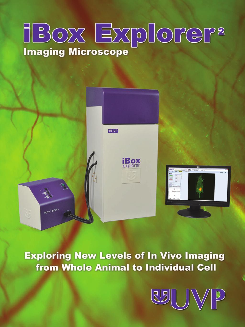
ibox Explorer TM Imaging Microscope
|
|
|
- Julian Sanders
- 6 years ago
- Views:
Transcription


1


 and red fluorescent protein (RFP)")


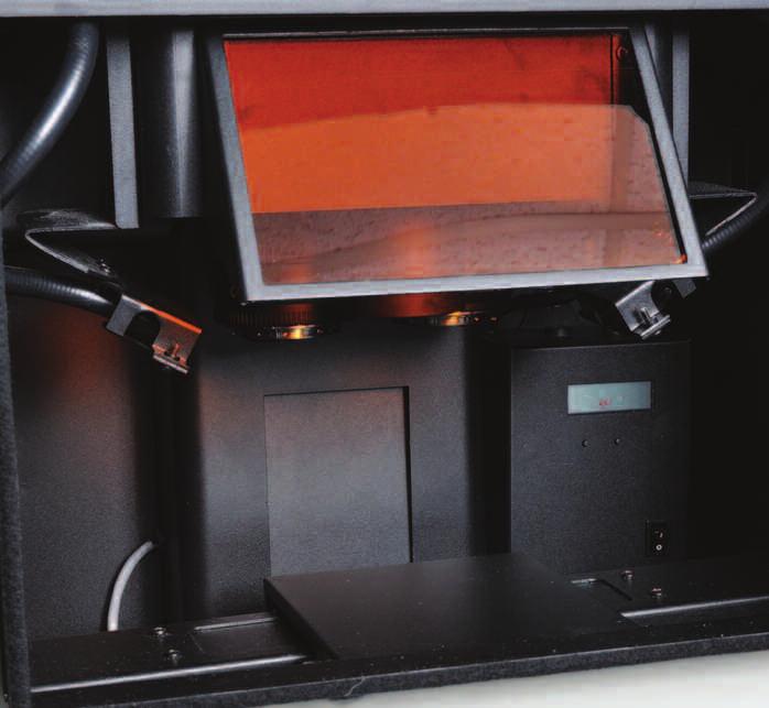
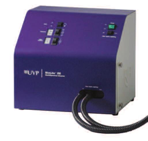
2 ibox Explorer TM Imaging Microscope Visible to NIR In Vivo Imaging for Macro to Micro Detection of Fluorescent Markers in Small Animals Capture images with the high sensitivity, cooled CCD camera and optics, ideal for in vivo imaging applications View whole animal down to single cell via motorized optics Maintain uniform animal temperature for up to two mice on the slide-out warming plate Select BioLite TM excitation filters and light intensity settings via the software interface Seemlessly navigate through the parcentered and parfocal magnification levels from 0.7X to 6.5X Detect a variety of fluorescent markers with emission filters optimized for visible to NIR imaging applications Acquire images and produce quantitative analysis results with VisionWorks LS Software Adjust the location of the motorized X, Y, Z platform via the external joystick; fine tune and bookmark stage positions using the software interface BioLite Xe MultiSpectral Light Source The BioLite is an external light engine for excitation of fluorescent stains. The unit features: Xenon light source with uniform and directed lighting via fiber optic light guides to samples inside the darkroom Matched excitation/emission green fluorescent protein (GFP) and red fluorescent protein (RFP) filter sets included; custom filter sets available Eight excitation filter positions for a variety of filters Emission filters are placed in the darkroom. The neutral density filter passes emission wavelengths from nm. This filter is used for white light or non-fluorescent imaging. The long pass filter blocks blue light excitation and passes fluorescence. The BioLite supplies dual excitation light paths: Coaxial lighting emits targeted illumination for use with high magnification ranges Side lighting emits a broader illumination area for use with the lower magnification ranges Amber viewing screen Side lighting onto the sample stage
3 Benefits of the ibox Explorer Nine discrete magnification levels maximize visualization from whole animal to individual cell, subcutaneously and within the body cavity of living mice Parcentered and parfocal optics allow seamless imaging throughout the magnification levels High sensitivity cooled CCD camera is ideal for visible to NIR small animal imaging applications High intensity xenon light source is optimized for excitation of visible through NIR markers in vivo User-defined software templates simplify image acquisition for consistent and reproducible results Software analysis capabilities enable area density, tumor size and volume measurements for research studies such as biodistribution, progression and regression ibox Explorer Applications Pre-Clinical Research Applications for the ibox Explorer Researchers can visualize in vivo cancer cell migration and tumor progression with the ibox Explorer. The system s upright optics provide an ultra long working distance and high numerical aperture (NA) which is ideal for animal fluorescent imaging studies from the macroscopic (cm) to microscopic (µm) scale. Tumor shedding Tumor/host margins and interactions Hematogenous trafficking Tumor angiogenesis Tumor micro environment Intralymphatic trafficking Micro/macro metastases Primary tumor growth or tumor tracking Biodistribution monitoring Fluorescent Cancer Cell Detection Migration of fluorescent cancer cells within vasculature of a mouse. HT-080 fluorescent cancer cells were injected into the epigastrica cranialis of a mouse. Immediately after injection the fluorescent signature around injection site identifies cells that have escaped into surrounding tissue. A magnified image highlights cancer cells migrating within the bloodstream. Biodistribution Monitoring Monitoring biodistribution of DyLight 755, a near infrared (NIR) dye, conjugated to anti CEA monoclonal antibody within the tissues of a nude mouse. Four hours after injection, the dye can be seen accumulating in the liver. Ex vivo analysis shows that the liver has the most intense NIR emission, suggesting the greatest accumulation of dye. Application Note FP-78. Detection of Multiplex NIR Dyes In Vivo View of a Qdot conjugated to anti-cd monoclonal antibody distribution within a surgically exposed abdomen. The image displays the presence of a GFP expressing tumor lesion within the liver and collection of conjugate in the abdominal organs. The image represents a three color multiplex channels. Application Note FP-7. Multiplex Dual Color Imaging Human osteosarcoma cells at 50% confluence in RPMI media. The multiplexed image is a composite of GFP expressing nuclei and RFP expressing cytoplasm.
4 ibox Explorer In Vivo Applications Detecting a Mouse Pancreatic Cancer Cell Line Mouse pancreatic tumor tissue sample imaged at two magnifications:.5x and 8.8x. A red fluorescent protein (RFP) filter was used to highlight the RFP-tagged cytoplasm to show the distinct morphologic characteristics of individual cells as well as the tissue microenvironment ex vivo. The cell line, XPA (Fig ) shows a histological sample viewed at.5x magnification, excited with light funneled through the optical components. At high magnification (8.8x, Fig ), more distinct detail can be seen, such as cell orientation, areas of high RFP concentration and cytoplasmic morphology. To read more about this research, see Application Note FP-69. Fig. Fig. Fig. Intravital Imaging of a Mouse Ventral Skin-Flap Real-time in vivo imaging of HT-080 cancer cells at several levels of magnification. Figure shows a multiplexed image of a skin flap at.5x magnification. The major vessel in the field corresponds to the epigastrica cranialis vein of a nude mouse. Clusters of dual-colored HT-080 cells can be visualized moving through the vasculature in the right-most aspect of the field. Figure shows an 8.8x magnification of a skin-flap. Migrated dual-colored HT-080 cells (bright yellow) within a distal vessel have begun to extravasate. To read more about this research, see Application Note FP-7. Tumor Targeting of NIR GFP-expressing tumor bearing CEA surface antigen. Human pancreatic cancer cell line with high levels of CEA surface antigen expressing the GFP reporter gene was implanted into the right flank of a mouse. After four weeks, a NIR dye was injected intravenously, conjugated to an anti-cea polyclonal antibody. 8 hours after injection, the dye antibody conjugates were co-localized within the tumor. Constitutively Fluorescent CFP Mouse Labeling of mouse thyroid tissue histology section with Alexa88 conjugated to anti- CEA antibody. Background fluorescent areas appear blue due to co-expression of cyan fluorescent protein (CFP) reporter with an intracellular protein. Areas of high CEA surface antigen expression readily bind to the Alexa88 conjugate and emit green fluorescence. Co-Localization of NIR-tagged Antibody Subcutaneous tumor implanted in a mouse. Qdot 800-CD antibody conjugate was injected into the vasculature of a hepatoma tumor-bearing mouse. High magnification of the tumor vascular shows co-localization of the Qdot conjugate in red within the GFP expressing tumor. Qdot800 signal can be seen within the perivascular region of the tumor vessel. Tumor Cells in the Colon Skin flap technique visualizing the MMT (breast cancer cell line) dual color tumor cells in the colon. GFP and RFP filters were used to isolate the green and red fluorescence. Fig.
5 Ordering Information and Specifications System Name Camera ibox Explorer 60 OptiChemi 60 Darkroom Interior: Darkroom Specifications: Emission Filters: Four-position interchangeable tray: 55nm Longpass, GFP, RFP, Neutral Density; Additional filters available Controls: Automated, software controlled Stage: Precision motorized X, Y, Z Travel: X=00mm, Y=00mm, Z=00mm Resolution: X=0μm, Y= 0μm, Z=μm Position/Focus: Controlled by joystick/software Warming Plate: 7 O C, user adjustable Darkroom Dim.: 7.5 W x 9.5 D x H (.5 x 9.5 x 0cm) Camera Specifications: Camera: OptiChemi 60 Type: Monochrome CCD Bit Depth: 6 bit File Bit Depth: 6 bit Pixel Resolution: 8 x 7 Megapixels:., extendable to 9.6 Cooling Type: -50 O C from ambient, Peltier cooling Peak QE: 86% (quantum efficiency) BioLite Xe MultiSpectral Source: Light Source: 50 watt xenon Fiber Optic: Coaxial and epi illumination; light heads mounted to the darkroom Excitation Filters: Eight position wheel includes filters: GFP and RFP Additional wavelength filters available Controls: Via software Dimensions: 8.75H x 7W x 0D in. (. x 7.8 x 5.cm) VisionWorksLS Software: Interfaces with: Camera, optics, darkroom, BioLite Tools: Macros and templates Analysis: Extensive functions including area density and volume measurements, tumor sizing and compositing tools Documentation: Create reports and export data Compliance: Supports CFR Part Compatibility: Win XP (SP), Win 7, 8 Adjustable amber viewing screen Optics Epi light guides Height adjustable stage with warming plate Excitation/Emission Filter Sets Included: Filter Type GFP RFP General Neutral Density BioLite Xe: BioLite Excitation Wavelength 55-95nm 50-57nm Microscope Emission Wavelength 50-5nm nm 55 long pass nm Additional filter sets are available to cover a wide spectra of fluorescent proteins. For an extensive list of filters, go to UVP.com/pdf/UVPFilterSelectionChart.pdf Variable intensity settings indicator Position filter indicator Access to wheel with 8 filter ports Fiber optic cables and light guides connect in the darkroom to supply directed side and coaxial lighting to the animal Magnification and Field of View Imaging Area: Optical Magnification: 0.7x 0.5x 0.5x.66x.5x.5x 7.5x 8.8X 6.5X FOV Range (mm): 90x90 60x60 0x0 9x9 6x6.x. x.7x.7 0.9x0.9
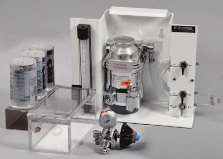
6 ibox Accessories ibox Anesthesia System The ibox Anesthesia system supplies a complete kit for safe anesthetizing of small animals with isoflurane or sevoflurane (gas not included with kit). While imaging is performed with the ibox Explorer, it is important that there is no movement of the animal. The anesthesia system minimizes movement and permits the animal to use less anesthesia, reduces stress and potential side-effects while imaging is performed. The anesthesia allows quick recovery time of the animal. Several components are combined in the anesthesia system to regulate and administer a combination of oxygen and isoflurane gas to the animal. The initial anesthesia is performed in the induction chamber. The animal is then moved to the warming plate inside the ibox darkroom and connected to a nose cone (two nose cones are included). The plate generates uniform temperature conditions which maintain a safe body temperature of the animal during the imaging process. A low profile breathing device on the plate connects to patented valves. The system s non-rebreathing technology safely prevents backflow of gases into the darkroom. The ibox Anesthesia System is a portable unit that is designed for maximum efficiency, ease of use and low anesthetic gas consumption. Cell Lines Well over 00 cancer cell lines are available that have been engineered to express GFP, RFP, or dual color (nucleus/ cytoplasm) for in vivo fluorescent imaging analysis. These cell lines represent a wide range of mainly human cancers such as breast, pancreas, prostate, ovarian, lung, colon, melanoma and many others. In addition, mouse and rat cell lines are also available as well as custom fluorescent cell lines through contract development work. Tumor Type Cell Line GFP RFP Dual-Color Prostate, Human PC X X X Prostate, Human DU5 X X Prostate, Human LNCap X X Technology UVP has developed and manufactured imaging systems for Life Science research since the 980s. Requirements for equipment in cancer research applications led to the development of the ibox Explorer Imaging Microscope. UVP s team of R&D and Life Scientists continue to expand system capabilities and provide economical equipment to researchers worldwide. UVP offers system and software training. Installation and Operation Qualification documents are available and enable researchers to comply with regulatory bodies. The choice of fluorescent protein used in the cell line will depend in part on the type of mouse being used. Nude mice which express GFP, RFP and CFP as normal background are also available so that the cell line fluorescent color can contrast with that of the normal tissue. For example, a RFP tumor cell line can be tracked with high contrast against a GFP background normal tissue using the GFP mouse. Contact UVP for further details. The chart shows a sampling of over 00 cancer cell lines available. Contact UVP for details. A variety of application notes and articles are available which discuss fluorescence in vivo imaging. Go to uvp.com for details. ibox Explorer is sold under license from AntiCancer, Inc. which allow scientists to optimize their research using an extensive array of patented fluorescent protein imaging application technologies. Go to UVP.com or contact UVP for details. For applications beyond in vivo imaging, contact UVP for information on BioImaging Systems for chemiluminescent, gel and plant imaging. For information contact: Web Site: UVP.com UVP, LLC 066 W. th St., Upland, CA 9786 Tel: (800) (909) Fax: (909) info@uvp.com Ultra-Violet Products Ltd. Unit, Trinity Hall Farm Estate, Nuffield Road, Cambridge CB TG UK Tel: +(0)-00 Fax: +(0) uvp@uvp.co.uk ibox and VisionWorks are registered trademarks of UVP, LLC. Explorer and BioLite are trademarks of UVP, LLC. All other trademarks are recognized as owned by their respective companies. Specifications subject to change without notice. Ó UVP, LLC 0 Lit: ibox Explorer R0
 APPLICATIONS OLYMPUS ANTICANCER, INC. TECHNOLOGY PARTNERSHIP OV100 In vivo imaging system OV100 TM fluorescence imaging system for visualizing cancer cell extravasation in live mice Detailed dynamic changes
NEWTON 7.0 BIOLUMINESCENCE & FLUORESCENCE IMAGING IN VIVO - IN VITRO IMAGING
 NEWTON 7.0 BIOLUMINESCENCE & FLUORESCENCE IMAGING IN VIVO - IN VITRO IMAGING The NEWTON s protocol driven image acquisition is as quick as it is intuitive: adjust your exposure, save, print or quantify.
NEWTON 7.0 BIOLUMINESCENCE & FLUORESCENCE IMAGING IN VIVO - IN VITRO IMAGING
 NEWTON 7.0 BIOLUMINESCENCE & FLUORESCENCE IMAGING IN VIVO - IN VITRO IMAGING SMART IMAGING SYSTEM The NEWTON 7.0 system combines high sensitivity with advanced animal-handling features and userfriendly
NEWTON 7.0 BIOLUMINESCENCE & FLUORESCENCE IMAGING IN VIVO - IN VITRO IMAGING
 NEWTON 7.0 BIOLUMINESCENCE & FLUORESCENCE IMAGING IN VIVO - IN VITRO IMAGING The NEWTON s protocol driven image acquisition is as quick as it is intuitive: adjust your exposure, save, print or quantify.
Azure cseries. A new way to see the light. c600 c500 c400 c300 c200
 Azure cseries A new way to see the light. c6 c5 c4 c3 c2 Infrared Laser Excitation for Quantitative Western Blot Imaging in the NIR Improve Your Data Quality Imaging with infrared dyes offers signal stability
Chemiluminescence Detection. Using UVP s ChemiDoc-It System
 Chemiluminescence Detection Using UVP s ChemiDoc-It System Protein Detection Agents Chemiluminescence Fluorescence Colorimetric Detection Radioisotopic Detection Chemiluminescence Generation of light through
Azure Biosystems Western Blotting Workflow
 Azure Biosystems Western Blotting Workflow PROBE PLAN SEPARATE ANALYZE VISUALIZE PLAN Plan your experiment and choose your detection method Chemiluminescent Western Blotting The most common method for
Azure cseries. A new way to see the light. c600 c500 c400 c300
 Azure cseries A new way to see the light. c600 c500 c400 c300 Infrared Laser Excitation for Quantitative Western Blot Imaging in the NIR Improve Your Data Quality Imaging with infrared dyes offers signal
MF-ChemiBIS. Today s most comprehensive solution for your bio-imaging needs and applications. Documenting Nature
 MF-ChemiBIS Today s most comprehensive solution for your bio-imaging needs and applications Documenting Nature MF-ChemiBIS Excellence in bio-imaging The DNR Advantage As pioneers in bio-imaging technologies
High-Throughput In Vivo Bioluminescence Imaging System
 IVIS SpectrumBL P R O D U C T N O T E Pre-clinical in vivo imaging Key Features Ultra high-sensitivity to support in vivo bioluminescence, chemiluminescence and Cerenkov imaging High-throughput (10 mice)
In Vivo Programs. Contract Research Services. Programs
 P r o d u c t N o t e Contract Research Services In Vivo Programs Key Features Evaluation of drug properties Drug efficacy studies Biodistribution studies Complementary tissue analysis services CDAS In
Sapphire. Biomolecular Imager THE NEXT GENERATION OF LASER-BASED IMAGING
 Sapphire Biomolecular Imager THE NEXT GENERATION OF LASER-BASED IMAGING Breakthrough image capture and analysis The Sapphire Biomolecular Imager is a next generation laser scanning system that provides
QImaging Camera Application Notes Multicolor Immunofluorescence Imaging
 QImaging Camera Application Notes Multicolor Immunofluorescence Imaging In order to image localization of intracellular proteins with high specificity, it is frequently necessary to multiplex antibody
Innovations To Meet Your Needs
 Innovations To Meet Your Needs Cooled CCD Camera 1340 x 1037 pixel resolution for greatest image quality 12-bit precision provides 3 orders of linear dynamic range Windows and Power Macintosh Software
2004 Debye Lecture 4 C. B. Murray. Quantum Dot Applications: Sun Screen. Solar Cells. Bio-tagging. Solid State Lighting?
 2004 Debye Lecture 4 C. B. Murray Quantum Dot Applications: Sun Screen Solar Cells Bio-tagging Solid State Lighting? Quantum Dot Solar cells Nanocrystal Solar Cells Double-labeling of mitochondria
Sapphire. Biomolecular Imager THE NEXT GENERATION OF LASER-BASED IMAGING
 Sapphire Biomolecular Imager THE NEXT GENERATION OF LASER-BASED IMAGING Breakthrough image capture and analysis The Sapphire Biomolecular Imager is a next generation laser scanning system that provides
OCTOPLUS QPLEX FLUORESCENCE IMAGER. for fast & powerful fast 2D Gel image acquisition
 OCTOPLUS QPLEX FLUORESCENCE IMAGER for fast & powerful fast 2D Gel image acquisition Octoplus QPLEX Fluorescence Imager The new Octoplus QPLEX fluorescence imager sets a novel standard fluorescence 2D
Odyssey Fc Imaging System Infrared fluorescent and chemiluminescent imaging in one system!
 Odyssey Infrared Imaging System A fundamental change in western blot analysis Odyssey Fc Imaging System Infrared fluorescent and chemiluminescent imaging in one system! Accurate Quantification Wide linear
Supplementary Figures
 Supplementary Figures Supplementary Figure S1. In vitro determination of the detection limit of vanco-800cw (a) Specificity of vanco-800cw for different clinical bacterial isolates. The in vitro imaging
Advanced preclinical optical imaging. Preclinical in vivo imaging. IVIS Spectrum
 IVIS Spectrum P R O D U C T N O T E Preclinical in vivo imaging Key Features High Sensitivity in vivo fluorescence and bioluminescence imaging 3D tomographic reconstruction Absolute calibration High throughput
Focal Points. Application Note FP-153. Next Generation Gel Imaging with GelRed and GelGreen Dyes and GelDoc-It Imaging System
 Focal Points Application Note FP-153 MIDSCI 800.227.9997 636.227.9997 Gel Doc Equipment Next Generation Gel Imaging with GelRed and GelGreen Dyes and GelDoc-It Imaging System Introduction By using state-of-the-art
DEPArray Technology. Sorting and Recovery of Rare Cells
 DEPArray Technology Sorting and Recovery of Rare Cells Delivering pure, single, viable cells The DEPArray system from Silicon Biosystems is the only automated instrument that can identify, quantify, and
Amnis ImageStream : Technical Reports & Applications
 Amnis ImageStream : Technical Reports & Applications ImageStream : Flow Cytometry and Microscopy in a Single Platform The ImageStream achieves true multispectral Imaging in Flow by combining microscopy
More on fluorescence
 More on fluorescence Last class Fluorescence Absorption emission Jablonski diagrams This class More on fluorescence Common fluorophores Jablonski diagrams to spectra Properties of fluorophores Excitation
Optical Observation - Hyperspectral Characterization of Nano-scale Materials In-situ
 Optical Observation - Hyperspectral Characterization of Nano-scale Materials In-situ Research at the nanoscale is more effective, when research teams can quickly and easily observe and characterize a wide
Tumor tissues or cells were homogenized and proteins were extracted using
 SUPPLEMENTAL MATERIALS AND METHODS Western Blotting Tumor tissues or cells were homogenized and proteins were extracted using T-PER tissue protein extraction buffer. Protein concentrations were determined
Ultra-Violet Products Ltd W 11th Street, Upland, CA Unit 1,Trinity Hall Farm Estate,
 FirstLight UV Transilluminators Instruction Manual UVP, LLC Ultra-Violet Products Ltd. 2066 W 11th Street, Upland, CA 91786 Unit 1,Trinity Hall Farm Estate, Tel: (800) 452-6788 / (909) 946-3197 Nuffield
Dino-Lite knowledge & education. Fluorescence Microscopes
 Dino-Lite knowledge & education Fluorescence Microscopes Dino-Lite Fluorescence models Smallest fluorescence microscope in the world Revolution to biomedical and educational applications Flexible Easy
Imaging the immune system with a Two photon (2P) microscope
 microscope") Imaging the immune system with a Two photon (2P) microscope QuickTime et un décompresseur Cinepak sont requis pour visionner cette image. Main advantages of 2P microscopy : 1/ Deep penetration into tissue
Imaging the immune system with a Two photon (2P) microscope QuickTime et un décompresseur Cinepak sont requis pour visionner cette image. Main advantages of 2P microscopy : 1/ Deep penetration into tissue
Kazuki N. Sugahara, Tambet Teesalu, Priya Prakash Karmali, Venkata Ramana Kotamraju, Lilach
 Cancer Cell, Volume 16 Supplemental Data Tissue-Penetrating Delivery of Compounds and Nanoparticles into Tumors Kazuki N. Sugahara, Tambet Teesalu, Priya Prakash Karmali, Venkata Ramana Kotamraju, Lilach
Insight Through In Vivo Imaging
 Insight Through In Vivo Imaging Resolution Revolution in Realtime The Vevo 770 provides anatomical, functional and molecular data in realtime, on an affordable, easy to use and translational platform.
Considerations Dynamic Range Detection Linearity Components Software Comparing qpcr Instrumentation Sensitivity Specificity of Detection sigma.
 Considerations When Higuchi et. al 1 designed a qpcr system, a conventional PCR block, a UV light source and a camera for signal detection were the instruments used. Since the design of the first qpcr
End of Year. Special Offers
 End of Year Special Offers 2018 Cell Biology TC20 Automated Cell Counter Automated and precise cell counts and viability assessment within 30 seconds Compatible with a broad range of cell sizes and types
1st Faculty of Medicine, Charles University in Prague Center for Advanced Preclinical Imaging (CAPI)
") ADVANTAGES Optical Imaging OI Optical Imaging is based on the detection of weak light by a highly sensitive and high resolution CCD camera DISADVANTAGES High sensitivity Limited penetration depth Easy
ADVANTAGES Optical Imaging OI Optical Imaging is based on the detection of weak light by a highly sensitive and high resolution CCD camera DISADVANTAGES High sensitivity Limited penetration depth Easy
Simple, intuitive and accessible MRI solution for preclinical research. M-Series Compact MRI Systems
 Simple, intuitive and accessible MRI solution for preclinical research M-Series Compact MRI Systems Application Oriented Imaging Anatomy and Morphology In vivo soft tissue imaging for morphological characterization.
Quantum Dot applications in Fluorescence Imaging for Calibration and Molecular Imaging
 Quantum Dot applications in Fluorescence Imaging for Calibration and Molecular Imaging Introduction In this application note, we will discuss the application of quantum dots in fluorescence imaging, both
Everything counts. But nothing counts like the
 Everything counts But nothing counts like the Countess II FL Automated Cell Counter The new Countess II FL Automated Cell Counter is a benchtop cell assay platform equipped with state-of-the-art optics
ab CytoPainter ER Staining Kit Red Fluorescence
 ab139482 CytoPainter ER Staining Kit Red Fluorescence Instructions for Use Designed to detect Human endoplasmic reticulum by microscopy. This product is for research use only and is not intended for diagnostic
NovoCyte Flow Cytometer
 NovoCyte Flow Cytometer The Flow Cytometer for Everyone 2 Experience the NovoCyte Advantage Focus on advancing your research. Let the flow cytometer do the rest. NovoCyte Flow Cytometer High Performance
HYPERSPECTRAL MICROSCOPE PLATFORM FOR HIGHLY MULTIPLEX BIOLOGICAL IMAGING. Marc Verhaegen
 HYPERSPECTRAL MICROSCOPE PLATFORM FOR HIGHLY MULTIPLEX BIOLOGICAL IMAGING Marc Verhaegen CMCS, MONTREAL, MAY 11 th, 2017 OVERVIEW Hyperspectral Imaging Multiplex Biological Imaging Multiplex Single Particle
Accelerating the Pace of Understanding
 Vectra 3 P R O D U C T N O T E Quantitative Pathology Imaging and Analysis Key Benefits Part of PerkinElmer's Phenoptics workflow solution for Cancer Immunology Research Detect and measure multiple expressed
Unrivalled Optical Imaging solutions
 Unrivalled Optical Imaging solutions high throughput high resolution x-ray TOMOFLUO multispectral analysis 3D And beyound... bioluminescence and fluorescence Imaging systems Real-time imaging capability
Intraoperative Fluorescence Surgical Goggle
 Intraoperative Fluorescence Surgical Goggle Ruogu Qin Abstract Background Tumor surgery is a great challenge to surgeons. Due to their disseminated characteristic, the small and varied foci and blurred
Skull optical clearing window for in vivo imaging of the mouse cortex at synaptic resolution
 SUPPLEMENTARY INFORMATION for Skull optical clearing window for in vivo imaging of the mouse cortex at synaptic resolution Yanjie Zhao 1,2, Tingting Yu 1,2, Chao Zhang 1,2, Zhao Li 1,2, Qingming Luo 1,2,
Ilya Turchin. Institute of Applied Physics of the RAS, Nizhny Novgorod, Russia.
 Fluorescence 3D imaging of small animals Ilya Turchin Institute of Applied Physics of the RAS, Nizhny Novgorod, Russia ilya@ufp.appl.sci-nnov.ru http://www.bioimaging.ru German-Russian Forum Biotechnology
Simple, intuitive and accessible MRI solution for preclinical research. M-Series Compact MRI Systems
 Simple, intuitive and accessible MRI solution for preclinical research M-Series Compact MRI Systems Application Oriented Imaging Molecular Imaging Using Contrast Agents Detection and quantification of
SpectraMax Paradigm Microplate Reader
 SpectraMax Paradigm Microplate Reader User-upgradeable Multi-Mode Detection Platform BENEFITS Flexible design allows for single or dual excitation and emission High speed detection and excellent sensitivity
ChemiDoc MP Imaging System
 Imaging ChemiDoc MP Imaging System Confidence in every step of your imaging workflow. ChemiDoc MP Imaging System Because results always matter. Bring a new level of capability and efficiency to your experiments
ab CytoPainter ER Staining Kit Red Fluorescence
 ab139482 CytoPainter ER Staining Kit Red Fluorescence Instructions for Use Designed to detect Human endoplasmic reticulum by microscopy. This product is for research use only and is not intended for diagnostic
Sale. Bio-Rad. See inside for an array of offers on: The
 Life Science Group The Bio-Rad 2013 Sale See inside for an array of offers on: Electrophoresis and Blotting Imaging Systems Thermal Cyclers Cell Counting PCR Reagents and Plastics Electroporation Immunoassay
Contents. SCHOOL of FLUORESCENCE. For more information, go to lifetechnologies.com/imagingbasics
 MPSF educator packet This packet contains illustrations and figures from the Molecular Probes School of Fluorescence website. They illustrate concepts from the basic physical properties that underlie fluorescence
BIO 315 Lab Exam I. Section #: Name:
 Section #: Name: Also provide this information on the computer grid sheet given to you. (Section # in special code box) BIO 315 Lab Exam I 1. In labeling the parts of a standard compound light microscope
Automatic detection of plasmonic nanoparticles in tissue sections
 Automatic detection of plasmonic nanoparticles in tissue sections Dor Shaviv and Orly Liba 1. Introduction Plasmonic nanoparticles, such as gold nanorods, are gaining popularity in both medical imaging
ab CFSE Fluorescent Cell Labeling Kit
 ab113853 CFSE Fluorescent Cell Labeling Kit Instructions for Use For the durable fluorescent labeling of live cells for fluorescent microscopy and flow cytometry, population growth studies and within sample
Boundary-breaking acoustic focusing cytometry
 Boundary-breaking acoustic focusing cytometry Introducing the Attune NxT Acoustic Focusing Cytometer a high-performance system that s flexible enough for any lab One of the main projects in my laboratory
Quantum dots in Molecular Imaging
 Quantum dots in Molecular Imaging Ivo Que 1 Introduction Quantum dots (Qdots ) are nanoparticles exhibiting fluorescent properties that can be used for cell staining. In the early 1980s the first quantum
PCR SYSTEMS. a new era in high-productivity qpcr. Applied Biosystems ViiA 7 Real-Time PCR System
 PCR SYSTEMS a new era in high-productivity qpcr Applied Biosystems ViiA 7 Real-Time PCR System a new era in high-productivity qpcr The ViiA 7 Real-Time PCR System delivers the proven reliability, sensitivity,
Confocal Microscopes. Evolution of Imaging
 Confocal Microscopes and Evolution of Imaging Judi Reilly Hans Richter Massachusetts Institute of Technology Environment, Health & Safety Office Radiation Protection What is Confocal? Pinhole diaphragm
Fluorescence Microscopy. Terms and concepts to know: 10/11/2011. Visible spectrum (of light) and energy
 and energy") Fluorescence Microscopy Louisiana Tech University Ruston, Louisiana Microscopy Workshop Dr. Mark DeCoster Associate Professor Biomedical Engineering 1 Terms and concepts to know: Signal to Noise Excitation
Fluorescence Microscopy Louisiana Tech University Ruston, Louisiana Microscopy Workshop Dr. Mark DeCoster Associate Professor Biomedical Engineering 1 Terms and concepts to know: Signal to Noise Excitation
TARGETED IMAGING. Maureen Chan and Ruwani Mahathantila
 TARGETED IMAGING Maureen Chan and Ruwani Mahathantila Overview 2 Introduction to fluorescent imaging Fluorescent agents Quantum Dots Physical properties How QDs work In Vivo QD imaging Future Video What
ClonePix 2. Screen and select more clones in less time. Genetix Now part of Molecular Devices.
 ClonePix 2 Screen and select more clones in less time www.moleculardevices.com/genetix Genetix Now part of Molecular Devices Screen more clones in less time Cut cell line and antibody development times
A histone H1-binding-aptide based apoptosis-imaging probe for monitoring
 Electronic Supplementary Material (ESI) for MedChemComm. This journal is The Royal Society of Chemistry 2017 Supporting Information A histone H1-binding-aptide based apoptosis-imaging probe for monitoring
SPHERO TM Fluorescent Particles
 The SPHERO TM fluorescent microparticles are prepared by either staining polystyrene particles with a fluorophore solution or by polymerizing a fluorophore in styrene in the presence of polystyrene core
APPLICATION NOTE Rev. 7/2017, v4.0 Fluorescent Nanodiamonds: Bio-applications. Physical and Fluorescence Properties
 APPLICATION NOTE Rev. 7/2017, v4.0 Fluorescent Nanodiamonds: Bio-applications Fluorescent nanodiamonds (FNDs) offer a unique alternative to currently existing fluorescent biomarkers. With exceptional photo
SUPPLEMENTARY INFORMATION
 VOLUME: 1 ARTICLE NUMBER: 0011 In the format provided by the authors and unedited. In situ Activation of Platelets with Checkpoint Inhibitors for Post-Surgical Cancer Immunotherapy Chao Wang 1, 2, Wujin
The New Wave in Spectroscopy FS-2 FluoroMate Fluorescence Spectrometer
 www.scinco.com The New Wave in Spectroscopy FluoroMate FS-2 FluoroMate FS-2 FluoroMate FS-2 The SCINCO FS-2 fluorescence spectrometer delivers exceptional sensitivity for the most accurate measurements.
Fluorescence Microscopy
 Fluorescence Microscopy Dr. Arne Seitz Swiss Institute of Technology (EPFL) Faculty of Life Sciences Head of BIOIMAGING AND OPTICS BIOP arne.seitz@epfl.ch Fluorescence Microscopy Why do we need fluorescence
A high-performance, easy-to-use multimode reader for luminescence, fluorescence, absorbance, BRET and FRET applications.
 A high-performance, easy-to-use multimode reader for luminescence, fluorescence, absorbance, BRET and FRET applications. The Perfect Partner for Promega Assays Easy-To-Use Choose from preloaded Promega
You ll never look at cells the same way again.
 E S S E N I N C U C Y T E T M You ll never look at cells the same way again. The number of cell-based experiments conducted every day is growing dramatically. Through these experiments, and leveraging
BIO 315 Lab Exam I. Section #: Name:
 Section #: Name: Also provide this information on the computer grid sheet given to you. (Section # in special code box) BIO 315 Lab Exam I 1. In labeling the parts of a standard compound light microscope
ENVAIR lab Real-Time PCR
 ENVAIR lab Real-Time PCR Laboratory Real-Time Thermal Cycler Our advanced technology highlight Introduction The ENVAIR lab Real-Time PCR system is a high-performance benchtop instrument giving you greater
LabChip 90 System with DataViewer Software
 Revolutionizing DNA and Protein Gel Electrophoresis LabChip 90 System with DataViewer Software The Proven Alternative to Slab Gels. The massive flow of information emerging from today s genomic and proteomic
Image Analysis. Tools for basic and translational research. visualize analyze interpret report archive customize
 Image Analysis Tools for basic and translational research visualize analyze interpret report archive customize Unlock the benefits of image analysis. Image analysis is an integrated part of Aperio s digital
SUPPLEMENTARY FIGURES
 SYNERGISTIC STRATEGY FOR MULTICOLOR TWO-PHOTON MICROSCOPY: APPLICATION TO THE ANALYSIS OF GERMINAL CENTER REACTIONS IN VIVO ASYLKHAN RAKHYMZHAN, RUTH LEBEN, HANNA ZIMMERMANN, ROBERT GÜNTHER, PEGGY MEX,
Microendoscopes for imaging of the pancreas
 Microendoscopes for imaging of the pancreas Angelique Kano *, Andrew R. Rouse, Shona M. Kroto, Arthur F. Gmitro Optical Sciences Center& Radiology Research Lab, University of Arizona, Tucson 85724 ABSTRACT
Anti-MOUSE IgG (H&L) (GOAT) Antibody DyLight 488 Conjugated (Min X Bv Ch Gt GP Ham Hs Hu Rb Rt & Sh Serum Proteins)
 (GOAT) Antibody DyLight 488 Conjugated (Min X Bv Ch Gt GP Ham Hs Hu Rb Rt & Sh Serum Proteins)") Anti-MOUSE IgG (H&L) (GOAT) Antibody DyLight 488 Conjugated (Min X Bv Ch Gt GP Ham Hs Hu Rb Rt & Sh Serum Proteins) - 610-141-121 Code: 610-141-121 Size: 100 µg Product Description: Anti-MOUSE IgG (H&L)
Anti-MOUSE IgG (H&L) (GOAT) Antibody DyLight 488 Conjugated (Min X Bv Ch Gt GP Ham Hs Hu Rb Rt & Sh Serum Proteins) - 610-141-121 Code: 610-141-121 Size: 100 µg Product Description: Anti-MOUSE IgG (H&L)
Fluorescence Light Microscopy for Cell Biology
 Fluorescence Light Microscopy for Cell Biology Why use light microscopy? Traditional questions that light microscopy has addressed: Structure within a cell Locations of specific molecules within a cell
NanoPro Characterize cell signaling in your smallest samples
 NanoPro 1000 Characterize cell signaling in your smallest samples Technology A NEW TAKE ON IMMUNOASSAYS The NanoPro 1000 is a capillary-based nano-immunoassay platform. As in Western blot analysis, proteins
NV High Brightness Series
 PRODUCT SHEET Rev. 8/17, v9 NV High Brightness Series The NV-High Brightness Series features fluorescent nanodiamonds ranging in size from 20 nm up to 150µm containing nitrogen vacancy (NV) centers with
NEW INSIGHTS. NEW DISCOVERIES. Real-time automated measurements of cell health, movement and function inside your incubator.
 THE NEXT GENERATION HAS ARRIVED IncuCyte S3 Live-Cell Analysis System Real-time automated measurements of cell health, movement and function inside your incubator. NEW INSIGHTS. NEW DISCOVERIES. See what
Reach greater highs. (and lows ) with new protein ladder choices. Fermentas now sold as. Thermo Scientific
 with new protein ladder choices. Fermentas now sold as. Thermo Scientific") Introducing Thermo Scientific Pierce Prestained and Unstained s for easy protein molecular weight determination. Fermentas now sold as Thermo Scientific Reach greater highs (and lows ) with new protein
Introducing Thermo Scientific Pierce Prestained and Unstained s for easy protein molecular weight determination. Fermentas now sold as Thermo Scientific Reach greater highs (and lows ) with new protein
C101-E112. BioSpec-nano. Shimadzu Spectrophotometer for Life Science
 C101-E112 BioSpec-nano Shimadzu Spectrophotometer for Life Science Power of small. BioSpec-nano BioSpec-nano Shimadzu Spectrophotometer for Life Science Quick and Simple Nucleic Acid Quantitation Drop-and-Click
Imaging Quantum Dots using FUJIFILM LAS 4000
 Imaging Quantum Dots using FUJIFILM LAS 4000 Application Note John Pizzonia, Ph.D. 9-28-07 Quantum dots (also known as nanocrystals) are a special class of materials known as semiconductors, which are
64 CuCl 2 in 50 µl 0.1N NaOAc buffer, and 20 µg of each DOTA-antibody conjugate in 40 µl
 Number of DOTA per antibody The average number of DOTA chelators per antibody was measured using a reported procedure with modifications (1,2). Briefly, nonradioactive CuCl 2 (80-fold excess of DOTA antibodies)
Cell Imaging. Cell Imaging 48
 Cell Imaging 48 bio-rad.com/zoe Cell Imaging Bio-Rad s suite of tools for fluorescence microscopy and cell imaging includes the ZOE fluorescent cell imager and nuclear dyes. See Also PureBlu Hoechst 33342
NanoPro 1000 System. characterize cell signaling in your smallest samples
 NanoPro 1000 System characterize cell signaling in your smallest samples Technology The NanoPro 1000 system is a capillary-based nano-immunoassay platform. As in Western blot analysis, proteins from complex
Quantum Dots and Carbon Nanotubes in Cancer diagnose EE453 Project Report submitted by Makram Abd El Qader
 Quantum Dots and Carbon Nanotubes in Cancer diagnose EE453 Project Report submitted by Makram Abd El Qader abdelqad@unlv.nevada.edu, Fall 2008 Abstract On the basis of research and cancer medical treatment,
[A complex community of T cells, B cells, NK, DC monocytes,neutrophils, etc.] Lymphocyte Communication Does not Obey at least one Aristotelian Ideal:
![[A complex community of T cells, B cells, NK, DC monocytes,neutrophils, etc.] Lymphocyte Communication Does not Obey at least one Aristotelian Ideal:](/thumbs/93/114465749.jpg "[A complex community of T cells, B cells, NK, DC monocytes,neutrophils, etc.] Lymphocyte Communication Does not Obey at least one Aristotelian Ideal:") [A complex community of T cells, B cells, NK, DC monocytes,neutrophils, etc.] Lymphocyte Communication Does not Obey at least one Aristotelian Ideal: Broadcast An entire city should be of a sufficiently
[A complex community of T cells, B cells, NK, DC monocytes,neutrophils, etc.] Lymphocyte Communication Does not Obey at least one Aristotelian Ideal: Broadcast An entire city should be of a sufficiently
Accessorize Your Imaging. Ian Clements Invitrogen Corp
 Accessorize Your Imaging Ian Clements Invitrogen Corp Imaging Tools and Accessories Antifade Reagents Prolong Gold SlowFade Gold Image-iT FX Signal Enhancer FocalCheck Microscope Test Slides Fluorescent
Special Techniques 1. Mark Scott FILM Facility
 Special Techniques 1 Mark Scott FILM Facility SPECIAL TECHNIQUES Multi-photon microscopy Second Harmonic Generation FRAP FRET FLIM In-vivo imaging TWO-PHOTON MICROSCOPY Alternative to confocal and deconvolution
Cancer inflammation research applications and products
 Cancer inflammation research applications and products Flow cytometry Immunoassays Cell imaging Instrumentation Invitrogen Attune NxT Flow Cytometer Antibodies RNA flow Conjugated antibodies for flow cytometry
Quality Matters: Bethyl DyLight Antibody Conjugates
 Quality Matters: Bethyl DyLight Antibody Conjugates Criteria for Fluorescent Probe Selection The use of fluorescent probes has become a mainstream technique to detect speci ic molecular targets in both
Development of an Immuno-PCR Assay
 Development of an Immuno-PCR Assay Innova Biosciences Guide Innova Biosciences Ltd. Babraham Research Campus, Cambridge, UK, CB22 3AT +44 (0)1223 661000 info@innovabiosciences.com Development of an Immuno-PCR
CytoPainter Golgi Staining Kit Green Fluorescence
 ab139483 CytoPainter Golgi Staining Kit Green Fluorescence Instructions for Use Designed for the detection of Golgi bodies by microscopy This product is for research use only and is not intended for diagnostic
PSC 4-Marker Immunocytochemistry Kit PSC (OCT4, SSEA4) Immunocytochemistry Kit PSC (SOX2, TRA-1-60) Immunocytochemistry Kit
 Immunocytochemistry Kit PSC (SOX2, TRA-1-60) Immunocytochemistry Kit") PSC 4-Marker Immunocytochemistry Kit PSC (OCT4, SSEA4) Immunocytochemistry Kit PSC (SOX2, TRA-1-60) Immunocytochemistry Kit Catalog no. A24881, A25526, A25525 Table 1 Contents and storage Kit component
PSC 4-Marker Immunocytochemistry Kit PSC (OCT4, SSEA4) Immunocytochemistry Kit PSC (SOX2, TRA-1-60) Immunocytochemistry Kit Catalog no. A24881, A25526, A25525 Table 1 Contents and storage Kit component
THE FREEDOM TO DISCOVER BOND RX FULLY AUTOMATED IHC AND ISH AND EMERGING TESTS RESEARCH USE ONLY. NOT FOR USE IN DIAGNOSTIC PROCEDURES.
 BOND RX FULLY AUTOMATED IHC AND ISH AND EMERGING TESTS RESEARCH USE ONLY. NOT FOR USE IN DIAGNOSTIC PROCEDURES. EXPLORE YOUR IDEAS PUSH THE BOUNDARIES OF WHAT IS POSSIBLE The BOND RX fully automated research
A legacy of innovation and discovery
 A legacy of innovation and discovery CellInsight CX7 LZR High Content Analysis Platform Quantifiably brilliant data Since the introduction of Thermo Scientific ArrayScan High Content Analysis (HCA) Readers
Supplementary Data. In-Vivo Tumor Targeting and Spectroscopic Detection with Surface- Enhanced Raman Nanoparticle Tags
 Supplementary Data In-Vivo Tumor Targeting and Spectroscopic Detection with Surface- Enhanced Raman Nanoparticle Tags X.-M. Qian, 1 Xiang-Hong Peng, 2 Dominic O. Ansari, 1 Qiqin Yin-Goen, 3 Georgia Z.
ab CFSE Fluorescent Cell Labeling Kit
 ab113853 CFSE Fluorescent Cell Labeling Kit Instructions for Use For the durable fluorescent labeling of live cells for fluorescent microscopy and flow cytometry, population growth studies and within sample
Development of a Red Fluorescent Labeled Agent for Assessing HER2 Expression
 APPLICATION NOTE Development of a Red Fluorescent Labeled Agent for Assessing HER2 Expression HER2Sense 645 Author Jeffrey D. Peterson, Ph.D. PerkinElmer, Inc. Waltham, MA USA Abstract Despite the clinical