INFECTIOUS DISEASES - BACTERIA: The good, the bad & the ugly.
|
|
|
- Jerome Stone
- 6 years ago
- Views:
Transcription
1 Bioscience MONTANA Unit 3 INFECTIOUS DISEASES - BACTERIA: The good, the bad & the ugly. Department of Microbiology and Immunology, Montana State University Dr. Jovanka Voyich-Kane, Assistant Professor, Fermin E. Guerra, PhD Candidate, Eli W. Sward, Research Assistant 5349 October 2017

2 Writer Department of Microbiology and Immunology, Montana State University: Dr. Jovanka Voyich-Kane, Assistant Professor Fermin E. Guerra, PhD Candidate Eli W. Sward, Research Assistant Editors Stephanie Davison Art Dani Bergey Layout and Design MSU Extension Communications and Publications Reviewers Emily Lindner, MSU student and former 4-H member Meghan Phillippi, 4-H Volunteer Specialist Brent Harlan, Belgrade Public Schools, MT Inverness Research Jamie Cornish, PhD Outreach and Education Specialist, MSU The project and this curriculum were developed as the result of a 5-year grant to Montana State University from the National Institutes of Health (NIH) Science Education Partnership Award (SEPA) and Extended University. Grant number R25RR A1. There are three units in this project. They are categorized as basic, applied and translational research. Basic research helps us develop general knowledge. In this project, the unit on neuroscience represents basic research. Applied research helps us to apply research results. In this project, the unit on metabolomics provides information we can apply in our everyday lives. Translational research examines how research results can be translated to clinical and community settings. The unit on infectious diseases is an example of translational research. UNIT 1 (Basic) Neuroscience: Getting to Know Your Inner Self Anatomy/Physiology Visual System Memory UNIT 2 (Applied) Metabolomics: You Are What You Eat Carbohydrates Proteins Fats Micronutrients (Vitamins/Minerals) UNIT 3 (Translational) Infectious Diseases: Bacteria The Good The Bad The Ugly The 4-H Experiential Learning Model provides the framework for this curriculum and reflects the design of the original Bioscience Montana project ( bioscience/). 1 Experience Youth do before being told or shown how MSU Extension The U.S. Department of Agriculture (USDA), Montana State University and Montana State University Extension prohibit discrimination in all of their programs and activities on the basis of race, color, national origin, gender, religion, age, disability, political beliefs, sexual orientation, and marital and family status. Youth share how they will use the project and life skill practiced in other parts of their lives. Youth relate the project and life skill practiced to their own everyday experiences. 5 Apply 4 Generalize Experiential Learning Model 3 Process 2 Share Pfeiffer, J.W., & Jones, J.E., Reference Guide to Handbooks and Annuals 1983 John Wiley & Sons, Inc. Reprinted with permission of John Wiley & Sons, Inc. Youth describe the experience and their reaction. Youth discuss what was most important about what they did.
3 Overview: Bioscience is the study of living organisms. The goal of this project is to study the world of biosciences, specifically neuroscience, metabolomics or the study of metabolism, and infectious diseases. When you finish this project, you will be able to design your own experiments. Age Group: The Bioscience units are written for middle and high-school students (approximately grades 6-12). What you Need: There is a list of recommended supplies in the appendix and at the beginning of some activities. In general, you will also need a sharpened pencil (or two), and scratch paper. Note to Facilitators: Facilitators can help youth with ideas that are hard to understand, suggest ways to research specific topics and help youth design experiments. Check the Bioscience Montana website ( edu/bioscience/) for additional resources to help your 4-H members. Some of the activities require the purchase of supplies - please see the appendix. Project Requirements: This project can be completed in 1-3 years. Complete at least three activities each year. At least two required activities should be completed each year, with all required activities completed by the end of the project. Fair Entry Suggestions: Submit educational posters or displays from your activities, videos related to your activities, and any other exhibits that share what you have learned through this project. Be creative! Activities: Each activity shows a content skill, the life skills that you are working on as well as the Montana Standard for Science associated with the topic. Some activities from each unit are required. These must be completed for your project to be finished. Some activities should be completed in a special order and they will be marked. Life Skills: The 4-H life skills this curriculum addresses are: Critical thinking, problem-solving, communication, planning/organizing, learning to learn, healthy lifestyle choices. Montana Office of Public Instruction - Montana Standards for Science (addressed in this curriculum): Science Content Standard 1 - Students, through the inquiry process, demonstrate the ability to design, conduct, evaluate, and communicate results and reasonable conclusions of scientific investigations. Science Content Standard 3 - Students, through the inquiry process, demonstrate knowledge of characteristics, structures and function of living things, the process and diversity of life, and how living organisms interact with each other and their environment. Science Content Standard 5 - Students, through the inquiry process, understand how scientific knowledge and technological developments impact communities, cultures and societies. INFECTIOUS DISEASES: The good, the bad and the ugly i
4 A Bit about the Scientific Method In science, a series of steps is used to reach a conclusion about a problem. This is called the scientific method. The main parts of the scientific method are: 1. research the topic of interest and what is already known about it; 2. decide what the question is to answer (your research question); 3. make a hypothesis based on what you know about the topic and what you think will happen; 4. carry out an experiment(s); 5. analyze results; 6. make conclusions. A hypothesis is a guess, based on information you already know about a topic or that you find out by researching a topic; it is a statement about your research question. The experiment is what you do to test a hypothesis. Trying to see if one thing causes another by controlling an experiment in such a way that you can tell if the thing you are changing (the independent variable) affects something else (the dependent variable). When designing an experiment, try to control all the factors that could affect the outcome For example, if you are trying to grow plants in a cave, and they keep dying, you might wonder why this is your research question. Then come up with a hypothesis based on what you know about how plants grow, maybe that they are dying because they are not getting enough light. To test this hypothesis, start with two or more of the same plants, potted in the same soil, placed in two different corners of the cave having the same temperature, and give the plants the same amount of water these steps are how to control the experiment, by keeping the variables of soil, temperature, and water the same. For the experiment, one group of the plants would be lit with a lamp and the other group would not. Then, test for other factors that affect the way plants grow, such as humidity, wind, etc. It is important to test one variable at a time, though, to clearly tell which variable is causing the plants to die or helping them to live. The results are the data collected from the experiment. For the plant example, you may choose to measure the size of the plant as it gets light (or doesn t), its number of leaves or flowers, or the size of its fruit, etc. No matter what is measured, it is important that the data is measurable, recorded carefully and correctly. Measurable data means that it is not an opinion. In the plant example, you can record the number of inches that the plants grow over time. The inches are measurable since inches are a standard measure of distance. The number of leaves or flowers can be counted and the size of the fruit s diameter can be measured. Scientists record their data in pen (not pencil) inside a laboratory journal. They keep a detailed record of the method they use for their experiment so that others can do the same experiment to see if they get the same results. You will analyze your results to find out if there is a causal relationship between the independent variable (the thing you change light or no light) and the dependent variable (the thing that is affected the life of the plants). Then, come up with conclusions based on what you have found. The most powerful method in science is called the method of multiple working hypotheses. In this method, think of all the possible hypotheses you can from what you know and then design experiments to disprove some of the hypotheses. From the findings, you may be able to think of additional hypotheses and then design more experiments to disprove some until arriving at the hypothesis that survives. ii Bioscience Montana - Unit 3
5 TABLE OF CONTENTS: Unit 3 INFECTIOUS DISEASES - BACTERIA 2 The Good 2 The Bad 4 The Ugly 6 1. Nasal Swabbing and Culturing Bacteria* 7 AGARS & BACTERIAL COLONIES The Agars** 12 USING BLOOD AGAR TO IDENTIFY TOXIN PRODUCTION Identify Hemolytic Reactions** Catalase Test* Design Your Own Experiment The Infection Game* 21 *Required Activities **Activity 2 or 3 required APPENDIX 23 PAGE PARENT INITIALS DATE EVALUATION: What do you know? At the end of this unit, evaluate your knowledge of the topic using the following scale: 0 = none, 1 = low, 2 = moderate, 3 = high Before I began Infectious Diseases After I know how to conduct an experiment I know what an agar is I know the difference between gram positive and gram negative I know the difference between exotoxins and endotoxins I know what a bacterial colony looks like I understand how diseases are spread I know what a catalese test is I know what a selective medium does I know the difference between viruses and bacteria I can distinguish bacteria by looking at their morphology I know the types of hemolytic reactions I know what a variable is in an experiment INFECTIOUS DISEASES: The good, the bad and the ugly iii
6 1 Bioscience Montana - Unit 3

7 INFECTIOUS DISEASES BACTERIA: The Good, the Bad, the Ugly This unit gives a basic introduction to bacteria and their role in illness. Learn some of the details about bacteria s structure, the way they reproduce, how they cause infection, and the role of antibiotics in fighting them. BACTERIA - The Good You probably know that the body hosts a diverse community of microorganisms that are happy to call it home. The majority of the non-you inhabitants living on and inside your body are bacteria. In fact, there are about 10 times as many bacterial cells living inside your body as there are cells that make up your body. For the most part, you are happy to have these tiny helpers on board. They help fulfill vital roles such as helping with digestion and absorbing nutrients in your digestive area, keeping a healthy immune system, reducing inflammation, and keeping your skin healthy. You and your bacteria usually live together peacefully, unaware of one another. Maybe you should know a little more about these bacteria that are such an important part of you. Just what are bacteria? Bacteria are organisms with only one cell that are in a wide variety of environments on our planet and on us too. Bacteria are classified as prokaryotes, which means they do not have a true center or nucleus that is surrounded by a skin or membrane. Instead, their genetic material is packed tightly into a ball-like structure called a nucleoid. They have a single chromosome that has about 3,000 genes, depending on the type of bacteria. Bacteria are usually divided into groups based on their shape. The three main shapes are spherical, rod-like, and spiral (Figure 1). Bacteria reproduce through a type of nonsexual reproduction called binary fission. Binary fission allows bacteria to copy themselves by copying their DNA and then dividing. Bacteria are very good at reproducing this way, and in the right conditions can do so quickly - it s one of the reasons why there are so many on our planet and in our bodies. For example, Escherichia coli (E. coli) bacteria is common in the intestines. E. coli can reproduce very quickly, dividing every 20 minutes under the best conditions. That means one E. coli in a petri dish (a shallow, plastic dish that has a plastic cover that fits loosely over the dish) can become two in 20 minutes. Those two become four in another 20 minutes, those four become eight in another 20 minutes, and so on and so on. This might not be so interesting when you are talking about numbers in the single digits but if you keep the clock running, the numbers get a lot more interesting! After two hours, the same petri dish will contain 64 bacteria. After three hours it will be 512, and at four hours it will be 4,096 bacteria. In an average human 20 billion E. coli are made each day. This is a huge number but it is also close to the amount lost every day. On average your natural digestive flora (the microbes that live in the digestive tract) should stay FIGURE 1. spherical (coccus) rod-like (bacillus) spiral (spirillum) INFECTIOUS DISEASES: The good, the bad and the ugly 2
8 FIGURE 2. constant. Which is good, because they do a lot of good things to help digest food and control the digestive tract. Another interesting characteristic about bacteria is that they have a cell wall made from a material called peptidoglycan that helps to provide structure for the cell and to fight pressure from osmosis, when fluid, usually water, passes through a cell wall. Also, the cell wall for some bacteria is on the outside of the cell skins or membranes while others are not. There are two different ways the cell walls of bacteria are laid out, and this difference is classified as: Gram positive Gram negative. Early in research on bacteria, scientists noticed that two groups of bacteria responded differently to the stains that scientists used to look at them. One group of bacteria held the stain and were called Gram positive. The other type of bacteria didn t, and were called Gram negative. cell wall layer between an inner and outer membrane. Because it is sandwiched between two membranes, it doesn t interact with the stain and the stain is not absorbed. In Figure 2 you can see the basic structural differences between Gram-positive and Gram-negative bacterial cell walls. The structural differences are important to the way that bacteria make us sick and how they respond to antibiotics. Unfortunately, when we think about bacteria, we usually think about how they harm us. Bacteria can and do harm the human body. Sometimes we get sick from outside invaders that are new to our system, but even our own helpful bacteria can grow out of control and harm us under the right circumstances, and that s when things get bad. The Gram-positive bacteria build a thick cell wall and it is this peptidoglycan layer that absorbs the stain. Gram-negative bacteria use the peptidoglycan as a FUN FACT In an average human being, 20 billion coli (bacteria in the intestine or colon) are copied each day. 3 Bioscience Montana - Unit 3


.")
9 BACTERIA - The bad Scientists have a specific word for the things that make us sick: pathogens. A pathogen is defined as a diseasecausing particle or microorganism. Our bodies are constantly exposed to foreign particles and microbes and usually we don t get sick. That s because we have an immune system and other defenses that do a fantastic job of defending us most of the time. Therefore, for us to get sick, a pathogen must invade us and resist our defenses enough that we become ill. There are four major classes of pathogens: 1. viruses 2. bacteria 3. parasites 4. fungi Each of them has its own special way to attack the body and cause disease. Viruses and bacteria are different in many ways, both in what they are, but also in how they reproduce. Viruses Viruses are not considered living things because they are not made of cells and can t reproduce on their own without a host. They also don t need to eat and don t grow or develop. Structurally, viruses are simple and surprisingly well-designed. Viruses have an outer coat, called a capsid that is made of proteins. Their capsids are made of geometric patterns, often in elaborate arrangements. Inside the capsid lies the heart of the virus, its DNA or RNA (Figure 3). This is what holds the genetic code for the virus and, more importantly, the directions for how to make more of the virus Viruses can t duplicate on their own so they must find a suitable host cell. They do this entirely by chance as they flow through the environment. If a virus happens to encounter a cell that is a good host, the virus particle attaches to the outside of the cell and injects its DNA or RNA into the cell through the cell membrane. The virus DNA or RNA then takes over the cell and forces it to make copies of the virus. The cell fills with new virus particles until it bursts, releasing more viruses to infect more cells in the host organism. A virus infects your cells and attacks, causing them to burst. This makes you feel icky. Your body reacts to this attack in a variety of ways, depending on the type of virus, and the result is that you feel sick. Bacteria Bacteria make you feel sick too, but for a different reason. There are two ways bacteria cause illness: 1) By destroying tissue in the host organism and 2) by making poisons or toxins. Bacteria that cause the diseases of tuberculosis (a disease that usually affects the lungs), gonorrhea (a disease of the genital area), and leprosy (a disease that affects the skin and peripheral nerves) invade the tissues they infect and destroy them, damaging the individual and the living tissues the bacteria have invaded. However, these are not the most common forms of pathogenic or disease-causing bacteria; more common are the types that produce toxins. FIGURE 3. INFECTIOUS DISEASES: The good, the bad and the ugly 4
10 There are two types of toxins made by bacteria: exotoxins and endotoxins. Exotoxins are released by bacteria; they are extremely potent and even small amounts can kill. Botulinum toxin (Botulism), a neurotoxin produced by the bacterium Clostridium botulinum is one of the world s most powerful toxins. One gram of the toxin is strong enough to kill a million people. Endotoxins are toxins that are present on the bacteria themselves. They are on the outside of a bacteria s outer membrane. Because the endotoxin is found on the outer membrane, endotoxins are found only in Gramnegative bacteria, for example Salmonella and E. coli. When they grow in your digestive system, you feel sick mainly because of endotoxins that are in your intestinal tract. The net result of the toxins, whether they are endotoxins or exotoxins, is that they make you sick and produce symptoms of illness. When a virus infects a host s cells, it hides inside the host s cells. This means it s hard to find ways to fight a virus without damaging the host cell. That s why your doctor will tell you that you must let a virus run its course; generally, there s not a drug you can take to help your body fight the infection. This is not the case for bacterial infections. For bacterial pathogens, we can take an antibiotic. Antibiotic comes from Greek words that means against life. There are different types of antibiotics that attack bacteria, but all types either attack the bacteria or interrupt the way it functions. To fight bacteria, we need a selective poison, one that works against bacterial cells but does no harm to your cells. Luckily, bacteria have a very distinct structural difference from your cells. Can you recall what it is? If you are thinking about a cell wall, you re right! Many antibiotics work by keeping the bacteria from assembling a cell wall that works properly, or by causing the peptidoglycan that makes up the cell wall to break apart. The bacterium succumbs to osmotic pressure, bursts, and is destroyed. Antibiotics that work this way are extremely effective against Gram positive bacteria, but tend to be less effective against Gram negative bacteria because Gram negative bacteria have their peptidoglycan layer sandwiched between two membranes. For Gram negative bacteria, other classes of antibiotics are more effective. The other classes of antibiotics work against bacteria in a variety of ways, by reducing the bacteria s ability to make proteins properly so the structure of their cell suffers, or by interfering with their ability to copy their DNA so they can t reproduce. Still others make it difficult for them to make energy from glucose, which slows down the reproductive rate of the bacteria and lets your immune system catch up and defeat the bacteria. In the end, your immune system wins and you feel better. The unfortunate side effect is that antibiotics target all the bacteria in your body, both the good and the bad, but it is a small price to pay when you don t feel well from a bacterial infection. In a perfect world, you get sick, you take a pill, and you get better. But bacteria are stubborn and they have some advantages that help them adapt to the drugs we throw at them. Without trying, they sometimes find a way to survive even as we try to wipe them out. That s when things get ugly. FIGURE 4. 5 Bioscience Montana - Unit 3


11 BACTERIA The Ugly The term antibiotic- or drug-resistant bacteria describes bacteria that have developed a way to deal with antibiotics. The whole idea of antibiotic resistance comes from bacteria s ability to change quickly to adapt to an environment. It is one of the characteristics that has helped them to be so successful on our planet. Bacteria s adaptability would be impressive if they weren t infecting us! Bacteria are extremely adaptable through natural selection because of their fast rates of reproduction, but also because they mutate or change. Because bacteria clone themselves (and do not reproduce sexually), their genetic variation comes from random mutations that occur when the DNA is copied in preparation for cell division. If one of those random mutations happens to help a bacterium to be resistant to the effects of an antibiotic, it will have a rapid impact on the genetics of the bacterial population. For example, let s say we had a group of bacteria living and replicating in a petri dish. Now say we introduced an antibiotic to that petri dish. Ideally, the antibiotic would kill all the bacteria in the dish, but what if it didn t? What if just one bacterium living in the petri dish had some mutation that made it resistant to the antibiotic? That single survivor would go on to replicate and after four hours it would have more than 4,000 antibiotic-resistant buddies to keep it company. In fact, the entire new population of bacteria in the petri dish would have this antibiotic-resistant genetic mutation. This petri dish scenario is like what happens in your body, except you are talking about much larger populations of bacteria with a much broader range of mutations and a lot more genetic diversity to start with. An example of a superbug that is hard to treat because of its resistance to antibiotics is Methicillinresistant Staphylococcus aureus (MRSA). Methicillin is a class of antibiotics related to Penicillin. Staphylococcus aureus is a type of bacterium that is very common on your skin and normally lives there without bothering you. Sometimes the bug overwhelms your immune system and you get a Staph infection, but these are commonly treated with antibiotics and rarely cause for concern. However, MRSA is different because it is resistant to a large class of antibiotics, so there aren t as many treatment options. Researchers are working hard to develop new lines of antibiotics, and doctors are aware that they need to use antibiotics conservatively and appropriately. FIGURE 5. A group of 4-Hers compared the cleanliness of a human s mouth (left) to a dog s mouth (right). INFECTIOUS DISEASES: The good, the bad and the ugly 6
: A. Pipette B. Conical Tube C. Cotton-tipped applicator D.")
12 ACTIVITY 1 Nasal Swabbing and Culturing Bacteria (Required) LIFE SKILLS: Critical Thinking, Planning/Organizing, Problem-Solving, Communication CONTENT SKILL: Learn how to conduct a scientific experiment MONTANA STANDARD FOR SCIENCE: 1, 5 NOTE: Complete this activity before completing Activity 2. MATERIALS NEEDED (Figure 6): A. Pipette B. Conical Tube C. Cotton-tipped applicator D. Blood agar petri dish E. Luria Broth (LB) agar petri dish Saline (not pictured) (see appendix for more information on where to purchase) FIGURE 6. We tend to think about bacteria and germs on our bodies when they make us sick. While bacteria can cause us harm, most of the time we do not feel their presence even though they are all over our body. In this activity, you will observe the bacteria living in your body. You will collect bacteria and perform experiments to show how they can survive your body s powerful immune system. A. B. C. D. E. STEP 1: Use a pipette to place 1 ml of fresh saline into a conical tube (Figure 7). STEP 2: Dip a sterile cotton-tipped applicator into the saline to moisten the tip. Squeeze the cotton tip on the side of the conical tube to remove excess saline (Figure 8). STEP 3: Carefully insert the damp cotton-tipped applicator into one of your nostrils. Do not be afraid to really get the cotton-tipped applicator into your nostril but make sure not to hurt yourself! Maintain a circular motion around the inside of your nostril for about 10 seconds to capture as much of the bacteria as possible (Figure 9). STEP 4: Remove the cotton-tipped applicator from your nose and place it back into the conical tube filled with saline. Shake the cotton-tipped applicator in the saline to remove as much bacteria as possible. Dispose of the cotton-tipped applicator. STEP 5: Take a fresh cotton-tipped applicator (Figure 10), soak it in the conical tube with saline and bacteria (Figure 11), and apply the soaked applicator to a fresh Luria Broth (LB) agar petri dish (Figure 12). Slide the cotton-tipped applicator on the petri dish using the streak plate technique (Figure 14) making sure not to rip into the agar. Let the petri dish dry. STEP 6: Take a fresh cotton-tipped applicator, soak it in the conical tube with saline and bacteria, and apply it to a fresh Blood Agar petri dish (Figure 13) in the same manner as for the LB petri dish. Slide the cotton-tipped applicator all over the petri dish using the streak plate technique (Figure 14) making sure not to rip into the agar. Let the petri dish dry. STEP 7: Place the dry covered petri dishes in a 37 C heated incubator. If you don t have access to an incubator, don t worry, just leave the plates in a warm spot. Most of the bacteria will likely grow at room temperature but slower than it will at 37 C. If you have access to a heated incubator, you will see bacterial growth within hours. If you re growing plates at room temperature, it may take a couple of days to see bacterial growth. STEP 8: Repeat Steps 1-7 for your throat (substituting your throat for your nostril in Step 3). STEP 9: Answer the questions on page 9 (see appendix for suggested answers). 7 Bioscience Montana - Unit 3


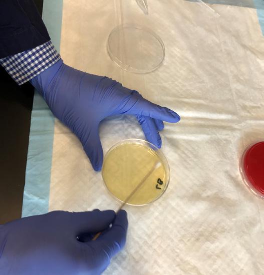
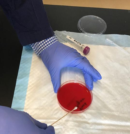


13 FIGURE 7. FIGURE 8. FIGURE 9. FIGURE 10. FIGURE 11. FIGURE 12. FIGURE 13. FIGURE 14. Streak plate technique. INFECTIOUS DISEASES: The good, the bad and the ugly 8
14 How many different types of bacteria do you expect to see on the agar plates? How would you differentiate one group of bacteria from another just by looking at your plates? Do you expect all bacteria to grow at similar rates? Why or why not? Your nasal passage and throat are connected. Do you expect to see similar bacterial growth on your throat and nasal swabs? What are some differences between your nose and throat that could affect the presence of different bacteria? Why do you think we have bacteria all over our body without any symptoms? Why do some bacterial colonies have a clear zone around them on the Blood Agar plates? Your nose and throat swabs probably show different types of bacteria. How would you have an agar plate grow multiple colonies of the same bacteria? 9 Bioscience Montana - Unit 3
15 AGARS AND BACTERIAL COLONIES In both research and clinical or scientific settings, scientists use agar, a jelly-like or gelatinous substance, to grow and identify different types of bacteria. Agar is a valuable tool because it allows visualization of bacteria that would not be possible without a microscope. Once a bacterium is transferred to an agar plate, it starts to undergo asexual replication through binary fission. Over time, the bacterium copies itself exponentially (doubles and then doubles again, and again, and so on) and forms a colony or group that is visible to the human eye. Colonies can be distinguished based on their shape, size, and color (Figure 16) when viewed under both reflected and transmitted light. Distinguishing these characteristics helps to identify specific types of bacteria. Bacteria have unique metabolisms and structures and can produce toxins by living cells. These bacterial characteristics can be identified using agar. In scientific terms, agar is referred to as primary isolation media, and there are approximately 100 of these media that are available commercially. Primary isolation media is further broken down into four categories: 1. Enrichment medium encourages the growth of specific bacteria while suppressing the growth of others in a large sample population. In a clinical setting, enrichment media is used to identify certain stool pathogens while suppressing other bacteria that would be considered part of our normal intestinal flora. 2. Selective medium selects for certain groups of bacteria such as Gram-positives or Gram-negatives. The method that is used in selective media to isolate a specific type of bacteria is the incorporation of antibiotics. Antibiotics target specific bacteria and prevent their growth and replication while allowing other organisms to survive. Thus, selective media is a valuable agar that can distinguish Gram-positives from Gram-negatives. 3. Supportive medium contains nutrients that allow most organisms to grow. This medium is useful for identifying different types of bacteria present in a sample, because this medium doesn t prevent the growth of different types of bacteria. 4. Differential medium is a combination of selective and supportive media. This medium not only selects for a certain group of bacteria but also differentiates between colony types, such as fermenting and nonfermenting colonies of gram-negative bacteria. After isolation of bacterial colonies on your agar plates you will notice unique characteristics. By learning which organisms will grow on a certain type of medium, you can start to recognize, through colony shape, size, and color certain groups of bacteria. Page 11 lists the different types of media that will be most useful for identifying different types of bacteria. FIGURE 16. Hudson, Barbara. Medical Bacteriology. Laboratory Manual. Montana State University. Montana State. December INFECTIOUS DISEASES: The good, the bad and the ugly 10

 FIGURE 18.")

 FIGURE 19.")
 FIGURE 20.")
 FIGURE 21.")

16 Mannitol Salt Agar (MSA) FIGURE 17. This is a selective and differential agar which contains 7.5% salt to select for certain Grampositive bacteria such as Staphylococci. The high salt concentration selects for organisms that like salt. It also contains mannitol sugar and a ph indicator, which will select for organisms that ferment or cause a chemical change to mannitol. If an organism can use mannitol as its energy source, the agar will turn yellow due to a drop in the ph which causes a color change. Blood Agar (BA) FIGURE 18. This is a nutrient-rich medium that contains 5% sheep s blood. This agar will grow most organisms and will help you identify which bacteria is growing based on the ability of the bacteria to break down blood cells. Different bacteria have different types of hemolysins or substances that cause red blood cells to dissolve: α (alpha), β (beta), and δ (gamma) hemolysin. The blood agar will look different depending on what type of hemolysins the bacteria have. MacConkey Agar (MAC) FIGURE 19. This is a selective and differential medium used to select Gram-negative bacteria. It can differentiate bacteria based on their ability to ferment lactose (large sugar molecule). If bacteria can use lactose, they will turn a pink color. If bacteria cannot use lactose, they will remain colorless or yellow. E. coli will ferment lactose but Salmonella sp. cannot. Eosin Methylene Blue Agar (EMB) FIGURE 20. This is a selective and differential medium that allows growth of Gram-negative bacteria. The agar contains lactose and two dyes: eosin and methylene blue. When bacteria ferment lactose, it produces acid and turns the bacterial colony a dark purple to black. Colonies that do not use lactose will be colorless. Colistin-Nalidixic Acid Agar (CNA) FIGURE 21. This is a selective medium containing the antibiotics colistin and nalidixic acid which selects for Gram-positive bacteria. S. aureus, S. epidermidis, and Group A Streptococcus will grow on this medium. Tryptic Soy Agar (TSA) This is a supportive medium which contains glucose or sugar made from food in your diet and amino acids for the growth of most micro-organisms. It will not select for Gram-positive or Gram-negative (both groups will grow on this agar). FIGURE 22. Lysogeny Broth Agar (LB) This is a nutrient-rich medium that supports the growth of both Gram-negative and Grampositive bacteria. This medium contains peptides (molecules that are structurally like proteins, but smaller), amino acids and carbohydrates in a low-salt formulation, which promotes the growth of all bacteria, but is widely used to grow E. coli. With the addition of antibiotics, LB agar can also be useful in identifying antibiotic-resistant bacteria. FIGURE 23. Bacteriological Media, 11 Bioscience Montana - Unit 3
17 ACTIVITY 2: The Agars (This activity or Activity 3 is required) LIFE SKILLS: Critical Thinking, Communication, Planning/Organizing CONTENT SKILL: Learn to use agars to identify bacteria MONTANA STANDARD FOR SCIENCE: 1, 3 NOTE: Complete Activity 1 before completing this activity In Activity 1, you isolated bacteria from your nose and throat. Now you will use the information you just learned about agars and put it into action. In Table 1 there are cells to enter information about nasal bacteria and throat bacteria. There are two rows, one for the LB agar and the other for the blood agar. You will examine the colonies on both agars, for nasal and throat cultures, and note what you see. Don t forget to note the colony morphology characteristics mentioned in Figure 16 and use caution when looking at the colonies: although a colony may only be one type of bacterium, it has replicated exponentially over time. A variety of types of bacteria will grow on the agars and those can produce unique colors or pigments which you should pay attention to. The pigment of the colony is a color, which should be recorded (see Table 1) as it helps in identification of the bacterium. The number of colonies is also important and they should be counted because they show the variety of bacteria in the nose compared to the throat. STEP 1: Refer to the LB and blood agars in Activity 1 for your nasal culture. STEP 2: Complete table as shown. STEP 3: Refer to the LB and blood agars in Activity 1 for your throat culture. STEP 4: Complete table as shown TABLE 1. Nasal Number of colonies Colony Morphologies Form Elevation Margins Color What else did you notice? Throat Number of colonies Colony Morphologies Form Elevation Margins Color What else did you notice? Blood agar LB agar Blood agar LB agar INFECTIOUS DISEASES: The good, the bad and the ugly 12
18 STEP 5: Answer the questions below (see appendix for suggested responses). What type of media (selective, supportive, differential) would be the most useful when comparing the number of bacteria between your nose and throat cultures? What primary isolation media would distinguish between a Gram-positive and a Gram-negative bacterium? Why? After you have an isolated bacterial colony, how might you further identify, using the materials you have, if the organism is Gram-positive or Gram-negative? Only using the LB agar, what experiments could you create to identify if a bacterium is sensitive to different household cleaning agents? FIGURE Bioscience Montana - Unit 3


 which are visualized by looking at the colonies using transmitted light. 1. Alpha (α)-hemolysis 2.")
19 USING BLOOD AGAR TO IDENTIFY TOXIN PRODUCTION Exotoxins are toxins that are released by bacteria and can be harmful in small amounts. Blood agar can identify bacterial toxin production. Blood agar is a supportive medium because it supports the growth of almost all common and medically-significant bacteria. The reason why it is called blood agar is that it is made of 5% sheep blood cells, also called erythrocytes. When the colonies are forming, the bacteria will produce toxins that lead to the breakdown or lysis of the erythrocytes present in the agar. The toxin production that leads to lysis of the erythrocytes is called hemolysis (which means blood breakdown). The hemolysis, or hemolytic reaction, can be categorized into three types (Figure 24) which are visualized by looking at the colonies using transmitted light. 1. Alpha (α)-hemolysis 2. Beta (β)-hemolysis 3. Gamma (γ)-hemolysis FIGURE 25. FIGURE 26. Beta (β)-hemolysis (Figure 26) is the complete breakdown of the red blood cells around the colony, producing a clear and transparent zone. Common bacteria that exhibit beta-hemolysis are Streptococcus pyogenes (Group A Strep) and Staphylococcus aureus. Both organisms are natural body flora and they can be differentiated based on the catalase test (Activity 4, page 17). Additionally, colony morphology can differ drastically between these organisms; thus, it is important to note characteristics, such as colony color, when describing the hemolysis. FIGURE 27. Alpha (α)-hemolysis (Figure 25) is an incomplete breakdown of the red blood cells producing a greenish to brownish discoloration of the medium. Common organisms known to display alpha-hemolysis are Streptococcus pneumonia and Streptococcus viridans. Don t be worried if your agar shows this characteristic, as these bacteria typically exist in the nose of healthy individuals. Also, it is important to observe hemolysis around individual colonies in Figure 25. Notice the greenish brown tint of individual colonies Gamma (γ)-hemolysis (Figure 27) is the term used when there is no apparent hemolytic activity of the bacterium and, therefore, no change in the blood agar around the colony. Many organisms will not produce exotoxins and they appear as a colony growing on the media but do not promote breakdown of the red blood cells. A common organism that produces this hemolysis is Staphylococcus epidermidis. INFECTIOUS DISEASES: The good, the bad and the ugly 14
20 ACTIVITY 3: Identify Hemolytic Reactions (This activity or Activity 2 is required) LIFE SKILLS: Communication, Critical Thinking CONTENT SKILL: Identify hemolytic reactions MONTANA STANDARD FOR SCIENCE: 1, 3 In this activity, you will refer to your nose and throat cultures again, but you will add the type of hemolysis that you observe. STEP 1: Observe your nose and throat blood agar plates using transmitted light. STEP 2: Record your observations of colony morphology, including shape, color, size, and the hemolytic reaction in Table 2. Combing these characteristics allows you to identify what organisms are present in your nose and throat. Did you know that these practices are still being used today? For example, when you go into a doctor s office with symptoms of coughing and a sore throat, the doctor will most likely take a sterile cotton swab and sample your throat. The doctor will run tests like those that you perform in your experiments and will identify what pathogen is causing the symptoms. After identification of the pathogen, the doctor will most likely prescribe an antibiotic to help fight the infection. Thus, the experiments that you are performing have real life applications and are used to help individuals in both clinical and research settings. TABLE 2. Nasal Number of colonies on agar Colony morphologies (form, elevation, color) Type of hemolysis (α, β, γ) Other observations Blood agar Throat Number of colonies on agar Colony morphologies (form, elevation, color) Type of hemolysis (α, β, γ) Other observations Blood agar 15 Bioscience Montana - Unit 3
21 STEP 3: Answer the questions below (see appendix for suggested responses). What are the bacteria producing that promote the lysis of red blood cells? If two colonies have the same hemolytic reaction, does that mean that they are the same bacterium? How else might one distinguish if the colonies are the same or different bacterium? Toxin production by bacteria is often associated with causing food poisoning. With your knowledge of how a bacterium grows, how could one prevent the process of toxins being produced? If your nose or throat samples are positive for alpha- or beta-hemolysis, what reasons can you think of as to why you are not becoming sick? Do you think your nose or throat bacteria would be like another person? How about a dog? Or a horse? Why or why not? INFECTIOUS DISEASES: The good, the bad and the ugly 16
22 ACTIVITY 4 Catalase Test (Required) LIFE SKILLS: Critical Thinking CONTENT SKILL: Learn if bacteria contain catalase MONTANA STANDARD FOR SCIENCE: 1 MATERIALS NEEDED: Inoculating loop (This is something that is used by microbiologists to get a culture. It is scraped across your tongue, for example, and then smeared onto an agar). You can also use a toothpick instead of an inoculating loop. Hydrogen Peroxide Our immune cells, the cellular defenders produced by our body to fight against foreign pathogens, have multiple ways to kill bacteria. One of these is to produce a reactive oxygen species (ROS). Once our immune cells are invaded by a pathogen, they use oxygen to produce superoxide which quickly turns into hydrogen peroxide and finally into hypochlorous acid/hypochlorite anion (the active ingredient in bleach!) (see Figure 28). Because bleach is very useful for killing bacteria, bacteria have developed ways to prevent the production of bleach. One of these is to produce an enzyme called catalase. Catalase breaks down hydrogen peroxide into water and oxygen preventing hydrogen peroxide from being turned into hypochlorous acid (bleach). In this experiment, we will use a simple procedure to test for the presence of catalase in the bacteria you cultured from your nose and throat. FIGURE 28. STEP 1: Your nose and throat swabs should show different types of bacterial morphologies. Use the pointy end of an inoculating loop (or clean toothpick) to pick out individual colonies from your agar plates and smear the colony on the inside of your agar s plastic cover. STEP 2: Place a drop of hydrogen peroxide onto the smeared colony. What do you see? Did bubbles form where the hydrogen peroxide met the smeared colonies? Note your observations in Table 3. STEP 3: Pick different colonies from your nose and throat swab plates. Perform the catalase-hydrogen peroxide test. Record in Table 3 the number of colonies producing bubbles when they meet hydrogen peroxide versus those that did not form bubbles. TABLE 3. Nasal Culture Throat Culture # Bubbles # None # Bubbles # None Colony 1 Colony 2 Colony 3 Colony 4 Colony 5 Colony 6 17 Bioscience Montana - Unit 3
23 STEP 4: Answer the questions below (see appendix for suggested responses). Were there any differences in the number of bacteria that produced catalase (those that formed bubbles when exposed to hydrogen peroxide) versus bacteria that did not when comparing colonies from nose and throat cultures? Why do bubbles form when bacteria encounter hydrogen peroxide? What is the gas that makes up the bubbles? Why do you think bubbles form when you pour hydrogen peroxide into a cut? ACTIVITY 5 Design Your Own Experiment LIFE SKILLS: Critical Thinking, Planning/Organizing, Communication CONTENT SKILL: Learn how to design an experiment MONTANA STANDARD FOR SCIENCE: 1, 3 Using what you ve learned you can now culture bacteria from anywhere you can think of and perform different experiments. Below is an example experiment, with questions to think about when designing experiments. Design a procedure where you test to see how effective the use of water and soap are to clean your hands, versus waterless hand sanitizer. STEP 1: Develop a hypothesis related to the use of soap and water versus waterless hand sanitizers. What do you think are the differences between the two? STEP 2: See next page. INFECTIOUS DISEASES: The good, the bad and the ugly 18
24 STEP 2: Establish a baseline, or a beginning level, before you conduct the experiment. How do you obtain a baseline of bacteria from your hands? Should you wash your hands before beginning the experiment? Explain. STEP 3: Identify your experimental or independent variables. In this experiment, these can be anything that will dirty your hands. Below are some suggestions. Touch raw chicken for 60 seconds. Brush a horse without gloves for 5 minutes Type on a public computer for 5 minutes (at a library or computer lab at school) Write down your experimental variables. STEP 4: Decide how to conduct your experiment or how to sample the bacteria on your hands. (Note: the tests should be done on the same day, and you should wash your hands after touching raw chicken. Not washing them is a potential biohazard.) What are your sampling methods: Will you swab each finger? Will you swab 3 fingers on each hand or only on one hand? Will you combine the swab material? If so how? Make sure you are consistent with how you collect samples. Write down sampling methods here: a) b) c) d) STEP 5: Standardize how you wash hands, for example, the length of time, and the vigor with which you wash. Make sure the motion to wash your hands with the soap and water is like the motion to wash them with the waterless hand sanitizer. Describe the method to wash your hands so if someone else wants to repeat the experiment, they know how you did it. (Note: researchers regularly repeat experiments to see if they get the same results as their peers. The more people that get similar results, the more valid the results, assuming the experiment was done correctly). 19 Bioscience Montana - Unit 3
25 STEP 6: Sample the bacteria on your hands 3 times: After you wash your hands the first time (to establish a baseline) After you dirty your hands After you rewash your hands STEP 7: Record your results in Table 4. TABLE 4 Soap and Water Waterless Hand Sanitizer Number of colonies Colony Morphologies Other Number of colonies Colony Morphologies Other Baseline Ex. 22 1st variable Ex nd variable 3rd variable Re-wash hands Ex. 30 STEP 8: Answer the questions below. Was your hypothesis correct? Explain. What are the implications of your findings? What do they mean? How do your findings add to the information that is already available on soap and water versus waterless hand sanitizers? If you were to perform your experiment again what would you change? INFECTIOUS DISEASES: The good, the bad and the ugly 20
26 ACTIVITY 6 The Infection Game* (Required) LIFE SKILLS: Critical Thinking, Problem-Solving, Communication CONTENT SKILL: Learn how diseases spread MONTANA STANDARD FOR SCIENCE: 1, 5 MATERIALS NEEDED: see appendix for worksheets Individual Record Sheet A, for one person Individual Record Sheet B, for all remaining people Organizer s record sheet, one for the organizer (you) Pens or pencils NOTE: Explain the rules without telling the name of the game In this activity, you will organize a game that shows how diseases are spread. The behavior you see in the game could represent bacteria spreading through an animal population, a virus spreading through a computer network, a rumor spreading through a school, or any other type of contagious agent. The more people you can organize the better you will be able to see patterns (recommended minimum is 35 people). Get two 4-H clubs to work together or ask permission to play this game at school. In this game, a disease is spread by the multiplication of numbers. One person is assigned the number zero (meaning they carry the disease), while the rest are assigned the number one (meaning they are healthy). As the players multiply their numbers together in pairs, the repeated multiplication process will cause more and more products to be zero. In other words, the number zero simulates the disease that is spreading through the population. Most diseases start with a very, very small number of the population - for example, the one individual given the number zero. If the disease succeeds, it will spread throughout the population. Secrecy is very important to this game. Do not give this background information to your players so they can discover how things work for themselves. STEP 1: Explain to the players that they will be exchanging numbers with each other and recording the numbers on Individual Record Sheets. Every sheet has a pre-written starting number on it (see appendix, p. 23). STEP 2: Designate the person that will start with the number zero. Choose a person who follows directions well. Give this person Individual Record Sheet A (page 26) without announcing that they were assigned zero (remember, secrecy is very important!) STEP 3: Give Individual Record Sheet B to all remaining players (page 27). STEP 4: Ask each person to find any other person and quietly tell each other their numbers. STEP 5: After a person has another s number, ask them to return to their sheet and secretly multiply the two numbers on their sheet together and record the product on the next line. This will be the number they use for the next round. STEP 6-10 (or more): Play more rounds until you decide to stop the game. STEP 11: When time is up, ask the players to notice the last product on their sheets. Most, if not all the students will have the number zero. STEP 12: Ask the student who was initially assigned 0 (this should be only one person) to raise their hand. Record this in the first column on your Organizer s Record Sheet (page 28). STEP 13: Ask how many players had zeros on their sheet when they started the second round. Record this on your sheet. Repeat this for all rounds and record the information until there are no new infections with zero. Do not discuss or reveal the results yet. STEP 14: In the second column of the Organizer s Record Sheet keep a running total of the number of zeros that occurred (Figure 29). STEP 15: Plot a graph using the numbers from the *Reprinted and modified from Shape of Change 2010 Permission granted by the Creative Learning Exchange (clexchange.org) 21 Bioscience Montana - Unit 3
27 Total Number of Zeros column of the Organizer s Record Sheet (Figure 29). You should expect to see a curve like the one below. This is called a sigmoidal curve, and is typical when plotting disease spread through a population STEP 16: Lead a discussion using the following questions: FIGURE 29. Round Number of NEW Zeros TOTAL Number Of Zeros Start What does the graph tell us? What happened to the number of players infected with zero during the game? At first only one student was infected, but the infection eventually spread to everyone in the game in a general pattern called S-shaped growth. Why does the line have an S shape? How does this relate to what was happening during the game? What was happening at letter A in the graph? Why is the line flatter? This is the initial spread. Very few people had the infection, so it spread very slowly at first. What was happening at B? Why is the line steeper? Growth was increasing. As more and more people were infected and they interacted with others, the disease spread at an increasing rate. What happened at C? Why does the curve change its shape? What happened at region D? Growth was decreasing. When most people already had the illness, there were fewer healthy people to infect, so the disease spread more slowly. The number of infected people was still increasing, but at a slower rate. What was happening at region E? Why is the line flat? There was no further growth. Everyone was infected. 4 FIGURE 30. INFECTIOUS DISEASES: The good, the bad and the ugly 22
28 APPENDIX ACTIVITY ANSWER KEY ACTIVITY 1 Nasal Swabbing and Culturing Bacteria How many different types of bacteria do you expect to see on the agar plates? This question has no right/ wrong answer (your best-educated guess!) How would you differentiate one group of bacteria from another just by looking at your plates? A good way to differentiate is to look at colony size, shape, and color. Do you expect all bacteria to grow at similar rates? Why or why not? Not all bacteria will have similar growth rates since the nutrients in the plates may not be optimized for ALL bacteria to take advantage of equally. Bacteria that like the nutrients in the plates will grow faster than bacteria that do not like the nutrients too much, but still use them to survive. Your nasal passage and throat are connected. Do you expect to see similar bacterial growth on your throat and nasal swabs? This is also a question that has no right/wrong answer. However, our noses and mouths take in different samples from the environment, for example, food and water versus air, so you should see some differences. But, don t be surprised if you find similar bacteria living in the mouth and nose, since they are connected. What are some differences between your nose and throat that could affect the presence of different bacteria? Nutrients! Your mouth likely has a lot more sugars allowing for better bacterial growth. At the same time, we brush our teeth daily to try to remove these bacteria and sugars. The process of brushing our teeth also affects the different bacteria that you might find in your nasal passage since we do not brush our noses! Why do you think we have bacteria all over our body without any symptoms? Our immune barriers such as skin and the gastrointestinal tract, defend against harmful bacteria from infiltrating our blood stream and sensitive tissues. Fully understanding how commensal bacteria interact with our immune system is an area of active research. Most bacteria are commensal organisms which means that they live in the same location of the body and one of the bacterium benefits from the other, but without harming it. This means that although the bacteria may produce toxins, such as alpha- or beta-hemolysis, you can remain healthy because they are part of your normal microbiota. Why do some bacterial colonies have a clear zone around them on the Blood Agar plates? Bacteria produce toxins that lyse the red blood cells. The plate becomes clear when the red blood cells lyse. Your nose and throat swabs probably show different types of bacteria. How would you have an agar plate grow multiple colonies of the same bacteria? Take a single colony from one plate and streak the colony on a new plate. 23 Bioscience Montana - Unit 3
29 ACTIVITY 2 The Agars What type of media (selective, supportive, differential) media would be the most useful when comparing the number of bacteria between your nose and throat cultures? Supportive media because it promotes the growth of all bacteria. What primary isolation media would distinguish a Gram-positive bacterium between a Gram-negative bacterium? Why? Selective media would distinguish Gram-positives from Gram-negatives because selective media has antibiotics that target either Gram-positives or Gram-negatives. For example, if an antibiotic prevents the growth of Gram-positives it will likely not affect the growth of Gram-negatives. However, some antibiotics are universal and can target both. After you have an isolated bacterial colony, how might you further identify, using the materials you have, if the organism is Gram-positive or Gram-negative? The colony could be suspended in a small amount of water then streaked on both selective and differential media using the streak plate technique (Figure 14). After incubation, colonies will appear on the media, which can be identified using the information provided in the agar media information section (page 11). Only using the LB agar (Figure 23) what experiments could you try to see if a bacterium is sensitive to different household cleaning agents? Divide the plate into sections and thoroughly swab the entire plate with the sample or isolated colony. In each of the sections add a small amount of household cleaning agent, dry then incubate. After incubation, colonies will either grow or be prevented from growing in the specific cleaning agent, which will show sensitivity of the bacterium to the specific cleaning agent. ACTIVITY 3 Identify Hemolytic Reactions What are the bacteria producing that promotes the lysis of red blood cells? Toxins called hemolysins. If two colonies have the same hemolytic reaction, does that mean that they are the same bacterium? How else might one distinguish if the colonies are the same or different bacterium? Different bacteria can produce similar toxins that have the same hemolysis reaction. Colony morphology (shape, color, elevation) will help in identifying if the bacteria are similar or different. To further identify if the bacteria are the same, they could both be streaked on selective and differential media. Toxin production by bacteria is often associated with causing food poisoning. With your knowledge of how a bacterium grows, how could one prevent the process of toxins being produced? These bacteria are incubated between room temperature and 37 degrees Celsius, which allows them to grow exponentially with the right nutrients. To prevent exponential growth and toxin production, media or food containing bacteria could be placed in a refrigerator or at lower temperatures, which slows the replication process and toxin production. If your nose or throat samples are positive for alpha- or beta-hemolysis, what reasons can you think of as to why you are not becoming sick. Most bacteria are commensal organisms which means that they live in the same location of the body and one of the bacterium benefits from the other, but without harming it. This means that although the bacteria may produce toxins, such as alpha- or beta-hemolysis, you can remain healthy because they are part of your normal microbiota. Do you think your nose or throat bacteria would be like another person? How about a dog? Or a horse? Why or why not? Bacteria could be similar between individuals within a family but different compared to other humans. Similarities within a family could happen from living in close quarters and being exposed to the same environment. Someone in another location might be exposed to different environmental conditions. Animals have their own unique nose and throat bacteria due to their living environments and the food they eat. However, there can also be similar bacteria between all species (humans, dogs, horses), but usually in different amounts. INFECTIOUS DISEASES: The good, the bad and the ugly 24
30 ACTIVITY 4 The Catalese Test Were there any differences in the number of bacteria that produced catalase (those that formed bubbles when exposed to hydrogen peroxide) versus bacteria that did not when comparing colonies from nose and throat cultures? This answer will depend on results. You will probably see some bacteria that produced catalase and others that did not. Why do bubbles form when bacteria encounter hydrogen peroxide? What is the gas that makes up the bubbles? The catalase breaks down hydrogen peroxide into oxygen and water. Oxygen makes up the bubbles as it floats into the air from water. Why do you think bubbles form when you pour hydrogen peroxide into a cut? Most of our skin cells contain their own catalase to protect themselves from harmful chemicals. Thus, the bubbles you see when you pour hydrogen peroxide into a cut come from your own catalase breaking down hydrogen peroxide. ACTIVITY 5, Step 2 Design Your Own Experiment How do you obtain a baseline of bacteria from your hands? The baseline is achieved from washing your hands then swabbing for bacteria. You could swab from unclean hands but by cleaning your hands first, you are excluding other potential variables, like bacteria already present on your hands. Should you wash your hands before beginning the experiment? Explain. You should wash your hands before the experiment to allow for good comparisons between the control (baseline, clean hands) and the experimental variables. SUGGESTIONS FOR FURTHER STUDY This virtual lab allows you to walk through laboratory steps using a virtual platform (for laptop): For phone or tablet (downloadable app): Another virtual lab: RECOMMENDED SUPPLIES Luria Broth Agar Plates Blood Agar Plates Cotton Tip Applicator Transfer Pipette Conical Tubes Inoculation Loops All of the above can be purchased online via amazon.com or a science/biology tools online store. Saline and hydrogen peroxide are easily available at pharmacies and supermarkets 25 Bioscience Montana - Unit 3
31 ACTIVITY 6 (The Infection Game) Worksheets: INDIVIDUAL RECORD SHEET FORM A NAME: Start with the number written below. Do not share this number! Once the game starts, select another student and exchange numbers. On your own, secretly MULTIPLY the two numbers and write the product on line 1 - this is your new number. Select another student and write the new number of line 2. Repeat this process until time is called. ROUND NUMBER Start INFECTIOUS DISEASES: The good, the bad and the ugly 26
32 INDIVIDUAL RECORD SHEET FORM B NAME: Start with the number written below. Do not share this number! Once the game starts, select another student and exchange numbers. On your own, secretly MULTIPLY the two numbers and write the product on line 1 - this is your new number. Select another student and write the new number of line 2. Repeat this process until time is called. ROUND NUMBER Start copy as many as needed 27 Bioscience Montana - Unit 3
33 ORGANIZER S RECORD SHEET Round Number of NEW Zeros TOTAL Number of Zeros Start INFECTIOUS DISEASES: The good, the bad and the ugly 28
34
BIOLOGY. Bacteria Growth Lab. Bacterial Growth. Slide 2 / 61. Slide 1 / 61. Slide 4 / 61. Slide 3 / 61. Slide 5 / 61. Slide 6 / 61
 Slide 1 / 61 Slide 2 / 61 New Jersey Center for Teaching and Learning Progressive Science Initiative This material is made freely available at www.njctl.org and is intended for the non-commercial use of
Bacteria and Evolution Junior Science
 Bacteria and Evolution Junior Science Micro-organisms Micro-organisms (or microbes) are very small organisms, which are usually only visible with the aid of a microscope. Sometimes a colony of micro-organisms
Inoculate: Media. Physical State of Media: Liquid. The Five I s: Basic Techniques to Culture Microbes Tools of the Microbiology Laboratory
 The Five I s: Basic Techniques to Culture Microbes Tools of the Microbiology Laboratory 1. Inoculate 2. Incubate 3. Isolate 4. Inspect 5. Identify The Five I s: Inoculate Inoculate: Media Classified according
GENUS STAPHYLOCOCCUS: Isolation and Identification
 GENUS STAPHYLOCOCCUS: Isolation and Identification Staphylococcus is a genus of Gram +, nonspore-forming cocci belonging to the family Micrococcaceae that are often found as normal human microbiota of
320 MBIO Microbial Diagnosis. Aljawharah F. Alabbad Noorah A. Alkubaisi 2017
 320 MBIO Microbial Diagnosis Aljawharah F. Alabbad Noorah A. Alkubaisi 2017 Primary Media for Isolation of Microorganisms As we know, many clinical specimens contain a mixed flora of microorganisms. Thus
some bacteria will have an outer capsule which gives them greater protection
 some bacteria will have an outer capsule which gives them greater protection Can be classified based on: Shape Arrangement Cell wall structure Energy source 1. Cocci (round); singular coccus resist drying
Immune System. Viruses vs. Bacteria
 Immune System Viruses vs. Bacteria Concept Map Section 19-1 Bacteria are classified into the kingdoms of Eubacteria Archaebacteria include a variety of lifestyles such as live in harsh environments such
Bacteria and other microbes have particular requirements for growth When they reside in and on our bodies or in the environment, they harvest their
 Bacteria and other microbes have particular requirements for growth When they reside in and on our bodies or in the environment, they harvest their food from us or from the environment When we grow bacteria
Shehab. Yousef... Omar. Yousef Omar. Anas
 3 Shehab Yousef Omar Yousef... Omar Anas Bacterial Growth and Survival After discussing the structure of a Bacteria, we must know how it survive and grow in a specific media. Firstly, the survival of any
Bacterial Plate Preparation. ~ Using aseptic techniques ~
 Bacterial Plate Preparation ~ Using aseptic techniques ~ Bacterial Plates Laboratory and research scientists have to prepare nutrient media to grow specific strains of bacteria for their research. To do
Project 7: Wound Cultures and Identification
 Project 7: Wound Cultures and Identification Readings: https://labtestsonline.org/understanding/analytes/wound-culture/tab/test Identification of Gram-Positive & Gram-Negative Bacteria Guide to laboratory
The Grand Prismatic Spring in Yellowstone National Park is the largest hot spring in the United States The vivid colors in the spring are the result
 The Grand Prismatic Spring in Yellowstone National Park is the largest hot spring in the United States The vivid colors in the spring are the result of pigmented bacteria in the microbial mats that grow
NATURE OF MICROBES WORKBOOK
 NATURE OF MICROBES WORKBOOK Name: Tutor Group: 1 Microbes and Mankind 4. NATURE OF MICROBES 1. OBJECTIVES: What are microbes and are there different types? How are they seen? How can they be grown? How
Microbial Biotechnology agustin krisna wardani
 Microbial Biotechnology agustin krisna wardani 1. The Structure of Microbes Microbes (microorganisms) are tiny organisms that are too small to be seen individually by the naked eye and must be viewed with
Preparing and Gram-staining a bacteriological smear
 College of Life Sciences and Technology Medical Laboratory Science (Applied Learning) AP 84-205-00 (42) Module 4: Practical 2014-16 Preparing and Gram-staining a bacteriological smear Date Time Class Venue
Isolation & Characterization of Bacteria
 PR025 G-Biosciences 1-800-628-7730 1-314-991-6034 technical@gbiosciences.com A Geno Technology, Inc. (USA) brand name Isolation & Characterization of Bacteria Teacher s Handbook (Cat. # BE 204) think proteins!
Preparing and Gram-staining a bacteriological smear
 College of Life Sciences and Technology Medical Laboratory Science (Applied Learning) AP 84-205-00 (52) Module 4: Practical 2015-17 Preparing and Gram-staining a bacteriological smear Date Time Class Venue
Identification of Indigenous Unknowns
 Unknown Lab Report III Identification of Indigenous Unknowns Introduction: In a scientific field with so much genetic variation and rapidly evolving species, it has become increasingly difficult to correctly
Bacterial Transformation Lab - pglo
 Bacterial Transformation Lab - pglo Name: Date: Pre-Lab Score: Lab Overview: In this investigation, you will gain an understanding of the techniques of culturing E. coli bacteria and transforming using
90927 Demonstrate understanding of biological ideas relating to micro-organisms COLLATED QUESTIONS
 90927 Demonstrate understanding of biological ideas relating to micro-organisms COLLATED QUESTIONS DIGESTION AND REPRODUCTION PROCESSES (2013:1) (a) Describe the processes of digestion and reproduction
Culture Media. Provide certain environmental conditions, nutrients & energy in order to grow and produce bacteria
 Culture Media Culture Media Provide certain environmental conditions, nutrients & energy in order to grow and produce bacteria Different categories of media can be made according to the type and combination
Bacteria Introduction Bacteria are unicellular micro-organisms ranging in length from a few micrometers to half a millimeter. They come in a variety
 Kingdom Bacteria Bacteria Introduction Bacteria are unicellular micro-organisms ranging in length from a few micrometers to half a millimeter. They come in a variety of different shapes (cocci, bacilli
GUIDELINES FOR WRITING A LAB REPORT FOR BIOL 215L (MICROBIOLOGY FOR HEALTHCARE PROFESSIONALS)
") GUIDELINES FOR WRITING A LAB REPORT FOR BIOL 215L (MICROBIOLOGY FOR HEALTHCARE PROFESSIONALS) Your lab report will focus only on your unknown bacteria, which you collected, cultured, isolated, analyzed,
GUIDELINES FOR WRITING A LAB REPORT FOR BIOL 215L (MICROBIOLOGY FOR HEALTHCARE PROFESSIONALS) Your lab report will focus only on your unknown bacteria, which you collected, cultured, isolated, analyzed,
Pet Microbe 1: Lab Guide
 Experiment 2: Pet microbes: A lesson in taxonomy This experiment will start with three lab periods and then continue throughout the semester. What's in a name? Names help us communicate a lot of information
Unit 12 Viruses & Bacteria
 Unit 12 Viruses & Bacteria Learning Goals Identify structures and characteristics of Viruses and Bacteria Explain how viruses and bacteria reproduce Recognize the importance of viruses and bacteria Explain
How to perform a Gram Stain. Jasleen Singh
 How to perform a Gram Stain Jasleen Singh Table of Contents iii Table of Contents Table of Contents... iii Introduction... 5 Terminology... 7 Terms to be familiar with... 7 Gram Staining... 8 What is
MODULE 1 NGSS TEACHER S GUIDE. Meet The Microbes! Keego Technologies LLC. All rights reserved.
 MODULE 1 NGSS TEACHER S GUIDE Meet The Microbes! Keego Technologies LLC. All rights reserved. NGSS Alignment CORE IDEAS Core Idea LS1: From Molecules to Organisms: Structures and Processes LS1.A: Structure
Cell Culture. Suggested Age: years old. Time: 45 minutes preparation, 3-4 days for data collection. What is a Cell Culture?
 Cell Culture Suggested Age: 10-14 years old Time: 45 minutes preparation, 3-4 days for data collection What is a Cell Culture? Cell Culture is a technique used to grow and identify microorganisms that
Viruses and Prokaryotes
 Viruses and Prokaryotes Viruses Are they living things? Viruses can reproduce, however, they cannot reproduce without a host cell. They also do not contain cytoplasmic materials and they do not have a
Name Block Desk # BACTERIA AND VIRUSES. 1. What are prokaryotes? They are -celled organisms with no
 Name Block Desk # BACTERIA AND VIRUSES Identifying Bacteria: 1. What are prokaryotes? They are -celled organisms with no - bound organelles. 2. True or false: prokaryotes are much larger that eukaryotes.
About Science Prof Online PowerPoint Resources
 About Science Prof Online PowerPoint Resources Science Prof Online (SPO) is a free science education website that provides fully-developed Virtual Science Classrooms, science-related PowerPoints, articles
Biology Lab Activity 4-5 DNA Transformation
 Biology Lab Activity 4-5 DNA Transformation Scientists can insert genes into bacteria. The genes inserted in the Indo-Blu process (this lab) are on a circular piece of DNA called a plasmid. (The plasmid
Biology Class The Evolution Unit Lessons 7-12
 Name: Hour: Teacher: ROZEMA Biology Class The Evolution Unit Lessons 7-12 Last Round We Figured Out: How common is Addie s problem? Could it happen to me? Where are bacteria? How do antibiotics work? How
MICROBIOLOGY #2 PREPERATION AND STERILIZATION OF CULTURE MEDIA
 MICROBIOLOGY #2 PREPERATION AND STERILIZATION OF CULTURE MEDIA When we receive a sample (ex. Urine sample) for detection, we cannot gram stain it right away if it requires to be inoculated because when
Cornell Institute for Biology Teachers in partnership with the Pseudomonas-Plant Interaction Project
 Cornell Institute for Biology Teachers in partnership with the Pseudomonas-Plant Interaction Project Copyright CIBT, 2004. This work may be copied by the original recipient from CIBT to provide copies
In order to do transformation, the gene to be transferred is placed into a plasmid. This is done with the help of restriction enzymes, 7
 Fluorescent Protein Transformation Student Background Genetic transformation occurs when a cell takes up (i.e. takes inside) and expresses a new piece of genetic material DNA. Genetic transformation literally
2/3/12. + Prokaryote: + Eubacteria. + How do the two groups of prokaryotes differ? + Unique characteristics Where found
 Prokaryote: unicellular organisms that lack a nucleus. Ch. 19 Bacteria and Viruses Eubacteria Eubacteria have a cell wall that protects the cell and determines its shape. The cell wall of eubacteria contain
Section A: Prokaryotes Types and Structure 1. What is microbiology?
 Section A: Prokaryotes Types and Structure 1. What is microbiology? 2. Compare and contrast characteristics of each bacterial type: Eubacteria and Archaebacteria. Eubacteria Both Archaebacteria 3. Label
Chapter 6: Microbial Growth
 Chapter 6: Microbial Growth 1. Requirements for Growth 2. Culturing Microorganisms 3. Patterns of Microbial Growth 1. Requirements for Growth Factors that affect Microbial Growth Microbial growth depends
Outline. This week s plans Introduction to Kirby Bauer and Antibiotics Introduction to Biochemical analysis of strains
 Outline This week s plans Introduction to Kirby Bauer and Antibiotics Introduction to Biochemical analysis of strains 1 This week 2 Monday Finish Exercise 10, part C. Run MspI restriction digest on a gel
Activity 5.1.4: Gram Staining
 Activity 5.1.4: Gram Staining Introduction In the last activity, you isolated the bacteria that was responsible for Anna s illness and performed a gross examination of the resultant colonies. While gross
4/7/2007. Key Concepts: Viruses, Bacteria and Diseases: Are We Ahead of Them? Objectives:
 Viruses, Bacteria and Diseases: Are We Ahead of Them? Objectives: 1. To learn the basic structures of viruses. 2. To understand the life cycles of viruses. 3. To learn the differences between various species
Chapter 03 - Tools of the Laboratory: Methods for the Culturing of Microscopic Analysis of microorganisms
 Microbiology: A Systems Approach 4th Edition Cowan Test Bank Completed download: https://testbankreal.com/download/microbiology-systems-approach-4thedition-test-bank-cowan/ (Downloadable package TEST BANK
PURE CULTURE TECHNIQUES
 PURE CULTURE TECHNIQUES Most specimens (from animal tissue, plant tissue, or environmental samples) will be mixed, with a variety of bacteria (or other microorganisms). A single gram of feces, for example,
Microbiological Methods
 Microbiological Methods Making Media Pouring Culture Plates Sterile Technique Inoculating Plates and Culture Tubes Use of a Plate Counter to Estimate Microbial Population Densities Sterile Technique Sterile
2 Creating Genetically Modified Bacteria Gl o w - i n-th e - d a r k rabbits, pigs, and mice may sound like something out
 2 Creating Genetically Modified Bacteria Gl o w - i n-th e - d a r k rabbits, pigs, and mice may sound like something out of a science fiction movie, but because of genetic modification, these animals
Chapter 13A: Viral Basics
 Chapter 13A: Viral Basics 1. Viral Structure 2. The Viral Life Cycle 3. Bacteriophages 1. Viral Structure What exactly is a Virus? Viruses are extremely small entities that are obligate intracellular parasites
BACTERIA. NO or membrane bound WHAT ARE THE TWO TYPES OF PROKARYOTES? TYPES EUBACTERIA ARCHAEBACTERIA. bilayer embedded with
 Bacteria and Virus Notes WHAT ARE PROKARYOTES? ALWAYS organisms BACTERIA NO or membrane bound Very compared to cells WHAT ARE THE TWO TYPES OF PROKARYOTES? TYPES EUBACTERIA ARCHAEBACTERIA MAJOR DIFFERENCES
Biotechnology In Your Mouth
 PR005 G-Biosciences 1-800-628-7730 1-314-991-6034 technical@gbiosciences.com A Geno Technology, Inc. (USA) brand name Biotechnology In Your Mouth Teacher s Guidebook (Cat. # BE 102) think proteins! think
Bacterial Transformation: Unlocking the Mysteries of Genetic Material
 PR009 G-Biosciences 1-800-628-7730 1-314-991-6034 technical@gbiosciences.com A Geno Technology, Inc. (USA) brand name Bacterial Transformation: Unlocking the Mysteries of Genetic Material Teacher s Guidebook
SAMPLE. NCFE Level 2. Certificate in the Principles of the Prevention and Control of Infection in Health Care Settings. Part A
 Level 2 Certificate in the Prevention and Control of Infection Introduction NCFE Level 2 Certificate in the Principles of the Prevention and Control of Infection in Health Care Settings 1 Part A These
Lab 5/5a Transformation of E. coli with a Recombinant Plasmid
 Lab 5/5a Transformation of E. coli with a Recombinant Plasmid Lab 2 Pre Lab Readiness Familiarity and Proper use of micropipettes Remember the 1 st and 2 nd stops Aseptic Technique Antibiotic Resistance
Microbiological Methods
 Microbiological Methods Making Media Pouring Culture Plates Sterile Technique Inoculating Plates and Culture Tubes Use of a Plate Counter to Estimate Microbial Population Densities Culturing Microorganisms
Project 5: Urine Cultures and Identification
 Project 5: Urine Cultures and Identification Readings: http://www.webmd.com/a-to-z-guides/urine-culture http://www.medscape.com/viewarticle/558845 (Listen to the two lectures by Dr. Robert A. Weinstein.)
Biology Test Review Microorganisms
 Name: Period: Biology Test Review Microorganisms Use your booklet, notes, & quizzes to complete this review. 1. Define the following terms using a few key words: a. Host cell - victim of the virus b. Retrovirus
Activity Sheet 2 BACTERIA TAKE-DOWN Problem: What metals are the most effective in inhibiting bacterial growth?
 Activity Sheet 2 BACTERIA TAKE-DOWN Problem: What metals are the most effective in inhibiting bacterial growth? Background: Germs are EVERYWHERE!!! We have hear this all before. Quite often illnesses and
ECOS Inquiry Template
 ECOS Inquiry Template 1. Contributor s Name: NATHAN GORDON 2. Name of Inquiry: NOT TOO HOT, NOT TOO COLD: THE EFFECTS OF TEMPERATURE ON SOIL BACTERIA 3. Goals and Objectives: a. Inquiry Questions: 1. How
20 Bacteria (Monera)
") 20 Bacteria (Monera) Mr. C Biology 1 Bacteria are very small. They are measured in µm (nanometres). There are 1000 nanometres in 1 mm. Bacteria can be 1µm in size. Bacteria have a cell wall that protects
20 Bacteria (Monera) Mr. C Biology 1 Bacteria are very small. They are measured in µm (nanometres). There are 1000 nanometres in 1 mm. Bacteria can be 1µm in size. Bacteria have a cell wall that protects
yeast cell virus fungal hypha (filament)
") Biology 1.3 AS 90927 Demonstrate understanding of biological ideas relating to micro-organisms Externally assessed 4 credits Copy correctly Up to 3% of a workbook Copying or scanning from ESA workbooks
Biology 1.3 AS 90927 Demonstrate understanding of biological ideas relating to micro-organisms Externally assessed 4 credits Copy correctly Up to 3% of a workbook Copying or scanning from ESA workbooks
PS: the present of organisms in a sterile part in our body like the blood or the inner tissue it means there is infection
 This lecture is an introduction about the bacteria cell. Microbiology specially is related to micro-organisms? We can't observe the microorganisms by our naked eyes but we could culture them in special
The genetic material is arranged differently in the cells of bacteria compared with animal and plant cells.
 Some infections are caused by bacteria. The genetic material is arranged differently in the cells of bacteria compared with animal and plant cells. Describe two differences. Tuberculosis (TB) is an infection
INTRODUCTION water-soluble Figure 1.
 INTRODUCTION Natural waters contain bacteria. The aerobic gram negative bacillus of the genera Psedomonas, Alcalignes, and Flavobacterium are common in natural waters. Many of these bacteria are able to
Biology 322 Fall 2010 Transfer of genetic information in the bacterium Escherichia coli: Part I
 Biology 322 Fall 2010 Transfer of genetic information in the bacterium Escherichia coli: Part I REQUIRED Reading Assignments: Superbugs on the Hoof http://fire.biol.wwu.edu/trent/trent/superbugs.pdf Triple
MiSP Evolution by Natural Selection / Bacterial Resistance Teacher Guide, L1 L3. Introduction
 MiSP Evolution by Natural Selection / Bacterial Resistance Teacher Guide, L1 L3 Introduction This unit uses the development of resistance to antimicrobials as an example of natural selection. The students
Lab Activity #14 - Bacteriological Examination Of Water and Milk (Adapted from Lab manual by Dr. Diehl)
") Lab Activity #14 - Bacteriological Examination Of Water and Milk (Adapted from Lab manual by Dr. Diehl) Some of the diseases that humans can contract from drinking polluted water include typhoid, dysentery,
Lab Activity #14 - Bacteriological Examination Of Water and Milk (Adapted from Lab manual by Dr. Diehl) Some of the diseases that humans can contract from drinking polluted water include typhoid, dysentery,
Sergeant System s Immune System Defense Team Webquest
 Name Sergeant System s Immune System Defense Team Webquest Hello! I m Sergeant Cell and I am in charge of the Special Operations Teams here at the ImmunoSystem Police Station. 2 Thanks so much for coming
INTRODUCTION Sanitization sterilization Antibiotics Bactericidal Bacteriostatic Antiseptics disinfectants
 INTRODUCTION Infectious agents on environmental surfaces, given the correct circumstances, may potentially find their way into an unsuspecting victim. Thus, it is important to keep the surfaces we regularly
How is the Global Increase in Carbon Dioxide Changing the Population Density of Soil. Bacteria? Soil Ecology Project. Madeline Kim. Katherine Harrison
 Carbon Dioxide Effect on Soil Bacteria 1 How is the Global Increase in Carbon Dioxide Changing the Population Density of Soil Bacteria? Soil Ecology Project Madeline Kim Katherine Harrison Foula Protopapas
National 5 Unit 1: Cell Biology Topic 1.1 Cell Structure. Which part of the cell is composed of cellulose? 1
 National 5 Unit 1: Cell Biology Topic 1.1 Cell Structure 1. The diagram below shows parts of a plant cell. Which part of the cell is composed of cellulose? 1 2. Which structural feature is found in a plant
Adapted from Biology 15 Laboratory Manual Supplement: Wrightsman, Ininns and Cannon-Moloznic, Saddleback College, CA 92692
 Biology 4B Laboratory Bacteriological Examination of Water Adapted from Biology 15 Laboratory Manual Supplement: Wrightsman, Ininns and Cannon-Moloznic, Saddleback College, CA 92692 Objectives Carry out
LAB NOTES FOR EXAM 1 SECTION
 LAB NOTES FOR EXAM 1 SECTION EX. 2-1: DIVERSITY AND UBIQUITY OF MICROOGANISMS Purpose: Microorganisms are found everywhere in the environment around us. To demonstrate this and to get a taste of the different
Sixth Grade Science BLOCK 2 ASSESSMENT Simple Organisms
 Sixth Grade Science BLOCK 2 ASSESSMENT Simple Organisms 2008-09 Directions: Choose the best answer for each question. Then mark your choice on your answer sheet. 1. Which of the following best describes
BYA3. HUMAN BIOLOGY (SPECIFICATION A) Unit 3 Pathogens and Disease. General Certificate of Education June 2005 Advanced Subsidiary Examination
 Unit 3 Pathogens and Disease. General Certificate of Education June 2005 Advanced Subsidiary Examination") Surname Other Names Leave blank Centre Number Candidate Number Candidate Signature General Certificate of Education June 2005 Advanced Subsidiary Examination HUMAN BIOLOGY (SPECIFICATION A) Unit 3 Pathogens
Surname Other Names Leave blank Centre Number Candidate Number Candidate Signature General Certificate of Education June 2005 Advanced Subsidiary Examination HUMAN BIOLOGY (SPECIFICATION A) Unit 3 Pathogens
pglo Transformation Lab Integrated Science 4 Redwood High School Name Per:
 pglo Transformation Lab Integrated Science 4 Redwood High School Name Per: n Introduction To Transformation In this lab you will perform a procedure known as a genetic transformation. Remember that a gene
Pathogenic Bacteria. culture media. Components of the Typical Culture Medium: Culture Media Importance:
 Level4 Lab2: Pathogenic Bacteria culture media Microorganisms, like all other living organisms, require basic nutrients for sustaining their life. All microorganisms have the same basic requirements but
Unit 2: Metabolism and Survival Sub-Topic (2.7) Genetic Control of Metabolism (2.8) Ethical considerations in the use of microorganisms
 Genetic Control of Metabolism (2.8) Ethical considerations in the use of microorganisms") Unit 2: Metabolism and Survival Sub-Topic (2.7) Genetic Control of Metabolism (2.8) Ethical considerations in the use of microorganisms Duncanrig Secondary JHM&MHC 2015 Page 1 of 18 On completion of this
Unit 2: Metabolism and Survival Sub-Topic (2.7) Genetic Control of Metabolism (2.8) Ethical considerations in the use of microorganisms Duncanrig Secondary JHM&MHC 2015 Page 1 of 18 On completion of this
EXPERIMENT. Biochemical Testing for Microbial Identification Methyl Red, Voges- Proskauer, and Catalase Testing
 EXPERIMENT Biochemical Testing for Microbial Identification Methyl Red, Voges- Proskauer, and Catalase Testing Hands-On Labs, Inc. Version 42-0246-00-02 Review the safety materials and wear goggles when
Identity and pathogenicity of bacteria isolated from a domestic feline claw
 Martushoff 1 Identity and pathogenicity of bacteria isolated from a domestic feline claw Introduction Microorganisms bacteria, archaea, viruses, fungi, and protozoa are the most genetically diverse group
Viruses & Bacteria. One is the ultimate bad guy. One is an ultimate good guy Gone bad guy. But still good guy.
 Viruses & Bacteria One is the ultimate bad guy. One is an ultimate good guy Gone bad guy. But still good guy. Viruses A virus is a non-cellular particle made up of genetic material (DNA or RNA) and protein
Microbiology Chapter 2 Laboratory Equipment and Procedures 2:1 The Light Microscope MICROSCOPE: any tool with a lens to magnify and observe tiny
 Microbiology Chapter 2 Laboratory Equipment and Procedures 2:1 The Light Microscope MICROSCOPE: any tool with a lens to magnify and observe tiny details of specimens Micro tiny, small Scope to see SIMPLE
ASEPTIC TRANSFER & PURE CULTURE TECHNIQUES
 ASEPTIC TRANSFER & PURE CULTURE TECHNIQUES GENERAL GUIDELINES & REMINDERS: SAFETY: NO EATING OR DRINKING IN THE LAB! Wash your hands with soap both BEFORE and AFTER lab, and, in addition, when you have
Requirements for Growth
 Requirements for Growth Definition: Bacterial growth defined as an increase in the number of cells. Physical Requirements: temperature, ph, tonicity Temperature: On the basis of growth range of temperature
FARM MICROBIOLOGY 2008 PART 2: BASIC STRUCTURE AND GENETICS OF BACTERIA. 1. Epulopiscium fishelsoni and Thiomargarita namibiensis.
 FARM MICROBIOLOGY 2008 PART 2: BASIC STRUCTURE AND GENETICS OF BACTERIA I. Basic Morphology (Shape) of Vegetative Cells. A. Microscopic. Example Escherichia coli (aka E. coli) is 1.3 µm (= 0.000052 inch)
Transduction of an Antibiotic Resistance Gene. Background
 I Student Guide 21-1128 Name------------ Date Transduction of an Antibiotic Resistance Gene Background Transduction is a natural method of gene transfer that occurs in bacteria. The key player in transduction
Exercise 23-C BACTERIOPHAGE REPRODUCTION AND PLAQUE FORMATION
 Exercise 23-C BACTERIOPHAGE REPRODUCTION AND PLAQUE FORMATION Introduction The reproductive cycle of a cytolytic bacteriophage called T2 begins with its adsorption onto a sensitive host cell. The phage
GENETICS - CLUTCH CH.5 GENETICS OF BACTERIA AND VIRUSES.
 !! www.clutchprep.com CONCEPT: WORKING WITH MICROORGANISMS Bacteria are easy to with in a laboratory setting They are fast dividing, take up little space, and are easily grown in a lab - Plating is when
Sample collection & laboratory methods for identification bacteria. Lab. 2 A.T. Samira
 Sample collection & laboratory methods for identification bacteria Lab. 2 A.T. Samira Learning objectives At the end of the presentation, participants should understand the: Procedures, preparation, processing
Heat Shock Proteins in Yeast (2012)
") MASSACHUSETTS INSTITUTE OF Technology Department of Biology Heat Shock Proteins in Yeast (2012) Summary Lydia Breen (Stoneham High School) Mary Brunson (Brookline High School) Yeast is a single-celled
MASSACHUSETTS INSTITUTE OF Technology Department of Biology Heat Shock Proteins in Yeast (2012) Summary Lydia Breen (Stoneham High School) Mary Brunson (Brookline High School) Yeast is a single-celled
PreLab Activity: Read through the entire lab! Come prepared to explain to your group what this lab is about
 BIO101Lab weeks 4 and 5 PreLab Activity: Read through the entire lab! Come prepared to explain to your group what this lab is about The Evolution of the bacterium, Pseudomonas fluorescens Introduction
Bacteria and Viruses
 Bacteria and Viruses 1 of 25 Boardworks Ltd 2012 2 of 25 Boardworks Ltd 2012 Comparing cell sizes 3 of 25 Boardworks Ltd 2012 Most plant and animal cells are between 10µm and 100µm in size around the diameter
Biology 123 Microbiology Spring 2015
 Parkland College Biology Courses Natural Sciences Courses 2015 Biology 123 Microbiology Spring 2015 Vikki Crnekovic Parkland College, vcrnekovic@parkland.edu Tiffany Gibson Parkland College, TGibson@parkland.edu
Cell Biology Homework
 Cell Biology Homework NAME: CLASS: 1 1. The diagram below shows two cells. Cell Structure a) Complete the table below to give the name and functions of the parts labelled A, B and C. (2) Letter Part Function
MICROBIOLOGICAL TOOLS FOR QUALITY ASSURANCE IN HATCHERY: Laboratory Methods
 Issue No.29 / March 2010 MICROBIOLOGICAL TOOLS FOR QUALITY ASSURANCE IN HATCHERY: Laboratory Methods By Dr Vincent TURBLIN, Deputy Regional Market Manager Poultry - CEVA Animal Health Asia Pacific Most
Biology 123 Microbiology Fall 2015
 Parkland College Biology Courses Natural Sciences Courses 2015 Biology 123 Microbiology Fall 2015 Vikki Crnekovic Parkland College, vcrnekovic@parkland.edu Recommended Citation Crnekovic, Vikki, "Biology
The diagrams below show a human ovum (egg) and a human sperm. Which part does a sperm use to swim towards an egg?
 and a human sperm. Which part does a sperm use to swim towards an egg?") Year 9 Exam Biology Practice Questions Q. The diagrams below show a human ovum (egg) and a human sperm. What are eggs and sperm? Tick the correct box. animals cells organs Which part does a sperm use to
Year 9 Exam Biology Practice Questions Q. The diagrams below show a human ovum (egg) and a human sperm. What are eggs and sperm? Tick the correct box. animals cells organs Which part does a sperm use to
SCHEDULE. Friday: Pet Investigations: Plate counts - how to know how many clones of your pet you have (pg. 9-10)
") SCHEDULE Wednesday: Pet Investigations: Phenol Red Broth with Durham tubes (pg. 3-4) Oxidation/Fermentation Agar (pg. 5-6) Anaerobic Growth (pg. 7) Growth in Liquid Culture (pg. 8-9) Friday: Pet Investigations:
SCHEDULE Wednesday: Pet Investigations: Phenol Red Broth with Durham tubes (pg. 3-4) Oxidation/Fermentation Agar (pg. 5-6) Anaerobic Growth (pg. 7) Growth in Liquid Culture (pg. 8-9) Friday: Pet Investigations:
Student Manual. pglo Transformation
 Student Manual pglo Transformation STUDENT MANUAL LESSON 1 Lesson 1 Introduction to Transformation In this lab you will perform a procedure known as genetic transformation. Remember that a gene is a piece
Transforming E. Coli with pglo Plasmids
 Name: Transforming E. Coli with pglo Plasmids AP Biology Transformation Background: Transformation is a process of transferring genetic information from one organism to another. In bacteria, a small circular