Comparing the Quality of Fixation for Gel-based Formalin (Formagel) versus Traditional Liquid-Based Formalin for Immunohistochemistry
|
|
|
- Abigayle Alexina Simpson
- 6 years ago
- Views:
Transcription
1 Comparing the Quality of Fixation for Gel-based Formalin (Formagel) versus Traditional Liquid-Based Formalin for Immunohistochemistry Brian H. Le, M.D., Reading Hospital Reviewed by Michael R. LaFrinere, HT (ASCP) Introduction As the first stage of a multistep process to prepare surgical specimens for microscopic analysis, tissue fixation must demonstrate the ability to preserve the architectural and morphologic integrity of cells and their supporting matrix. Additional criteria for an effective fixative medium includes the ability to withstand the treatments involved in all steps associated with tissue preparation, including specimen transport, and the processing steps that encompass tissue dehydration, embedding, microtomy and staining. For specialized techniques, such as immunohistochemistry, an effective fixative must also demonstrate the ability to retain tissue antigenicity such that the subsequent steps of antigen retrieval can be optimally executed. Currently, 10% neutral buffered formalin serves as the gold standard for fixation of tissue in preparation for routine histologic, histochemical, and immunohistochemical assessments. However, as a non-viscous liquid preparation, it is prone to leakage during transport of pre-filled specimen containers, thus posing potential hazards for laboratory couriers, as well as for commercial ground and air transportation carriers. Additionally, in a liquid form, volatility of the substance is high, and this may further increase the health hazards to laboratory personnel. To address these concerns, a 10% formalin gel preparation (Formagel) was formulated by Azer Scientific, Inc. (Morgantown, PA) as a substitute to the traditional liquid based, 10% neutral buffered formalin. This formalin preparation is more viscous, less prone to leakage in
2 prefilled specimen containers, and has less volatility as a mucosal irritant. Validation testing performed by two independent laboratories (LaFriniere and Lu, unpublished data) has demonstrated equal efficacy between Formagel and 10% neutral buffered formalin with respect to hematoxylin uptake, eosin uptake, cellular preservation and detail, staining as well as background cleanliness. This study attempts to additionally characterize the efficacy of Formagel as a fixative for the purpose of immunohistochemistry, an important and commonly employed technique in modern anatomic pathology. Materials and Methods A prospective, side-by-side study was undertaken to compare the quality and acceptability of tissue fixation with 10% neutral buffered formalin versus Formagel for the purpose of immunohistochemistry. Twenty cases were randomly selected for study, sampling tissues commonly encountered in surgical pathology. For each specimen that arrived in the laboratory without fixative, a block of tissue was obtained from a representative area, demonstrating either grossly normal organs, or part of a large neoplasm. The block of tissue, measuring approximately 1 cm cubed, was divided in half; one half was placed in a jar prefilled with approximately 10 ml of Formagel, while the other half was placed in a jar with approximately 10 ml of 10% neutral buffered formalin. Tissue was allowed to fix, with fixation times (varying from 5 hours to 26 hours) matched for the Formagel and formalin samples. Subsequent to fixation, Formagel and formalin treated samples were treated identically and placed in histologic cassettes for processing using the Sakura Tissue-
3 Tek VIP processor. No special techniques were required in the handling of the Formageltreatment samples. Following routine histologic processing, an H&E stained section of each sample (20 formalin fixed, 20 Formagel fixed) was examined to ensure acceptable morphology. Subsequently, based on the origin of the tissue, commonly utilized antibodies were selected for comparative assessment. The immunoreactivity for each tissue section under study (20 Formagel-fixed, 20 formalin-fixed) was examined and scored for intensity on a tier-system (0, 1, 2, 3) by a pathologist who was blinded to the fixation medium. Results A summary of the immunoreactivity (intensity) of tissues fixed with Formagel versus traditional neutral buffered formalin is presented in Table 1. In terms of the quality of histology, formalin and Formagel treated samples yielded tissue sections on slides demonstrating clean backgrounds. No excess stains or chromogenic substrates were noted on slides with tissue treated by either methods, neither around the tissue edges or in blank areas of the slides. With respect to the various intermediate filaments associated with epithelial differentiation (pan-cytokeratin, AE1/AE3, CK 5/6, CK7, CK20, K903), there was no difference in immunoreactivity for Formagel-fixed versus formalin-fixed samples. In addition, no


, and selected")

4 significant difference in immunoreactivity for Formagel-fixed and formalin-fixed samples was observed with mesenchymal markers (vimentin, desmin, smooth muscle actin). Additionally, the immunoreactivity for membrane-associated antigens (epithelial membrane antigen, placental alkaline phosphatase), nuclear antigens (p63, estrogen receptor, progesterone receptor), and selected clustered differentiation hematolymphoid markers (CD34, CD45) was identical for Formagel-treated samples versus formalin-treated samples. Image Results Image 1: Breast Tissue, ER Stain Image 2: Breast Tissue, PR Stain


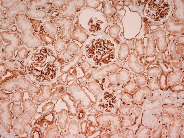
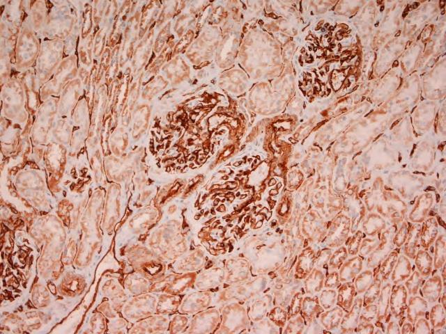
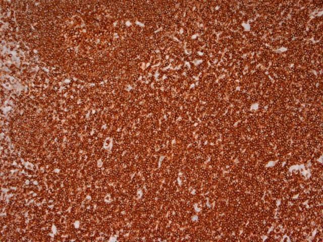
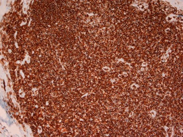
5 Image 3: Fallopian Tube Tissue, Pan-Cytokeratin Stain Image 4: Kidney Tissue, CD34 Stain Image 5: Tonsil Tissue, LCA Stain
6 Discussion This is a prospective, side-by-side study comparing the quality and acceptability of 10% gel-based formalin preparation (Formagel) versus the traditional, liquid-based 10% neutral buffered formalin as it pertains to immunohistochemical assessments. For each sample, unfixed tissue arriving from the operating room was procured and divided into two small, similarly sized fragments, each of which was allowed to fix in either of the formalin preparation for an identical period of time prior to further processing; tissue origin, sample size, fixative amount and fixation times were thus variables controlled across the two fixatives. Based on site of origin, antibodies were selected to accentuate key cellular constituents. Tissue immunoreactivity was assessed with regards to intensity on a tiered grading system. The global results demonstrate that Formagel is just as effective as the traditional liquidbased, 10% neutral buffered formalin in terms of intensity of immunoreactivity across all major classes of antibodies commonly employed in diagnostic surgical pathology. This includes antibodies directed against intermediate filaments (cytokeratins, vimentin), and other mesenchymal markers (smooth muscle actin, desmin), hematolymphoid markers (LCA, CD34). Immunoreactivity was also similar among liquid formalin treated specimens versus gel-based formalin fixed specimen with respect to antigen localization within a cellular structure, such as membrane-bound (epithelial membrane antigen, placental alkaline phosphatase), intracytoplasmic (keratins), and nucleus-associated (ER, PR, Ki-67). As a formalin product with identical concentration to its traditional liquid counterpart, Formagel is expected to facilitate retention of antigenicity and immunoreactivity. It is possible that as a gel-based preparation with less volatility, Formagel may be better able to prevent
7 tissue disintegration and antigenic dissipation; as a result, this physical property may in fact even enhance morphologic and antigenic preservation for the purpose of routine microscopic examination, and for specialized techniques such as immunohistochemistry. Conclusion Formagel is an effective and practical alternate fixative to traditional, neutral buffered formalin for the purpose of antigenicity retention and preservation. The immunoreactivity of tissue fixed in Formagel is at least equivalent to that of tissue fixed in neutral buffered formalin across a wide spectrum of antibody/antigen types and classes.
LAMININ. For Immunohistochemical Demonstration of Laminin in Paraffin-embedded and Frozen Human Tissue Sections Stock No. IMMH-7
 LAMININ For Immunohistochemical Demonstration of Laminin in Paraffin-embedded and Frozen Human Tissue Sections Stock No. IMMH-7 TABLE OF CONTENTS BACKGROUND AND PRINCIPLE... 4 REAGENTS AND EQUIPMENT PROVIDED...
Actin, Smooth Muscle (1A4)
") Product Identification Cat. No. Description 45121 IMPATH Actin Smooth Muscle RTU M (1A4) Symbol Definitions P A E S DOC# DIS ready-to-use ascites serum supernatant document number distributed by Intended
Product Identification Cat. No. Description 45121 IMPATH Actin Smooth Muscle RTU M (1A4) Symbol Definitions P A E S DOC# DIS ready-to-use ascites serum supernatant document number distributed by Intended
Actin, Muscle Specific (HHF35)
") Product Identification Cat. No. Description 45235 IMPATH Actin Muscle Specific RTU M (HHF35) Symbol Definitions P A E S DOC# DIS ready-to-use ascites serum supernatant document number distributed by Intended
Product Identification Cat. No. Description 45235 IMPATH Actin Muscle Specific RTU M (HHF35) Symbol Definitions P A E S DOC# DIS ready-to-use ascites serum supernatant document number distributed by Intended
Immunohistochemistry: Basics and Methods
 Immunohistochemistry: Basics and Methods Igor B. Buchwalow l Werner Böcker Immunohistochemistry: Basics and Methods Prof. Dr. Igor B. Buchwalow Prof. Dr. Werner Böcker Gerhard-Domagk-Institut für Pathologie
Materials and Methods Materials Required for Fixing, Embedding and Sectioning. OCT embedding matrix (Thermo Scientific, LAMB/OCT)
") Page 1 Introduction Tissue freezing and sectioning is a rapid method of generating tissue samples (cryosections) for histological analysis, and obviates the need for wax embedding. The method is popular
Page 1 Introduction Tissue freezing and sectioning is a rapid method of generating tissue samples (cryosections) for histological analysis, and obviates the need for wax embedding. The method is popular
The Children s Hospital of Philadelphia Department of Pathology and Laboratory Medicine
 TheChildren shospitalofphiladelphia DepartmentofPathologyandLaboratoryMedicine Muscle Biopsy - General Instructions The Division of Neuropathology, Department of Pathology and Laboratory Medicine, Children
PREPARATION OF HISTOLOGICAL SPECIMENS
 PREPARATION OF HISTOLOGICAL SPECIMENS Histo-techniques Preparation of tissue for microscopic examination Series of processes Ultimate aim to make tissue visible as it is Pathology Vs Anatomy Steps vary
Immunohistochemistry: Basics and Methods
 Immunohistochemistry: Basics and Methods Bearbeitet von Igor B Buchwalow, Werner Böcker 1st Edition. 2010. Buch. x, 153 S. Hardcover ISBN 978 3 642 04608 7 Format (B x L): 15,5 x 23,5 cm Gewicht: 445 g
Epithelial cell-cell adhesion molecule (Ep-CAM)
") Assessment Run 45 205 Epithelial cell-cell adhesion molecule (Ep-CAM) Material The slide to be stained for Ep-CAM comprised:. Appendix, 2. Kidney, 3. Basal cell carcinoma, 4. Colon adenocarcinoma, 5-6.
Assessment Run 45 205 Epithelial cell-cell adhesion molecule (Ep-CAM) Material The slide to be stained for Ep-CAM comprised:. Appendix, 2. Kidney, 3. Basal cell carcinoma, 4. Colon adenocarcinoma, 5-6.
Histopathological techniques The adoption of routine fixation and paraffin wax embedding.
 CIHRT Exhibit P-3359 Page 1 Routine tissue preparation in modern diagnostic histopathology. Bryan R. Hewlett ART, MLT. Histopathological techniques The adoption of routine fixation and paraffin wax embedding.
Web Based Promotion! 10% Off any initial product order. Mention promo code 1204!
 Web Based Promotion! 10% Off any initial product order. Mention promo code 1204! Volume 2, Number 2 1998 FROM PATIENT TO EMBEDDING CENTER IN TWO HOURS OR LESS The single biggest factor in health care today
Human Placental Lactogen (hpl) (Polyclonal)
 (Polyclonal)") Product Identification Cat. No. Description 45312 IMPATH Hpl RTU R (Poly) Symbol Definitions P A E S DOC# DIS ready-to-use ascites serum supernatant document number distributed by Intended Use This antibody
Product Identification Cat. No. Description 45312 IMPATH Hpl RTU R (Poly) Symbol Definitions P A E S DOC# DIS ready-to-use ascites serum supernatant document number distributed by Intended Use This antibody
Manufactured by. Zyagen Barnes Canyon Road San Diego, CA 92121, USA
 Alkaline Phosphatase Immunohistochemistry Detection kits For detection of mouse, rabbit, goat, rat, sheep, chicken, guinea pig, and human primary antibodies Size: 500 Tests Catalog #: AK-011, Mouse Kit
Fluorescent in-situ Hybridization
 Fluorescent in-situ Hybridization Presented for: Presented by: Date: 2 Definition In situ hybridization is the method of localizing/ detecting specific nucleotide sequences in morphologically preserved
Boost turnaround time for faster diagnosis
 Boost turnaround time for faster diagnosis Tissue-Tek Xpress x120 Rapid Tissue Processor True rapid continuous tissue processing The Tissue-Tek Xpress x120 is a standardized, turnkey solution combining
Principles And Procedures
 Product Identification Cat. No. Description 44837 Vimentin 0,1 M (V9) 44838 Vimentin 1 M (V9) 44409 Vimentin RTU M (V9) Symbol Definitions P C A E S DIL DOC# DIS ready-to-use concentrate ascites serum
ab Mouse and Rabbit Specific HRP/DAB (ABC) Detection IHC Kit
 Detection IHC Kit") ab64264 - Mouse and Rabbit Specific HRP/DAB (ABC) Detection IHC Kit Instructions for Use For the detection of a specific antibody bound to an antigen in tissue sections. This product is for research use
ab64264 - Mouse and Rabbit Specific HRP/DAB (ABC) Detection IHC Kit Instructions for Use For the detection of a specific antibody bound to an antigen in tissue sections. This product is for research use
HAM-56 antibody reacts with monocytes, but is unreactive with B and T-lymphocytes. Principles And Procedures
 Product Identification Cat. No. Description 44682 Macrophage Marker 0,1 M (HAM- 56) 44683 Macrophage Marker 1 M (HAM-56) 44330 Macrophage Marker RTU M (HAM- 56) Symbol Definitions P C A E S DIL DOC# DIS
Novocastra Liquid Mouse Monoclonal Antibody Nitric Oxide Synthase-1
 Novocastra Liquid Mouse Monoclonal Antibody Nitric Oxide Synthase-1 Product Code: NCL-L-NOS-1 Leica Biosystems Newcastle Ltd Balliol Business Park West Benton Lane Newcastle Upon Tyne NE12 8EW United Kingdom
Androgen Receptor (SP107)
") Product Identification Cat. No. Description 45164 IMPATH Androgen Receptor RTU M (SP107) Symbol Definitions P A E S DOC# DIS ready-to-use ascites serum supernatant document number distributed by Intended
Product Identification Cat. No. Description 45164 IMPATH Androgen Receptor RTU M (SP107) Symbol Definitions P A E S DOC# DIS ready-to-use ascites serum supernatant document number distributed by Intended
Cell & Tissue Staining Kit
 Cell & Tissue Staining Kit For the detection of goat, mouse, rabbit, rat, or sheep primary IgG Antibodies Size: 50 Tests HRP-DAB System Goat Kit (Catalog Number CTS008) Mouse Kit (Catalog Number CTS002)
KCC Path-Core Page 1 of 5
 Instructions for Sample preparation for Paraffin embedding PLEASE NOTE: There is no one-size-fits-all method of tissue preparation for all experimental designs. Before harvesting tissue, you need to assess
ab TripleStain IHC Kit: M&M&R on human tissue (DAB, Red/AP & DAB/Ni)
") ab183287 TripleStain IHC Kit: M&M&R on human tissue (DAB, Red/AP & DAB/Ni) Instructions for Use For the detection of Rabbit and Mouse Primary antibodies on Human tissue or cell samples. This product is
ab183287 TripleStain IHC Kit: M&M&R on human tissue (DAB, Red/AP & DAB/Ni) Instructions for Use For the detection of Rabbit and Mouse Primary antibodies on Human tissue or cell samples. This product is
Overview of Immunohistochemistry. (with a focus on wax-embedded sections)
") Overview of Immunohistochemistry (with a focus on wax-embedded sections) Overview of Immunohistochemistry (with a focus on wax-embedded sections) Overview of Immunohistochemistry IHC is like cooking. There
Overview of Immunohistochemistry (with a focus on wax-embedded sections) Overview of Immunohistochemistry (with a focus on wax-embedded sections) Overview of Immunohistochemistry IHC is like cooking. There
ab Mouse and Rabbit AP/Fast-Red (ABC) Detection IHC Kit
 Detection IHC Kit") ab128967 - Mouse and Rabbit AP/Fast-Red (ABC) Detection IHC Kit Instructions for Use For the detection of a specific antibody bound to an antigen in tissue sections. This product is for research use only
ab128967 - Mouse and Rabbit AP/Fast-Red (ABC) Detection IHC Kit Instructions for Use For the detection of a specific antibody bound to an antigen in tissue sections. This product is for research use only
KOS The microwave multifunctional tissue processor you were waiting for. MILESTONE
 KOS The microwave multifunctional tissue processor you were waiting for. Tissue processing Decalcification Special stains Fixation Gross hardening Antigen retrieval In a fraction of the time MILESTONE
Assessing the Quality of Tissue Processing and the Performance of PelorisTM using the Leica Microsystems Scoring System
 Assessing the Quality of Tissue Processing and the Performance of PelorisTM using the Leica Microsystems Scoring System Geoffrey Rolls, Neville Farmer, Fiona Tarbet Leica Microsystems, Biosystems Division,
Effect of Fixation Time and Microwave Oven Heating Time on Retrieval of the Ki-67 Antigen from Paraffin-embedded Tissue
 22-1554/93/$3.3 The Journal of Histochemistry and Cytochemistry Copyright 1993 by The Histochemical Society, Inc Vol. 41, No. 8. pp. 1241-1246, 1993 Printed in US.A. Original Article I Effect of Fixation
Immunofluorescence Staining Protocol for 3 Well Chamber, removable
 Immunofluorescence Staining Protocol for 3 Well Chamber, removable This Application Note presents a simple protocol for the cultivation, fixation, and staining of cells using the 3 Well Chamber, removable.
Histology Pattern Recognition Software in Investigative Pathology
 Histology Pattern Recognition Software in Investigative Pathology J. Webster, DVM, PhD, DACVP Laboratory of Cancer Biology and Genetics National Cancer Institute, Bethesda, MD Pathology Visions 2011 November
Varicella Zoster Virus (SG1-1, SG1- SG4, NCP-1 & IE-62)
") Varicella Zoster Virus (SG1-1, SG1- SG4, Product Identification Cat. No. Description 44833 Varicella-zoster virus 0,1 M 44834 Varicella-zoster virus 1 M 44407 Varicella-zoster virus RTU M Symbol Definitions
Varicella Zoster Virus (SG1-1, SG1- SG4, Product Identification Cat. No. Description 44833 Varicella-zoster virus 0,1 M 44834 Varicella-zoster virus 1 M 44407 Varicella-zoster virus RTU M Symbol Definitions
Varicella Zoster Virus (SG1-1, SG1- SG4, NCP-1 & IE-62)
") Varicella Zoster Virus (SG1-1, SG1- SG4, Product Identification Cat. No. Description 45269 IMPATH Varicella Zoster Virus RTU M (Monocl CKT) Symbol Definitions P A E S DOC# DIS ready-to-use ascites serum
Varicella Zoster Virus (SG1-1, SG1- SG4, Product Identification Cat. No. Description 45269 IMPATH Varicella Zoster Virus RTU M (Monocl CKT) Symbol Definitions P A E S DOC# DIS ready-to-use ascites serum
Survey of Neural Networks in Digital Pathology and Pathology Workflow
 Survey of Neural Networks in Digital Pathology and Pathology Workflow Christianne Dinsmore 1 1 Department of Computing and Digital Media DePaul University Chicago, IL Abstract Digital pathology has already
Principles And Procedures
 Product Identification Cat. No. Description 44482 CD25 (Interleukin-2 Receptor) 0,1 M (4C9) 44483 CD25 (Interleukin-2 Receptor) 1 M (4C9) 44230 CD25 (Interleukin-2 Receptor) RTU M (4C9) Symbol Definitions
CME/SAM. Formaldehyde Substitute Fixatives Analysis of Macroscopy, Morphologic Analysis, and Immunohistochemical Analysis
 Anatomic Pathology / Formaldehyde Substitute Fixatives Formaldehyde Substitute Fixatives Analysis of Macroscopy, Morphologic Analysis, and Immunohistochemical Analysis Cathy B. Moelans, PhD, Natalie ter
The Effect of Antigen Retrieval on Cells Fixed in 10% Neutral Buffered Formalin Followed by Transfer to 70% Ethanol
 The Effect of Antigen Retrieval on Cells Fixed in 10% Neutral Buffered Formalin Followed by Transfer to 70% Ethanol Dennis Otali 1,2, Qinghua He 3, William E. Grizzle 1 * 1 Department of Pathology, University
Ep-CAM/Epithelial Specific Antigen (Ber-EP4)
") Ep-CAM/Epithelial Specific Antigen (Ber- Product Identification Cat. No. Description 45648 IMPATH Ep-CAM RTU M (Ber- Symbol Definitions P A E S DOC# DIS ready-to-use ascites serum supernatant document
Ep-CAM/Epithelial Specific Antigen (Ber- Product Identification Cat. No. Description 45648 IMPATH Ep-CAM RTU M (Ber- Symbol Definitions P A E S DOC# DIS ready-to-use ascites serum supernatant document
Microarray Industry Products
 Via Nicaragua, 12-14 00040 Pomezia (Roma) Phone: +39 06 91601628 Fax: +39 06 91612477 info@lifelinelab.com www.lifelinelab.com Microarray Industry Products Page 10 NBT / BCPIP Chromogenic phosphatase
Application Note Tissue reconstruction using ThinCert Cell Culture Inserts and Plates
 Application Note Tissue reconstruction using ThinCert Cell Culture Inserts and Plates www.gbo.com/bioscience Introduction For many years live-animal experimentation provided the singular means to access
Immunohistochemistry. How does it look like? When do we need IHC? When do we need IHC? In clinic: In research:
 Introduction How does it look like? Immunohistochemistry Smooth muscle actin Parvalbumin Distrophyn Sandrine Bichet Head of Molecular Histology Platform Signal versus background 06.03.2012 IHC basics Introduction
ERG (Ets-Related Gene)
") Assessment Run 50 2017 ERG (Ets-Related Gene) Material The slide to be stained for ERG comprised: 1. Tonsil, 2. Appendix, 3. Prostate with hyperplasia, 4-5. Prostate adenocarcinoma. All tissues were fixed
Assessment Run 50 2017 ERG (Ets-Related Gene) Material The slide to be stained for ERG comprised: 1. Tonsil, 2. Appendix, 3. Prostate with hyperplasia, 4-5. Prostate adenocarcinoma. All tissues were fixed
Isolation, culture, and transfection of primary mammary epithelial organoids
 Supplementary Experimental Procedures Isolation, culture, and transfection of primary mammary epithelial organoids Primary mammary epithelial organoids were prepared from 8-week-old CD1 mice (Charles River)
ZytoDot. 2C SPEC HER2/CEN 17 Probe
 ZytoDot 2C SPEC HER2/CEN 17 Probe C-3032-400 C-3032-100 40 (0.4 ml) 10 (0.1 ml) For the detection of the human HER2 gene and alphasatellites of chromosome 17 by chromogenic in situ hybridization (CISH)....
August 2017 Changes. Anatomic Pathology Checklist. CAP Accreditation Program
 August 2017 Changes Anatomic Pathology Checklist CAP Accreditation Program College of American Pathologists 325 Waukegan Road Northfield, IL 60093-2750 www.cap.org 08.21.2017 Disclaimer and Copyright Notice
Automation in the Histology Lab
 Automation in the Histology Lab Investing in Cost Savings, Faster Turnaround Times, Reduced Contamination, and Standardization Meghan J. Cuddihy, Ph.D. Abigail G. Garrity, M.S. In this white paper Automation
Effects of Prolonged Formalin Fixation on Diagnostic Immunohistochemistry in Domestic Animals
 Volume 57(8): 753 761, 2009 Journal of Histochemistry & Cytochemistry http://www.jhc.org ARTICLE Effects of Prolonged Formalin Fixation on Diagnostic Immunohistochemistry in Domestic Animals Joshua D.
Polyclonal Anti-PSA Is More Sensitive but Less Specific Than Monoclonal Anti-PSA Implications for Diagnostic Prostatic Pathology
 Anatomic Pathology / PSA IMMUNOHISTOCHEMICAL ANALYSIS Polyclonal Anti-PSA Is More Sensitive but Less Specific Than Monoclonal Anti-PSA Implications for Diagnostic Prostatic Pathology Murali Varma, FRCPath,
Evaluating the impact of ISO on an Irish histopathology laboratory
 Evaluating the impact of ISO 15189 on an Irish histopathology laboratory Laboratory accreditation is a valuable asset that delivers real results. Here, Linda O Connor, Alison Malkin and Breffnie Carroll
Matthias S. Matter 1,2, Esther Schwarz 1, Teresa Marafioti 3, Peter Schraml 1, Holger Moch 1
 Immunohistochemical detection of CD3 in T- cell lymphomas: superior sensitivity of rabbit monoclonal 2GV6 antibody compared to mouse monoclonal F7.2.38 antibody Matthias S. Matter 1,2, Esther Schwarz 1,
Paired box gene-8 protein (PAX8)
") Assessment Run 42 2014 Paired box gene-8 protein (PAX8) Material The slide to be stained for PAX8 comprised: 1. Fallopian tube, 2. Tonsil, 3. Renal clear cell carcinoma, 4. Kidney, 5. Lung adenocarcinoma
Assessment Run 42 2014 Paired box gene-8 protein (PAX8) Material The slide to be stained for PAX8 comprised: 1. Fallopian tube, 2. Tonsil, 3. Renal clear cell carcinoma, 4. Kidney, 5. Lung adenocarcinoma
Notice of Faculty Disclosure
 Pathologist Extenders: Current and Future Amy Clayton, M.D. Vice Chair Clinical Practice and Quality Department of Laboratory Medicine and Pathology Mayo Clinic, Rochester MN Notice of Faculty Disclosure
Nerve Growth Factor Receptor (NGFR) (MRQ-21)
 (MRQ-21)") Nerve Growth Factor Receptor (NGFR) Product Identification Cat. No. Description 45244 IMPATH NGFR RTU M Symbol Definitions P A E S DOC# DIS ready-to-use ascites serum supernatant document number distributed
Nerve Growth Factor Receptor (NGFR) Product Identification Cat. No. Description 45244 IMPATH NGFR RTU M Symbol Definitions P A E S DOC# DIS ready-to-use ascites serum supernatant document number distributed
MEDICAL LABORATORY SCIENCE (MLS)
") MEDICAL LABORATORY SCIENCE (MLS) Explanation of Course Numbers Courses in the 1000s are primarily introductory undergraduate courses Those in the 2000s to 4000s are upper-division undergraduate courses
MEDICAL LABORATORY SCIENCE (MLS) Explanation of Course Numbers Courses in the 1000s are primarily introductory undergraduate courses Those in the 2000s to 4000s are upper-division undergraduate courses
The diagnosis of biopsy and surgical tissue samples
 Special Article Uniform Labeling of Blocks and Slides in Surgical Pathology Guideline From the College of American Pathologists Pathology and Laboratory Quality Center and the National Society for Histotechnology
Immunohistochemical pitfalls in the demonstration of insulindegrading enzyme in normal and neoplastic human tissues
 Immunohistochemical pitfalls in the demonstration of insulindegrading enzyme in normal and neoplastic human tissues RAZVAN T. RADULESCU 1*, ANGELIKA JAHN 1, DANIELA HELLMANN 1, AND GREGOR WEIRICH 2 1 Clinical
RNAscope 2.5 LS Duplex Reagent Kit User Manual for BDZ 11. For use with Leica Biosystems BOND RX System
 RNAscope 2.5 LS Duplex Reagent Kit User Manual for BDZ 11 For use with Leica Biosystems BOND RX System For Research Use Only. Not for diagnostic use. 322440-USM Effective Date 03312017 For Research Use
Procedure for Immunohistochemical Examination
 Procedure for Immunohistochemical Examination 1. Preparation of Paraffin Block (Separate Attachment 2-2) Disposable bench protection sheet, cutting board, sectioning blade, tweezers, stainless
STANDARD OPERATING PROCEDURE
 Effective Date: 12/1/2011 Page 1 of 12 Approval Name/Title Signature Date Authors: Jessica Landis Trial Implementation Manager Immune Tolerance Network Mary C. Philogene Associate Director, QA And Cell
Converting your ELISA from horseradish peroxidase to alkaline phosphatase using NovaBright chemiluminescence detection reagents
 Application note DynaLight Substrate with RapidGlow Enhancer Converting your ELISA from horseradish peroxidase to alkaline phosphatase using NovaBright chemiluminescence detection reagents Introduction
Troubleshooting Made Easy with Education
 Indiana Society for Histotechnology Spring Symposium Troubleshooting Made Easy with Education March 6-7, 2015 Meeting: Fishers Banquet Center 9775 North by Northeast Blvd. Fishers, IN 46037 http://fishersbanquetandconferencecenter.com
UltraMap Alk Phos. UltraMap anti-ms Alk Phos
 UltraMap anti-rb Alk Phos UltraMap anti-ms Alk Phos Biotin-free Alkaline Phosphatase Detection Systems for the Ventana DISCOVERY Series of Instruments Catalog Number: s 760-4314 (UltraMap anti-rb Alk Phos)
Pinpoint Slide DNA Isolation System Catalog No. D3001
 INSTRUCTIONS Pinpoint Slide DNA Isolation System Catalog No. D3001 Highlights Easily isolates genomic DNA in any targeted microscopic tissue area on a slide. The simple procedure combines Pinpoint tissue
ZytoFast HPV type 6/11/16/18/31/33/35 Screening Probe (Bio
 ZytoFast HPV type 6/11/16/18/31/33/35 Screening Probe (Bio iotin in-labeled labeled) T-1044-400 40 (0.4 ml) For the detection of Human Papilloma Virus (HPV) type 6/11/16/18/31/33/35 DNA by chromogenic
SANTA CRUZ BIOTECHNOLOGY, INC.
 TECHNICAL SERVICE GUIDE: Western Blotting 2. What size bands were expected and what size bands were detected? 3. Was the blot blank or was a dark background or non-specific bands seen? 4. Did this same
Detection of DNA by scraping the formalin-fixed paraffin-embedded tissue
 999 Detection of DNA by scraping the formalin-fixed paraffin-embedded tissue M H Bukhari 1 *, S Niazi 1, R Ghani 2, Z Rathore 1, R Basharat 1, N A Chaudhry 1, and S Naeem 1 1 Department of Pathology, King
ReliaPrep FFPE gdna Miniprep System
 TECHNICAL MANUAL ReliaPrep FFPE gdna Miniprep System Instructions for Use of Products A2351 and A2352 Revised 12/15 TM352 ReliaPrep FFPE gdna Miniprep System All technical literature is available at: www.promega.com/protocols/
August 2017 Changes. Flow Cytometry Checklist. CAP Accreditation Program
 August 2017 Changes Flow Cytometry Checklist CAP Accreditation Program College of American Pathologists 325 Waukegan Road Northfield, IL 60093-2750 www.cap.org 08.21.2017 Disclaimer and Copyright Notice
The Influence of Protease Digestion and Duration of Fixation on the Immunostaining of Keratins.
 0022-1554/86/$3 30 The Journal of Histochemistry and Cytochemistry Copyright 1986 by The Histochemical Society, inc Vol 34, No 8, pp 1095-1100, 1986 Printed in U S A Original Article The Influence of Protease
Electron microscopy technology of reticulocytes after sorting with
 Electron microscopy technology of reticulocytes after sorting with magnetic beads The Cell Analysis Center Scientific Bulletin Part 2 For efficient analysis of cells, sorting of the target cells is crucial.
SAMPLE SENDING MANAGEMENT
 SAMPLE SENDING MANAGEMENT CIBOMA/2004-01 Phase IV.III, multicenter, open, randomized treatment study to evaluate the efficacy of maintenance therapy with capecitabine (X) after standard adjuvant chemotherapy
Imagine... fully automated embedding with the highest quality blocks. Tissue-Tek AutoTEC a120 & Paraform. Automated Embedding System
 Imagine... fully automated embedding with the highest quality blocks Tissue-Tek AutoTEC a120 & Paraform Automated Embedding System KEEP UP WITH THE GROWING NUMBER OF SAMPLES AutoTEC a120, part of SMART
Osteogenic Differentiation and Analysis of MSC
 Osteogenic Differentiation and Analysis of MSC Application Note Background Mesenchymal Stem Cells (MSC) are fibroblastoid multipotent adult stem cells with a high capacity for self-renewal. So far, these
Baraa Ayed AL-Odat. Israa Ayed. Heba kalbouneh
 1 Baraa Ayed AL-Odat Israa Ayed Heba kalbouneh Introduction: "lecture #1" The name " histology " is derived from the Greek words: "histo" means a tissue and "logos" means the study of. So, Histology mean
celldatasci.com/rnastorm RNAstorm RNA Isolation Kit for FFPE Tissue Samples Sample Kit (20 extractions)
") celldatasci.com/rnastorm info@celldatasci.com RNAstorm RNA Isolation Kit for FFPE Tissue Samples Sample Kit (20 extractions) Support: Email: support@celldatasci.com Phone: 650.285.2376 (option 2) Toll-free:
celldatasci.com/rnastorm info@celldatasci.com RNAstorm RNA Isolation Kit for FFPE Tissue Samples Sample Kit (20 extractions) Support: Email: support@celldatasci.com Phone: 650.285.2376 (option 2) Toll-free:
Atlas Antibodies AB and The Human Protein Atlas. Hong Kong, August 10, 2012
 Atlas Antibodies AB and The Human Protein Atlas Hong Kong, August 10, 2012 Company Overview Founded in February 2006 Spinout from The Human Protein Atlas (HPA) project Exclusive commercial rights to HPA
Digital Cytopathology & Interactive Case Discussion
 Digital Cytopathology & Interactive Case Discussion Marilyn M. Bui, MD, PhD Senior Member & Scientific Director of Analytic Microscopy Core Section Head, Bone and Soft Tissue Pathology Program Leader,
#$% &%" ' " ( ) * % '-'
 * % '-'") !" #$% &%" ' " ( ) * '++, % '-' '"%!./,0012 DISPOSITION INSTRUCTIONS: Destroy this report when no longer needed. Do not return to the originator. DISCLAIMERS: The opinions or assertions contained herein
!" #$% &%" ' " ( ) * '++, % '-' '"%!./,0012 DISPOSITION INSTRUCTIONS: Destroy this report when no longer needed. Do not return to the originator. DISCLAIMERS: The opinions or assertions contained herein
Integrated Radiology & Pathology Diagnostic Services: The Time is Now at UCLA
 Integrated Radiology & Pathology Diagnostic Services: The Time is Now at UCLA Scott W Binder MD Pritzker Professor of Clinical Pathology and Dermatology Senior Vice Chair Director, Pathology Clinical Services
ROUTINE SPECIMEN COLLECTION BY SPECIMEN TYPE
 1. AUTOPSY EYE ROUTINE SPECIMEN COLLECTION BY SPECIMEN TYPE a. Arrangements: No special arrangements necessary. i. Immerse the specimen in a sufficient quantity of fixative so that the eye is covered completely
IMMUNOPRECIPITATION TROUBLESHOOTING TIPS
 IMMUNOPRECIPITATION TROUBLESHOOTING TIPS Creative Diagnostics Abstract Immunoprecipitation (IP) is the technique of precipitating a protein antigen out of solution using an antibody that specifically binds
Optimized Covaris truxtrac FFPE RNA Protocol for Isolating RNA from Laser-Capture Microdissected (LCM) FFPE Tissue for Next Generation Sequencing
 FFPE Tissue for Next Generation Sequencing") Optimized truxtrac FFPE RNA Protocol for Isolating RNA from Laser-Capture Microdissected (LCM) FFPE Tissue for Next Generation Sequencing Enni Markkanen Institute of Veterinary Pharmacology and Toxicology,
Optimized truxtrac FFPE RNA Protocol for Isolating RNA from Laser-Capture Microdissected (LCM) FFPE Tissue for Next Generation Sequencing Enni Markkanen Institute of Veterinary Pharmacology and Toxicology,
Strep-tag detection in Western blots
 Strep-tag detection in Western blots General protocol for the detection of Strep-tag fusion proteins Last date of revision April 2012 Version PR07-0010 www.strep-tag.com For research use only Important
Updated April 27, PRODUCT INSERT
 Updated April 27, 2009 1 PRODUCT INSERT Hypoxyprobe, Inc. 121 Middlesex Turnpike Burlington, MA 01803 USA www.hypoxyprobe.com Hypoxyprobe Gemini Kit Kit Contents: Solid pimonidazole HCl (Hypoxyprobe -1)
Ariol. Clinical IHC and FISH Breast Marker Analysis Quality. Efficiency. Confidence.
 Ariol Clinical IHC and FISH Breast Marker Analysis Quality. Efficiency. Confidence. 3 Behind every breast slide in your laboratory is a woman waiting for life changing answers With over 1.25 million women
Quick Ray Manual Tissue Microarrayer. Manual Tissue Microarrayer Quick-Ray
 Manual Tissue Microarrayer Quick-Ray 1 NOTE This user guide describes the instruction of Quick Ray. Review this user guide to avoid injury and prevent damage to this product or any products connected to
The Evaluation of Estrogen Receptor in Primary Breast Carcinoma by Computer-Assisted Image Analysis
 The Evaluation of Estrogen Receptor in Primary Breast Carcinoma by Computer-Assisted Image Analysis SARA BACUS, PH.D., JULIE L. FLOWERS, B.S., MICHAEL F. PRESS, PH.D., JAMES W. BACUS, PH.D., AND KENNETH
TECHNOLOGIES & SERVICES FOR THERAPEUTIC ANTIBODY DEVELOPMENT
 Specialist in tissue analysis by Histology, Immunohistochemistry and In Situ Hybridization TECHNOLOGIES & SERVICES FOR THERAPEUTIC ANTIBODY DEVELOPMENT CONTENTS Histalim: who we are Our areas of expertise
Identification of red and white blood cells from whole blood samples using the Agilent 2100 bioanalyzer. Application Note
 Identification of red and white blood cells from whole blood samples using the Agilent 2100 bioanalyzer Application Note Sylvie Veriac Valérie Perrone Madeleine Avon Abstract Agilent Equipment: 2100 bioanalyzer
Immunoassay Kit Catalog # KCA0021. Canine. C-Reactive Protein
 Immunoassay Kit Catalog # KCA0021 Canine C-Reactive Protein BioSource International, Inc. 542 Flynn Road Camarillo, California 93012 USA Tel: 805-987-0086 800-242-0607 FAX: 805-987-3385 email: tech.support@biosource.com
High Pure PCR Template Preparation Kit for preparation of 100 nucleic acid samples Cat. No
 for preparation of 100 nucleic acid samples Cat. No. 1 796 88 Principle Cells are lysed during a short incubation with Proteinase K in the presence of a chaotropic salt (guanidine HCl), which immediately
HUMAN CONNECTIVE TISSUE GROWTH FACTOR (CTGF) ELISA KIT
 ELISA KIT") HUMAN CTGF ELISA KIT PAGE 1 HUMAN CONNECTIVE TISSUE GROWTH FACTOR (CTGF) ELISA KIT PURCHASE INFORMATION: ELISA NAME HUMAN CTGF ELISA FOR THE QUANTITATIVE DETERMINATION OF HUMAN CTGF CONCENTRATIONS IN SERUM
HUMAN CTGF ELISA KIT PAGE 1 HUMAN CONNECTIVE TISSUE GROWTH FACTOR (CTGF) ELISA KIT PURCHASE INFORMATION: ELISA NAME HUMAN CTGF ELISA FOR THE QUANTITATIVE DETERMINATION OF HUMAN CTGF CONCENTRATIONS IN SERUM
Cytomics in Action: Cytokine Network Cytometry
 Cytomics in Action: Cytokine Network Cytometry Jonni S. Moore, Ph.D. Director, Clinical and Research Flow Cytometry and PathBioResource Associate Professor of Pathology & Laboratory Medicine University
PreAnalytiX Supplementary Protocol
 PreAnalytiX Supplementary Protocol Purification of full-length proteins from sections of PAXgene Tissue fixed, paraffin-embedded (PFPE) tissue This protocol describes using the Qproteome FFPE Tissue kit
Streamline Pre-Analytic Orchard Pathology Processes
 Orchard Pathology Orchard Pathology is a complete diagnostic information system where clinical and anatomic data share the same database and where pathology data is stored in discrete data fields to simplify
Chapter 15. Cytoskeletal Systems. Lectures by Kathleen Fitzpatrick Simon Fraser University Pearson Education, Inc.
 Chapter 15 Cytoskeletal Systems Lectures by Kathleen Fitzpatrick Simon Fraser University Table 15-1 - Microtubules Table 15-1 - Microfilaments Table 15-1 Intermediate Filaments Table 15-3 Microtubules
Pathology Solution IVD US. Philips IntelliSite HER2/neu IHC Digital Manual Read
 Pathology Solution IVD US Philips IntelliSite HER2/neu IHC Digital Manual Read Multisite configuration Role based User access control Single worklist view across the sites Real-time Collaboration & Sharing
NOTE- HUMAN BREAST CANCER SERIALLY TRANSPLANTABLE IN NUDE MICE IN ASCITES FORM*1
 NOTE- HUMAN BREAST CANCER SERIALLY TRANSPLANTABLE IN NUDE MICE IN ASCITES FORM*1 Setsuo HIROHASHI,*2 Yukio SHIMOSATO, Kanji NAGAI,*3 Tsutomu KOIDE, and Toru KAMEYA Pathology Division, National Cancer Center