How To Optimize Your IMMUNOHISTOCHEMISTRY EXPERIMENT
|
|
|
- Gertrude Jones
- 6 years ago
- Views:
Transcription

1 How To Optimize Your IMMUNOHISTOCHEMISTRY EXPERIMENT

2 2 How To Optimize Your IHC Experiment
3 ptglab.com 3 CONTENTS 4 5 Introduction To Immunohistochemistry 6 9 General Protocols Antigen Retrieval 12 Blocking 13 Featured Product Immunostaining Visualization Tips, Tricks, and Troubleshooting Guide 22 Contact Us
4 4 How To Optimize Your IHC Experiment Introduction To IMMUNHOHISTOCHEMISTRY Immunohistochemistry (IHC) allows you to visualize proteins in tissue while retaining its microstructure. It helps to demonstrate the exact position and distribution of the protein of interest in the analyzed tissue section. The advantage of this visualization is that it allows for comparison between, for example, healthy and diseased tissues. Briefly, in an IHC experiment, the antigen of interest is localized by the binding of an antibody. The antibody-antigen interaction is then further visualized via chromogenic or fluorescent detection. The IHC protocol contains many steps that may require optimization to ensure specific antibody binding and optimal visualization of the target protein. As IHC protocol contains many different variable factors, it is challenging to find the best working conditions to obtain strong and specific staining. The following guide outlines some tips and hints for each of these steps. Below, we also provide our general staining protocols. Factors to consider in IHC experimental design IHC Factor Sample Type Antigen Epitope Primary Antibody Blocking Secondary Antibody Labelling Counterstaining Analysis Controls To Consider Fixed, frozen Species, level of expression, subcellular location Conformation, post-translational modification Monoclonal vs. Polyclonal Sera, BSA, commercial buffer, temperature, ph, dilution, incubation time Species, label type Chromogenic, enzymatic, fluorescent Chromogenic, fluorescent Microscope, software-based analysis, evaluation by eye Secondary antibody only, antigen positive tissue, isotype control
5 ptglab.com 5 The Main Steps Of IHC Overview of the main steps of IHC using a chromogenic labeled secondary antibody on paraffin-embedded tissue slides.
6 6 How To Optimize Your IHC Experiment GENERAL PROTOCOLS Materials And Equipment Frozen or paraffin-embedded tissue Cryo-embedded media (e.g., OCT) Sucrose 4% Paraformaldehyde (PFA) Microtome Glass slides Coverslips Refrigerator Incubator Xylene Ethanol Boiling source Primary and secondary antibodies Hematoxylin Serum Citrate buffer Tris-EDTA buffer Diaminobenzidine tetrachloride (DAB) Microscope Membrane Transfer Citrate Buffer For 1000 ml 1x TBS For 1000 ml 10 mm Tris-sodium citrate ₂H₂O 2.9 g 20 mm Tris-base 2.4 g 1.9 mm citric acid H₂O Adjust ph to 6.0 Add ddh₂o to 1000 ml 0.4 g 150 mm NaCl 8.7 g Adjust ph to 7.6 Add ddh₂o to 1000 ml
7 ptglab.com 7 Sample Preparation Frozen sections of clinical samples For fast processing of clinical samples, eliminating the fixation step by directly freezing and embedding with OCT, followed by cutting (6-8 μm thickness), is recommended as it would be timesaving and avoid the increased difficulty of sectioning caused by fixation. As for the temperature in the cryostat for unfixed tissues, please refer to the table below: Cryostat Temperature For Unfixed Tissues Brain, liver and lymph node tissues Thyroid, spleen, kidney and muscle tissues Tissue containing fat Tissue containing plenty of fat -10ºC/-15ºC -15ºC/-20ºC -25ºC -30ºC Tip: Fresh tissue and fixation using 4% PFA in 4ºC overnight are recommended. 1. Wash tissue 3 times with PBS for 5 minutes each. 2. Immerse tissue in 20 30% sucrose for hours. 3. Place the tissue block onto a pre-labeled tissue base mold. 4. Cover the entire tissue block with cryo-embedding media (e.g., OCT). 5. Slowly place the base mold containing the tissue block into liquid nitrogen until the entire tissue block is submerged into liquid nitrogen to ensure tissue is completely frozen. 6. Store the frozen tissue block at -80ºC until ready for sectioning. 7. Transfer the frozen tissue block to a cryotome cryostat (e.g., -20ºC) prior to sectioning and allow the temperature of the frozen tissue block to equilibrate to the temperature of the cryotome cryostat. 8. Section the frozen tissue block into a desired thickness. Keep the section apparatus, including the blade and the blade holder, and clean by polishing with soft paper tissue. 9. Place the tissue sections onto glass slides suitable for immunohistochemistry. Sections can be stored in a sealed slide box at -80ºC for later use. Before further use, equilibrate the tissue first at -20ºC for 30 minutes.
8 8 How To Optimize Your IHC Experiment Tissue slide handling Tissue sections should be placed on a glass slide. Allow adhesion of tissue to slide by air drying and additional baking in an incubator. Cut and unbaked slides can be stored at 4 C for later use. However, storage might have an effect on the antigentic potential and this varies for each protein. Therefore, it is recommended to minimize storage time or to use freshly prepared slides. Immunohistochemistry On Paraffin Sections Please note: All steps in the following protocol are carried out at room temperature unless stated otherwise. Deparaffinizing and rehydration 1. Immerse slides in xylene for 10 minutes. Repeat this step again in fresh xylene for a further 10 minutes. (If required, repeat a third time in fresh xylene for another 10 minutes.) 2. Rehydrate sections by sequentially incubating with 100%, 95%, 80%, and 60% ethanol for 5 minutes each. 3. Rinse sections with distilled water three times for 3 minutes each. Antigen retrieval (optional) 4. Transfer slides to a microwave-proof container and cover with citrate buffer. 5. Heat in the microwave on medium power for 10 minutes. 6. Allow slides to cool in citrate buffer for approximately 35 minutes each. Primary antibody incubation 7. Rinse slides three times with 1x TBS for 3 minutes each. 8. Incubate slides with 3% H₂O₂ solution (diluted in distilled water) for 10 minutes to quench endogenous peroxidase activity. 9. Rinse slides three times with 1x TBS for 3 minutes each. 10. Prepare 5% normal blocking serum in 1x TBS. The serum should be derived from the same species in which the secondary antibody was raised. Block the sections for 1 hour. (Alternatively, use 5% BSA in 1x TBS for blocking if the corresponding serum is not available.) 11. Incubate sections with the primary antibody diluted in 1x TBS for 1 hour, or overnight at 4ºC; the optimal antibody dilution ratio should be pre-determined by experimentation. Set up negative controls by omitting the primary antibody incubation step for one slide per each experimental condition. 12. Following primary antibody incubation, rinse slides three times with 1x TBS for 3 minutes each.
9 ptglab.com 9 Signal Detection Please note: Proteintech * routinely uses EnVision Kit (Dako) for this step. 13. Apply sufficient peroxidase labeled polymer and incubate for 30 minutes. 14. Rinse slides three times with 1x TBS for 3 minutes each. 15. Prepare an appropriate volume of substrate solution prior to use by mixing one drop of Liquid DAB plus chromogen immediately with 1 ml of substrate buffer. Apply the substrate carefully and incubate for 5 10 minutes until a brown colour develops. 16. Rinse sections gently with sufficient distilled water. Hematoxylin counterstaining (optional) 17. To stain nuclei, immerse slides in a bath of hematoxylin for 3 minutes. 18. Rinse slides gently with distilled water. 19. Transfer slides into a 1% HCI, 99% ethanol solution for 10 seconds; transfer to distilled water immediately. Dehydration and mounting 20. Immerse slides sequentially into 60%, 80%, 95%, and 100% ethanol baths for 5 minutes each. 21. Immerse slides in xylene for 5 minutes. Repeat this step again in fresh xylene for another 5 minutes. 22. Mount the section with sufficient mounting media and cover with a coverslip Air-dry in a well-ventilated area (e.g., fume hood). Immunohistochemistry On Frozen Tissue Unlike paraffin samples, frozen samples are not treated with fixative, so the antigens are not cross-linked with other proteins and therefore do not require an antigen retrieval step to unmask them for recognition by antibodies. *Proteintech and the Proteintech logo are trademarks of Proteintech Group registered in the US Patent and Trademark Office.
10 10 How To Optimize Your IHC Experiment ANTIGEN RETRIEVAL The fixation process during paraffinization cross-links proteins. This may result in masking the epitopes, resulting in weak or false negative staining. However, this challenge can be overcome by heat-induced epitope retrieval (HIER) or proteolytic-induced epitope retrieval (PIER). Which of these to use depends on the tissue type and primary antibody. Heat-induced Epitope Retrieval (HIER) Antigen retrieval is carried out when the slide is heated up for a specific time in a specific buffer. Different sources such as a microwave, pressure cooker, water bath, steamer, etc are normally used for this step. The most commonly used buffer is the citrate buffer (see recipe in general protocols). However, the choice of buffer depends on the antibody in use. Also, follow the instructions on the specific datasheet and recommendations for retrieval buffers. As a rule of thumb, an EDTA buffer is favorable when working with antibodies against phospho-tyrosines. Proteolytic-induced Epitope Retrieval (PIER) In the proteolytic-induced epitope retrieval (PIER) technique, epitopes are unmasked by peptidases. Trypsin is a popular enzyme that breaks the protein cross-links, unmasks the hidden antigen, and thus increases the staining intensity and specificity of the primary antibody. 1. Prepare the trypsin and pre-heat to 37ºC. Pipette the enzyme solution onto the section. 2. Place the slides in a humidified container and then into the 37ºC incubator. 3. After 15 minutes, remove the slides from the incubator and transfer to a rack in a container with tap water. Rinse by running water for 3 minutes. 4. Continue with the immunohistochemical staining. (Other enzymes used are proteinase k, pepsin, protease, or pronase.)
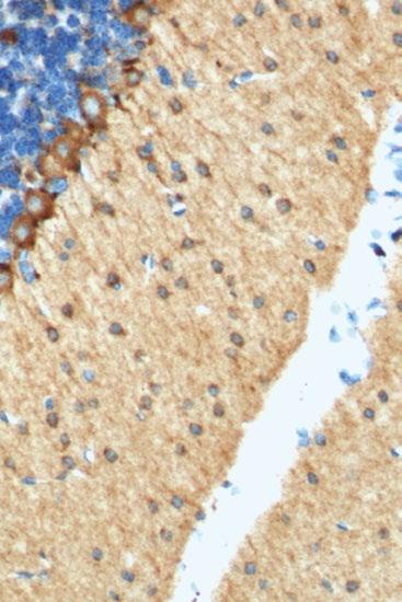
11 ptglab.com 11 Advantage Disadvantage Difficulties ph* Heat-induced Smooth epitope recovery No impact on cell morphology Unequal retrieval due to unequal heating Typically ph 6 (citrate), ph 9 (Tris-EDTA) Proteolytic-induced Preferred for difficult-torecover epitopes Impacts and damages, also a harsh method Concentration calibration Typically ph 7.4 Incubation time* Around 20 mins Around 10 mins Temperature* Around 100ºC Around 37ºC *Optimal conditions always have to be determined by each laboratory and in accordance with the specific product information. Comparison of heat-induced epitope retrieval (HIER) and proteolytic-induced epitope retrieval (PIER) No Retrieval Tris-EDTA Sodium Citrate Antigen retrieval optimization of CD3 gamma antibody ( AP) on paraffin-embedded tonsillitis tissue slides.
12 12 How To Optimize Your IHC Experiment BLOCKING Blocking is essential for preventing non-specific binding of antibodies or other reagents to the tissue. Even if the antibody has high specificity towards the target, intermolecular forces can promote non-specific binding to other molecules. Consequently, non-specific binding prevents visualization of the antigen-antibody binding of interest. To mitigate nonspecific binding, a blocking step should be carried out before incubation with the primary antibody. Different commercial buffer systems are available. Protein Blocking In general, serum (same species as secondary antibody) or bovine serum albumin (BSA) are used for blocking. Sera and BSA can help to prevent unspecific binding to the many hydrophobic side chains of proteins present in tissue. If you are staining with multiple antibodies, you need to use blocking serum against all used secondaries. If BSA is used, the addition of % Trition-X or Tween can help to prevent unspecific binding. Blocking Non-Specific Ionic Bindings Non-specific ionic bindings are due to, for example, Van der Waals interactions, dipole-dipole interactions or net charges of specific amino acid groups. In this case, altering the ionic strength of the antibody dilution buffer can help to reduce unspecific ionic bindings. Endogenous Enzyme Blocking When using a horseradish peroxidase (HRP)- or alkaline phosphatase (AP)-conjugated antibody for detection, the endogenous levels of the enzyme have to be blocked. This normally applies for tissues such as kidney, liver, intestine, lymphoide tissue, etc. Peroxidase can be blocked with buffers containing H₂O₂ and AP can be blocked with buffers containing acetic acid or Levamisole. Endogenous Biotin Blocking Endogenous biotin is found to be especially high in tissue of the liver, kidney, or brain. Blocking is necessary if working with an avidin-biotin detection system. A direct incubation with the ABC complex or streptavidin-hrp and then DAB is also possible.
13 ptglab.com 13 FEATURED PRODUCT IHC Detection System, Peroxidase/DAB+, Rabbit/Mouse (KIHC-1, KIHC-5) Two-step immunohistochemical staining technique: This system is based on an HRP labeled polymer that is conjugated to secondary antibodies. All reagents are ready to use, thus making it time- and cost-effective. The secondary antibodies are mixed with anti-mouse IgG and anti-rabbit IgG. This universal reagent can be used to detect any tissue slides bound with primary antibody of mouse or rabbit origin. Main Features Low background Strong staining Easy protocol IHC Kit System 1st Step Legend Tissue Slides Primary Antibody Secondary Antibody HRP 2nd Step Polymer Kit Content HRP-conjugated secondary antibody DAB Substrate Buffer DAB
14 14 How To Optimize Your IHC Experiment IMMUNOSTAINING Selecting And Optimizing Antibodies For IHC When deciding on an antibody, the following points should be considered to increase the likelihood of high specificity and low cross-reaction and a successful staining; further optimization will then help to achieve a reliable signal: Consulting literature and antibody comparison resources, checking in-house validation data of the antibody seller, searching for independent validation data, and consulting the original manufacturer for technical service and protocol support. Primary Antibodies For IHC Selecting a primary antibody for IHC The initial choice of the primary antibody can affect the whole outcome of the experiment. Thus, it is important to search for an antibody that is well validated to increase the chance of a successful experiment. Most importantly, it is helpful to purchase the antibody from the original manufacturer and not from a vendor, as only the original manufacturer has access to all validation data and can assist with superior technical support. Main questions to ask when choosing a primary antibody supplier 1 How many times has the antibody been cited? 2 How has the antibody been validated? 3 Has the application of interest already been tested? 4 Who is the original manufacturer? Is all the data and information about the antibody 5 publicly available? What is the vendor s refund policy, delivery time, 6 and stock availability? 7 What is the price?
15 ptglab.com 15 Polyclonal Vs. Monoclonal Antibody A final question to consider is whether a monoclonal or polyclonal antibody would be most suitable. For IHC, polyclonal antibodies tend to give the best results. Benefits of polyclonal antibodies in IHC Heterogenous population. Recognise multiple epitopes. Stable to changes (ph, tissue, buffer, protein confirmation), stable detection. Drawbacks of polyclonal antibodies in IHC Not specific to one epitope. Benefits of monoclonal antibodies in IHC Homogenous population. Specific to a single epitope. Detects a single protein with high affinity, even though it shares sequences/similarites with other proteins. Drawbacks of monoclonal antibodies in IHC Sensitive to even small changes (ph, tissue, buffer, protein confirmation), no stable detection. Optimizing A Primary Antibody For IHC The optimal conditions for the primary antibody depend on each individual experiment and therefore have to be optimized to gain a staining of high quality. General steps on optimizing conditions for a primary antibody in IHC Keep incubation time and temperature, titrate different antibody dilutions. Specific staining, but background signal: Vary incubation time and temperature. High-affinity antibody with a high concentration: Incubation with a high concentration for a short time. High-affinity antibody with a low concentration: Increase incubation time, lower incubation temperature. Polyclonal antibodies, in general, can be used at a higher working dilution than monoclonal antibodies.
16 16 How To Optimize Your IHC Experiment Selecting And Optimizing Secondary Antibodies For IHC In immunohistochemistry (IHC), it can be beneficial to reduce the signal-to-noise ratio when distinguishing between antibody subclass, purification, and fragments of the secondary antibody. Subclass specificity Polyclonal primary antibodies are mainly IgG isotypes. Primary monoclonal antibodies are occasionally of a different isotype and therefore need an isotype-specific antibody. Cross-absorption Secondary antibodies can go through an additional purification step to reduce potential cross-reactions with other species. Therefore, the secondary antibody solution is passed over different columns containing sera proteins of different species to filter out the non-specific secondary antibody. F(ab )₂ fragments High background staining can be due to the presence of Fc receptors in special tissue or cells (lymph nodes, spleen, macrophages, etc.). A whole antibody can bind to the Fc region, while an F(ab )₂ fragment does not ensure specificity of the antibody binding. Also, tissue penetration is facilitated when working with a smaller secondary antibody fragment and not with the whole antibody. Examples of IgG Fragments Whole Antibody F(ab )₂ Heavy Chain Light Chain

17 ptglab.com 17 IHC Controls It is essential to perform control stainings to ensure that the observed staining pattern is specific and authentic and not due to any, for example, cross-reaction or unspecific binding. Positive controls Control tissue that is known to express the protein of interest will prove that the staining protocol and antibody are working properly. Negative controls Control tissue that is known not to express the protein of interest will ensure that the observed staining pattern is due to specific signals. Incubate the tissue with the secondary antibody only. This control ensures that no cross-reactions or non-specific background signals are observed due to the secondary antibody and the detection reagents. Observing the unstained tissue under the microscope in the brightfield/fluorescence channel gives an idea about biological background signal/autofluorescence that mainly comes from mitochondria, lysosomes, and aromatic amino acid components and could be misinterpreted with positive staining. Incubating the tissue with a non-immune immunoglobulin of the same isotype will ensure that the observed staining is not due to unspecific binding of immunoglobulins. THE BENCHMARK IN ANTIBODIES Antibodies validated with sirna knockdown to demonstrate specificity.
18 18 How To Optimize Your IHC Experiment VISUALIZATION After incubation with the primary antibody, the staining can be visualized using different detection methods. The detection technique can be direct or indirect and the signal generation can be chromogenic or fluorescent. Direct Versus Indirect Detection In direct detection, the primary antibody contains the label. On the other hand, in indirect detection, the secondary is labeled. The choice of detection method depends on the expression level of the antigen. As primary labeled antibodies show a comparable low signal generation, they are suitable for highly expressed antigens. Direct detection is also advantageous for multi-labeling experiments. Medium expressed antigens instead show the best signal when analyzed via a secondary labeled antibody that helps to amplify the signal intensity. Indirect labeling, however, always shows a higher background noise. For very low-expressed proteins the indirect detection plus an enhancer (e.g., avidin, streptavidin) helps to further amplify the signal. This type of detection often needs some additional optimization steps to get the best signal-to-noise ratio. Different Detection Systems and Signal Amplification Expression level of protein of interest High Medium Low Direct Labeled First Antibody Secondary Antibody Labeled Secondary Antibody Labeled Plus Enhancer
19 ptglab.com 19 Chromogenic Versus Fluorescent Signal Generation Signal is generated by fluorophores or enzymes. The detection method and available microscope determine the choice of label. If working with fluorescent labels, the fluorophore can be conjugated either to the primary or secondary antibody. Especially for multi-color staining experiments, fluorescent labels are beneficial for detecting different cellular compartments at the same time. A disadvantage of fluorophores is their short lifetime. Thus, enzyme labels are often the preferred choice for visualization of tissue stainings. If working with enzymatic labels, the enzyme is attached to the primary or secondary antibody and then forms an insoluble colored product when an organic substrate is added. The most commonly used enzyimatic labels are horseradish peroxidase and alkaline phosphatase. Immunohistochemical staining of paraffin-embedded human colon tissue using KLF4 antibody ( AP) at a dilution of 1:50 (40x objective). THE BENCHMARK IN ANTIBODIES Proteintech products now feature in over 20,000 publications worldwide.

20 20 How To Optimize Your IHC Experiment Tips, Tricks, and TROUBLESHOOTING GUIDE No/Weak Staining Potential cause The primary/secondary antibody lost its activity. Conditions of antibody are not optimized. Protein of interest is not expressed in used tissue. Protein of interest is low expressed in used cells. Damaged epitope. Antibody is not suitable for this application. Suggested test or solution Use a new lot of antibody. Improper storage of antibody. Follow manufacturer s instruction. Normally, prepare single-use aliquots and store at -20 C. Extensive thaw-freezing cycles have damaged the antibody. Titrate the antibody concentration to optimize best working conditions. Incubate the primary antibody at room temperature or at 4 C overnight. Run a positive control. Use signal amplification when visualizing. Change to another antigen retrieval buffer/technique for paraffin-embedded samples. Check validation data of manufacturer. REQUEST COPIES OF OTHER PROTEINTECH TECHNICAL GUIDES ONLINE
21 ptglab.com 21 Background Staining/ Non-specific Staining Potential cause Too high primary/secondary antibody concentration. Non-specific binding of primary/ secondary antibodies. Non-specific binding of secondary antibodies. The sample is poorly washed. The antibody incubation temperature/time is not suitable. Damaged epitope. Suggested solution Titration of antibodies to determine optimal working concentration. Prolong blocking step and increase concentration of blocking solution. Run positive and negative controls. Run control with secondary antibody only. Change to a cross-adsorbed secondary antibody or a fragment antibody. Repeat or prolong washing step. Optimize conditions. Change to another antigen retrieval buffer/technique for paraffin-embedded samples. Inappropriate Cell Morphology Potential cause Harsh antigen retrieval conditions Unclear tissue structure Tissue is not adhesive to glass slide. Physically damaged cell shape. Suggested solution Optimize buffers, temperature, ph, incubation time, concentration. Optimize thickness of tissue slides. Cut new sections. Optimize fixation. Decrease heating time or temperature during HIER. Under-fixation. Change fixative or fixation time.
22 22 How To Optimize Your IHC Experiment CONTACT US Proteintech Group US Head Office Phone 1 (888) 4PTGLAB ( ) (toll free in USA), or 1(312) (outside USA) proteintech@ptglab.com Proteintech Europe United Kingdom Phone europe@ptglab.com Proteintech Europe Germany germany@ptglab.com Sales and technical support only. Proteintech China Office Phone or service@ptglab.com We are ISO 9001 and ISO accredited. Support Available 24 hours a day via or 9-5 via phone. You can also speak to a representative at any time via Live chat: Live Chat Blog YouTube
23 ptglab.com 23

24 24 How To Optimize Your IHC Experiment
ab TripleStain IHC Kit: M&M&R on human tissue (DAB, Red/AP & DAB/Ni)
") ab183287 TripleStain IHC Kit: M&M&R on human tissue (DAB, Red/AP & DAB/Ni) Instructions for Use For the detection of Rabbit and Mouse Primary antibodies on Human tissue or cell samples. This product is
ab183287 TripleStain IHC Kit: M&M&R on human tissue (DAB, Red/AP & DAB/Ni) Instructions for Use For the detection of Rabbit and Mouse Primary antibodies on Human tissue or cell samples. This product is
Manufactured by. Zyagen Barnes Canyon Road San Diego, CA 92121, USA
 Alkaline Phosphatase Immunohistochemistry Detection kits For detection of mouse, rabbit, goat, rat, sheep, chicken, guinea pig, and human primary antibodies Size: 500 Tests Catalog #: AK-011, Mouse Kit
Immunohistochemistry. How does it look like? When do we need IHC? When do we need IHC? In clinic: In research:
 Introduction How does it look like? Immunohistochemistry Smooth muscle actin Parvalbumin Distrophyn Sandrine Bichet Head of Molecular Histology Platform Signal versus background 06.03.2012 IHC basics Introduction
ab Mouse and Rabbit Specific HRP/DAB (ABC) Detection IHC Kit
 Detection IHC Kit") ab64264 - Mouse and Rabbit Specific HRP/DAB (ABC) Detection IHC Kit Instructions for Use For the detection of a specific antibody bound to an antigen in tissue sections. This product is for research use
ab64264 - Mouse and Rabbit Specific HRP/DAB (ABC) Detection IHC Kit Instructions for Use For the detection of a specific antibody bound to an antigen in tissue sections. This product is for research use
ab Mouse and Rabbit AP/Fast-Red (ABC) Detection IHC Kit
 Detection IHC Kit") ab128967 - Mouse and Rabbit AP/Fast-Red (ABC) Detection IHC Kit Instructions for Use For the detection of a specific antibody bound to an antigen in tissue sections. This product is for research use only
ab128967 - Mouse and Rabbit AP/Fast-Red (ABC) Detection IHC Kit Instructions for Use For the detection of a specific antibody bound to an antigen in tissue sections. This product is for research use only
Overview of Immunohistochemistry. (with a focus on wax-embedded sections)
") Overview of Immunohistochemistry (with a focus on wax-embedded sections) Overview of Immunohistochemistry (with a focus on wax-embedded sections) Overview of Immunohistochemistry IHC is like cooking. There
Overview of Immunohistochemistry (with a focus on wax-embedded sections) Overview of Immunohistochemistry (with a focus on wax-embedded sections) Overview of Immunohistochemistry IHC is like cooking. There
BrdU IHC Kit. For the detection and localization of bromodeoxyuridine incorporated into newly synthesized DNA of actively proliferating cells
 K-ASSAY BrdU IHC Kit For the detection and localization of bromodeoxyuridine incorporated into newly synthesized DNA of actively proliferating cells Cat. No. KT-077 For Research Use Only. Not for Use in
ab BrdU Immunohistochemistry Kit
 ab125306 - BrdU Immunohistochemistry Kit Instructions for Use For the detection and localization of bromodeoxyuridine incorporated into newly synthesized DNA of actively proliferating cells. This product
Cell & Tissue Staining Kit
 Cell & Tissue Staining Kit For the detection of goat, mouse, rabbit, rat, or sheep primary IgG Antibodies Size: 50 Tests HRP-DAB System Goat Kit (Catalog Number CTS008) Mouse Kit (Catalog Number CTS002)
Combined Digoxigenin-labeled in situ hybridization/ Immunohistochemistry protocol (for fixed frozen cryostat sections)
") Combined Digoxigenin-labeled in situ hybridization/ Immunohistochemistry protocol (for fixed frozen cryostat sections) A. Digoxigenin-UTP labeling of crna antisense probe Refer to laboratory protocol and
Combined Digoxigenin-labeled in situ hybridization/ Immunohistochemistry protocol (for fixed frozen cryostat sections) A. Digoxigenin-UTP labeling of crna antisense probe Refer to laboratory protocol and
ab In situ Apoptosis Detection Kit
 ab206386 In situ Apoptosis Detection Kit Instructions for Use For detection of apoptotic cells. This product is for research use only and is not intended for diagnostic use. Version 8 Last Updated 6 February
Materials and Methods Materials Required for Fixing, Embedding and Sectioning. OCT embedding matrix (Thermo Scientific, LAMB/OCT)
") Page 1 Introduction Tissue freezing and sectioning is a rapid method of generating tissue samples (cryosections) for histological analysis, and obviates the need for wax embedding. The method is popular
Page 1 Introduction Tissue freezing and sectioning is a rapid method of generating tissue samples (cryosections) for histological analysis, and obviates the need for wax embedding. The method is popular
LAMININ. For Immunohistochemical Demonstration of Laminin in Paraffin-embedded and Frozen Human Tissue Sections Stock No. IMMH-7
 LAMININ For Immunohistochemical Demonstration of Laminin in Paraffin-embedded and Frozen Human Tissue Sections Stock No. IMMH-7 TABLE OF CONTENTS BACKGROUND AND PRINCIPLE... 4 REAGENTS AND EQUIPMENT PROVIDED...
Anti-Human IDH1 R132H Astrocytoma, Oligodendroglioma Tumor Cell Marker Mouse Monoclonal Antibody Clone H09
 1 of 5 Technical Note 1 Procedure: Automated Immunostaining Ventana Benchmark XT XT ultraview DAB DIA--M (20µg) Reconstitution: DIA- (100µg), restore to 500µl Summary 1. Cut sections to 4 µm (Microm HM
IMMUNOPRECIPITATION (IP)
") 1 IMMUNOPRECIPITATION (IP) Overview and Technical Tips 2 CONTENTS 3 7 8 9 12 13 17 18 19 20 Introduction Factors Influencing IP General Protocol Modifications Of IP Protocols Troubleshooting Contact Us
1 IMMUNOPRECIPITATION (IP) Overview and Technical Tips 2 CONTENTS 3 7 8 9 12 13 17 18 19 20 Introduction Factors Influencing IP General Protocol Modifications Of IP Protocols Troubleshooting Contact Us
INOS. Colorimetric Cell-Based ELISA Kit. Catalog #: OKAG00807
 INOS Colorimetric Cell-Based ELISA Kit Catalog #: OKAG00807 Please read the provided manual entirely prior to use as suggested experimental protocols may have changed. Research Purposes Only. Not Intended
Phosphohistone-H3 (PHH3)
") Rev. 0.0 v3 Phosphohistone-H3 (PHH3) Rabbit Polyclonal Antibody PRODUCT AVAILABILITY Cat. No. Description 369A-14 0.1 ml, concentrate 369A-15 0.5 ml, concentrate 369A-16 1.0 ml, concentrate 369A-17 1.0
Rev. 0.0 v3 Phosphohistone-H3 (PHH3) Rabbit Polyclonal Antibody PRODUCT AVAILABILITY Cat. No. Description 369A-14 0.1 ml, concentrate 369A-15 0.5 ml, concentrate 369A-16 1.0 ml, concentrate 369A-17 1.0
Anti-White Spot Syndrome Virus (WSSV) monoclonal antibody. Product no: P13
 monoclonal antibody. Product no: P13") Anti-White Spot Syndrome Virus (WSSV) monoclonal antibody Product no: P13 Product Description The monoclonal antibody (Mab) against the Vp28 protein of White Spot Syndrome Virus (WSSV) is specific for
Anti-White Spot Syndrome Virus (WSSV) monoclonal antibody Product no: P13 Product Description The monoclonal antibody (Mab) against the Vp28 protein of White Spot Syndrome Virus (WSSV) is specific for
Immunohistochemistry: Basics and Methods
 Immunohistochemistry: Basics and Methods Bearbeitet von Igor B Buchwalow, Werner Böcker 1st Edition. 2010. Buch. x, 153 S. Hardcover ISBN 978 3 642 04608 7 Format (B x L): 15,5 x 23,5 cm Gewicht: 445 g
EGFR (Phospho-Ser695)
") Assay Biotechnology Company www.assaybiotech.com Tel: 1-877-883-7988 Fax: 1-877-610-9758 EGFR (Phospho-Ser695) Colorimetric Cell-Based ELISA Kit Catalog #: OKAG02090 Please read the provided manual entirely
Assay Biotechnology Company www.assaybiotech.com Tel: 1-877-883-7988 Fax: 1-877-610-9758 EGFR (Phospho-Ser695) Colorimetric Cell-Based ELISA Kit Catalog #: OKAG02090 Please read the provided manual entirely
For research use only. Not for use in diagnostic procedures.
 Dako LSAB 2 System-HRP for use on Rat Specimens Code K0609 Intended use For research use only. Not for use in diagnostic procedures. These instructions apply to the LSAB 2 System-HRP for use on RAT SPECIMENS,
Human IgG Antigen ELISA Kit
 Human IgG Antigen ELISA Kit Catalog No: IHUIGGKT Lot No: SAMPLE INTENDED USE This human immunoglobulin G antigen assay is intended for the quantitative determination of total human IgG antigen in serum,
KPL SignaLOCK ChemiWestern Kits (Film and Imager Analysis)
") KPL SignaLOCK ChemiWestern Kits (Film and Imager Analysis) SignaLOCK HRP ChemiWestern Kit (Film) Catalog No. 54-53-00 SignaLOCK HRP ChemiWestern Kit (Imager) Catalog No. 54-54-00 SignaLOCK AP ChemiWestern
KPL SignaLOCK ChemiWestern Kits (Film and Imager Analysis) SignaLOCK HRP ChemiWestern Kit (Film) Catalog No. 54-53-00 SignaLOCK HRP ChemiWestern Kit (Imager) Catalog No. 54-54-00 SignaLOCK AP ChemiWestern
HAM-56 antibody reacts with monocytes, but is unreactive with B and T-lymphocytes. Principles And Procedures
 Product Identification Cat. No. Description 44682 Macrophage Marker 0,1 M (HAM- 56) 44683 Macrophage Marker 1 M (HAM-56) 44330 Macrophage Marker RTU M (HAM- 56) Symbol Definitions P C A E S DIL DOC# DIS
Pinpoint Slide DNA Isolation System Catalog No. D3001
 INSTRUCTIONS Pinpoint Slide DNA Isolation System Catalog No. D3001 Highlights Easily isolates genomic DNA in any targeted microscopic tissue area on a slide. The simple procedure combines Pinpoint tissue
Mouse ICAM-1 / CD54 ELISA Pair Set
 Mouse ICAM-1 / CD54 ELISA Pair Set Catalog Number : SEK50440 To achieve the best assay results, this manual must be read carefully before using this product and the assay is run as summarized in the General
QImaging Camera Application Notes Multicolor Immunofluorescence Imaging
 QImaging Camera Application Notes Multicolor Immunofluorescence Imaging In order to image localization of intracellular proteins with high specificity, it is frequently necessary to multiplex antibody
In-Gel Western Detection Using Near-Infrared Fluorescence
 In-Gel Western Detection Using Near-Infrared Fluorescence Developed for: Aerius, and Odyssey Family of Imagers Please refer to your manual to confirm that this protocol is appropriate for the applications
IMMUNOPRECIPITATION TROUBLESHOOTING TIPS
 IMMUNOPRECIPITATION TROUBLESHOOTING TIPS Creative Diagnostics Abstract Immunoprecipitation (IP) is the technique of precipitating a protein antigen out of solution using an antibody that specifically binds
Immunohistochemistry: Basics and Methods
 Immunohistochemistry: Basics and Methods Igor B. Buchwalow l Werner Böcker Immunohistochemistry: Basics and Methods Prof. Dr. Igor B. Buchwalow Prof. Dr. Werner Böcker Gerhard-Domagk-Institut für Pathologie
Whole Mount IHC Protocol
 Whole Mount IHC Protocol Authors: Ruth Sullivan, Ryan Trevena and Kyle Wegner Creation Date: 03/17/2016 All steps should be conducted with gentle agitation on an orbital shaker, unless otherwise instructed.
Actin, Muscle Specific (HHF35)
") Product Identification Cat. No. Description 45235 IMPATH Actin Muscle Specific RTU M (HHF35) Symbol Definitions P A E S DOC# DIS ready-to-use ascites serum supernatant document number distributed by Intended
Product Identification Cat. No. Description 45235 IMPATH Actin Muscle Specific RTU M (HHF35) Symbol Definitions P A E S DOC# DIS ready-to-use ascites serum supernatant document number distributed by Intended
UltraMap Alk Phos. UltraMap anti-ms Alk Phos
 UltraMap anti-rb Alk Phos UltraMap anti-ms Alk Phos Biotin-free Alkaline Phosphatase Detection Systems for the Ventana DISCOVERY Series of Instruments Catalog Number: s 760-4314 (UltraMap anti-rb Alk Phos)
PROTOCOL TO PREPARE PLANTAR FOOTSKIN FOR MORPHOMETRY. I. Removal and Fixation of Plantar Skin (see video)
") PROTOCOL TO PREPARE PLANTAR FOOTSKIN FOR MORPHOMETRY I. Removal and Fixation of Plantar Skin (see video) 1. Sacrifice the animal a. Anaesthetize the animal by placing in a closed chamber with isoflurane.
PROTOCOL TO PREPARE PLANTAR FOOTSKIN FOR MORPHOMETRY I. Removal and Fixation of Plantar Skin (see video) 1. Sacrifice the animal a. Anaesthetize the animal by placing in a closed chamber with isoflurane.
Anti-Asian Sea bass (Lates calcarifer) IgM monoclonal antibody labelled with horseradish peroxidase. Product no: C2-HRP
 IgM monoclonal antibody labelled with horseradish peroxidase. Product no: C2-HRP") Anti-Asian Sea bass (Lates calcarifer) IgM monoclonal antibody labelled with horseradish peroxidase Product no: C2-HRP Product Description This monoclonal antibody (Mab) reacts with Asian Sea bass (Lates
Anti-Asian Sea bass (Lates calcarifer) IgM monoclonal antibody labelled with horseradish peroxidase Product no: C2-HRP Product Description This monoclonal antibody (Mab) reacts with Asian Sea bass (Lates
Mouse Monoclonal Antibody Isotyping Reagents
 Mouse Monoclonal Antibody Isotyping Reagents Catalog Number: SEK003 Storage Temperature: 2-8 C Fax : +86-10-58628220 Tel : +86-400-890-9989 http://www.sinobiological.com Description Mouse Monoclonal Antibody
Human IL10RB ELISA Pair Set ( CRFB4 )
") Human IL10RB ELISA Pair Set ( CRFB4 ) Catalog Number : SEK10945 To achieve the best assay results, this manual must be read carefully before using this product and the assay is run as summarized in the
Human IL10RB ELISA Pair Set ( CRFB4 ) Catalog Number : SEK10945 To achieve the best assay results, this manual must be read carefully before using this product and the assay is run as summarized in the
Bovine Prostaglandin E2 (PG-E2) ELISA Kit
 ELISA Kit") Bovine Prostaglandin E2 (PG-E2) ELISA Kit Catalog Number. CSB-E14237B For the quantitative determination of endogenic bovine prostaglandin E2 (PG-E2) concentrations in serum, plasma, tissue homogenates.
Bovine Prostaglandin E2 (PG-E2) ELISA Kit Catalog Number. CSB-E14237B For the quantitative determination of endogenic bovine prostaglandin E2 (PG-E2) concentrations in serum, plasma, tissue homogenates.
1. Cross-linking and cell harvesting
 ChIP is a powerful tool that allows the specific matching of proteins or histone modifications to regions of the genome. Chromatin is isolated and antibodies to the antigen of interest are used to determine
ab VEGF R1 (FLT1) Human ELISA Kit
 Human ELISA Kit") ab119613 VEGF R1 (FLT1) Human ELISA Kit Instructions for Use For quantitative detection of Human soluble VEGF R1 (FLT1) in cell culture supernatants, serum and plasma (EDTA). This product is for research
ab119613 VEGF R1 (FLT1) Human ELISA Kit Instructions for Use For quantitative detection of Human soluble VEGF R1 (FLT1) in cell culture supernatants, serum and plasma (EDTA). This product is for research
Product Information. Before you begin. Component A 1 vial of 30 ul vial of 300 ul each Glycerol. Tris
 Glowing Products for Science Mix-n-Stain Antibody Labeling Kits Size: 1 labeling per kit Storage: -20 o C Stability: Stable for at least 1 year from date of receipt when stored as recommended. Components:
Segments of the obstructed intestinal loops were fixed in 4% paraformaldehyde
 Supplementary text Supplementary materials and methods Histopathological examination Segments of the obstructed intestinal loops were fixed in 4% paraformaldehyde (PFA) and embedded in paraffin wax with
KCC Path-Core Page 1 of 5
 Instructions for Sample preparation for Paraffin embedding PLEASE NOTE: There is no one-size-fits-all method of tissue preparation for all experimental designs. Before harvesting tissue, you need to assess
EZ-10 SPIN COLUMN GENOMIC DNA MINIPREPS KIT HANDBOOK
 EZ-0 SPIN COLUMN GENOMIC DNA MINIPREPS KIT HANDBOOK (Bacteria, Plant, Animal, Blood) Version 8 Rev 05/0/03 EZ-0 Genomic DNA Kit Handbook Table of Contents Introduction Limitations of Use Features Applications
Mouse Factor XII Total ELISA Kit
 Mouse Factor XII Total ELISA Kit Catalog No: IMFXIIKT-TOT Lot No: SAMPLE INTENDED USE This mouse coagulation Factor XII antigen assay is intended for the quantitative determination of total Factor XII
!! PLEASE READ BEFORE USE!!
 In situ Proximity Ligation Assay protocols!! PLEASE READ BEFORE USE!! The test protocol is a guideline, user need to determine their optimal experimental condition for best performance. The following protocol
SANTA CRUZ BIOTECHNOLOGY, INC.
 TECHNICAL SERVICE GUIDE: Western Blotting 2. What size bands were expected and what size bands were detected? 3. Was the blot blank or was a dark background or non-specific bands seen? 4. Did this same
Human IgM Ready-SET-Go!
 PRODUCT INFORMATION & MANUAL Human IgM Ready-SET-Go! 88-50620 Ready-SET-Go! Enzyme-linked Immunosorbent Assay for quantitative detection of human IgM. For research use only. Human IgM Ready-SET-Go! ELISA
Human IgG ELISA Quantitation Set
 Human IgG ELISA Quantitation Set Cat. No. E80-104 Components Supplied Affinity purified Goat anti-human IgG-Fc Coating Antibody A80-104A, 1 ml at 1 mg/ml Human Reference Serum, RS10-110-4, 0.1 ml HRP Conjugated
Updated April 27, PRODUCT INSERT
 Updated April 27, 2009 1 PRODUCT INSERT Hypoxyprobe, Inc. 121 Middlesex Turnpike Burlington, MA 01803 USA www.hypoxyprobe.com Hypoxyprobe Gemini Kit Kit Contents: Solid pimonidazole HCl (Hypoxyprobe -1)
Human alpha-galactosidase A / GLA ELISA Pair Set
 Human alpha-galactosidase A / GLA ELISA Pair Set Catalog Number : SEK12078 To achieve the best assay results, this manual must be read carefully before using this product and the assay is run as summarized
Human connective tissue growth factor (CTGF) ELISA Kit. MyBioSource.com. This package insert must be read in its entirety before using this product.
 ELISA Kit. MyBioSource.com. This package insert must be read in its entirety before using this product.") Human connective tissue growth factor (CTGF) ELISA Kit Catalog Number. For the quantitative determination of human connective tissue growth factor (CTGF) concentrations in serum, plasma, tissue homogenates.
Human connective tissue growth factor (CTGF) ELISA Kit Catalog Number. For the quantitative determination of human connective tissue growth factor (CTGF) concentrations in serum, plasma, tissue homogenates.
Human Granulin / GRN / Progranulin ELISA Pair Set
 Human Granulin / GRN / Progranulin ELISA Pair Set Catalog Number : SEKA10826 To achieve the best assay results, this manual must be read carefully before using this product and the assay is run as summarized
Human Placental Lactogen (hpl) (Polyclonal)
 (Polyclonal)") Product Identification Cat. No. Description 45312 IMPATH Hpl RTU R (Poly) Symbol Definitions P A E S DOC# DIS ready-to-use ascites serum supernatant document number distributed by Intended Use This antibody
Product Identification Cat. No. Description 45312 IMPATH Hpl RTU R (Poly) Symbol Definitions P A E S DOC# DIS ready-to-use ascites serum supernatant document number distributed by Intended Use This antibody
ab66110 In situ BrdU-Red DNA Fragmentation (TUNEL) Assay Kit
 Assay Kit") Version 9b Last updated 28 November 2017 ab66110 In situ BrdU-Red DNA Fragmentation (TUNEL) Assay Kit For the detection of DNA fragmentation in apoptosis by flow cytometry and fluorescence microscopy in
Version 9b Last updated 28 November 2017 ab66110 In situ BrdU-Red DNA Fragmentation (TUNEL) Assay Kit For the detection of DNA fragmentation in apoptosis by flow cytometry and fluorescence microscopy in
celldatasci.com/rnastorm RNAstorm RNA Isolation Kit for FFPE Tissue Samples Sample Kit (20 extractions)
") celldatasci.com/rnastorm info@celldatasci.com RNAstorm RNA Isolation Kit for FFPE Tissue Samples Sample Kit (20 extractions) Support: Email: support@celldatasci.com Phone: 650.285.2376 (option 2) Toll-free:
celldatasci.com/rnastorm info@celldatasci.com RNAstorm RNA Isolation Kit for FFPE Tissue Samples Sample Kit (20 extractions) Support: Email: support@celldatasci.com Phone: 650.285.2376 (option 2) Toll-free:
Mouse TNF alpha ELISA Kit
 Mouse TNF alpha ELISA Kit Catalog No. GWB-ZZD049 Size 96 wells/kit Sandwich ELISA kit for quantitative detection of mouse TNF alpha in cell culture supernates, serum and plasma(heparin, EDTA). Typical
1 MICROTITER PLATE 96 wells 2 ENZYME CONJUGATE 10.0 ml 1 vial 3 STANDARD.1 0 pg/ml 1 vial 4 STANDARD pg/ml 1 vial 5 STANDARD.
 Rabbit Thyrotropin Releasing Hormone (TRH) Elisa Kit 96 Tests Catalog Number: MBS725153 Store all reagents at 2-8 C Valid Period:six months FOR LABORATORY RESEARCH USE ONLY. NOT FOR THERAPEUTIC OR DIAGNOSTIC
Rat IGF-1 ELISA Kit (rigf-1-elisa)
") Rat IGF-1 ELISA Kit (rigf-1-elisa) Cat. No. EK0377 96 Tests in 8 x 12 divisible strips Background Insulin-like growth factor 1 (IGF-1), also known as somatomedin C, is a polypeptide protein hormone similar
Rat IGF-1 ELISA Kit (rigf-1-elisa) Cat. No. EK0377 96 Tests in 8 x 12 divisible strips Background Insulin-like growth factor 1 (IGF-1), also known as somatomedin C, is a polypeptide protein hormone similar
WesternMAX Alkaline Phosphatase Chemiluminescent Detection Kits
 WesternMAX Alkaline Phosphatase Chemiluminescent Detection Kits Code N221-KIT N220-KIT Description WesternMAX Chemiluminescent AP Kit, Anti-Mouse Includes: Alkaline Phosphatase (AP) Conjugated Anti-Mouse
Actin, Smooth Muscle (1A4)
") Product Identification Cat. No. Description 45121 IMPATH Actin Smooth Muscle RTU M (1A4) Symbol Definitions P A E S DOC# DIS ready-to-use ascites serum supernatant document number distributed by Intended
Product Identification Cat. No. Description 45121 IMPATH Actin Smooth Muscle RTU M (1A4) Symbol Definitions P A E S DOC# DIS ready-to-use ascites serum supernatant document number distributed by Intended
Microarray Industry Products
 Via Nicaragua, 12-14 00040 Pomezia (Roma) Phone: +39 06 91601628 Fax: +39 06 91612477 info@lifelinelab.com www.lifelinelab.com Microarray Industry Products Page 10 NBT / BCPIP Chromogenic phosphatase
I n s t ru c t i o n s D AKO LSAB 2 System, P e r o x i d a s e
 I n s t ru c t i o n s D AKO LSAB 2 System, P e r o x i d a s e Universal Code No. K0672 K0673 K0675 Size 15 ml 15 ml 110 ml For Laboratory Use. 1. INTENDED USE FOR LABORATORY USE These instructions apply
PeliClass human IgG subclass ELISA kit Enzyme-linked immunosorbent assay
 PeliClass human IgG subclass ELISA kit Enzyme-linked immunosorbent assay Catalog No: M1551 Size: six pre-coated 8-well strips for each of the four IgG subclasses Test description The PeliClass human subclass
ab Optiblot Fluorescent Western Blot Kit
 ab133410 Optiblot Fluorescent Western Blot Kit Instructions for Use For quantitative, multi-color fluorescent Western blotting. This product is for research use only and is not intended for diagnostic
Purification Kits. Fast and Convenient PROSEP -A and PROSEP-G Spin Column Kits for Antibody Purification DATA SHEET
 Â Montage Antibody Purification Kits Fast and Convenient PROSEP -A and PROSEP-G Spin Column Kits for Antibody Purification DATA SHEET Available with immobilized Protein A or Protein G Easy-to-use Antibody
 Montage Antibody Purification Kits Fast and Convenient PROSEP -A and PROSEP-G Spin Column Kits for Antibody Purification DATA SHEET Available with immobilized Protein A or Protein G Easy-to-use Antibody
PREPARATION OF HISTOLOGICAL SPECIMENS
 PREPARATION OF HISTOLOGICAL SPECIMENS Histo-techniques Preparation of tissue for microscopic examination Series of processes Ultimate aim to make tissue visible as it is Pathology Vs Anatomy Steps vary
in-situ PCR Presented for: Presented by: Date:
 in-situ PCR Presented for: Presented by: Date: 2 in situ Hybridization - Definition in situ PCR is a method in which the polymerase chain reaction actually takes place in the cell on a slide, and the product
COLORIMETRIC SANDWICH ELISA KIT INSTRUCTION MANUAL
 Page 1 of 7 COLORIMETRIC SANDWICH ELISA KIT INSTRUCTION MANUAL This product is for research use ONLY and not for human or animal therapeutic or diagnostic use. Page 2 of 7 Contents Page 3 I. Supplied Materials:
Principles And Procedures
 Product Identification Cat. No. Description 44837 Vimentin 0,1 M (V9) 44838 Vimentin 1 M (V9) 44409 Vimentin RTU M (V9) Symbol Definitions P C A E S DIL DOC# DIS ready-to-use concentrate ascites serum
Human Haptoglobin ELISA Quantitation Kit. Manual
 Human Haptoglobin ELISA Quantitation Kit Manual Catalog number: 40-288-20080F For the quantitative determination of human Haptoglobin levels in serum or other biological samples GenWay Biotech, Inc. 6777
Store samples to be assayed within 24 hours at 2-8 C. For long-term storage, aliquot and freeze samples at -20 C. Avoid repeated freeze-thaw cycles.
 Human Retinol Binding Protein 4, RBP4 ELISA Kit Preparation Plate Washing Discard the solution in the plate without touching the side walls. Blot the plate onto paper towels or other absorbent material.
Anti-8-hydroxyguanine antibody (Anti-8-oxo-dG)
") Anti-8-hydroxyguanine antibody (Anti-8-oxo-dG) Monoclonal Antibody for Detection of 8-oxo-dG Cat# 4359-MC-100 v.4359/0511a Anti-8-hydroxyguanine antibody (Anti-8-oxo-dG) Cat# 4359-MC-100 Monoclonal Antibody
Anti-8-hydroxyguanine antibody (Anti-8-oxo-dG) Monoclonal Antibody for Detection of 8-oxo-dG Cat# 4359-MC-100 v.4359/0511a Anti-8-hydroxyguanine antibody (Anti-8-oxo-dG) Cat# 4359-MC-100 Monoclonal Antibody
SAMPLE LITERATURE Please refer to included weblink for correct version.
 REVISED & UPDATED Edvo-Kit #269 Introduction to ELISA Reactions Experiment Objective: This experiment introduces concepts and methodologies of enzyme-linked immunosorbent assays (ELISA). See page 3 for
Mouse Luteinizing Hormone (LH) ELISA
 ELISA") Mouse Luteinizing Hormone (LH) ELISA For the quantitative determination of mouse LH in serum, plasma and tissue homogenates Cat. No. KU-222 For Research Use Only. Not for use in diagnostic procedures.
Mouse Luteinizing Hormone (LH) ELISA For the quantitative determination of mouse LH in serum, plasma and tissue homogenates Cat. No. KU-222 For Research Use Only. Not for use in diagnostic procedures.
Human Myostatin, ELISA Kit (MSTN)
") Human Myostatin, ELISA Kit (MSTN) 96 Tests Catalog Number: MBS733837 Store all reagents at 2-8 C Valid Period: six months For samples: Cell culture fluid & body fluid & tissue homogenate Serum or blood
Human Myostatin, ELISA Kit (MSTN) 96 Tests Catalog Number: MBS733837 Store all reagents at 2-8 C Valid Period: six months For samples: Cell culture fluid & body fluid & tissue homogenate Serum or blood
Mouse Peptide YY (PYY) ELISA
 ELISA") Mouse Peptide YY (PYY) ELISA For the quantitative determination of mouse PYY in serum, plasma, cell culture fluid and other biological fluids Cat. No. KT-58705 For Research Use Only. Not for use in diagnostic
Mouse Peptide YY (PYY) ELISA For the quantitative determination of mouse PYY in serum, plasma, cell culture fluid and other biological fluids Cat. No. KT-58705 For Research Use Only. Not for use in diagnostic
One-Step Western TM Kit using TMB
 Technical Manual No. 0203 Version 03272008 I Description.. 1 II Kit Contents.. 2 III Applications 3 IV Key Features.. 3 V Storage.. 3 VI One-Step Western TM Protocol. 3 VII Examples. 3 VIII Troubleshooting..
USER MANUAL. Fluorescence
 USER MANUAL Fluorescence The protocols in this manual are compatible with all Duolink II PLA probes, Duolink II Detection Reagents Green (art no 92014), Orange (art no. 92007), Red (art no. 92008) and
Anti-8-hydroxyguanine antibody (Anti-8-oxo-dG) Cat #: 4355-MC-100 Monoclonal Antibody for Detection of 8-oxo-dG. Contents
 Cat #: 4355-MC-100 Monoclonal Antibody for Detection of 8-oxo-dG. Contents") Instructions for Use For Research Use Only Anti-8-hydroxyguanine antibody (Anti-8-oxo-dG) Cat #: Monoclonal Antibody for Detection of 8-oxo-dG Contents Section Page I Introduction 2 II Reagents Required
Instructions for Use For Research Use Only Anti-8-hydroxyguanine antibody (Anti-8-oxo-dG) Cat #: Monoclonal Antibody for Detection of 8-oxo-dG Contents Section Page I Introduction 2 II Reagents Required
The Biotechnology Education Company. Quantitative ELISA. Storage: See Page 3 for specific storage instructions EXPERIMENT OBJECTIVE:
 The Biotechnology Education Company Revised and Updated Quantitative ELISA Storage: See Page 3 for specific storage instructions EXPERIMENT OBJECTIVE: EDVO-Kit # 278 The objective of this experiment is
RayBio Phospho- Akt (Ser473) ELISA Kit
 ELISA Kit") RayBio Phospho- Akt (Ser473) ELISA Kit For Measuring Phosphorylated Akt (Ser473) in Human, Mouse and Rat Cell Lysates User Manual (Revised Mar 1, 2012) RayBio Akt (Ser473) ELISA Kit Protocol (Cat#: PEL-Akt-S473-001)
RayBio Phospho- Akt (Ser473) ELISA Kit For Measuring Phosphorylated Akt (Ser473) in Human, Mouse and Rat Cell Lysates User Manual (Revised Mar 1, 2012) RayBio Akt (Ser473) ELISA Kit Protocol (Cat#: PEL-Akt-S473-001)
RayBio Phospho- Stat 3 (Tyr705) ELISA Kit
 ELISA Kit") RayBio Phospho- Stat 3 (Tyr705) ELISA Kit For Measuring Phosphorylated Stat3 (Tyr705) in Human, Mouse and Rat Cell Lysates User Manual (Revised Mar 1, 2012) RayBio Stat3 (Tyr705) ELISA Kit Protocol (Cat#:
RayBio Phospho- Stat 3 (Tyr705) ELISA Kit For Measuring Phosphorylated Stat3 (Tyr705) in Human, Mouse and Rat Cell Lysates User Manual (Revised Mar 1, 2012) RayBio Stat3 (Tyr705) ELISA Kit Protocol (Cat#:
Human Angiotensin 2 (Ang2) ELISA
 ELISA") Human Angiotensin 2 (Ang2) ELISA For the quantitative determination of human Ang2 in serum, plasma, cell culture fluid and other biological fluids Cat. No. KT-52748 For Research Use Only. Not for use in
Human Angiotensin 2 (Ang2) ELISA For the quantitative determination of human Ang2 in serum, plasma, cell culture fluid and other biological fluids Cat. No. KT-52748 For Research Use Only. Not for use in
ab Albumin Human ELISA Kit
 ab108787 Albumin Human ELISA Kit Instructions for Use For the quantitative measurement of Human Albumin in plasma and serum. This product is for research use only and is not intended for diagnostic use.
Mouse Gonadotropin Releasing Hormone (GnRH) ELISA
 ELISA") KAMIYA BIOMEDICAL COMPANY Mouse Gonadotropin Releasing Hormone (GnRH) ELISA For the quantitative determination of mouse GnRH in serum, plasma, cell culture fluid and other biological fluids Cat. No. KT-58182
KAMIYA BIOMEDICAL COMPANY Mouse Gonadotropin Releasing Hormone (GnRH) ELISA For the quantitative determination of mouse GnRH in serum, plasma, cell culture fluid and other biological fluids Cat. No. KT-58182
ab HO-1 Human ELISA development set
 ab133058 HO-1 Human ELISA development set Instructions for Use For the quantitative measurement of HO-1 concentrations in cell lysates and microsomes. This product is for research use only and is not intended
TACS XL TM. In Situ Apoptosis Detection Kit. Catalog Number: TA200. DAB Kit. 30 tests FOR RESEARCH USE ONLY. NOT FOR USE IN DIAGNOSTIC PROCEDURES.
 TACS XL TM In Situ Apoptosis Detection Kit Catalog Number: TA200 DAB Kit 30 tests This package insert must be read in its entirety before using this product. FOR RESEARCH USE ONLY. NOT FOR USE IN DIAGNOSTIC
Human Collagen Type III (COL3) ELISA
 ELISA") Human Collagen Type III (COL3) ELISA For the quantitative determination of human COL3 in serum, plasma, cell culture fluid and other biological fluids Cat. No. KT-61018 For Research Use Only. Not for use
Human Collagen Type III (COL3) ELISA For the quantitative determination of human COL3 in serum, plasma, cell culture fluid and other biological fluids Cat. No. KT-61018 For Research Use Only. Not for use
Human IL-6 ELISA Set
 Human IL-6 ELISA Set Catalog No. CDK082B Quantity: 10 x 96 tests PRODUCT SPECIFICATIONS : Specificity: Recognizes both natural and recombinant human IL-6 Range: 6.25 pg / ml - 200 pg / ml Sensitivity:
Rat α-melanocyte stimulating hormone (α-msh) ELISA Kit
 ELISA Kit") Rat α-melanocyte stimulating hormone (α-msh) ELISA Kit For the quantitative determination of rat α-melanocyte stimulating hormone (α-msh) concentrations in serum, plasma, tissue homogenates. This package
Rat α-melanocyte stimulating hormone (α-msh) ELISA Kit For the quantitative determination of rat α-melanocyte stimulating hormone (α-msh) concentrations in serum, plasma, tissue homogenates. This package
ab PD-L1 RabMAb antibody (clone 28-8) Protocol Booklet For Immunohistochemistry, Western Blot and Flow Cytometry Applications.
 Protocol Booklet For Immunohistochemistry, Western Blot and Flow Cytometry Applications.") ab205921 PD-L1 RabMAb antibody (clone 28-8) Protocol Booklet For Immunohistochemistry, Western Blot and Flow Cytometry Applications. This product is for research use only and is not intended for diagnostic
ab205921 PD-L1 RabMAb antibody (clone 28-8) Protocol Booklet For Immunohistochemistry, Western Blot and Flow Cytometry Applications. This product is for research use only and is not intended for diagnostic
Orexin A (HUMAN, MOUSE, RAT, PORCINE, OVINE,
 Orexin A (HUMAN, MOUSE, RAT, PORCINE, OVINE, BOVINE) Western Blot Kit Protocol (Catalog #WBK-003-30) PHOENIX PHARMACEUTICALS, INC. TABLE OF CONTENTS 1. Kit Contents...2 2. Storage...2 3. Introduction...3
Sandwich High Sensitivity ELISA kit for Quantitative Detection of Human VEGF in cell culture supernates, serum, and plasma (heparin, EDTA, citrate).
.") GenWay Biotech, Inc. 6777 Nancy Ridge Drive San Diego, CA 92121 Phone: 858.458.0866 Fax: 858.458.0833 Email: techline@genwaybio.com http://www.genwaybio.com Human VEGF ELISA Kit Catalog Number: Size: GWB-ZZK029
GenWay Biotech, Inc. 6777 Nancy Ridge Drive San Diego, CA 92121 Phone: 858.458.0866 Fax: 858.458.0833 Email: techline@genwaybio.com http://www.genwaybio.com Human VEGF ELISA Kit Catalog Number: Size: GWB-ZZK029
ImmuLux Human IL-6 Fluorescent ELISA Kit
 INSTRUCTIONS ImmuLux Human IL- Fluorescent ELISA Kit 3747 N. Meridian Road P.O. Box 7 Rockford, IL 05 840 53w Product Description Number Description 840 ImmuLux Human IL- Fluorescent ELISA Kit The Pierce
Contents Introduction... 3 General ELISA Procedure... 3 ELISA Types*... 4
 Contents Introduction... 3 General ELISA Procedure... 3 ELISA Types*... 4 1. Direct ELISA... 5 2. Indirect ELISA... 5 3. Sandwich ELISA... 6 4. Competitive ELISA... 6 *For Cell-based ELISA principles and
Human VEGF-C ELISA. For the precise measurement of VEGF-C in human serum, plasma, body fluids, tissue homogenate or cell culture supernates.
 Product information User s Manual Human VEGF-C ELISA For the precise measurement of VEGF-C in human serum, plasma, body fluids, tissue homogenate or cell culture supernates. BE69217 Storage: 96 2-8 C RUO
In-Cell Western Kits I and II
 Odyssey and Aerius Infrared Imaging Systems In-Cell Western Assay Kits I and II Published November, 2006. The most recent version of this protocol is posted at http://biosupport.licor.com/protocols.jsp
Anti- Tissue Transglutaminase TTG IgG ELISA Kit
 Anti- Tissue Transglutaminase TTG IgG ELISA Kit Catalog number: NR-R10142 (96 wells) The kit is designed to quantitatively detect TTG IgG in Human serum or plasma. FOR RESEARCH USE ONLY. NOT FOR DIAGNOSTIC
ab Human TNFSF11 ELISA Kit (RANKL)
") Version 1 Last updated 24 March 2017 ab213841 Human TNFSF11 ELISA Kit (RANKL) For the quantitative detection of Human TNFSF11 in cell culture supernatants, cell lysates, tissue homogenates, serum and plasma
Version 1 Last updated 24 March 2017 ab213841 Human TNFSF11 ELISA Kit (RANKL) For the quantitative detection of Human TNFSF11 in cell culture supernatants, cell lysates, tissue homogenates, serum and plasma