Designing and Validating a Multicolor Flow Cytometry Assay. Brent Wood MD PhD Department of Laboratory Medicine University of Washington
|
|
|
- Gary Richard
- 6 years ago
- Views:
Transcription
1 Designing and Validating a Multicolor Flow Cytometry Assay Brent Wood MD PhD Department of Laboratory Medicine University of Washington
2 Specimen Handling Sample Requirements 5 ml Peripheral blood (EDTA, Heparin, ACD) 1-2 ml Bone marrow aspirate (EDTA, Heparin) Ideally 1 st pull to limit hemodilution Body Fluids Tissues in RPMI (1 cm 3 ) Bone marrow biopsy Lymph node biopsies Tissue biopsies (GI, Skin, etc.)
3 Specimen Transport Rapid delivery to lab is important Assays require viable cells Deterioration is significant after ~48 hours. Cells more stable in heparin than EDTA Heparin commonly used for reference testing Refrigeration retards degradation Heating/Cooling cycles probably not good Useful for storage when transport is delayed Prolonged transport leads to poor viability


4 Specimen Transport Australian COG B-ALL MRD sample - 4 days old Seattle MRD = 0.015% Local MRD = 0.13% HP


5 Transfix Myelomonocyt ic Myelomonocyt ic Myelomonocyt ic CD14 PE-Cy55 CD14 PE-Cy55 CD14 PE-Cy55 CD16 APC-A700 CD16 APC-A700 CD16 APC-A700 Day 0 Day 3 Day 3 Transfix RT Myelomonocyt ic Myelomonocyt ic Myelomonocyt ic CD14 PE-Cy55 CD14 PE-Cy55 CD14 PE-Cy55 CD16 APC-A700 CD16 APC-A700 CD16 APC-A700 Day 0 Day 4 Day 4 - Transfix 4C
6 Principle Specimen Processing Assay as rapidly as possible with minimal manipulation and stabilize early Method Stain / lyse / wash NH 4 Cl % formaldehyde FACSlyse (some loss of compromised cells) Versalyse + formaldehyde Pre-lysis or Bulk lysis Facilitates plasma removal Used to concentrate cells Cell recovery reduced Activates some cell populations (monocytes)
7 Assay Design and Validation Purpose Why? Design What? Verification What? Validation - What? Implementation How? Documentation If it isn t documented, it didn t happen
8 Assay Life Cycle Design Validation Plan Execute Validation Failure Validation Report Implementation Plan Yes Revision? No Retirement
9 Define Purpose of Assay Most important question What information is required? What information is optional? What information is most important? Prioritize Compromises are inevitable Simplest assay is best Less likely to fail Easier to maintain
10 Assay Design Target Identification Population(s) Minimal antigens required to obtain information Redundancy? Fluorochrome matching Intensity and background Instrument capabilities Intrinsic expression level of target Reagent availability Reagent performance Fluorochrome performance Conjugate performance Antibody titration Compensation effects Specimen processing Lyse/stain vs. Stain/lyse vs. No lyse Wash vs. No wash Verification Iterative process
11 Example - Progenitor evaluation Objective Evaluate novel antigen expression on progenitor subpopulations of defined lineages Antigens to be evaluated: Progenitor gating - CD45 Early progenitors - CD34, CD117, CD38 Erythroid - CD71 B cell - CD19 pdcs, basophils - CD123, HLA-DR Myelomonocytic - CD15, HLA-DR Test reagents - CD45RA, CD133, CD7, etc.
12 Possibilities PB / V450 DR CD15 CD19 CD123 CD117 CD38 CD34 CD71 CD45 X FITC PE PE-TR P-X PE- Cy7 A594 APC APC- A700 APC- X7
13 Possibilities - Availability PB / V450 DR CD15 CD19 CD123 CD117 CD38 CD34 CD71 CD45 X FITC PE PE-TR P-X PE- Cy7 A594 APC APC- A700 APC- X7
14 Fluorochrome Matching Match level of expression with fluorochrome intensity Bright expression = Dim fluorochrome Dim expression = Bright fluorochrome Note increased background from other fluorochromes

15 Fluorochrome Intensity Small molecule Phycobiliproteins and tandems CD4 FITC = Green PE = Dark Blue PE-TR = Light blue PerCP-Cy5.5 = Magenta PE-Cy7 = Orange Pacific Blue = Red A594 = Yellow green APC = Light green A700 = Blue APC-Cy7 = Purple
16 CD34
17 Surface Antigen Titration 2 ul + 5 ul + 10 ul Titer for signal to noise Saturation desirable Use same total volume as assay Use 5 ul


18 Cytoplasmic Antigen Titration 10 ul, 5 ul, 2 ul, 1ul of neat Titer for signal to noise Not saturation Particularly important for cytoplasmic antigens 10 ul, 5 ul, 2 ul, 1ul of 1:10 Use 5 ul of 1:10 Use 10 ul of 1:10
19 Possibilities PB / V450 DR CD15 CD19 CD123 CD117 CD38 CD34 CD71 CD45 X FITC PE PE-TR P-X PE- Cy7 A594 APC APC- A700 APC- X7
20 Compensation Spectral overlap between fluorochromes Critical to success of method For 10 color experiment Need to determine 90 values for Comp Matrix Software compensation required Maximum flexibility Non-destructive
21 FITC = Green PE = Orange Excitation = Dotted Emission = Solid
22 Compensation - Method Single stained controls used One for each individual fluorochrome One for each individual tandem As bright as brightest reagent to be used Samples run without compensation Compensation calculated in software Applied either at acquisition or analysis
23 Compensation Correct Undercompensated Overcompensated
24 Compensation
25 Compensation

26 Compensation Don t worry unduly about PMT voltage 245% 103% 47.5% 23.0% 12.2% 1.9% 4.7% 10.3% 21.0% 41.0% PE = 400 volts PE-TR = 550 volts 450 volts 500 volts 550 volts 600 volts Compensation values should reflect relative spectral overlap, i.e. detector gains should be equal

27 Compensation Validation Fluorescence minus one (FMO) control Removal of one reagent from the antibody combination

28 Compensation Validation Fluorescence minus one FMO control for PE Intensity of PE signal should be reduced to background
29 Compensation Background Avoid increased background due to fluorochromes Adjacent with longer wavelength emission PE / PE-TR, PE-TR / PE-Cy5, PE-Cy5.5 or PerCP-Cy5.5/ PE-Cy7 APC / APC-A700, APC-A700 / APC-Cy7 Primary fluorochrome of tandem PE and PE-TR, PE-Cy5, PE-Cy5.5, or PE-Cy7 APC and APC-A700 or APC-Cy7 Interlaser excitation and emission PE-Cy5 and APC PE-Cy5.5 or PerCP-Cy5.5 and APC-A700 PE-Cy7 and APC-Cy7 PE-TR and A594

30 Adjacent fluorochromes Fluorochromes cause increased background that is dependent on emission spectra and signal intensity

31 Primary of Tandems Antibody Aggregates 10.5% 1.6% Tandem leakage causes increased background

32 Interlaser compensation Uncompensated Compensated Interlaser compensation is dependent on excitation and emission spectra of fluorochrome
33 Strategies to deal with compensation background Avoid detection of dim expression in presence of high background Avoid bright fluorescence Put fluorochromes on different populations Put fluorochromes brightly on same population
34 Avoid bright fluorescence
35 Different populations
36 Bright dual positive
37 Possibilities PB / V450 DR CD15 CD19 CD123 CD117 CD38 CD34 CD71 CD45 X FITC PE PE-TR P-X PE- Cy7 A594 APC APC- A700 APC- X7
38 Possibilities PB / V450 DR CD15 CD19 CD123 CD117 CD38 CD34 CD71 CD45 X FITC PE PE-TR PECy5 PE- Cy7 A594 APC APC- A700 APC- X7
39 Final PB / V450 DR CD15 CD19 CD123 CD117 CD38 CD34 CD71 CD45 X FITC PE PE-TR PECy5 PE- Cy7 A594 APC APC- A700 APC- X7
40 Compromises PB / V450 DR CD15 CD19 CD123 CD117 CD38 CD34 CD71 CD45 X FITC PE PE-TR PECy5 PE- Cy7 A594 APC APC- A700 APC- X7

41 Compensation Compromises
42 PE background
43 Verification Fluorescence minus one controls Reveal antibody interactions Avoid IgG2 class unless plasma removed Understand background Troubleshooting Run samples Under same conditions as to be used Representative samples Positive and negative controls
44 Validation Objective Document, through the use of specific laboratory investigations, that the performance characteristics of a method are suitable and reliable for the intended analytical applications. Two main components Validation Plan Define validation approach and methods Define acceptance criteria Review and sign prior to execution Validation Report Assess if acceptance criteria met Identify deviations in performance Review and sign at completion
45 Categories of Methods Category Quantitative Relative Quantitative Quasi-Quantitative Qualitative Definition Calibration standard, Reference material Calibration standard, Reference material not representative No calibration standard, continuous numeric data Lacks proportionality, categorical data reported Flow cytometry Quantitative (MESF) = Relative quantitative Cell enumeration (CD34, T cells subsets, MRD) = Quasi-quantitative Immunophenotyping = Qualitative Wood B, et al (2013) Validation of Cell-based Fluorescence Assays: Practice Guidelines from the ICSH and ICCS - Part V - Assay performance criteria. Cytometry Part B 84B:
46 Validation Plan Methodology Accuracy Specificity Sensitivity Limit of Detection (LOD) Limit of Quantitation (LOQ) Linearity Imprecision Within Run Between Run Intermediate (analysts, equipment) Carryover Robustness
47 Accuracy
48 Quasi-Quantitative Methods Accuracy Assay average vs. reference value No reference material for cell-based assays Cannot determine accuracy Surrogates Reference method Rare Differences may reflect methodologies Inter-laboratory comparison Samples from patients with known condition Assay must be uniquely diagnostic Recommendation Minimum 10 samples, 90% concordance

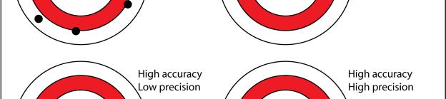
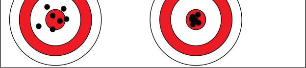
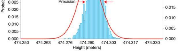
49 Accuracy vs. Precision
50 Quasi-Quantitative Methods Imprecision Intra-assay Use sample of same composition as to be assayed Imprecision related to number of events % CV = SQRT(N) / N, where N = number of events Recommendation Inter-assay 5 samples run in triplicate on same run Span range for analytical decisions Use % CV as the acceptance criterion» Target of 10% good for many assays Sample stability is major issue May use stabilized control material, if available and appropriate May perform on same day if instrument powered down Recommendation 2-3 levels of material assayed in triplicate over 3-5 runs
51 Quasi-Quantitative Methods Specificity Analytical Reagent specificity Provided by manufacturer Based on Leukocyte Antigen Differentiation Workshop Lab developed reagents require supporting data Gating specificity Clinical Distinguish population of interest Correlation with clinical situation of interest and not others Recommendation Assay material containing population of interest and other populations expected to be in sample
52 Quasi-Quantitative Methods Sensitivity Analytical (LOD, LOB) Limit of Blank = Highest signal in absence of measurand Mean (blank) SD (blank) Limit of Detection = 95% of signal above LOB LOB SD (low positive) Functional (LLOQ) Lower Limit of Quantitation Principle LOD with total error (bias + imprecision) meeting clinical criteria May >= LOD, but never lower Replicate assay of samples with dim fluorescence of low population frequency
53 Sensitivity
54 Quasi-Quantitative Methods Sensitivity (analytical) Ideal (soluble analytes) 100 low positive and 60 negative samples X samples assayed X times over X days, X = # samples / 10 Not realistic due to sample availability and reagent cost Recommendation Listmode file consists of many individual measurements 5 low positive and 5 negative samples run in 5 replicates Assay over minimum of 3 days Effect of daily QC and instrument start up LOB confirmed if < 5% exceed target LOD confirmed if < 5% below target
55 Quasi-Quantitative Methods Sensitivity (analytical) Alternate methods for LOB FMO control Does not take into account non-specific reagent binding Isotype control Accounts for isotype-mediated non-specific binding Must be carefully matched to binding characteristics of reagent Known negative populations Must be matched for autofluorescence and background binding Alternate method for LOD Each file consists of numerous measurements Minimum 1 low positive sample + 1 negative sample Only relevant for antigen intensity measurements 5 samples of each type will improve confidence
56 Quasi-Quantitative Methods Sensitivity (clinical) Definitions (LLOQ = bias + imprecision) Bias = difference of mean from true value Total error = Bias + 2SD For cell assays assume Bias = 0 and use %CV Ideal 40 replicates of 3-5 samples over 3 days Recommendation Assay 5 replicates near LLOQ Acceptable if imprecision meets acceptance criterion Serial dilution to create samples with low population frequency near LLOQ Dilution with unlabeled antibody to create low level intensity
57 Linearity
58 Quasi-Quantitative Methods Linearity Not directly applicable for quasi-quantitative assays Exceptions Enumeration of population frequency Quantitation of fluorescence intensity Recommendations Assess instrument linearity semi-annually Enumeration of population frequency Serial dilution of positive sample into negative background Can be performed as part of LLOQ experiment Quantitation of fluorescence intensity Serial dilution with increasing unlabeled antibody Can use calibrated fluorescence beads Must confirm equivalence of emission spectrum and environmental influences
59 Quasi-Quantitative Methods Stability Determine stability of fresh specimen, processed specimen, reagents Recommendations Fresh specimen stability 5 healthy or disease state specimens Assay at baseline and at intervals to desired stability limit Must perform for each processing or storage condition Accept < 20% variation from baseline or 80% within assay imprecision Processed specimen stability Same as fresh specimens Reagent stability Stability data for at least 3 lots of reagent Inter-lot CVs < 10% Validate under conditions to be used
60 Quasi-Quantitative Methods Carryover More an instrument rather than assay performance issue Recommendation Sequentially measure specimens in a low-high-low sequence Beads can substitute in part for cells, but may not be as sticky Sporadic release of accumulated cellular material very difficult to assess Consistent acquisition procedures best prevention Run water/buffer between tubes and samples until background is low Periodic use of cleaning solutions, e.g. bleach, detergent, etc.
61 Quasi-Quantitative Methods Robustness A measure of the assay capacity to remain unaffected by small but deliberate changes in test conditions. Robustness provides an indication of the ability of the assay to perform under normal usage. Robustness measures the effect of deliberate changes (incubation time, temperature, sample preparation, buffer ph) that can be controlled through specifications in the assay protocol. Recommendation No recommendation Often not assessed Ruggedness The reproducibility of the assay under a variety of normal, but variable, test conditions. Variable conditions might include different machines, operators, and reagent lots. Ruggedness provides an estimate of experimental reproducibility with unavoidable error. Also called Intermediate Imprecision when within laboratory.
62 Example AML MRD Assay Three tube assay for AML MRD detection Reference method = Serial dilution and Clinical outcome Sensitivity + Imprecision + Linearity Serial dilution of 3 samples (2 BM, 1 PB): 0%, 0.01%, 0.1%, 1% Each positive run in triplicate Accuracy Serial dilution of known leukemia, see above. Clinical outcome Specificity 3 negative normal marrows + 3 MRD positive samples Stability Same as those used for sensitivity assessment 3 samples run at baseline and daily for 3 days
63 Results AML MRD Specificity 3 MRD negative marrows confirmed assay negative 3 MRD positive marrows confirmed assay positive Sensitivity LOB = 0%, no false positives (0.009% LAIP) LOD = 0.003% LLOQ = LOD Imprecision Within run % CV = 3.5 at 1%, 6.6% at 0.1%, 6.4% at 0.01% Between run % CV = 3.7 at 1%, 6.9% at 0.1%, 4.2% at 0.01% Linearity Mean R 2 = , Slope mean = Accuracy Concordance on 100%. Stability 15% deviation after 4 days, 2 days judged to be stability limit
64 Example AML CD123 Assay Two tube assay for AML CD123 quantitation + Quantibrite beads Reference method = None Accuracy No reference materials Sensitivity + Imprecision (within run) 3 PB + 3 BM AML run in 5 replicates (% and ABC) FMO used to estimate LOB, CD123+ blasts (low) used to estimate LOD Imprecision + Specificity Between run: Immunotrol on 5 sequential days (Lymphs=Neg, Mono=dim, pdc=bright) Linearity Quantibrite beads + CS&T instrument linearity Stability See Imprecision
65 Results AML CD123 Accuracy Not done Specificity Lymphocyte confirmed negative, Monocytes dim positive, pdc bright Sensitivity LOB = 406 ABC BM and 242 PB, Median = 347 LOD = 433 ABC BM and 351 PB, Median = 439 LLOQ = LOD Imprecision CD123 % = 0.58 % CV BM, 0.18 % CV BM CD123 ABC = 1.48 % CV BM, 1.02 % CV BM Linearity Instrument linearity confirmed Stability Confirmed assay stability over 5 days
66 Qualitative Methods Accuracy Correlation with clinical or laboratory findings Recommendation 20 normal and 20 abnormal samples assayed Spiked specimens are acceptable for rare specimen types Specificity Analytical, same as for quasi-quantitative assays Clinical, specificity = TN / (TN + FP) Can determine at same time as accuracy Sensitivity Ability to recognize finding above background Highly variable depending on assay Sensitivity = TP / (TP + FN)
67 Qualitative Methods Imprecision Recommendation Linearity Assay 3 replicates each of positive and negative sample Not relevant Stability Recommendation Concordance on 4 of 5 samples (80%) at each time point or condition
68 Example Mast cell Disease Assay Two tube assay for mast cell disease diagnosis Reference method = morphology + clinical findings Accuracy Run all samples and compare with morphology + clinical findings until 10 true + Specificity Specificity = TN / (TN + FP) Sensitivity Sensitivity = TP / (TP + FN) Precision 5 positive and 5 negative samples run in triplicate Stability 5 positive and 5 negative samples run at baseline and daily for 3 days
69 Example Mast cell Disease Accuracy 108 samples assayed over ~ 9 months, results compared with morphology + clinical findings Specificity + Sensitivity Stability 40% decrease in # mast cells after hours, slight decrease in CD2 & CD25 No increase in CD2 or CD25 on normal mast cells Precision (%CV) Flow + Flow - Mast Cell Disease Mast Cell Disease Positive CD117+ = 11.5%, CD2+ = 15.5%, CD25+ = 2.6% Negative CD117+ = 7.5%, CD2+ = 50.4%, CD25+ = 52.2% Sensitivity 100% Specificity 83% PPV 37% NPV 100%
70 Example Mast cell Disease Mastocytosis (N=10) % Mast cells % CD2+ % CD25+ Range Average % cases Not mastocytosis false positives (N=17) % Mast cells % CD2+ % CD25+ Range Average % cases % + both 6% + both All cases have history of treatment for a hematopoeitic neoplasm
71 Mast Cell Disease Discovered CD2 and CD25 expression not specific Present following therapy for hematopoietic neoplasms Sensitivity and Specificity = 100% Using 10% threshold for positivity Accounting for prior therapy Assessment of CD2 is unnecessary CD25 alone detects all positive cases Mast cells are progressively lost after 48 hours Precision is within expectation for level of expression Cherian, et al (2016) Expression of CD2 and CD25 on mast cell populations can be seen outside the setting of systemic mastocytosis. Cytometry B Clin Cytom. 90(4): doi: /cyto.b.21336
72 Conclusions Assay validation is critical Assay validation requires careful experimental design Assay validation in flow cytometry is often inadequate Laboratories with insufficient resources to validate an assay should not perform it
73 Example B-ALL MRD Assay Single tube assay for B-ALL MRD detection Reference method = COG assay Accuracy 5 positive and 5 negative MRD samples sent to Johns Hopkins University Specificity 10 negative normal pediatric marrows + 10 MRD positive samples 5 negatives were those used for accuracy assessment Sensitivity + Imprecision + Linearity Serial dilution of 5 samples: 0%, 0.01%, 0.1%, 1% Each run in triplicate on 5 different days Stability 1 sample run at baseline and daily for 5 days
74 Results B-ALL MRD Accuracy Concordance on 90% (1 false positive) Specificity 10 MRD negative marrows confirmed assay negative 10 MRD positive marrows confirmed assay positive Sensitivity LOB = 0%, no false positives LOD = 0.007% LLOQ = LOD Imprecision % CV = 2.0 at 1%, 5.0% at 0.1%, 11.0% at 0.01% Linearity R 2 = Stability 5% deviation after 5 days using WBC denominator 16% deviation after 4 days using mononuclear cell denominator
Quality Control in Flow. Dr David Westerman Head of Haematopathology Peter MacCallum Cancer Centre
 Quality Control in Flow Dr David Westerman Head of Haematopathology Peter MacCallum Cancer Centre Aims Quality Assurance Quality Control Literature In house competencies SHOT DATA 1996-2009 Ref: SHOT Annual
Flow Cytometry in the Diagnosis of Hematopoietic Neoplasia. Brent Wood MD, PhD Professor, Laboratory Medicine University of Washington, Seattle
 Flow Cytometry in the Diagnosis of Hematopoietic Neoplasia Brent Wood MD, PhD Professor, Laboratory Medicine University of Washington, Seattle 1 Flow Cytometer 2 The Power of Flow Cytometry Single cell
Application Note. Assay Portability on the BD FACSVerse System. Summary. Maria Jaimes, Yibing Wang, Catherine McIntyre, and Dev Mittar
 September Assay Portability on the BD FACSVerse System Maria Jaimes, Yibing Wang, Catherine McIntyre, and Dev Mittar Contents Summary Introduction 3 Objective 4 Methods 6 Results Discussion Conclusions
FLOW CYTOMETRY. CyAn ADP. Analyzer
 FLOW CYTOMETRY CyAn ADP Analyzer Experience the Power of the CyAn ADP and its optimal performance The Power of Detection The Power of Speed The Power of Ease The CyAn ADP Analyzer is the next step in Advanced
Visualization of digital data Interpretation of the visual
 Technical aspects of 8-color flow cytometry in the diagnosis and classification of hematopoietic malignancies Tomas Kalina!"#$%&'()*+,&$'+-./(0 *1 (2#34%-.(56(7&1+3+*&/( 8$#94&/(!:&3"(;&
CLEARLLAB LS LYMPHOID SCREEN REAGENT
 CLEARLLAB LS LYMPHOID SCREEN REAGENT CE MARKED ANTIBODY COMBINATION FOR LEUKEMIA / LYMPHOMA ANALYSIS Because Your Patient is Her Everything BECAUSE YOUR PATIENT IS HER EVERYTHING ClearLLab LS Lymphoid
Flow Cytometry - The Essentials
 Flow Cytometry - The Essentials Pocket Guide to Flow Cytometry: 1. Know your Cytometer 2. Understanding Fluorescence and Fluorophores 3. Gating Process 4. Controls 5. Optimization 6. Panel Building 7.
Flow cytometry has become an increasingly important
 9-Color and 10-Color Flow Cytometry in the Clinical Laboratory Brent Wood, MD, PhD Context. The development of commercial flow cytometers capable of detecting more than 10 simultaneous fluorescent signals
Practical Guidelines and Tips for Sensitive and Accurate Identification of PNH Clones
 Practical Guidelines and Tips for Sensitive and Accurate Identification of PNH Clones AFCG Meeting Nov 28 Dec 1, 2013 Wellington, New Zealand Andrea Illingworth, MS, H(ASCP), QCYM Dahl-Chase Diagnostic
Detecting human circulating endothelial cells using the Attune Acoustic Focusing Cytometer
 APPLICATION NOTE Attune Acoustic Focusing Cytometer Detecting human circulating endothelial cells using the Attune Acoustic Focusing Cytometer Circulating endothelial cells (CECs) are mature cells shed
Identification of red and white blood cells from whole blood samples using the Agilent 2100 bioanalyzer. Application Note
 Identification of red and white blood cells from whole blood samples using the Agilent 2100 bioanalyzer Application Note Sylvie Veriac Valérie Perrone Madeleine Avon Abstract Agilent Equipment: 2100 bioanalyzer
Flow Cytometry SOP: 14 color flow for immune activation, senescence, and exhaustion
 Flow Cytometry SOP: 14 color flow for immune activation, senescence, and exhaustion Purpose This SOP standardizes the procedure for measuring immune cells using flow cytometry in ACTG Immunology Laboratories.
TITLE: LIVE/DEAD VIABILITY FOR CLINICAL SAMPLES
 Paul K. Wallace, Ph.D. Director Roswell Park Cancer Institute Elm & Carlton Streets Voice:(716) 845-8471 Buffalo, NY 14263 Fax:(716) 845-8806 FILE NAME FL-SRP-2090.00 Live/Dead Viability for Clinical Samples
TECHNICAL BULLETIN. QUANTUM SIMPLY CELLULAR KIT Product Numbers QSC-20 AND QSC-100 Storage Temperature 2-8 C. Do Not Freeze
 QUANTUM SIMPLY CELLULAR KIT Product Numbers QSC-20 AND QSC-100 Storage Temperature 2-8 C. Do Not Freeze TECHNICAL BULLETIN Product Description The Quantum Simply Cellular Kit provides a convenient method
PURPOSE: To delineate the subsets of human lymphocytes based on the expression profiles of different phenotypic markers by FACS analysis
 LABORATORY PROCEDURE: IMMUNOPHENOTYPING: Lymphocyte Staining for FACS Analysis Date: April 29 2014 Authors: Jennifer Hossler PURPOSE: To delineate the subsets of human lymphocytes based on the expression
Immunophenotyping of Peripheral Blood and Bone Marrow Cells by Flow Cytometry *Akanni EO and # Palini A.
 Immunophenotyping of Peripheral Blood and Bone Marrow Cells by Flow Cytometry *Akanni EO and # Palini A. * Department of Haematology & Blood Transfusion,College of Health Science, Ladoke Akintola University
Flow Cytometry Support Reagents
 Excite and inspire Flow Cytometry Support Reagents Introduction Miltenyi Biotec is a leading supplier of flow cytometry products, offering one of the broadest ranges of antibodies, kits, assays, and support
2. SUMMARY AND EXPLANATION
 English Stem-Trol Control Cells 1-10 1. INTENDED USE 2 2. SUMMARY AND EXPLANATION 2 3. PRINCIPLE OF TEST 2 4. REAGENT CONTENTS 2 5. STATEMENT OF WARNINGS 2 6. STORAGE CONDITIONS AND STABILITY 3 6.1 Evidence
TECHNICAL BULLETIN. QUANTUM FLUORESCENCE KITS FOR MESF UNITS OF FITC Product Numbers QMF-2 AND QMF-10 Storage Temperature 2-8 C.
 QUANTUM FLUORESCENCE KITS FOR MESF UNITS OF FITC Product Numbers QMF-2 AND QMF-10 Storage Temperature 2-8 C. Do Not Freeze TECHNICAL BULLETIN Product Description Quantum Fluorescence Kits for MESF Units
Boundary-breaking acoustic focusing cytometry
 Boundary-breaking acoustic focusing cytometry Introducing the Attune NxT Acoustic Focusing Cytometer a high-performance system that s flexible enough for any lab One of the main projects in my laboratory
Titration of Fluorochrome-Conjugated Antibodies for Labeling Cell Surface Markers on Live Cells
 Titration of Fluorochrome-Conjugated Antibodies for Labeling Cell Surface Markers on Live Cells Ruud Hulspas 1 UNIT 6.29 1 Cytonome/ST, Boston, Massachusetts ABSTRACT Nonspecific antibody binding is best
a Beckman Coulter Life Sciences: White Paper
 a Beckman Coulter Life Sciences: White Paper CytoFLEX Instrument Evaluation Using Biological Specimens Authors: James Tung 1, Dan Condello 3, Albert Donnenberg 4, Erika Duggan 3, Jesus Lemus 1, John Nolan
NovoCyte Flow Cytometer
 NovoCyte Flow Cytometer The Flow Cytometer for Everyone 2 Experience the NovoCyte Advantage Focus on advancing your research. Let the flow cytometer do the rest. NovoCyte Flow Cytometer High Performance
Application Information Bulletin: Set-Up of the CytoFLEX Set-Up of the CytoFLEX* for Extracellular Vesicle Measurement
 Application Information Bulletin: Set-Up of the CytoFLEX Set-Up of the CytoFLEX* for Extracellular Vesicle Measurement Andreas Spittler, MD, Associate Professor for Pathophysiology, Medical University
PDGFRB FISH PRODUCT DATASHEET. Proprietary Name: PDGFRB FISH for Gleevec Eligibility in Myelodysplastic Syndrome/Myeloproliferative Disease (MDS/MPD)
") PDGFRB FISH PRODUCT DATASHEET Proprietary Name: PDGFRB FISH for Gleevec Eligibility in Myelodysplastic Syndrome/Myeloproliferative Disease (MDS/MPD) Established Name: PDGFRB FISH for Gleevec in MDS/MPD
PDGFRB FISH PRODUCT DATASHEET Proprietary Name: PDGFRB FISH for Gleevec Eligibility in Myelodysplastic Syndrome/Myeloproliferative Disease (MDS/MPD) Established Name: PDGFRB FISH for Gleevec in MDS/MPD
determine optimum instrument settings for their own instruments and establish their own daily values.
 PC7 (770/488) SETUP KIT 6607121 PN 4299504-C FLOW CYTOMETER ALIGNMENT VERIFICATION FLUOROSPHERES FLOW CYTOMETER DETECTOR STANDARDIZATION FLUOROSPHERES INTENDED USE For Research Use Only. Not for use in
PERFECT-COUNT MICROSPHERES
 PERFECT-COUNT MICROSPHERES Perfect-Count Microspheres-Product code PCB-100 for 100 tests Introduction In recent years, the determination of absolute cell counts has been shown to be relevant in different
Attune TM Acoustic Focusing Cytometer Training. Manik Punj Attune Training
 Attune TM Acoustic Focusing Cytometer Training Manik Punj Attune Training Attune Training Agenda Section 1 An Introduction to Flow Cytometry Section 2 An Introduction to Acoustic Focusing Hydrodynamic
A2LA. R231 Specific Requirements: Threat Agent Testing Laboratory Accreditation Program. December 6, 2017
 Laboratory Page 1 of 17 Laboratory December 6, 2017 2017 by A2LA All rights reserved. No part of this document may be reproduced in any form or by any means without the prior written permission of A2LA.
a Beckman Coulter Life Sciences: White Paper
 a Beckman Coulter Life Sciences: White Paper Long Term Stabilization of Tandem Dyes for Use in High Content, Multi Variant Flow Cytometry Authors: Snehita Sattiraju 1, Tewfik Miloud 2, Neha Girish 1, Murthy
Strategies for Assessment of Immunotoxicology in Preclinical Drug Development
 Strategies for Assessment of Immunotoxicology in Preclinical Drug Development Rebecca Brunette, PhD Scientist, Analytical Biology SNBL USA Preclinical Immunotoxicology The study of evaluating adverse effects
Supplementary Figure. S1
 Supplementary Figure. S1 Supplementary Figure S1. Correlation of phagocytic ability measured with YG and YO beads. Fresh human monocytes (2 10 6 /ml) were labelled with APC conjugated anti CD14 mab alone
Rat α-melanocyte stimulating hormone (α-msh) ELISA Kit
 ELISA Kit") Rat α-melanocyte stimulating hormone (α-msh) ELISA Kit For the quantitative determination of rat α-melanocyte stimulating hormone (α-msh) concentrations in serum, plasma, tissue homogenates. This package
Rat α-melanocyte stimulating hormone (α-msh) ELISA Kit For the quantitative determination of rat α-melanocyte stimulating hormone (α-msh) concentrations in serum, plasma, tissue homogenates. This package
Flow Cytometry. Flow Cytometry Basics Guide
 Flow Cytometry Flow Cytometry Basics Guide Table of Contents Chapter 1 Chapter 2 Chapter 3 Chapter 4 Chapter 5 Principles of the Flow Cytometer Fluidics System.... 3 Optics and Detection.... 4 Signal and
HSC enumeration of fresh samples
 HSC enumeration of fresh samples Claude LEMARIÉ QC Facility Medical Director Cell Therapy Facility, Centre de Thérapie Cellulaire, Département de Biologie du Cancer, Institut Paoli-Calmettes, Comprehensive
Single Tube, Six-Color Flow Cytometric Analysis Is a Sensitive and Cost-Effective Technique for Assaying Clonal Plasma Cells
 Hematopathology / Flow Cytometric Analysis of Clonal Plasma Cells Single Tube, Six-Color Flow Cytometric Analysis Is a Sensitive and Cost-Effective Technique for Assaying Clonal Plasma Cells Derek K. Marsee,
ImmuLux Human IL-6 Fluorescent ELISA Kit
 INSTRUCTIONS ImmuLux Human IL- Fluorescent ELISA Kit 3747 N. Meridian Road P.O. Box 7 Rockford, IL 05 840 53w Product Description Number Description 840 ImmuLux Human IL- Fluorescent ELISA Kit The Pierce
Your Research, Revolutionized
 Your Research, Revolutionized Drive Your Research Forward Your research needs are evolving and with the CytoFLEX flow cytometer you ll see just how far your data can take you. CytoFLEX has the advanced
Bovine prolactin/luteotropic hormone (PRL/LTH) ELISA Kit
 ELISA Kit") Bovine prolactin/luteotropic hormone (PRL/LTH) ELISA Kit Catalog Number. MBS703224 For the quantitative determination of bovine prolactin/luteotropic hormone (PRL/LTH) concentrations in serum, plasma.
Bovine prolactin/luteotropic hormone (PRL/LTH) ELISA Kit Catalog Number. MBS703224 For the quantitative determination of bovine prolactin/luteotropic hormone (PRL/LTH) concentrations in serum, plasma.
Neutrophil/Monocyte Respiratory Burst Assay Kit
 Neutrophil/Monocyte Respiratory Burst Assay Kit Item No. 601130 www.caymanchem.com Customer Service 800.364.9897 Technical Support 888.526.5351 1180 E. Ellsworth Rd Ann Arbor, MI USA TABLE OF CONTENTS
SANTA CRUZ BIOTECHNOLOGY, INC.
 TECHNICAL SERVICE GUIDE: Western Blotting 2. What size bands were expected and what size bands were detected? 3. Was the blot blank or was a dark background or non-specific bands seen? 4. Did this same
CRITICAL ASPECTS OF STAINING FOR FLOW CYTOMETRY
 CRITICAL ASPECTS OF STAINING FOR FLOW CYTOMETRY From Givan, A.L. (2000), chapter in In Living Color: Protocols in Flow Cytometry and Cell Sorting (R. Diamond and S. DeMaggio, eds). Springer, Berlin, pp
Human connective tissue growth factor (CTGF) ELISA Kit. MyBioSource.com. This package insert must be read in its entirety before using this product.
 ELISA Kit. MyBioSource.com. This package insert must be read in its entirety before using this product.") Human connective tissue growth factor (CTGF) ELISA Kit Catalog Number. For the quantitative determination of human connective tissue growth factor (CTGF) concentrations in serum, plasma, tissue homogenates.
Human connective tissue growth factor (CTGF) ELISA Kit Catalog Number. For the quantitative determination of human connective tissue growth factor (CTGF) concentrations in serum, plasma, tissue homogenates.
Current Best Practices in Commercial Kit Evaluation and Validation for Biomarker Assays
 Current Best Practices in Commercial Kit Evaluation and Validation for Biomarker Assays 10th Workshop on Recent Issues in Bioanalysis, 2016 Paul Rhyne, Ph.D. - Director of Immunoassay Services Copyright
a Beckman Coulter Life Sciences: White Paper
 a Beckman Coulter Life Sciences: White Paper Flow Cytometric Analysis of Endothelial Progenitor Cells Authors: Affiliation: Dorota Sadowicz, Vasilis Toxavidis, John Tigges Beth Israel Deaconess Medical
Flow cytometry tools for characterization of GMP cell products
 Flow cytometry tools for characterization of GMP cell products Alexandra Rizzitelli, PhD Project manager Cell therapies Pty Ltd, Melbourne, Australia What is product characterization? Identity (phenotype)
Bovine Prostaglandin E2 (PG-E2) ELISA Kit
 ELISA Kit") Bovine Prostaglandin E2 (PG-E2) ELISA Kit Catalog Number. CSB-E14237B For the quantitative determination of endogenic bovine prostaglandin E2 (PG-E2) concentrations in serum, plasma, tissue homogenates.
Bovine Prostaglandin E2 (PG-E2) ELISA Kit Catalog Number. CSB-E14237B For the quantitative determination of endogenic bovine prostaglandin E2 (PG-E2) concentrations in serum, plasma, tissue homogenates.
Advances in B Lymphblastic Leukemia MRD. Brent Wood MD PhD Departments of Laboratory Medicine and Pathology University of Washington.
 Advances in B Lymphblastic Leukemia MRD Brent Wood MD PhD Departments of Laboratory Medicine and Pathology University of Washington Measures of Response Clinical outcome OS, EFS, RFS, etc. Blast count
Mouse Luteinizing Hormone (LH) ELISA
 ELISA") Mouse Luteinizing Hormone (LH) ELISA For the quantitative determination of mouse LH in serum, plasma and tissue homogenates Cat. No. KU-222 For Research Use Only. Not for use in diagnostic procedures.
Mouse Luteinizing Hormone (LH) ELISA For the quantitative determination of mouse LH in serum, plasma and tissue homogenates Cat. No. KU-222 For Research Use Only. Not for use in diagnostic procedures.
No-wash, no-lyse detection of phagocytic cells via a phrodo BioParticles functional assay in human whole blood on the
 APPLICATION NOTE Attune NxT Flow Cytometer No-wash, no-lyse detection of phagocytic cells via a phrodo BioParticles functional assay in human whole blood on the Attune NxT Flow Cytometer Introduction Analysis
Different Potential of Extracellular Vesicles to Support Thrombin Generation: Contributions of Phosphatidylserine, Tissue Factor, and Cellular Origin
 Different Potential of Extracellular Vesicles to Support Thrombin Generation: Contributions of Phosphatidylserine, Tissue Factor, and Cellular Origin Carla Tripisciano 1, René Weiss 1, Tanja Eichhorn 1,
ENUMERATION OF LYMPHOCYTES SUBSETS (IMMUNOPHENOTYPING-IPT) (CD3, CD4, CD8, CD19 & CD16/56)
 (CD3, CD4, CD8, CD19 & CD16/56)") Hospital Universiti Sains Malaysia for ENUMERATION OF LYMPHOCYTES SUBSETS (IMMUNOPHENOTYPING-IPT) (CD3, CD4, CD8, CD19 & CD16/56) Prepared by: Checked by: Approved by: En. Jamaruddin Mat Asan Date: Dr.
Hospital Universiti Sains Malaysia for ENUMERATION OF LYMPHOCYTES SUBSETS (IMMUNOPHENOTYPING-IPT) (CD3, CD4, CD8, CD19 & CD16/56) Prepared by: Checked by: Approved by: En. Jamaruddin Mat Asan Date: Dr.
NETosis Assay Kit. Item No Customer Service Technical Support
 NETosis Kit Item No. 601010 www.caymanchem.com Customer Service 800.364.9897 Technical Support 888.526.5351 1180 E. Ellsworth Rd Ann Arbor, MI USA TABLE OF CONTENTS GENERAL INFORMATION 3 Materials Supplied
Porcine IgM (Immunoglobulin M) ELISA Kit
 ELISA Kit") Porcine IgM (Immunoglobulin M) ELISA Kit Catalogue No: EP0085 Size: 48T/96T Reactivity: Porcine Detection Range: 0.156-10ng/ml Sensitivity:
Porcine IgM (Immunoglobulin M) ELISA Kit Catalogue No: EP0085 Size: 48T/96T Reactivity: Porcine Detection Range: 0.156-10ng/ml Sensitivity:
Human CNTF ELISA Kit
 Human CNTF ELISA Kit Catalog No. K0331254 Lot No. 31202 Quantity 96 tests Storage 4 C Standard Range 31.25 2000 pg/ml [Important Notice] Please read this User Manual carefully prior to performing the assay.
Your Research, Revolutionized
 Your Research, Revolutionized Drive Your Research Forward Your research needs are evolving and with the CytoFLEX flow cytometer you ll see just how far your data can take you. CytoFLEX has the advanced
Human vascular endothelial cell growth factor A (VEGF-A) ELISA Kit
 ELISA Kit") Human vascular endothelial cell growth factor A (VEGF-A) ELISA Kit For the quantitative determination of human vascular endothelial cell growth factor A (VEGF-A) concentrations in serum, plasma, tissue
Human vascular endothelial cell growth factor A (VEGF-A) ELISA Kit For the quantitative determination of human vascular endothelial cell growth factor A (VEGF-A) concentrations in serum, plasma, tissue
CytoPainter Golgi Staining Kit Green Fluorescence
 ab139483 CytoPainter Golgi Staining Kit Green Fluorescence Instructions for Use Designed for the detection of Golgi bodies by microscopy This product is for research use only and is not intended for diagnostic
Human cross linked N-telopeptide of type I collagen (NTX) ELISA Kit
 ELISA Kit") Human cross linked N-telopeptide of type I collagen (NTX) ELISA Kit Cat.No: DEIA4005 Lot. No. (See product label) Size 96T Intended use For the quantitative determination of human cross linked N-telopeptide
Human cross linked N-telopeptide of type I collagen (NTX) ELISA Kit Cat.No: DEIA4005 Lot. No. (See product label) Size 96T Intended use For the quantitative determination of human cross linked N-telopeptide
Cytomics in Action: Cytokine Network Cytometry
 Cytomics in Action: Cytokine Network Cytometry Jonni S. Moore, Ph.D. Director, Clinical and Research Flow Cytometry and PathBioResource Associate Professor of Pathology & Laboratory Medicine University
Human immunoglobulin G(IgG) ELISA Kit
 ELISA Kit") Human immunoglobulin G(IgG) ELISA Kit For the quantitative determination of human immunoglobulin G (IgG) concentrations in serum, plasma, cell culture supernates, urine, tissue homogenates, cell lysates.
Human immunoglobulin G(IgG) ELISA Kit For the quantitative determination of human immunoglobulin G (IgG) concentrations in serum, plasma, cell culture supernates, urine, tissue homogenates, cell lysates.
Bringing the EuroFlow Concept
 Bringing the EuroFlow Concept Cytognos - EuroFlow Supporting Company Company Overview Cytognos provides through worldwide distribution a broad range of reagents and software for flow cytometry applications
Resolving Stem Cell Heterogeneity Using Flow Cytometry
 Resolving Stem Cell Heterogeneity Using Flow Cytometry Mirko Corselli, PhD Senior Scientist We have not fully utilized the power of flow cytometry to address biological questions in other systems Flow
COMPONENT NAME COMPONENT # QUANTITY STORAGE SHELF LIFE FORMAT. Do not freeze. Store at 2-8 C. Do not freeze. Store at 2-8 C.
 This document is available at www.stemcell.com/pis Positive Selection Catalog #7896 EasySep Human Cord Blood CD3 Positive For processing 000 ml of cord blood Description Isolate highly purified CD3+ cells
Human placenta lactogen (HPL) ELISA Kit
 ELISA Kit") Human placenta lactogen (HPL) ELISA Kit Catalog Number. CSB-E09665h For the quantitative determination of human placenta lactogen (HPL) concentrations in serum, plasma. This package insert must be read
Human placenta lactogen (HPL) ELISA Kit Catalog Number. CSB-E09665h For the quantitative determination of human placenta lactogen (HPL) concentrations in serum, plasma. This package insert must be read
In vivo BrdU Incorporation Assay for Murine Hematopioetic Stem Cells Ningfei An, Yubin Kang *
 In vivo BrdU Incorporation Assay for Murine Hematopioetic Stem Cells Ningfei An, Yubin Kang * Division of Hematology-Oncology, Department of Medicine, Medical University of South Carolina, Charleston,
Evaluation of the Automated Immature Granulocyte Count (IG) on Sysmex XE-2100 Automated Haematology Analyser vs. Visual Microscopy (NCCLS H20-A)
 on Sysmex XE-2100 Automated Haematology Analyser vs. Visual Microscopy (NCCLS H20-A)") Evaluation of the Automated Immature Granulocyte Count (IG) on Sysmex XE-21 Automated Haematology Analyser vs. Visual Microscopy (NCCLS H2-A) Th WEILAND *1, H KALKMAN *2, and H HEIHN *1 *1 Central Laboratory,
Evaluation of the Automated Immature Granulocyte Count (IG) on Sysmex XE-21 Automated Haematology Analyser vs. Visual Microscopy (NCCLS H2-A) Th WEILAND *1, H KALKMAN *2, and H HEIHN *1 *1 Central Laboratory,
BD FACSCanto II. A proven research platform for maximum reliability and the highest quality results
 BD FACSCanto II A proven research platform for maximum reliability and the highest quality results A proven platform for maximum reliability and the highest quality results Built on more than 25 years
Human Amyloid Beta Peptide 1-42 (Aβ1-42) ELISA Kit
 ELISA Kit") Human Amyloid Beta Peptide 1-42 (Aβ1-42) ELISA Kit Catalog Number. For the quantitative determination of human amyloid beta peptide 1-42 (Aβ1-42) concentrations in serum, plasma, tissue homogenates, cerebrospinal
Human Amyloid Beta Peptide 1-42 (Aβ1-42) ELISA Kit Catalog Number. For the quantitative determination of human amyloid beta peptide 1-42 (Aβ1-42) concentrations in serum, plasma, tissue homogenates, cerebrospinal
BD IntraSure Kit IVD. Cell fixation and permeabilization reagents. 50 tests per kit Catalog No
 03/2015 23-8992-01 IVD BD IntraSure Kit Cell fixation and permeabilization reagents 50 tests per kit Catalog No. 641778 BD, BD Logo and all other trademarks are property of Becton, Dickinson and Company.
MSD 96-Well MULTI-ARRAY and MULTI-SPOT Mouse Cytokine Assays: Tissue Culture Kit
 MSD 96-Well MULTI-ARRAY and MULTI-SPOT Mouse Cytokine Assays: Tissue Culture Kit Summary MSD Cytokine Assays measure one to ten cytokines in a 96-well MULTI-ARRAY or MULTI-SPOT plate. The assays employ
Optimization of flow cytometric crossmatch assay to expedite pre-transplant immunologic risk assessment
 1 Optimization of flow cytometric crossmatch assay to expedite pre-transplant immunologic risk assessment Dr. Robert Liwski, MD, PhD, FRCPC Medical Director, HLA Laboratory Division of Hematopathology
Bio-Rad Laboratories BIOPLEX 2200 SYSTEM. BioPlex 2200 ANA Screen with MDSS. The first and only fully-automated, multiplexed ANA Screen
 Bio-Rad Laboratories BIOPLEX 2200 SYSTEM BioPlex 2200 ANA Screen with MDSS The first and only fully-automated, multiplexed ANA Screen Bio-Rad Laboratories BIOPLEX 2200 SYSTEM Like no other The BioPlex
XS-1000i New Sysmex 5-part diff haematology analyser with fluorescence technology
 XS-1000i New Sysmex 5-part diff haematology analyser Fig. 1: sysmex xs-1000i Since the introduction of fluorescence technology for leukocyte differentiation by sysmex in the year 1999, thousands of laboratories
Bio-Plex suspension array system
 Multiplex Assays Bio-Plex suspension array system tech note 64 Profiling of Human, Canine, and Rat Urine Samples Using Bio-Plex Pro RBM Kidney Toxicity Assays L. Stephen, H. Schnaars, K. Marshall, H. Akey,
Cyfra 21-1 IRMA. Product information Information about other products is available at: Userś Manual DE52100
 Product information Information about other products is available at: www.demeditec.com Userś Manual Cyfra 21-1 IRMA The CYFRA 21.1 IRMA system provides a direct in vitro quantitative determination of
arc lamp is substituted. Before
 CE update [cytology hematology generalist] The Principles of Flow Cytometry Antony C. Bakke, PhD From the Department of Pathology, Oregon Health Sciences University, Portland, OR On completion of this
J. Philip McCoy, Jr., Ph.D., H.C.L.D. J. Philip McCoy, Jr., PhD
 J. Philip McCoy, Jr., Ph.D., H.C.L.D. Email: mccoyjp@mail.nih.gov 1 Introduction 2 How It All Works A very brief description of how a flow cytometer works. Please read it it will help you design your experiments.
Cellometer Vision CBA
 Features of the Vision CBA Image Cytometry System All-in-One System Basic cell counting, primary cell viability, and cellbased assays. See for Yourself Why the Top Ten Pharmaceutical Companies Trust Cellometer
ab Optiblot Fluorescent Western Blot Kit
 ab133410 Optiblot Fluorescent Western Blot Kit Instructions for Use For quantitative, multi-color fluorescent Western blotting. This product is for research use only and is not intended for diagnostic
Human myelin basic protein(mbp) antibody ELISA Kit
 antibody ELISA Kit") Human myelin basic protein(mbp) antibody ELISA Kit Catalog Number.... For the quantitative determination of human myelin basic protein (MBP) antibody concentrations in serum, cerebrospinal fluid (CSF).
Human myelin basic protein(mbp) antibody ELISA Kit Catalog Number.... For the quantitative determination of human myelin basic protein (MBP) antibody concentrations in serum, cerebrospinal fluid (CSF).
Human Myostatin, ELISA Kit (MSTN)
") Human Myostatin, ELISA Kit (MSTN) 96 Tests Catalog Number: MBS733837 Store all reagents at 2-8 C Valid Period: six months For samples: Cell culture fluid & body fluid & tissue homogenate Serum or blood
Human Myostatin, ELISA Kit (MSTN) 96 Tests Catalog Number: MBS733837 Store all reagents at 2-8 C Valid Period: six months For samples: Cell culture fluid & body fluid & tissue homogenate Serum or blood
MSD 96-Well MULTI-ARRAY and MULTI-SPOT Human Cytokine Assays: Base Kit
 MSD 96-Well MULTI-ARRAY and MULTI-SPOT Human Cytokine Assays: Base Kit Summary MSD Cytokine Assays measure one to ten cytokines in a 96-well MULTI-ARRAY or MULTI-SPOT plate. The assays employ a sandwich
The Story of the Platelet Clump. Wayne Hall MTN Network Laboratory Magee-Womens Research Institute Pittsburgh, PA
 The Story of the Platelet Clump Wayne Hall MTN Network Laboratory Magee-Womens Research Institute Pittsburgh, PA Reason for Alarm? Platelet Clumping was occurring in multiple African sites 1-2 times per
Application Note. Authors. Abstract
 Automated, High Precision Tryptic Digestion and SISCAPA-MS Quantification of Human Plasma Proteins Using the Agilent Bravo Automated Liquid Handling Platform Application Note Authors Morteza Razavi, N.
Mouse ALP (Alkaline Phosphatase) ELISA Kit
 ELISA Kit") Mouse ALP (Alkaline Phosphatase) ELISA Kit Catalogue No.:MBS765655 Size:48T/96T Reactivity:Mouse Range:0.313-20ng/ml Sensitivity:
Mouse ALP (Alkaline Phosphatase) ELISA Kit Catalogue No.:MBS765655 Size:48T/96T Reactivity:Mouse Range:0.313-20ng/ml Sensitivity:
Immunoassay Kit Catalog # KCA0021. Canine. C-Reactive Protein
 Immunoassay Kit Catalog # KCA0021 Canine C-Reactive Protein BioSource International, Inc. 542 Flynn Road Camarillo, California 93012 USA Tel: 805-987-0086 800-242-0607 FAX: 805-987-3385 email: tech.support@biosource.com
SAMPLE. Enumeration of Immunologically Defined Cell Populations by Flow Cytometry; Approved Guideline Second Edition
 May 2007 Enumeration of Immunologically Defined Cell Populations by Flow Cytometry; Approved Guideline Second Edition This document provides guidance for the immunophenotypic analysis of non-neoplastic
ab CytoPainter ER Staining Kit Red Fluorescence
 ab139482 CytoPainter ER Staining Kit Red Fluorescence Instructions for Use Designed to detect Human endoplasmic reticulum by microscopy. This product is for research use only and is not intended for diagnostic
Method validation and reference range values for a peripheral blood immunophenotyping assay in non-human primates
 Journal of Immunotoxicology ISSN: 1547-691X (Print) 1547-6901 (Online) Journal homepage: http://www.tandfonline.com/loi/iimt20 Method validation and reference range values for a peripheral blood immunophenotyping
Converting your ELISA from horseradish peroxidase to alkaline phosphatase using NovaBright chemiluminescence detection reagents
 Application note DynaLight Substrate with RapidGlow Enhancer Converting your ELISA from horseradish peroxidase to alkaline phosphatase using NovaBright chemiluminescence detection reagents Introduction
Canine Symmetric dimethylarginine ELISA Kit
 Distribuito in ITALIA da Li StarFish S.r.l. Via Cavour, 35 20063 Cernusco S/N (MI) telefono 02-92150794 fax 02-92157285 info@listarfish.it www.listarfish.it Optimize Your Research Canine Symmetric dimethylarginine
Human rotavirus (RV) antibody (IgG) ELISA kit
 antibody (IgG) ELISA kit") Human rotavirus (RV) antibody (IgG) ELISA kit For the qualitative determination of human rotavirus (RV) antibody (IgG) concentrations in serum. This package insert must be read in its entirety before using
Human rotavirus (RV) antibody (IgG) ELISA kit For the qualitative determination of human rotavirus (RV) antibody (IgG) concentrations in serum. This package insert must be read in its entirety before using
EdU Flow Cytometry Kit. User Manual
 User Manual Ordering information: (for detailed kit content see Table 2) EdU Flow Cytometry Kits for 50 assays: Product number EdU Used fluorescent dye BCK-FC488-50 10 mg 6-FAM Azide BCK-FC555-50 10 mg
Complement C4 Universal Kit
 Order references Reagents CONT C4TUR-B00 Universal kit 1 x 50 ml R1 + 1 x 5 ml R2 C4TUR-00 Universal kit 2 x 50 ml R1 + 1 x 15 ml R2 C4TUR-C00 Universal kit 1 x 12 ml R1 + 1 x 2 ml R2 Other necessary products
Ligand Binding Assay strategies to support early drug development. Sarah Childs, Tina Panchal, Rose Edwards GlaxoSmithkline
 Ligand Binding Assay strategies to support early drug development Sarah Childs, Tina Panchal, Rose Edwards GlaxoSmithkline Overview PK Study Design Bioanalytical Strategy Method Development Validation
Flow Cytometry. Flow Cytometry Basics Guide
 Flow Cytometry Flow Cytometry Basics Guide Table of Contents Chapter 1 Chapter 2 Chapter 3 Chapter 4 Principles of the Flow Cytometer Fluidics System.... 3 Optics and Detection.... 4 Signal and Pulse
Document Title Versalyse Whole Blood Staining Method Department. Type Status CA
 1 of 6 1.0 PURPOSE This procedure describes process for preparing whole blood samples and washed blood samples with the VersaLyse method for cell surface staining and with 7AAD for viability measurement.
Human Angiotensin 2 (Ang2) ELISA
 ELISA") Human Angiotensin 2 (Ang2) ELISA For the quantitative determination of human Ang2 in serum, plasma, cell culture fluid and other biological fluids Cat. No. KT-52748 For Research Use Only. Not for use in
Human Angiotensin 2 (Ang2) ELISA For the quantitative determination of human Ang2 in serum, plasma, cell culture fluid and other biological fluids Cat. No. KT-52748 For Research Use Only. Not for use in