MICROSCOPY. "micro" (small) "scopeo" (to watch)
|
|
|
- Brook Bates
- 6 years ago
- Views:
Transcription
 \"scopeo\" (to")
1 MICROSCOPY "micro" (small) "scopeo" (to watch)

2 THE RELATIVE SIZES OF MOLECULES, CELLS AND ORGANISMS

3 THE RELATIVE SIZES OF MOLECULES, CELLS AND ORGANISMS
4 MICROSCOPY
5 MICROSCOPY

6 THE LIGHT Light: electromagnetic radiation of a wavelength that is visible to the human eye (about nm). Light can exhibit properties of both waves and particles (photons). This property is referred to as wave particle duality. Monochromatic light: light ray possessing one single wavelength Complex light: mixture of light rays with more different wavelengths.
7 THE LIGHT Characteristic parameters of light waves: Wavelength: the distance between repeating units of a propagating wave of a given frequency. (λ, expressed in nm). Frequency: The number of oscillations within a minute. Amplitude: distance from the center y position to the peak

8 COMPONENTS OF VISIBLE LIGHT

9 PARTS OF A LIGHT MICROSCOPE
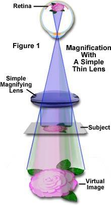
10 MAGNIFICATION

11 MAGNIFICATION Can we apply the maximal magnification? Total visual magnification of the microscope is derived by multiplying the magnification values of the objective and the eyepiece. Ocular: 5-30 x magnification Objective: x magnification Maximum magnification: 3000X
12 The resolution ~0.25 mm Resolution: the shortest distance between two points on a specimen that can still be distinguished by the observer or camera system as separate entities. The resolution of our optical equipment is better as closer points can be seen as separate ones.
.")
13 The resolution of a light microscope The resolution of the microscope, ( ) : ( ) = 2 A wavelength numeric aperture objective object A = n sinα The letter n is the refraction index of the media between the cover slip and the objective (air n=1, distilled water n=1.33, cedar oil n=1.51). α labels the angle closed by the main optical axis and the outermost light beam (half angle of the objective)
14 Possibilities of resolution improvement (i)reduction of the numerator, i.e. application of light beam with a shorter wavelength. With UV light the resolution can be reduced to 0.1 μm, but special quartz lenses and UV-light detector are needed, therefore the light microscope with UV light source is only a theoretical possibility.
15 Possibilities of resolution improvement (ii) To increase the value of the numeric aperture (A = n sinα). Increasing n (refraction index) : The resolution of the microscope can be enhanced by dropping a solution with higher refractive index between the front lens and the coverslip. Those lenses (mainly objectives with 100x magnification) are named as immersion objectives. The immersion liquid mentioned above is cedar oil, thus these lenses are objectives with oil immersion (they are labeled with HI). For the WI labeled objectives distilled water is the liquid which should be used. Increasing α: The half angle of a lens can be increased only until 72, since at larger angle than this the light beams became totally reflected.

16 MICROSCOPIC MEASUREMENT objective micrometer ocular micrometer
17 MICROSCOPIC MEASUREMENT A piece aof hair objective micrometer scale ocular micrometer scale ocular micrometer scale
18 ADVANCED MICROSCOPY 1. Phase contrast microscopy 2. Fluorescence microscopy 3. Laser scanning confocal microscopy 4. Electron microscopy

19 Phase contrast microscopy A large spectrum of living biological specimens are virtually transparent when observed in the optical microscope under brightfield illumination. Phase contrast microscopy provides an excellent method of improving contrast in unstained biological specimens without significant loss in resolution, and is widely utilized to examine dynamic events in living cells. Fritz Zernike received a Nobel prize in 1953 for his discovery of phase contrast. In a phase-contrast microscope, the annular rings in the objective lens and the condenser separate the light. The light that passes through the central part of the light path is recombined with the light that travels around the periphery of the specimen. The interference produced by these two paths produces images in which the dense structures appear darker than the background.

20 Phase contrast microscopy

21 Phase contrast microscopy

22 Fluorescence microscopy/ Laser scanning confocal microscopy

23 Fertilization: Sperm aster, male and female pronuclei MT, DNA, CS


24 Transfected tissue culture cells DNA microtubules F-actin


25 Microscopy: the past and present intestine cross section, haematoxilin/eosin staining intestine cross section, multicolor fluorescence image


26 Microscopy: the past and present testis cross section, haematoxilin/eosin staining testis cross section, multicolor fluorescence image
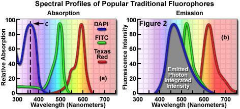
, releases the absorbed energy as a photon having a wavelength longer than the absorbed energy.")
27 Fluorescence Fluorescence - The process by which a suitable atom or molecule, which is transiently excited by absorption of external radiation at the proper energy level (usually ultraviolet or visible light), releases the absorbed energy as a photon having a wavelength longer than the absorbed energy. The fluorescence excitation and emission processes usually occur in less than a nanosecond.

28 Fluorescence Microscopy Fluorescence Microscopy is the most rapidly expanding microscopy technique employed today, both in the medical and biological sciences. When coupled to the optical microscope, fluorescence enables investigators to study a wide spectrum of phenomena in cellular biology. Foremost is the analysis of intracellular distribution of specific macromolecules in sub-cellular assemblies, such as the nucleus, membranes, cytoskeletal filaments, mitochondria, Golgi apparatus, and endoplasmic reticulum. In addition to steady state observations of cellular anatomy, fluorescence is also useful to probe intracellular dynamics and the interactions between various macromolecules, including diffusion, binding constants, enzymatic reaction rates, and a variety of reaction mechanisms, in time-resolved measurements. For example, fluorescent probes have been employed to monitor intracellular ph and the localized concentration of important ions.

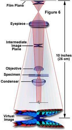
29 Fluorescence Microscope Structure

30 Fluorescence Filter Cube Structure Exciter filter: permits only selected wavelengths from the illuminator to pass through on the way toward the specimen. Barrier filter: blocks the excitation wavelengths and permit only selected emission wavelengths to pass toward the eye or other detector. Dichromatic beamsplitter (dichroic mirror): reflects excitation wavelengths and passes emission wavelengths.
31 Fluorescence Microscope Structure Microscopes with an inverted-style frame are designed primarily for tissue culture applications and are capable of producing fluorescence illumination through an episcopic and optical pathway. Epiilluminators usually consist of a mercury or xenon lamphouse (or laser system) stationed in a port at the rear of the microscope frame. Fluorescence illumination from the arc lamp passes through a collector lens and into a cube that contains a set of interference filters, including a dichroic mirror, barrier filter, and excitation filter. Light reflected from the dichroic mirror is restricted in wavelength by the excitation filter and enters the objective (now acting as a condenser) to bathe the specimen with a cone of illumination whose size and shape is determined by the objective numerical aperture. Secondary fluorescence, emitted by the specimen, returns through the objective, dichroic mirror and barrier filter before being routed through the microscope optical train. The microscope presented above contains a trinocular observation tube that is equipped with a port and extension tube for mounting a traditional or CCD camera system (a Peltier-cooled CCD camera is illustrated). Another port, located near the base at the front of the microscope, can also serve as an attachment point for a camera system (a traditional 35-millimeter camera is shown in the figure). In the figure presented above, wide-spectrum fluorescence illumination is filtered to produce a narrow bandwidth of green excitation wavelengths, which are capable of exciting specific fluorophores in the specimen. Secondary fluorescence (red light) passes back through the objective and is distributed throughout the microscope optical system. Transmitted illumination is provided by a tungsten-halogen lamphouse that is positioned on the microscope pillar, above the stage. Light from the lamphouse passes through a collector lens, a series of filters, and the field diaphragm before entering the condenser front aperture. After being focused by the condenser lens elements, transmitted illumination is projected onto the specimen, which is placed on the stage. The light that is diffracted, refracted, and not absorbed by the specimen continues through the objective and into the microscope optical train where it can be directed to the eyepieces or to a camera system.
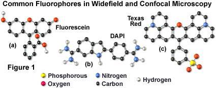
32 Fluorochromes Fluorochrome - A natural or synthetic dye or molecule that is capable of exhibiting fluorescence. Fluorochromes (also termed fluorescent molecules, probes, or fluorescent dyes) are usually polynuclear heterocyclic molecules containing nitrogen, sulfur, and/or oxygen with delocalized electron systems and reactive moieties that enable the compounds to be attached to a biological species.
33 Fluorochromes
 DAPI and Alexa Fluor 680 phalloidin, (top right) SYTOX Green dye and Alexa Fluor 568 phalloidin, (bottom left) 7-AAD and Alexa Fluor 488")
34 Fluorochromes Bovine pulmonary artery endothelial cells visualized using components of the SelectFX Nuclear Labeling Kit and Alexa Fluor phalloidin conjugates. Nuclei and F-actin were stained, respectively, with (top left) DAPI and Alexa Fluor 680 phalloidin, (top right) SYTOX Green dye and Alexa Fluor 568 phalloidin, (bottom left) 7-AAD and Alexa Fluor 488 phalloidin, and (bottom right) TO-PRO-3 dye and Alexa Fluor 350 phalloidin.

35 Organelle probes: Organelle/DNA/Ca 2+ probes a fluorochrome nucleus attached to a target-specific moiety that assists in localizing the fluorophore Mitochondrium: MitoTracker and MitoFluor Lysosome: LysoTracker and LysoSensor Golgi apparatus: BODIPY Endoplasmic reticulum: DiOC, Blue-White DPX DNA: Acridine orange, Propidium iodide, DAPI, Hoechst Ca 2+ : fura-2 and indo-1, fluo-3, fura red Bovine pulmonary artery endothelial cell labeled with probes to visualize mitochondria, peroxisomes, and the nucleus. Mitochondria were stained with the MitoTracker Red CMXRos reagent. Peroxisomal labeling was achieved with a primary antibody directed against PMP70, visualized using green-fluorescent Alexa Fluor 488 goat anti rabbit IgG. The nucleus was stained with blue-fluorescent DAPI.
36 Immunofluorescence Microscopy A targeted molecular species (protein, nucleic acid, membrane, etc.) in a specimen is labeled with a highly specific fluorescent antibody. After the labeled antibodies have been excited by a selected region of wavelengths, secondary fluorescence emission is gathered by the objective to form an image of the specimen. Antibodies are labeled either by coupling directly with a fluorochrome (fluorescent dye; termed direct immunofluorescence), or with a second fluorescent antibody that recognizes epitopes on the primary antibody (indirect immunofluorescence).
37 Immunofluorescence Microscopy

38 Immunofluorescence Microscopy MT DNS CS

39 Fluorescent Proteins Green Fluorescent Protein (GFP) - A naturally occurring protein fluorescent probe derived from the jellyfish Aequorea victoria, which is commonly employed to determine the location, concentration, interactions, and dynamics of a target protein in living cells and tissues. The excitation and emission spectra of enhanced GFP (a genetic derivative) have maxima at 489 nanometers and 508 nanometers, respectively. In order to incorporate the GFP (or any of its genetic derivatives) into a cell, the DNA sequence for the gene is ligated to the DNA encoding the protein of interest. After cultured cells have been transfected with the modified DNA, they are able to express chimeric fluorescent proteins for observation in the microscope. There are genetically modified variants of GFP such as blue fluorescent protein (BFP), cyan fluorescent protein (CFP), yellow fluorescent protein (YFP) DsRed fluorescent protein

40 Fluorescent Proteins Tubulin-GFP
41 GFP-FUSION PROTEINS gene of interest plasmid with GFP gene transfection fusion protein emits green light upon excitation by blue light, intracellular localization determined GFP transcription, translation
 is sensed by a photomultiplier tube and displayed in pixels on a computer monitor.")
42 Confocal Laser Scanning Microscopy - A popular mode of optical microscopy in which a focused laser beam is scanned laterally along the x and y axes of a specimen in a raster pattern. The emitted fluorescence (reflected light signal) is sensed by a photomultiplier tube and displayed in pixels on a computer monitor. The pixel display dimensions are determined by the sampling rate of the electronics and the dimensions of the raster. Signal photons that are emitted away from the focal plane are blocked by a pinhole aperture located in a plane confocal with the specimen. This technique enables the specimen to be optically sectioned along the z axis. Confocal Laser Scanning Microscopy

43 Confocal Laser Scanning Microscope EURO

44 Light sources for fluorescence/confocal microscopy As opposed to traditional arc-discharge lamps used with the shortest range (10-20 nanometers) bandpass interference filters in widefield fluorescence microscopy, the laser systems used for fluorophore excitation in scanning confocal microscopy restrict excitation to specific laser spectral lines that encompass only a few nanometers.
45
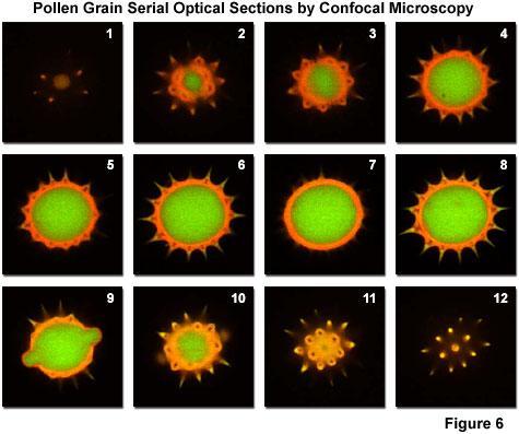
46 Z-sectioning

47 Z-sectioning Drosophila Egg Chamber

48 Z-sectioning Drosophila Egg Chambers
49 3D Reconstruction After Z-sectioning, the computer attached to the confocal microscope can generate virtual images of the specimen what can be viewed from any desired angles.

50 3D Reconstruction Tubulin-GFP

51 3D Reconstruction Drosophila Egg Chamber

52 3D Reconstruction Drosophila Egg Chambers
53 4D imaging The confocal microscope can be programmed to take pictures of the living sample at any desired time intervals. The resulting pictures are put together as movie files.

54 4D imaging Tubulin-GFP C. Elegans embryonic divisions

55 4D imaging Tubulin-GFP, Histon-RFP Drosophila embryonic divisions
56 FRET Fluorescence Resonance Energy Transfer (FRET) - An adaptation of the resonance energy transfer phenomenon to fluorescence microscopy in order to obtain quantitative temporal and spatial information about the binding and interaction of proteins, lipids, enzymes, and nucleic acids in living cells.

57 FRET: fluorescence resonance energy transfer CFP CFP YFP YFP
 to ascertain the location of specific genetic sequences.")
58 Fluorescence in situ Hybridization (FISH) - The fluorescence FISH technique is based on hybridization between target sequences of chromosomal DNA with fluorescently labeled single-stranded complementary sequences (termed cdna) to ascertain the location of specific genetic sequences. In medical practice, the most important application of FISH is the prenatal diagnosis of chromosome number abnormalities (and other chromosomal mutations). FISH
 Space charge e - e - e - +++++++++++ +++++++++++ Anode (positive")
59 ELECTRON MICROSCOPY Elecron ray source (Electron gun) Filament Heat e Wehnelt cap (negative potential) Space charge e - e - e Anode (positive potential)


60 Transmission electron microscope (TEM) Electron ray source Condenser lenses Condenser aperture Sample OBJECTIVE Objective apertures Intermediate lenses PROJECTOR Detector

61 Transmission electron microscope (TEM)

62 Scanning Electron Microscope (SEM) Electron ray source 1st condenser lens Condenser aperture 2nd condenser lens Objective aperture Scan coils OBJECTIVE Sample Backscattered electrons Amplifier/Detector 2nd electrons

63 Scanning Electron Microscope (SEM)
Visualizing Cells Molecular Biology of the Cell - Chapter 9
 Visualizing Cells Molecular Biology of the Cell - Chapter 9 Resolution, Detection Magnification Interaction of Light with matter: Absorbtion, Refraction, Reflection, Fluorescence Light Microscopy Absorbtion
Resolution of Microscopes Visible light is nm Dry lens(0.5na), green(530nm light)=0.65µm=650nm for oil lens (1.4NA) UV light (300nm) = 0.13µm f
, green(530nm light)=0.65µm=650nm for oil lens (1.4NA) UV light (300nm) = 0.13µm f") Microscopes and Microscopy MCB 380 Good information sources: Alberts-Molecular Biology of the Cell http://micro.magnet.fsu.edu/primer/ http://www.microscopyu.com/ Approaches to Problems in Cell Biology
Microscopes and Microscopy MCB 380 Good information sources: Alberts-Molecular Biology of the Cell http://micro.magnet.fsu.edu/primer/ http://www.microscopyu.com/ Approaches to Problems in Cell Biology
Partha Roy
 Fluorescence microscopy http://micro.magnet.fsu.edu/primer/index.html Partha Roy 1 Lecture Outline Definition of fluorescence Common fluorescent reagents Construction ti of a fluorescence microscope Optical
Practical light microscopy: an introduction
 Practical light microscopy: an introduction Dr. Mark Leake, Oxford University www.physics.ox.ac.uk/users/leake Aim of today s talk: Explanation of the very (very) basics of how a light microscope works
Spectral Separation of Multifluorescence Labels with the LSM 510 META
 Microscopy from Carl Zeiss Spectral Separation of Multifluorescence Labels with the LSM 510 META Indians living in the South American rain forest can distinguish between almost 200 hues of green in their
Con-focal and Multi-photon Microscope Experiment Fundamental. Qian Hu, Lab of Laser Scanning Confocal & Two-Photon Microscopy, ION, CAS
 Con-focal and Multi-photon Microscope Experiment Fundamental Qian Hu, Lab of Laser Scanning Confocal & Two-Photon Microscopy, ION, CAS 1. Light is Electromagnetic Wave ν = c / λ 2. Image of a Point Source
Confocal Microscopes. Evolution of Imaging
 Confocal Microscopes and Evolution of Imaging Judi Reilly Hans Richter Massachusetts Institute of Technology Environment, Health & Safety Office Radiation Protection What is Confocal? Pinhole diaphragm
Nodes of regulation in cellular systems
 Nodes of regulation in cellular systems cell membrane signal transduction ligands receptors oligomerization transport signal transduction modified protein Golgi transcription factor transport ER transport
Confocal Microscopy Analyzes Cells
 Choosing Filters for Fluorescence A Laurin Publication Photonic Solutions for Biotechnology and Medicine November 2002 Confocal Microscopy Analyzes Cells Reprinted from the November 2002 issue of Biophotonics
QImaging Camera Application Notes Multicolor Immunofluorescence Imaging
 QImaging Camera Application Notes Multicolor Immunofluorescence Imaging In order to image localization of intracellular proteins with high specificity, it is frequently necessary to multiplex antibody
Super-resolution Microscopy
 Semr oc kwhi t epaperser i es : 1. Introduction Super-resolution Microscopy Fluorescence microscopy has revolutionized the study of biological samples. Ever since the invention of fluorescence microscopy
Chapter 10: Classification of Microorganisms
 Chapter 10: Classification of Microorganisms 1. The Taxonomic Hierarchy 2. Methods of Identification 1. The Taxonomic Hierarchy Phylogenetic Tree of the 3 Domains Taxonomic Hierarchy 8 successive taxa
Automated Digital Microscopy
 A p p l i c a t i o n G u i d e Peter Banks, Ph.D. and Peter J. Brescia, Applications Department, BioTek Instruments, Inc., Winooski, VT Table of Contents Introduction ----------------------------------------------------------------------------------------------------------------------
Methods of Culturing Microorganisms. Chapter 3. Five Basic Techniques of Culturing Bacteria. Topics
 Chapter 3 Topics Methods of Culturing Microorganisms Microscope (History, Types, Definitions) Staining (Gram s) Methods of Culturing Microorganisms Five basic techniques of culturing Media Microbial growth
Lab 1A: Microscopy I. Name: Section:
 Lab 1A: Microscopy I A response is required for each item marked: (# ). Your grade for the lab 1 report (1A and 1B combined) will be the fraction of correct responses on a 50 point scale[(# correct/# total)
CytoPainter Golgi Staining Kit Green Fluorescence
 ab139483 CytoPainter Golgi Staining Kit Green Fluorescence Instructions for Use Designed for the detection of Golgi bodies by microscopy This product is for research use only and is not intended for diagnostic
ab CytoPainter ER Staining Kit Red Fluorescence
 ab139482 CytoPainter ER Staining Kit Red Fluorescence Instructions for Use Designed to detect Human endoplasmic reticulum by microscopy. This product is for research use only and is not intended for diagnostic
Transmission Electron Microscopy (TEM) Prof.Dr.Figen KAYA
 Prof.Dr.Figen KAYA") Transmission Electron Microscopy (TEM) Prof.Dr.Figen KAYA Transmission Electron Microscope A transmission electron microscope, similar to a transmission light microscope, has the following components along
Transmission Electron Microscopy (TEM) Prof.Dr.Figen KAYA Transmission Electron Microscope A transmission electron microscope, similar to a transmission light microscope, has the following components along
Advanced fluorescence microscopy techniques
 Practice-oriented, student-friendly modernization of the biomedical education for strengthening the international competitiveness of the rural Hungarian universities TÁMOP-4.1.1.C-13/1/KONV-2014-0001 Advanced
Introduction to Fluorescence Jablonski Diagram
 ntroduction to Fluorescence Jablonski Diagram Excited Singlet Manifold S1 internal conversion S2 k -isc k isc Excited riplet Manifold 1 S0 k nr k k' f nr fluorescence k p phosphorescence Singlet round
Simultaneous multi-color, multiphoton fluorophore excitation using dual-color fiber lasers
 Multiphoton Microscopy / Fiber Laser Simultaneous multi-color, multiphoton fluorophore excitation using dual-color fiber lasers Matthias Handloser, Tim Paasch-Colberg, Bernhard Wolfring TOPTICA Photonics
Innovations To Meet Your Needs
 Innovations To Meet Your Needs Cooled CCD Camera 1340 x 1037 pixel resolution for greatest image quality 12-bit precision provides 3 orders of linear dynamic range Windows and Power Macintosh Software
Confocal Microscopy of Electronic Devices. James Saczuk. Consumer Optical Electronics EE594 02/22/2000
 Confocal Microscopy of Electronic Devices James Saczuk Consumer Optical Electronics EE594 02/22/2000 Introduction! Review of confocal principles! Why is CM used to examine electronics?! Several methods
Introduction to N-STORM
 Introduction to N-STORM Dan Metcalf Advanced Imaging Manager Outline Introduction Principles of STORM Applications N-STORM overview Biological Scale Mitochondrion Microtubule Amino Acid 1Å Kinesin 1nm
Challenges to measuring intracellular Ca 2+ Calmodulin: nature s Ca 2+ sensor
 Calcium Signals in Biological Systems Lecture 3 (2/9/0) Measuring intracellular Ca 2+ signals II: Genetically encoded Ca 2+ sensors Henry M. Colecraft, Ph.D. Challenges to measuring intracellular Ca 2+
QuantaMaX and standard filters for fluorescence
 QuantaMaX and standard filters for fluorescence Our fluorescence filter product line is comprised of Stock QuantaMAX and Standard Vivid and Basic excitation, emission and dichroic interference filters,
Fundamentals and Applications of Biofilms Analysis, Structure and Physiology of Bacterial Biofilms Ching-Tsan Huang ( 黃慶璨 ) Office: Agronomy
 Office: Agronomy") 1 Fundamentals and Applications of Biofilms Analysis, Structure and Physiology of Bacterial Biofilms Ching-Tsan Huang ( 黃慶璨 ) Office: Agronomy Building, Room 111 Tel: (02) 33664454 E-mail: cthuang@ntu.edu.tw
1 Fundamentals and Applications of Biofilms Analysis, Structure and Physiology of Bacterial Biofilms Ching-Tsan Huang ( 黃慶璨 ) Office: Agronomy Building, Room 111 Tel: (02) 33664454 E-mail: cthuang@ntu.edu.tw
Flow Cytometry. Flow Cytometry Basics Guide
 Flow Cytometry Flow Cytometry Basics Guide Table of Contents Chapter 1 Chapter 2 Chapter 3 Chapter 4 Chapter 5 Principles of the Flow Cytometer Fluidics System.... 3 Optics and Detection.... 4 Signal and
Rice/TCU REU on Computational Neuroscience. Fundamentals of Molecular Imaging
 Rice/TCU REU on Computational Neuroscience Fundamentals of Molecular Imaging June 2, 2009 Neal Waxham 713-500-5621 m.n.waxham@uth.tmc.edu Objectives Introduction to resolution in light microscopy Brief
A Survey of Laser Types. Gas Lasers
 Mihail Pivtoraiko Andrei Rozhkov Applied Optics Winter 2003 A Survey of Laser Types Laser technology is available to us since 1960 s, and since then has been quite well developed. Currently, there is a
ER-ID Red Assay Kit (GFP-Certified )
") Enabling Discovery in Life Science ER-ID Red Assay Kit (GFP-Certified ) for detection of endoplasmic reticulum by microscopy Instruction Manual Cat. No. ENZ-51026-K500 500 assays For research use only.
Enabling Discovery in Life Science ER-ID Red Assay Kit (GFP-Certified ) for detection of endoplasmic reticulum by microscopy Instruction Manual Cat. No. ENZ-51026-K500 500 assays For research use only.
A legacy of innovation and discovery
 A legacy of innovation and discovery CellInsight CX7 LZR High Content Analysis Platform Quantifiably brilliant data Since the introduction of Thermo Scientific ArrayScan High Content Analysis (HCA) Readers
Fluorescent Imaging in Cell Biology. Invitrogen Cellular Analysis
 Fluorescent Imaging in Cell Biology Invitrogen Cellular Analysis Topics Fluorescent Dyes and Dots Cellular Structure. Multicolor Immuno-Labeling. rganelle Stains Cellular Physiology Intracellular Ions
Interferometric optical biosensor. Xingwei Wang
 Interferometric optical biosensor Xingwei Wang 1 Light Transverse electromagnetic wave Reflection Refraction Diffraction Interference 2 Fabry-Perot interferometer 3 Interferometer Two waves that coincide
Azure Biosystems Western Blotting Workflow
 Azure Biosystems Western Blotting Workflow PROBE PLAN SEPARATE ANALYZE VISUALIZE PLAN Plan your experiment and choose your detection method Chemiluminescent Western Blotting The most common method for
Microscopy Reagents. for Immunocytochemistry and Immunohistochemistry. World-Class Quality Superior Customer Support Outstanding Value
 Microscopy Reagents for Immunocytochemistry and Immunohistochemistry BioLegend is ISO 9001:2008 and ISO 13485:2003 Certified Toll-Free Tel: (US & Canada): 1.877.BIOLEGEND (246.5343) Tel: 858.768.5800 biolegend.com
The analysis of fluorescence microscopy images for FRET detection
 The analysis of fluorescence microscopy images for FRET detection Ela Claridge, Dale J. Powner and Michael J.O. Wakelam School of Computer Science, The University of Birmingham B5 2TT Institute for Cancer
Applicability of Hyperspectral Fluorescence Imaging to Mineral Sorting
 Institute of Industrial Information Technology Applicability of Hyperspectral Fluorescence Imaging to Mineral Sorting Optical Characterization of Materials, March 19, 2015 Sebastian Bauer, M.Sc. (Head:
Fig1: Melt pool size of LAMP vs. µlamp. The LAMP process s melt pool is x the area of the LAMP s melt pool.
 Proceedings of the 4th Annual ISC Research Symposium ISCRS 2010 April 21, 2010, Rolla, Missouri LOW COST IMAGING OF MELTPOOL IN MICRO LASER AIDED MANUFACTURING PROCESS (µlamp) ABSTRACT This paper describes
Basic Principles in Flow Cytometry. Flow Cytometry
 Basic Principles in Flow Cytometry Flow Cytometry» Flow Cytometry is the technological process that allows for the individual measurements of cell fluorescence and light scattering. This process is performed
A quantitative protocol for intensity-based live cell FRET imaging.
 A quantitative protocol for intensity-based live cell FRET imaging. Kaminski CF, Rees EJ, Schierle GS. Methods Mol Biol. 2014; 1076:445-454. Department of Chemical Engineering and Biotechnology, Pembroke
Bioinstrumentation Light Sources Lasers or LEDs?
 Bioinstrumentation Light Sources Lasers or LEDs? A comprehensive analysis of all the factors involved in designing and building life sciences instrumentation reveals that lasers provide superior performance
Segments of the obstructed intestinal loops were fixed in 4% paraformaldehyde
 Supplementary text Supplementary materials and methods Histopathological examination Segments of the obstructed intestinal loops were fixed in 4% paraformaldehyde (PFA) and embedded in paraffin wax with
Experiment 2b X-Ray Diffraction* Optical Diffraction Experiments
 * Experiment 2b X-Ray Diffraction* Adapted from Teaching General Chemistry: A Materials Science Companion by A. B. Ellis et al.: ACS, Washington, DC (1993). Introduction Inorganic chemists, physicists,
Optical Observation - Hyperspectral Characterization of Nano-scale Materials In-situ
 Optical Observation - Hyperspectral Characterization of Nano-scale Materials In-situ Research at the nanoscale is more effective, when research teams can quickly and easily observe and characterize a wide
4.1. Genetics as a Tool in Anthropology
 4.1. Genetics as a Tool in Anthropology Each biological system and every human being is defined by its genetic material. The genetic material is stored in the cells of the body, mainly in the nucleus of
CF Dyes Next Generation Fluorescent Dyes Secondary antibody
 CF Dyes Next Generation Fluorescent Dyes Secondary antibody OZYME 10 AVENUE AMPÈRE - CS 30268-78053 ST QUENTIN EN YVELINES CEDEX Tél. : 01 34 60 24 24 - Fax : 01 34 60 92 12 - www.ozyme.fr/info CF Dyes
Answers to the multiple choice questions are at the bottom of the last page of this document.
 Review for Unit Test #2: Cell Parts, Functions and Protein Synthesis, Answers Answers to the multiple choice questions are at the bottom of the last page of this document. 1. Know all of the material on
The best and brightest
 Labeling and Detection The best and brightest Alexa Fluor 488 dye Labeling and Detection A superior alternative to FITC Brighter conjugate fluorescence Unequalled photostability Perfect spectral match
Modification of Glass by FS Laser for Optical/Memory Applications
 Modification of Glass by FS Laser for Optical/Memory Applications Kazuyuki Hirao and Kiyotaka Miura Department of Material Chemistry Kyoto University International Workshop on Scientific Challenges of
arc lamp is substituted. Before
 CE update [cytology hematology generalist] The Principles of Flow Cytometry Antony C. Bakke, PhD From the Department of Pathology, Oregon Health Sciences University, Portland, OR On completion of this
Electron microscopy II
 Electron microscopy II Nanomaterials characterization I RNDr. Věra Vodičková, PhD. Interaction ction: electrons solid matter Signal types SE.secondary e - AE Auger s e - BSE back scattered e - X-ray photons,
OPTIQUE et BIOLOGIE. Cycle ingénieur 2A mars/mai Nathalie Westbrook Karen Perronet Groupe Biophotonique, Institut d Optique
 OPTIQUE et BIOLOGIE Cycle ingénieur 2A mars/mai 2017 Nathalie Westbrook Karen Perronet Groupe Biophotonique, Institut d Optique Outline of the course (18h) 14 & 21 / 03 / 2017: Molecular and Cellular Biology
Flow Cytometry - The Essentials
 Flow Cytometry - The Essentials Pocket Guide to Flow Cytometry: 1. Know your Cytometer 2. Understanding Fluorescence and Fluorophores 3. Gating Process 4. Controls 5. Optimization 6. Panel Building 7.
Flow Cytometry. Marta Argenti, PhD student. Department of Biomedical Sciences Padua
 Flow Cytometry Marta Argenti, PhD student Department of Biomedical Sciences Padua 14.12.12 Flow ~ cells in motion Cyto ~ cell Metry ~ measure Physical properties: Flow Cytometry is the measurement of cells
BIOIMAGING AND OPTICS PLATFORM EPFL SV PTBIOP FLUORESCENCE MICROSCOPY
 FLUORESCENCE MICROSCOPY Internal course 2014 January 14 th FLUORESCENCE MICROSCOPY Why do we need it? - 2- UNSTAINED SPECIMEN Missing specificity 3 DIFFERENT STAINING STRATEGIES Histological stain (Absorption)
Baraa Ayed AL-Odat. Israa Ayed. Heba kalbouneh
 1 Baraa Ayed AL-Odat Israa Ayed Heba kalbouneh Introduction: "lecture #1" The name " histology " is derived from the Greek words: "histo" means a tissue and "logos" means the study of. So, Histology mean
Total Test Questions: 66 Levels: Grades Units of Credit: 1.0 STANDARD 2. Demonstrate appropriate use of personal protective devices.
 DESCRIPTION Biotechnology is designed to create an awareness of career possibilities in the field of biotechnology. Students are introduced to diagnostic and therapeutic laboratory procedures that support
Spectral imaging fluorescence microscopy
 Oxford, GTC Genes 1356-9597 September 79 Blackwell to UK Cells Science, 2002 Ltd Ltd COMMENTARY Spectral imaging fluorescence microscopy T Haraguchi imaging et al. fluorescence microscopy Tokuko Haraguchi
Time-resolved Measurements Using the Agilent Cary Eclipse Fluorescence Spectrophotometer A Versatile Instrument for Accurate Measurements
 Time-resolved Measurements Using the Agilent Cary Eclipse Fluorescence Spectrophotometer A Versatile Instrument for Accurate Measurements Technical Overview Authors Dr. Fabian Zieschang, Katherine MacNamara,
What is SEM? TM-1000 Tabletop Microscope
 What is SEM? TM-1000 Tabletop Microscope SEM vs OM SEM (Scanning Electron Microscope) OM (Optical Microscope) Backscattered (BSE) Image (Vacuum : 5Pa) Sample : Bulb filament Magnification : 500X Hitachi
Basic Fluorescence Microscopy and Sample Preparation. Eva Wegel
 Basic Fluorescence Microscopy and Sample Preparation Eva Wegel eva.wegel@bioch.ox.ac.uk Visible Light 390 700 nm visible to the human eye White light is split into its components through a prism Reason:
Principles of optical measurement used for the determination of the leukocyte differential part on Pentra series
 Principles of optical measurement used for the determination of the leukocyte differential part on Pentra series P. Nerin, Research Director HORIBA Medical E. Tournier, Hematology Product Manager HORIBA
Nucleic Acid Staining. Fluorophores & Applica6ons
 Nucleic Acid Staining Fluorophores & Applica6ons Types of Nucleic Acid Stains Intercala)ng dyes- - ethidium bromide and propidium iodide Minor- groove binders- - DAPI and the Hoechst dyes Miscellaneous-
Multiphoton Microscopy: Seeing deeper and clearer
 Multiphoton Microscopy: Seeing deeper and clearer Since the invention of simple microscope by Leuwenhoek and Hooke in the 17th century, different types of light microscopy techniques (such as phase contrast,
Fluorescence Nanoscopy
 Fluorescence Nanoscopy Keith A. Lidke University of New Mexico panda3.phys.unm.edu/~klidke/index.html Optical Microscopy http://en.wikipedia.org/wiki/k%c3%b6hler_illumination 30 µm Fluorescent Probes Michalet
Immunofluorescence Staining Protocol for 3 Well Chamber, removable
 Immunofluorescence Staining Protocol for 3 Well Chamber, removable This Application Note presents a simple protocol for the cultivation, fixation, and staining of cells using the 3 Well Chamber, removable.
SUPPLEMENTARY FIGURES
 SYNERGISTIC STRATEGY FOR MULTICOLOR TWO-PHOTON MICROSCOPY: APPLICATION TO THE ANALYSIS OF GERMINAL CENTER REACTIONS IN VIVO ASYLKHAN RAKHYMZHAN, RUTH LEBEN, HANNA ZIMMERMANN, ROBERT GÜNTHER, PEGGY MEX,
Chapter 03 - Tools of the Laboratory: Methods for the Culturing of Microscopic Analysis of microorganisms
 Microbiology: A Systems Approach 4th Edition Cowan Test Bank Completed download: https://testbankreal.com/download/microbiology-systems-approach-4thedition-test-bank-cowan/ (Downloadable package TEST BANK
Fs- Using Ultrafast Lasers to Add New Functionality to Glass
 An IMI Video Reproduction of Invited Lectures from the 17th University Glass Conference Fs- Using Ultrafast Lasers to Add New Functionality to Glass Denise M. Krol University of California, Davis 17th
Fluorescent in-situ Hybridization
 Fluorescent in-situ Hybridization Presented for: Presented by: Date: 2 Definition In situ hybridization is the method of localizing/ detecting specific nucleotide sequences in morphologically preserved
Using ULS24 CMOS Bio-imager as a Readout Sensor for Chemiluminescence Immunoassay and DNA Hybridization Assay
 Using ULS24 CMOS Bio-imager as a Readout Sensor for Chemiluminescence Immunoassay and DNA Hybridization Assay Updated: Nov 11, 2016 Introduction Immunoassay is a widely used method for detecting the presence
Immunohistochemistry: Basics and Methods
 Immunohistochemistry: Basics and Methods Bearbeitet von Igor B Buchwalow, Werner Böcker 1st Edition. 2010. Buch. x, 153 S. Hardcover ISBN 978 3 642 04608 7 Format (B x L): 15,5 x 23,5 cm Gewicht: 445 g
Thermo Scientific GENESYS 10S Bio spectrophotometer
 PRODUCT SPECIFICATIONS GENESYS 10S Bio UV-Visible Spectrophotometer Thermo Scientific GENESYS 10S Bio spectrophotometer Accurate and convenient life science UV-Visible measurements Designed for performance
Next Level of Super Resolution Fluorescence Microscopy
 Work in your familiar GFP/YFP/RFP system from the first experiment to the nanoimage Bwcon business award winner: Inventor Prof Christoph Cremer Next Level of Super Resolution Fluorescence Microscopy Resolution:
Lecture Four. Molecular Approaches I: Nucleic Acids
 Lecture Four. Molecular Approaches I: Nucleic Acids I. Recombinant DNA and Gene Cloning Recombinant DNA is DNA that has been created artificially. DNA from two or more sources is incorporated into a single
Post-expansion antibody delivery, after epitope-preserving homogenization.
 Supplementary Figure 1 Post-expansion antibody delivery, after epitope-preserving homogenization. (a, b) Wide-field fluorescence images of Thy1-YFP-expressing mouse brain hemisphere slice before expansion
FLUORESCENT PEPTIDES. Outstanding Performance and Wide Application Range
 FLUORESCENT PEPTIDES Peptides and amino acids labeled with and Tide Quencher TM We offer peptides and amino acids tagged with fluorescent dyes. They meet highest demands in fluorescence intensity and photo-stability,
Flow Cytometry for Ploidy and Genome Size analysis - 1. Functional principles
 Flow Cytometry for Ploidy and Genome Size analysis - 1. Functional principles The technique of plant ploidy and genome size analysis in a flow cytometer is based on the fluorescent labelling of the nuclear
Imaging & analysis with the LSM780 NLO Discover the secrets beyond the twilight zone
 Imaging & analysis with the LSM780 NLO Discover the secrets beyond the twilight zone Sven Terclavers LSM780 System overview The Scan Module - Core of the LSM 780 1 V/tunable PTC laser ports (405/440, cw/ps;
Assays for studying mitochondrial health and function
 APPLICATION NOTE Fluorescence labeling and detection Assays for studying mitochondrial health and function Introduction Mitochondria play a critical role in maintaining normal cellular activities. Mitochondria
How to perform-control immunostaining experiment - microscopist subjective point of view. Pawel Pasierbek
 How to perform-control immunostaining experiment - microscopist subjective point of view. Pawel Pasierbek Immunolabeling and fluorescent detection became such a standard procedure in the biomedical research
Cavity Filters. KIGRE, INC., 100 Marshland Road, Hilton Head, SC 29926, USA PH: FAX: Web:
 Cavity Filters Kigre, Inc. s expertise in laser glass technology has led the way in the company s development of a wide range of cavity filter glass for solid-state lasers. Filters are used inside the
Physical structure of matter. Monochromatization of molybdenum X-rays X-ray Physics. What you need:
 X-ray Physics Physical structure of matter Monochromatization of molybdenum X-rays What you can learn about Bremsstrahlung Characteristic radiation Energy levels Absorption Absorption edges Interference
Name Date Block LAB: Exploring Plant & Animal Cells
 Name Date Block LAB: Exploring Plant & Animal Cells Background Information: One of the first scientists to look at cells under a microscope was an English scientist by the name of Robert Hooke. He viewed
Monitoring and Optimizing the Lipopolysaccharides-plasmid DNA interaction by FLIM-FRET
 Transactions on Science and Technology Vol. 4, No. 3-3, 342-347, 2017 Monitoring and Optimizing the Lipopolysaccharides-plasmid DNA interaction by FLIM-FRET Nur Syahadatain Abdul Razak 1#, Clarence M.
16.2 Scanning Infrared Spectrometers
 16.2 Scanning Infrared Spectrometers it's difficult to find materials transparent in the infrared water vapor and atmospheric CO 2 can cause problems there are three common sources high diffraction orders
Lecture 25 (11/15/17)
") Lecture 25 (11/15/17) Reading: Ch9; 328-332 Ch25; 990-995, 1005-1012 Problems: Ch9 (study-guide: applying); 1,2 Ch9 (study-guide: facts); 7,8 Ch25 (text); 1-3,5-7,9,10,13-15 Ch25 (study-guide: applying);
Lecture 25 (11/15/17) Reading: Ch9; 328-332 Ch25; 990-995, 1005-1012 Problems: Ch9 (study-guide: applying); 1,2 Ch9 (study-guide: facts); 7,8 Ch25 (text); 1-3,5-7,9,10,13-15 Ch25 (study-guide: applying);
Cell Growth and Reproduction
 Cell Growth and Reproduction Robert Hooke was the first person to describe cells, in the year 1665. He was looking through his microscope at a piece of cork when he noticed a lot of repeating honeycomb
Mystery microscope images
 Mystery microscope images Equipment: 6X framed microscopy images 6X cards saying what each microscope image shows 6X cards with the different microscopy techniques written on them 6X cards with the different
Immunostaining Protocols
 Immunostaining Protocols Lula L. Hilenski, Ph.D. Director Microscopy in Medicine Core Emory University Variables in standard immunostaining protocol 2-step or indirect immunofluorescence 1. Substrate on
Chapter 10 Analytical Biotechnology and the Human Genome
 Chapter 10 Analytical Biotechnology and the Human Genome Chapter Outline Enzyme tests and biosensors DNA-based tests DNA analysis technologies Human genome and genome-based analytical methods 1 Enzyme-based
Conquer cell counting
 Conquer cell counting Cell Countess II Automated Cell Counters Fast Accurate Affordable Countess II Automated Cell Counters Advanced technology at an affordable price Accurate counts in as little as 10
including, but not limited to:
 *This Section is part of the original Request for Proposal # P08 080. The Contractor should provide the following eligible Scientific Biomedical Research Equipment, Reagents & Supplies including, but not
Recombinant DNA Technology. The Role of Recombinant DNA Technology in Biotechnology. yeast. Biotechnology. Recombinant DNA technology.
 PowerPoint Lecture Presentations prepared by Mindy Miller-Kittrell, North Carolina State University C H A P T E R 8 Recombinant DNA Technology The Role of Recombinant DNA Technology in Biotechnology Biotechnology?
Sketch the light paths from object to image in a single lens system in following situations.
 Solutions o Chapter I 1.1. There are three rules governing light path or a simple lens: 1) light ray passing through the center o a lens is not deviated. ) Light ray parallel with optic axis will pass
Confocal immunofluorescence microscopy
 Confocal immunofluorescence microscopy HL-6 and cells were cultured and cytospun onto glass slides. The cells were double immunofluorescence stained for Mt NPM1 and fibrillarin (nucleolar marker). Briefly,
average diameter = 3 nm, from PlasmaChem) was mixed in NLCs to produce QDembedded
 was mixed in NLCs to produce QDembedded") Electronic Supplementary Material (ESI) for RSC Advances. This journal is The Royal Society of Chemistry 2014 Supporting information Experimental Section The blended CLC-monomer materials used to fabricate
Electronic Supplementary Material (ESI) for RSC Advances. This journal is The Royal Society of Chemistry 2014 Supporting information Experimental Section The blended CLC-monomer materials used to fabricate
Multiplex Fluorescent Western Blot Starter Kit for the Bio- Rad ChemiDoc MP
 Page 1 of 7 INSTRUCTIONS: Z-310 Multiplex Fluorescent Western Blot Starter Kit for the Bio- Rad ChemiDoc MP Rockland Immunochemicals and Bio-Rad Laboratories have jointly developed an easy to use multiplex