MULTI-PHOTON MICROSCOPY: APPLICATIONS AND THEORY PART II
|
|
|
- Blaze Hutchinson
- 6 years ago
- Views:
Transcription
1 MULTI-PHOTON MICROSCOPY: APPLICATIONS AND THEORY PART II JAMES LOPEZ PH.D. NATIONAL SALES APPLICATIONS SPECIALIST FLUOVIEW LASER SCANNING CONFOCAL AND MULTIPHOTON

2 SINGLE PHOTON VERSUS TWO PHOTON IMAGING
. MATHEMATICALLY PREDICTED.")

3 TWO-PHOTON DISCOVERY MARIA GOEPPERT-MAYER FIRST DESCRIBED THE PROCESS OF TWO-PHOTON ABSORPTION IN HER DISSERTATION IN 1931 (UNIVERSITY OF GÖTTINGEN). MATHEMATICALLY PREDICTED. 2P LASERS WERE NOT INVENTED UNTIL 1960 S. LATER RECEIVED NOBEL PRIZE FOR WORK ON ATOMIC NUCLEAR SHELL THEORY. 2-P MICROSCOPY PIONEERED BY WINFRIED DENK IN THE LABORATORY OF WATT WEBB AT CORNELL. 2-P PATENT WAS HELD BY DENK, STRICKLER, WEBB 1991 (RECENTLY EXPIRED).
4 GM UNITS FOR 2P-CROSS SECTIONS

5 NUMERICAL APERATURE NA = n sin µ 4 Fluorescence NA 2 Mag Intensity Super 25x Water Objective MPE NA = 1.05 Mag = 25x UPLANFL 20X Dry Objective NA = 0.5 Mag = 20x n refractive index Air = 1.0 Water = 1.3 Oil = 1.5 Fluorescence Intensity 12.4x Resolution 2.1x (R = λ/2na)
6

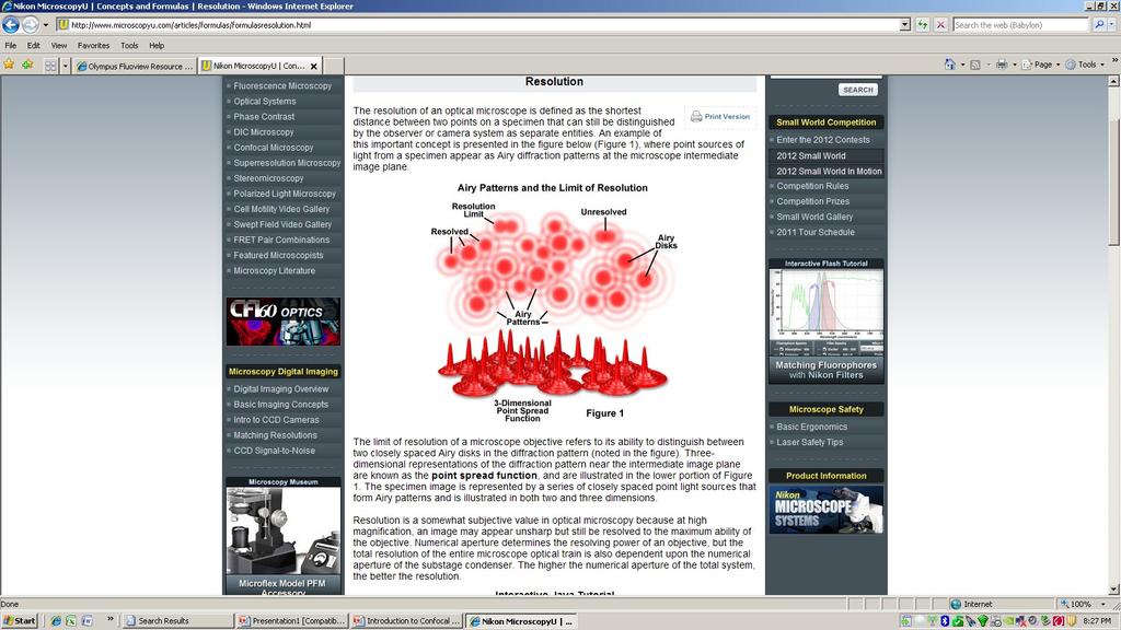
7 EMISSION WAVELENGTH AND RESOLUTION

8 FV1000MPE components Confocal detectors Reflected detector Pulsed TiS laser AOM Motorized beam expander Forward detector SEG-MRKT-PP-11
VS. MULTI-PHOTON")
9 SINGLE PHOTON (CONFOCAL )VS. MULTI-PHOTON EXCITATION

10 Lasers for MPE microscopy Almost all lasers used for MPE microscopy are Titanium sapphire or Ti:sapphire lasers. Typical pulse durations range from 80 femptoseconds (fsec) to 140 fsec. Typical bandwidth is from 690 to 1040nm, limited by the falloff in laser power available at the extremes of the range and other factors. At a typical repetition frequency of 80MHz, there are 134 pulses per pixel at 1 frame/sec at 512 x 512 pixels resolution.
11 Image Collection In Confocal vs MP Confocal Microscope Multi-Photon Microscope No need for confocal aperture Excitation only at the focal plane


12 Simple optical path and optimized optics for efficient 2P detection.

13 RELATIONSHIP BETWEEN IMAGE INTENSITY AND LASER POWER AND PULSE WIDTH
14

15 RELATIONSHIP BETWEEN IMAGE INTENSITY AND LASER POWER AND PULSE WIDTH

16 RELATIONSHIP BETWEEN IMAGE INTENSITY AND LASER POWER AND PULSE WIDTH
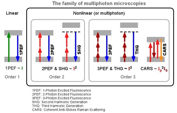
17 LINEAR AND NONLINEAR IMAGING MODALITIES

18 SECOND HARMONIC GENERATION (SHG) HIGHLY ORDERED MOLECULAR STRUCTURE NONCENTROSYMMETRIC MOLECULAR STRUCTURE -NO INVERSION CENTER LIGHT EXITING SHG MATERIAL IS: ½ λ AND 2E OF LIGHT ENTERING SHG MATERIAL 860nm excitation -> 430nm emission at 2x ENERGY MUSCLE, COLLAGEN, POLYSACCHARIDES ENERGY CONSERVED - MOLECULES ARE NOT EXCITED NO PHOTOBLEACHING AND NO PHOTOTOXICITY LABEL FREE COLLAGEN

19 Second Harmonic Generation Collagen Emission with 800nm TPE
20 SECOND HARMONIC GENERATION
 COLLAGEN INSIDE OF INTACT MOUSE")
21 SECOND HARMONIC GENERATION IMAGING MIP xyz 25X 860nm excitation (LABEL FREE) COLLAGEN INSIDE OF INTACT MOUSE LYMPHNODE
 AND PULSE")
22 RELATIONSHIP BETWEEN IMAGE INTENSITY (SHG) AND PULSE WIDTH

23 SECOND HARMONIC GENERATION Polysaccharide (starch) packets in a slice of raw potato 25x MIP xyz LABEL FREE 860nm

24 C. elegans Second Harmonic Generation Imaging Epi-SHG Forward-SHG


25 SECOND HARMONIC GENERATION MOUSE MUSCLE SHG 25X LABEL FREE INTRAVITAL 860nm EXCITATION
26 SHG AND TPEF/2PEF

27 Second Harmonic Generation and TPEF 860nm excitation Skin graft infected with GFP encoding virus SHG Emission in violet and green 25x DEEP

28 Rat skin 800nm E-SHG TRITC AUTO AUTO 25x

29 Second Harmonic Generation and TPEF-AUTOFLUORESCENCE 800nm excitation Emission in violet and green MIP XYZ Human skin section 25x Label free

30 Second Harmonic Generation and TPEF-AUTOFLUORESCENCE 800nm excitation Emission in violet and green MIP XYZ C. elegans 25x Label free


31 Alzheimer s Plaques Amyloid in red SHG in grey DAPI in white/grey Stitching upgrade

32 FV1000MPE Exclusive Super 25x Transgenic zebrafish expressing GFP in pancreatic beta cells. GFP Anti-insulin E-SHG F-SHG Dr. Stef Nalle Dr.Vicky Prince
33 Membrane Potential using FM4-64 SHG MPE 950nm DM505 Fluorescence at nm SHG at 475nm overlay
 WITH IMAGING ph (2P FLUORESCENCE)")
34 COMBINING IMAGING OF MEMBRANE POTENTIAL (SHG) WITH IMAGING ph (2P FLUORESCENCE)
35
36 2PEF/TPEF

37 GFP-LABELED GLOMERULUS DEXTRAN-TEXASRED VESSELS INTACT KIDNEY FROM A TRANSGENIC MOUSE FOLLOWING CARDIAC PERFUSION WITH DEXTRAN-TEXASRED
38 Drosophila testes

39 Transgenic GFP-IP3R3 in olfactory bulb

40 Human intestine

41 GFP Neuron Spines

42 INTRAVITAL GFP TUMOR CELLS 2PEF GFP LABELED TUMOR CELLS INJECTED INTO MOUSE 960nm Z-series Optical sectioning MIP XYZ

43 ZEBRAFISH HEAD 920nm GFP 2PEF LABELED ZEBRAFISH HEAD RACHEL WONG LABORATORY 25X

44 INTRAVITAL IMAGING CONSIDERATIONS MOVEMENT HEART BEAT BREATHING RUNNING ANIMAL SURVIVAL BODY WINDOWS VENTILATOR ANESTHESIA UPRIGHT VS. INVERTED DEPTH OF IMAGING TTL TRIGGERING OF ACQUISITION TIME RESOLUTION
 1.")
45 MICROPROBE OBJECTIVES wide-angle 6X magnification (NA 0.14) 1.3 mm-diameter lens high-resolution 20X magnification (NA 0.5) 1.3 mm-diameter lens high resolution 27X magnification (NA 0.7) 3.5 mm-diameter

46 INTRAVITAL IMAGING DEXTRAN-TEXAS RED LABELED BLOOD VESSELS TUMOR CELLS LABELED WITH GFP VISUALIZING METASTASIS 25X LIVE MOUSE UNDER ANESTHESIA 920nm excitation


47 - compensation + compensation Dr. Ken Dunn IUPUI Animal Imaging Facility
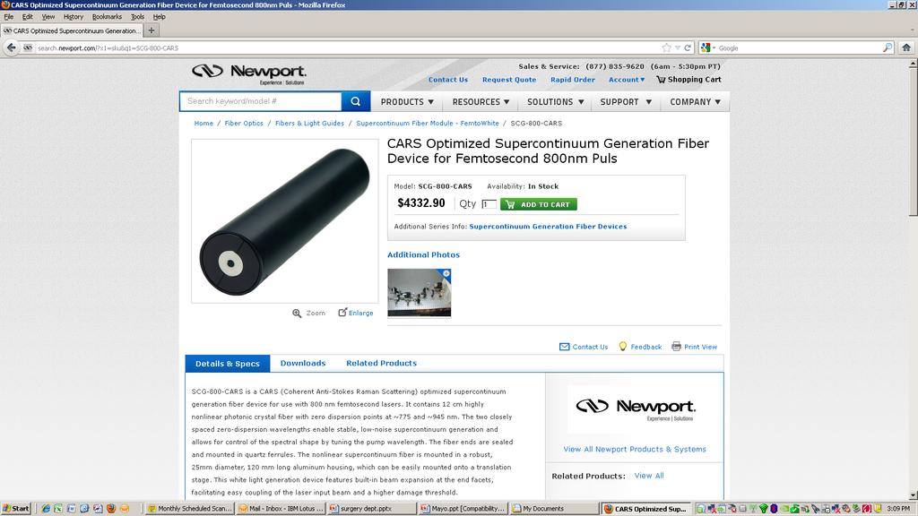
48 LABEL FREE LIPID IMAGING USING CARS (UPGRADE FOR FV1000MPE) COHERENT ANTI-STOKES RAMAN SCATTERING (CARS) MICROSCOPY IMAGING LIPIDS (CH2) WITHOUT STAINING OR FLUORESCENT TAGS COMBINE WITH OTHER (LABEL FREE) TECHNIQUES INTRINSIC FLUORESCENCE FLUORESCENT PROTEIN OR DYE SHG
49 COHERENT ANTI-STOKES RAMAN SCATTERING (CARS) PRINCIPLE p s p p s Two excitation wavelength, ωp (pump) and ωs (Stokes). The wavelength difference ( ) matches vibration frequency. It is coherent. The signal appears in a specific direction (forward or backward) The first two light-matter interaction prepared the coherent state. The third interaction create Raman scattering. NIR excitation, high optical resolution
50 Coherent Anti-Stokes Raman Spectroscopy (CARS)


51 PROPERTIES OF CARS SIGNAL CARS signal can be detected in Epi and Forward direction. Called E-CARS and F-CARS signal. E-CARS signal is weak relative to FCARS, but sensitive to small features F-CARS is strong due to coherent addition
52 RAMAN SPECTROSCOPY INSIDE A CELL Intenisty CH2 (lipid) Phosphate (DNA) Amide 1 (protein) Wavenumber (cm-1) Strong CH2 signal
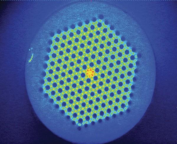
53 GENERATING THE PUMP AND STOKES BEAMS WITH A PCF

54 LOW DISPERSION WITH THE CARS PCF
55 Simultaneous Forward CARS/Second Harmonic Generation Detection Excitation: Pump 800nm Stokes 1040nm PMT SHG: nm DM 485nm PMT Emission CARS: nm
 GREEN : TPE WHITE : SHG")
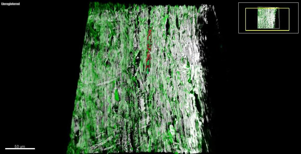
56 Multimodal Imaging using FV1000MPE Porcine muscle tissue with no staining RED : CARS (lipid) GREEN : TPE WHITE : SHG (collagen)
 of rat")

57 CARS imaging of lipids in vivo Myelin sheath (70% lipid) of rat dorsal root nerve Image courtesy of: Dr. Albert Stolow (NRC,Canada)
58

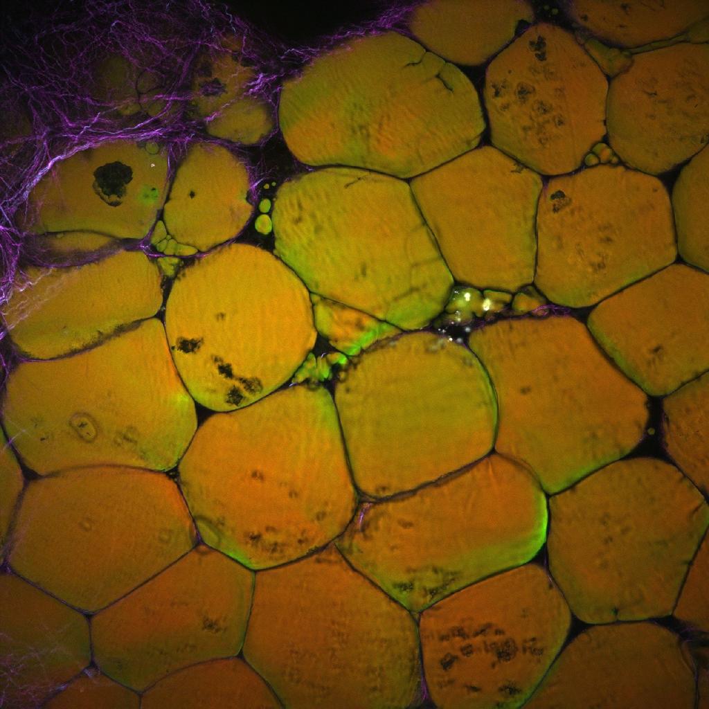
59 E-CARS F-CARS Epi-TPEF High-fat fed mouse fat imaged with F-CARS, E-CARS, and TPEF. Maximum Intensity Projections. 800nm Pump 1040nm Stokes Super 25X 1.05NA Scale Bar = 100µm Olympus FV1000MPE-CARS

60 CARS AND SHG Porcine Super 25x Epi-CARS SHG

61 CARS AND SHG Porcine Super 25x Epi-CARS SHG

62 CARS AND SHG Salmonoides Super 25x Epi-CARS Second Harmonic Generation
 and second harmonic generation (BLUE) in a nude")
63 SHG / TPEF / F-CARS Early cancer metasis labeled with GFP (GREEN) invading liver tissue visualized with Forward-CARS (PURPLE) and second harmonic generation (BLUE) in a nude mouse.

64 Breast cancer tissue 800nm pump 1040 stokes E-SHG F-SHG CARS 25x Scale bar = 100um
65

66 E-CARS F-CARS

67 Intestinal villi from olive oil gavaged mouse. E-CARS Autofluorescence Label free 25x MIP
68 CARS Lipid Imaging Applications Lipids are hard to image Extracted during fixation Extrinsic dyes can disrupt structure No antibodies Biological Relevance, areas of Study lipid-droplet biology lipid metabolism in simple organisms obesity-cancer-lipids in metastasis relationship atherosclerotic lesions lipid-rich structures including myelin sheath, skin, and brain
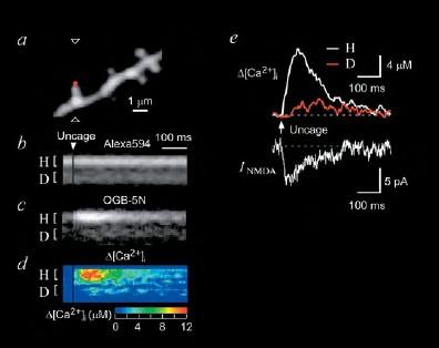
69 Combining Electrophysiology with Imaging
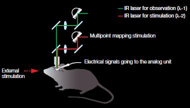
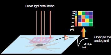
70 Combining Electrophysiology with Imaging


71 NEW OLYMPUS 25X MPE OBJECTIVE Super 20x Dipping Objective 0.95NA 2mm WD Super 25x MP Dipping Objective 1.05NA 2mm WD
72 Nature Neuroscience - epub Aug. 2011
73 OLYMPUS 25x MPE Objective Olympus 25x MPE Objective NA 1.05 Water WD 2.0
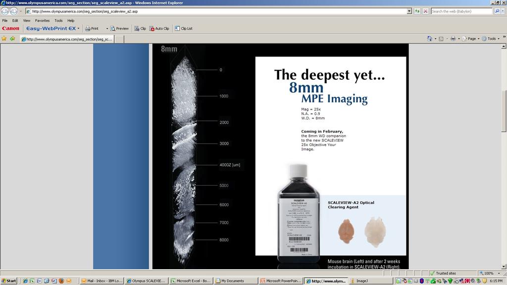
74 OLYMPUS Scaleview 25x MPE Objective NEW! Olympus Scaleview 25x MPE Objective NA 1.0 Scale-A2 WD 4.0


75 Olympus Scaleview 25x MPE Objective NA 1.0 Scale-A2 WD 4.0

76 NEW Olympus Scaleview-A2 Clearing Reagent

77 Clearing Reagent Comparison: Olympus Scaleview-A2 vs. BABB
78
79

80
81 FV1000MPE IMAGING SYSTEM OFFERS NEW CAPABILITIES Visible Confocal Visible spectral confocal imaging for multi-color high resolution optical sectioning. MPEIntravital imaging in upright and inverted configurations. Deep tissue imaging. Extended timelapse. SHGCollagen, muscles, electrical potential, Alzheimer s plaques. *ALL FORWARD AND REFLECTED MPE IMAGING
82 Transgenic mouse eyes expressing tdtomato
Rice/TCU REU on Computational Neuroscience. Fundamentals of Molecular Imaging
 Rice/TCU REU on Computational Neuroscience Fundamentals of Molecular Imaging June 2, 2009 Neal Waxham 713-500-5621 m.n.waxham@uth.tmc.edu Objectives Introduction to resolution in light microscopy Brief
Visualizing Cells Molecular Biology of the Cell - Chapter 9
 Visualizing Cells Molecular Biology of the Cell - Chapter 9 Resolution, Detection Magnification Interaction of Light with matter: Absorbtion, Refraction, Reflection, Fluorescence Light Microscopy Absorbtion
Simultaneous multi-color, multiphoton fluorophore excitation using dual-color fiber lasers
 Multiphoton Microscopy / Fiber Laser Simultaneous multi-color, multiphoton fluorophore excitation using dual-color fiber lasers Matthias Handloser, Tim Paasch-Colberg, Bernhard Wolfring TOPTICA Photonics
Genetically targeted all-optical electrophysiology with a transgenic Credependent
 Genetically targeted all-optical electrophysiology with a transgenic Credependent Optopatch mouse Short title: Transgenic Optopatch mouse Shan Lou 1, Yoav Adam 1, Eli N. Weinstein 1,4, Erika Williams 2,
SUPPLEMENTARY FIGURES
 SYNERGISTIC STRATEGY FOR MULTICOLOR TWO-PHOTON MICROSCOPY: APPLICATION TO THE ANALYSIS OF GERMINAL CENTER REACTIONS IN VIVO ASYLKHAN RAKHYMZHAN, RUTH LEBEN, HANNA ZIMMERMANN, ROBERT GÜNTHER, PEGGY MEX,
Confocal Microscopes. Evolution of Imaging
 Confocal Microscopes and Evolution of Imaging Judi Reilly Hans Richter Massachusetts Institute of Technology Environment, Health & Safety Office Radiation Protection What is Confocal? Pinhole diaphragm
Confocal Microscopy of Electronic Devices. James Saczuk. Consumer Optical Electronics EE594 02/22/2000
 Confocal Microscopy of Electronic Devices James Saczuk Consumer Optical Electronics EE594 02/22/2000 Introduction! Review of confocal principles! Why is CM used to examine electronics?! Several methods
Confocal Microscopy Analyzes Cells
 Choosing Filters for Fluorescence A Laurin Publication Photonic Solutions for Biotechnology and Medicine November 2002 Confocal Microscopy Analyzes Cells Reprinted from the November 2002 issue of Biophotonics
Resolution of Microscopes Visible light is nm Dry lens(0.5na), green(530nm light)=0.65µm=650nm for oil lens (1.4NA) UV light (300nm) = 0.13µm f
, green(530nm light)=0.65µm=650nm for oil lens (1.4NA) UV light (300nm) = 0.13µm f") Microscopes and Microscopy MCB 380 Good information sources: Alberts-Molecular Biology of the Cell http://micro.magnet.fsu.edu/primer/ http://www.microscopyu.com/ Approaches to Problems in Cell Biology
Microscopes and Microscopy MCB 380 Good information sources: Alberts-Molecular Biology of the Cell http://micro.magnet.fsu.edu/primer/ http://www.microscopyu.com/ Approaches to Problems in Cell Biology
Multiphoton Microscopy: Seeing deeper and clearer
 Multiphoton Microscopy: Seeing deeper and clearer Since the invention of simple microscope by Leuwenhoek and Hooke in the 17th century, different types of light microscopy techniques (such as phase contrast,
A simple introduction to multiphoton microscopy
 Journal of Microscopy, Vol. 243, Pt 3 2011, pp. 221 226 Received 29 April 2011; accepted 28 June 2011 doi: 10.1111/j.1365-2818.2011.03532.x A simple introduction to multiphoton microscopy A. USTIONE &
High-throughput three-dimensional (3D) lithographic microfabrication in biomedical applications
 lithographic microfabrication in biomedical applications") High-throughput three-dimensional (3D) lithographic microfabrication in biomedical applications The MIT Faculty has made this article openly available. Please share how this access benefits you. Your story
High-throughput three-dimensional (3D) lithographic microfabrication in biomedical applications The MIT Faculty has made this article openly available. Please share how this access benefits you. Your story
Optical Observation - Hyperspectral Characterization of Nano-scale Materials In-situ
 Optical Observation - Hyperspectral Characterization of Nano-scale Materials In-situ Research at the nanoscale is more effective, when research teams can quickly and easily observe and characterize a wide
Automated Digital Microscopy
 A p p l i c a t i o n G u i d e Peter Banks, Ph.D. and Peter J. Brescia, Applications Department, BioTek Instruments, Inc., Winooski, VT Table of Contents Introduction ----------------------------------------------------------------------------------------------------------------------
CENTER FOR BRAIN EXPERIMENT
 CENTER FOR BRAIN EXPERIMENT Section of Brain Structure Associate Professor: ARII, Tatsuo, PhD 1967 Graduated from Tohoku University, Faculty of Science. Completed the doctoral course in Engineering, Nagoya
Microendoscopes for imaging of the pancreas
 Microendoscopes for imaging of the pancreas Angelique Kano *, Andrew R. Rouse, Shona M. Kroto, Arthur F. Gmitro Optical Sciences Center& Radiology Research Lab, University of Arizona, Tucson 85724 ABSTRACT
Going deeper than microscopy: the optical imaging frontier in biology
 Going deeper than microscopy: the optical imaging frontier in biology Vasilis Ntziachristos Optical microscopy has been a fundamental tool of biological discovery for more than three centuries, but its
ibox Explorer TM Imaging Microscope
 ibox Explorer TM Imaging Microscope Visible to NIR In Vivo Imaging for Macro to Micro Detection of Fluorescent Markers in Small Animals Capture images with the high sensitivity, cooled CCD camera and optics,
Multiphoton Redox Ratio Imaging for Metabolic Monitoring In Vivo
 Chapter 11 Multiphoton Redox Ratio Imaging for Metabolic Monitoring In Vivo Melissa Skala and Nirmala Ramanujam Summary Metabolic monitoring at the cellular level in live tissues is important for understanding
Innovations To Meet Your Needs
 Innovations To Meet Your Needs Cooled CCD Camera 1340 x 1037 pixel resolution for greatest image quality 12-bit precision provides 3 orders of linear dynamic range Windows and Power Macintosh Software
Spectral Separation of Multifluorescence Labels with the LSM 510 META
 Microscopy from Carl Zeiss Spectral Separation of Multifluorescence Labels with the LSM 510 META Indians living in the South American rain forest can distinguish between almost 200 hues of green in their
Introduction to N-STORM
 Introduction to N-STORM Dan Metcalf Advanced Imaging Manager Outline Introduction Principles of STORM Applications N-STORM overview Biological Scale Mitochondrion Microtubule Amino Acid 1Å Kinesin 1nm
Mystery microscope images
 Mystery microscope images Equipment: 6X framed microscopy images 6X cards saying what each microscope image shows 6X cards with the different microscopy techniques written on them 6X cards with the different
Direct visualization, sizing and concentration measurement of fluorescently labeled nanoparticles using NTA
 Direct visualization, sizing and concentration measurement of fluorescently labeled nanoparticles using NTA NANOSIGHT RANGE Visualize and Measure Nanoparticle Size and Concentration PARTICLE SIZE PARTICLE
SI8000 Live Cell Imaging System
 SI8000 Live Cell Imaging System Sony Biotechnology Inc. SI8000 Cell Motion Imaging System The Sony SI8000 Live Cell Imaging System detects and quantifies cellular motion using proprietary video processing
AURORA AIRY BEAM LIGHT SHEET IMAGING SYSTEM THE CUSTOM DEVELOPMENT PROGRAMME
 AURORA AIRY BEAM LIGHT SHEET IMAGING SYSTEM THE CUSTOM DEVELOPMENT PROGRAMME The Custom Development Programme Collaboration breeds innovation Our aim at M Squared Life, a new Biophotonics division within
Supplementary Information and Figures
 Supplementary Information and Figures Multicolor two-photon imaging of endogenous fluorophores in living tissues by wavelength mixing Chiara Stringari 1, Lamiae Abdeladim 1, Guy Malkinson 1, Pierre Mahou
Supporting information. Single-cell and subcellular pharmacokinetic imaging allows insight into drug action in vivo
 Supporting information Single-cell and subcellular pharmacokinetic imaging allows insight into drug action in vivo Greg Thurber 1, Katy Yang 1, Thomas Reiner 1, Rainer Kohler 1, Peter Sorger 2, Tim Mitchison
LUPAS Luminescent Polymers for in vivo Imaging of Amyloid Signatures
 LUPAS Luminescent Polymers for in vivo Imaging of Amyloid Signatures A research project for innovative diagnostics for neurodegenerative disorders Funded by the European Union under the 7 th Framework
Simple, intuitive and accessible MRI solution for preclinical research. M-Series Compact MRI Systems
 Simple, intuitive and accessible MRI solution for preclinical research M-Series Compact MRI Systems Application Oriented Imaging Anatomy and Morphology In vivo soft tissue imaging for morphological characterization.
NovoCyte Flow Cytometer
 NovoCyte Flow Cytometer The Flow Cytometer for Everyone 2 Experience the NovoCyte Advantage Focus on advancing your research. Let the flow cytometer do the rest. NovoCyte Flow Cytometer High Performance
Post-expansion antibody delivery, after epitope-preserving homogenization.
 Supplementary Figure 1 Post-expansion antibody delivery, after epitope-preserving homogenization. (a, b) Wide-field fluorescence images of Thy1-YFP-expressing mouse brain hemisphere slice before expansion
Electron microscopy technology of reticulocytes after sorting with
 Electron microscopy technology of reticulocytes after sorting with magnetic beads The Cell Analysis Center Scientific Bulletin Part 2 For efficient analysis of cells, sorting of the target cells is crucial.
Fs- Using Ultrafast Lasers to Add New Functionality to Glass
 An IMI Video Reproduction of Invited Lectures from the 17th University Glass Conference Fs- Using Ultrafast Lasers to Add New Functionality to Glass Denise M. Krol University of California, Davis 17th
BioMater Centre. - the equipment and services at the university - Virpi Tiitu Translational research-kuh and UEF opportunities
 BioMater Centre - the equipment and services at the university - Virpi Tiitu 21.1. 2011 Translational research-kuh and UEF opportunities Basic Role of BioMater Centre "BioMater Centre acts as an independent
Super-resolution Microscopy
 Semr oc kwhi t epaperser i es : 1. Introduction Super-resolution Microscopy Fluorescence microscopy has revolutionized the study of biological samples. Ever since the invention of fluorescence microscopy
Multimodal microscopy with sub-30 fs Yb fiber laser oscillator
 Multimodal microscopy with sub-30 fs Yb fiber laser oscillator Bai Nie, 1,4 Ilyas Saytashev, 1,4 Andy Chong, 2 Hui Liu, 2 Sergey N. Arkhipov, 1 Frank W. Wise, 2 and Marcos Dantus 1,3,* 1 Department of
Nature Methods: doi: /nmeth Supplementary Figure 1. Retention of RNA with LabelX.
 Supplementary Figure 1 Retention of RNA with LabelX. (a) Epi-fluorescence image of single molecule FISH (smfish) against GAPDH on HeLa cells expanded without LabelX treatment. (b) Epi-fluorescence image
Flow Cytometry - The Essentials
 Flow Cytometry - The Essentials Pocket Guide to Flow Cytometry: 1. Know your Cytometer 2. Understanding Fluorescence and Fluorophores 3. Gating Process 4. Controls 5. Optimization 6. Panel Building 7.
Nodes of regulation in cellular systems
 Nodes of regulation in cellular systems cell membrane signal transduction ligands receptors oligomerization transport signal transduction modified protein Golgi transcription factor transport ER transport
Quantum Dot applications in Fluorescence Imaging for Calibration and Molecular Imaging
 Quantum Dot applications in Fluorescence Imaging for Calibration and Molecular Imaging Introduction In this application note, we will discuss the application of quantum dots in fluorescence imaging, both
User guide and application notes for Zeiss LSM7 Multiphoton intravital microscope
 User guide and application notes for Zeiss LSM7 Multiphoton intravital microscope Biomedicum Imaging Unit (BIU) University of Helsinki Written by Dmitry Molotkov, BIU Edited by Marja Lohela, BIU September
Multiphoton fluorescence excitation Requires an enormous density of photons. Intravital multiphoton microscopy Principles and challenges
 Multiphoton fluorescence excitation Requires an enormous density of photons Intravital multiphoton microscopy Principles and challenges Fluorescence can be stimulated by the absorption of one photon of
Imaging of protein crystals with two photon microscopy
 Supporting Information Imaging of protein crystals with two photon microscopy Pius Padayatti,*, Grazyna Palczewska,*, Wenyu Sun, Krzysztof Palczewski,# and David Salom Polgenix Inc., Cleveland, Ohio 44106,
Basic Principles in Flow Cytometry. Flow Cytometry
 Basic Principles in Flow Cytometry Flow Cytometry» Flow Cytometry is the technological process that allows for the individual measurements of cell fluorescence and light scattering. This process is performed
Gamma detectors for molecular imaging with radionuclides: design and applications
 Gamma detectors for molecular imaging with radionuclides: design and applications M. Ballerini, E. Cisbani, F. Cusanno, F. Garibaldi, M.L. Magliozzi, S. Torrioli, S. Majewski (JLab/Newport News), B.M.W.
Two-photon excitation (TPE) microscopy 1 has evolved as. Two-Photon Microscopy of Cells and Tissue. Michael Rubart
 microscopy 1 has evolved as. Two-Photon Microscopy of Cells and Tissue. Michael Rubart") This Review is part of a thematic series on Imaging of Cardiovascular Cells and Tissues, which includes the following articles: Use of Chimeric Fluorescent Proteins and Fluorescence Resonance Energy Transfer
This Review is part of a thematic series on Imaging of Cardiovascular Cells and Tissues, which includes the following articles: Use of Chimeric Fluorescent Proteins and Fluorescence Resonance Energy Transfer
Practical light microscopy: an introduction
 Practical light microscopy: an introduction Dr. Mark Leake, Oxford University www.physics.ox.ac.uk/users/leake Aim of today s talk: Explanation of the very (very) basics of how a light microscope works
Bioinstrumentation Light Sources Lasers or LEDs?
 Bioinstrumentation Light Sources Lasers or LEDs? A comprehensive analysis of all the factors involved in designing and building life sciences instrumentation reveals that lasers provide superior performance
TARGETED IMAGING. Maureen Chan and Ruwani Mahathantila
 TARGETED IMAGING Maureen Chan and Ruwani Mahathantila Overview 2 Introduction to fluorescent imaging Fluorescent agents Quantum Dots Physical properties How QDs work In Vivo QD imaging Future Video What
QImaging Camera Application Notes Multicolor Immunofluorescence Imaging
 QImaging Camera Application Notes Multicolor Immunofluorescence Imaging In order to image localization of intracellular proteins with high specificity, it is frequently necessary to multiplex antibody
Azure Biosystems Western Blotting Workflow
 Azure Biosystems Western Blotting Workflow PROBE PLAN SEPARATE ANALYZE VISUALIZE PLAN Plan your experiment and choose your detection method Chemiluminescent Western Blotting The most common method for
APPLICATION NOTE Rev. 7/2017, v4.0 Fluorescent Nanodiamonds: Bio-applications. Physical and Fluorescence Properties
 APPLICATION NOTE Rev. 7/2017, v4.0 Fluorescent Nanodiamonds: Bio-applications Fluorescent nanodiamonds (FNDs) offer a unique alternative to currently existing fluorescent biomarkers. With exceptional photo
IWAN W. SCHIE, CENTER FOR BIOPHOTONICS, SCIENCE, AND TECHNOLOGY, UNIVERSITY OF CALIFORNIA DAVIS, SACRAMENTO, CA 95817, USA
 IWAN W. SCHIE, CENTER FOR BIOPHOTONICS, SCIENCE, AND TECHNOLOGY, UNIVERSITY OF CALIFORNIA DAVIS, SACRAMENTO, CA 95817, USA THOMAS HUSER DEPARTMENT OF PHYSICS AND CENTER FOR BIOPHOTONICS, UNIVERSITY OF
How to perform-control immunostaining experiment - microscopist subjective point of view. Pawel Pasierbek
 How to perform-control immunostaining experiment - microscopist subjective point of view. Pawel Pasierbek Immunolabeling and fluorescent detection became such a standard procedure in the biomedical research
Lab 1A: Microscopy I. Name: Section:
 Lab 1A: Microscopy I A response is required for each item marked: (# ). Your grade for the lab 1 report (1A and 1B combined) will be the fraction of correct responses on a 50 point scale[(# correct/# total)
Lighting research Toulouse team (France) Ludovic VANQUIN Ikbal MARGHAD Lydie AREXIS BOISSON
 Ludovic VANQUIN Ikbal MARGHAD Lydie AREXIS BOISSON") 1 Lighting research Toulouse team (France) Ludovic VANQUIN Ikbal MARGHAD Lydie AREXIS BOISSON Plan 2 INTRODUCTION I. Medical Imaging for the diagnosis of the Alzheimer s disease (Ludovic) II.Lighting display:
1 Lighting research Toulouse team (France) Ludovic VANQUIN Ikbal MARGHAD Lydie AREXIS BOISSON Plan 2 INTRODUCTION I. Medical Imaging for the diagnosis of the Alzheimer s disease (Ludovic) II.Lighting display:
Chemiluminescence Detection. Using UVP s ChemiDoc-It System
 Chemiluminescence Detection Using UVP s ChemiDoc-It System Protein Detection Agents Chemiluminescence Fluorescence Colorimetric Detection Radioisotopic Detection Chemiluminescence Generation of light through
Imagerie et spectroscopie de fluorescence par excitation non radiative
 Imagerie et spectroscopie de fluorescence par excitation non radiative comment s affranchir de la limite de diffraction Rodolphe Jaffiol, Cyrille Vézy, Marcelina Cardoso Dos Santos LNIO, UTT, Troyes NanoBioPhotonics
TWO-PHOTON EXCITED FLUORESCENCE OF THE LENS FOR THE DIAGNOSIS OF PRESBYOPIA
 ОПТИКА И СПЕКТРОСКОПИЯ, 29, том 17, 3, с. 495 499 УДК 621.373:375 НАНОФОТОНИКА И БИОМЕДИЦИНА TWO-PHOTON EXCITED FLUORESCENCE OF THE LENS FOR THE DIAGNOSIS OF PRESBYOPIA 29 г. R. Steiner, M. Kessler, O.
Standard Optics Information
 INFRASIL 301, 302 1. GENERAL PRODUCT DESCRIPTION Heraeus INFRASIL 301 and 302 are optical quartz glass grades manufactured by fusion of natural quartz crystals in an electrically heated furnace. They combine
Chapter 10: Classification of Microorganisms
 Chapter 10: Classification of Microorganisms 1. The Taxonomic Hierarchy 2. Methods of Identification 1. The Taxonomic Hierarchy Phylogenetic Tree of the 3 Domains Taxonomic Hierarchy 8 successive taxa
CyFlow Space Your flexible flow cytometer
 CyFlow Space Your flexible flow cytometer www.sysmex-partec.com CyFlow Space its flexibility gives you the space you need for your work Analysing cells and particles, be it from blood, plasma, tissue,
Imaging & analysis with the LSM780 NLO Discover the secrets beyond the twilight zone
 Imaging & analysis with the LSM780 NLO Discover the secrets beyond the twilight zone Sven Terclavers LSM780 System overview The Scan Module - Core of the LSM 780 1 V/tunable PTC laser ports (405/440, cw/ps;
Ultrasound and Photoacoustics
 Biomedical Imaging: From Drug Target Discovery to Medical Diagnostics FRAP and FLIP Fluorescence recovery after photobleaching (FRAP) and fluorescence loss in photobleaching (FLIP) exploit the principle
Scintillating Optical Fibers
 Scintillating Optical Fibers Plastic Scintillating Fibers Saint-Gobain Crystals manufactures a variety of plastic scintillating, wavelength-shifting and light-transmitting fibers used for research and
ab CytoPainter ER Staining Kit Red Fluorescence
 ab139482 CytoPainter ER Staining Kit Red Fluorescence Instructions for Use Designed to detect Human endoplasmic reticulum by microscopy. This product is for research use only and is not intended for diagnostic
Performance of the Micro Photon Devices PDM 50CT SPAD detector with PicoQuant TCSPC systems
 Technical Note Performance of the Micro Photon Devices PDM 5CT SPAD detector with PicoQuant TCSPC systems Rolf Krahl, Andreas Bülter, Felix Koberling, PicoQuant GmbH These measurements were performed to
The Nuclear Area Factor (NAF): a measure for cell apoptosis using microscopy and image analysis
: a measure for cell apoptosis using microscopy and image analysis") The Nuclear Area Factor (NAF): a measure for cell apoptosis using microscopy and image analysis Mark A. DeCoster Department of Biomedical Engineering and Institute for Micromanufacturing, Louisiana Tech
The Nuclear Area Factor (NAF): a measure for cell apoptosis using microscopy and image analysis Mark A. DeCoster Department of Biomedical Engineering and Institute for Micromanufacturing, Louisiana Tech
MICROSCOPY. "micro" (small) "scopeo" (to watch)
 scopeo (to watch)") MICROSCOPY "micro" (small) "scopeo" (to watch) THE RELATIVE SIZES OF MOLECULES, CELLS AND ORGANISMS THE RELATIVE SIZES OF MOLECULES, CELLS AND ORGANISMS MICROSCOPY 1590 2012 MICROSCOPY THE LIGHT Light:
MICROSCOPY "micro" (small) "scopeo" (to watch) THE RELATIVE SIZES OF MOLECULES, CELLS AND ORGANISMS THE RELATIVE SIZES OF MOLECULES, CELLS AND ORGANISMS MICROSCOPY 1590 2012 MICROSCOPY THE LIGHT Light:
Qdot nanocrystal. wide range of biological investigations, Qdot nanocrystals are powerful complements
 Feature nanocrystal conjugates for flow cytometry Take the easy route to multicolor flow cytometry. With applications across a wide range of biological investigations, nanocrystals are powerful complements
AFM-Raman Characterization of Pharmaceutical Tablets
 AFM-Raman Characterization of Pharmaceutical Tablets Compound Distribution Studies in Pharmaceutical Tablets by Integrated AFM-Raman Instrument 1,2 1 Sergey Shashkov and Pavel Dorozhkin, 1 NT-MDT Co.,
BioScope Catalyst Life Science Atomic Force Microscope
 BioScope Catalyst Life Science Atomic Force Microscope New Standard for AFM and Light Microscope Integration Uncompromised Performance from Both Techniques Increased Productivity and Ease of Use Simple,
Lecture 13: Analysis of 2D gels
 Lecture 13: Analysis of 2D gels A complete proteomic analysis aims at collecting quantitative information about all protein in a sample. A normal 2 Dimensional gel electrophoresis results are analyzed
Non-linear Optical Microscopy and Spectroscopy for Biomedical Studies
 Doctoral Thesis Non-linear Optical Microscopy and Spectroscopy for Biomedical Studies Stina Guldbrand Department of Physics University of Gothenburg SE-412 96 Göteborg, Sweden 2012 Non-linear Optical Microscopy
Attune TM Acoustic Focusing Cytometer Training. Manik Punj Attune Training
 Attune TM Acoustic Focusing Cytometer Training Manik Punj Attune Training Attune Training Agenda Section 1 An Introduction to Flow Cytometry Section 2 An Introduction to Acoustic Focusing Hydrodynamic
Monitoring and Optimizing the Lipopolysaccharides-plasmid DNA interaction by FLIM-FRET
 Transactions on Science and Technology Vol. 4, No. 3-3, 342-347, 2017 Monitoring and Optimizing the Lipopolysaccharides-plasmid DNA interaction by FLIM-FRET Nur Syahadatain Abdul Razak 1#, Clarence M.
CyFlow Space Your flexible flow cytometer
 CyFlow Space Your flexible flow cytometer www.sysmex-partec.com CyFlow Space its flexibility gives you the space you need for your work Analysing cells and particles, be it from blood, plasma, tissue,
arxiv: v3 [physics.med-ph] 10 Aug 2015
![arxiv: v3 [physics.med-ph] 10 Aug 2015](/thumbs/75/71994741.jpg "arxiv: v3 [physics.med-ph] 10 Aug 2015") 3D super-resolved in vitro multiphoton microscopy by saturation of excitation arxiv:1507.01408v3 [physics.med-ph] 10 Aug 2015 Anh Dung Nguyen, 1, Franois Duport, 1 Arno Bouwens, 1 Frédérique Vanholsbeeck,
3D super-resolved in vitro multiphoton microscopy by saturation of excitation arxiv:1507.01408v3 [physics.med-ph] 10 Aug 2015 Anh Dung Nguyen, 1, Franois Duport, 1 Arno Bouwens, 1 Frédérique Vanholsbeeck,
Chapter 8 Cell Diversity
 Chapter 8 Cell Diversity Mr. C. Biology 1 Future? Chapter 8 Cell Diversity Cells, Tissues, Organs and Systems Cells have different shapes because they have different jobs to do. A nerve cell is very different
SIL-based confocal fluorescence microscope for investigating individual nanostructures
 Cent. Eur. J. Phys. 9(2) 2011 293-299 DOI: 10.2478/s11534-010-0098-5 Central European Journal of Physics SIL-based confocal fluorescence microscope for investigating individual nanostructures Research
Transport of Potato Lipoxygenase into the Vacuole Larsen, Mia Kruse Guldstrand; Welinder, Karen Gjesing; Jørgensen, Malene
 Aalborg Universitet Transport of Potato Lipoxygenase into the Vacuole Larsen, Mia Kruse Guldstrand; Welinder, Karen Gjesing; Jørgensen, Malene Publication date: 2009 Document Version Publisher's PDF, also
SAPIENZA Università di Roma Laurea magistrale in Ingegneria delle Nanotecnologie A.A Biophotonics Laboratory Course
 SAPIENZA Università di Roma Laurea magistrale in Ingegneria delle Nanotecnologie A.A. 2016-2017 Biophotonics Laboratory Course Prof. Francesco Michelotti SAPIENZA Università di Roma Facoltà di Ingegneria
CREOL, The College of Optics & Photonics, University of Central Florida
 Metal Substrate Induced Control of Ag Nanoparticle Plasmon Resonances for Tunable SERS Substrates Pieter G. Kik 1, Amitabh Ghoshal 1, Manuel Marquez 2 and Min Hu 1 1 CREOL, The College of Optics and Photonics,
Basic Fluorescence Microscopy and Sample Preparation. Eva Wegel
 Basic Fluorescence Microscopy and Sample Preparation Eva Wegel eva.wegel@bioch.ox.ac.uk Visible Light 390 700 nm visible to the human eye White light is split into its components through a prism Reason:
Fluorescence Nanoscopy
 Fluorescence Nanoscopy Keith A. Lidke University of New Mexico panda3.phys.unm.edu/~klidke/index.html Optical Microscopy http://en.wikipedia.org/wiki/k%c3%b6hler_illumination 30 µm Fluorescent Probes Michalet
Imaging with second-harmonic radiation probes in living tissue
 Imaging with second-harmonic radiation probes in living tissue Rachel Grange, 1,2,* Thomas Lanvin, 1 Chia-Lung Hsieh, 1,3 Ye Pu, 1 and Demetri Psaltis 1 1 Laboratory of Optics, School of Engineering, EPFL,
A Level. A Level Biology. Cells, Microscopes, Cell Cycle and Immunity Questions. AQA, OCR, Edexcel. Name: Total Marks: Page 1
 AQA, OCR, Edexcel A Level A Level Biology Cells, Microscopes, Cell Cycle and Immunity Questions Name: Total Marks: Page 1 Q1.The diagram shows a eukaryotic cell. (a) Complete the table by giving the letter
Partha Roy
 Fluorescence microscopy http://micro.magnet.fsu.edu/primer/index.html Partha Roy 1 Lecture Outline Definition of fluorescence Common fluorescent reagents Construction ti of a fluorescence microscope Optical
64 CuCl 2 in 50 µl 0.1N NaOAc buffer, and 20 µg of each DOTA-antibody conjugate in 40 µl
 Number of DOTA per antibody The average number of DOTA chelators per antibody was measured using a reported procedure with modifications (1,2). Briefly, nonradioactive CuCl 2 (80-fold excess of DOTA antibodies)
Methods of Culturing Microorganisms. Chapter 3. Five Basic Techniques of Culturing Bacteria. Topics
 Chapter 3 Topics Methods of Culturing Microorganisms Microscope (History, Types, Definitions) Staining (Gram s) Methods of Culturing Microorganisms Five basic techniques of culturing Media Microbial growth
E N G I N E E R I N G M 1 3 BAC T E R I O P H AGE N I R - I I P L AT F O R M S F O R T U M O R I M A G I N G A P P L I C AT I O N S
 E N G I N E E R I N G M 1 3 BAC T E R I O P H AGE N I R - I I P L AT F O R M S F O R T U M O R I M A G I N G A P P L I C AT I O N S U Y A N G A T S E D E V B I O M O L E C U L A R M A T E R I A L S G R
Microscopy Reagents. for Immunocytochemistry and Immunohistochemistry. World-Class Quality Superior Customer Support Outstanding Value
 Microscopy Reagents for Immunocytochemistry and Immunohistochemistry BioLegend is ISO 9001:2008 and ISO 13485:2003 Certified Toll-Free Tel: (US & Canada): 1.877.BIOLEGEND (246.5343) Tel: 858.768.5800 biolegend.com
Picosecond Transient Absorption Spectroscopy System. picotas
 Picosecond Transient Absorption Spectroscopy System Can Easily Measure Short-Lived Intermediates. In most of light induced phenomena, intermediates (transient species) play important roles to determine
Melanin Fluorescence Spectra by Step-wise Three Photon Excitation
 Melanin Fluorescence Spectra by Step-wise Three Photon Excitation Zhenhua Lai a,c, Josef Kerimo a,c, Charles A. DiMario a,b,c a Electrical and Computer Engineering Department,Northeastern University, Boston,
average diameter = 3 nm, from PlasmaChem) was mixed in NLCs to produce QDembedded
 was mixed in NLCs to produce QDembedded") Electronic Supplementary Material (ESI) for RSC Advances. This journal is The Royal Society of Chemistry 2014 Supporting information Experimental Section The blended CLC-monomer materials used to fabricate
Electronic Supplementary Material (ESI) for RSC Advances. This journal is The Royal Society of Chemistry 2014 Supporting information Experimental Section The blended CLC-monomer materials used to fabricate
Corning Microplates for Microscopy and High Content Imaging. Improve results with microplates for high resolution cell imaging
 Corning Microplates for Microscopy and High Content Imaging Improve results with microplates for high resolution cell imaging High Performance for Cell-based Assays Within the drug discovery process, high