Methodology for Immunohistochemistry. Learning Objectives:
|
|
|
- Lorraine Barton
- 6 years ago
- Views:
Transcription


1 Proteomics Methodology for Immunohistochemistry Methodology for Immunohistochemistry A staining process for identifying the proteins location in cells, tissues by using antigen-antibody property. Immuno means antibodies which are used to bind and histo means tissue sample to be detected. Learning Objectives: After interacting with this learning object, the learner will be able to: Perform cutting and mounting of the sections. Define rehydrating of the sections. Choose antigen retrieval process. Carry out immunohistochemical staining process. Infer the sections under microscope. Assess the troubleshooting steps involved in the experiments. Note: The current IDD exists in two modes- interactive and automatic. Students taking lab course should select interactive (set as default), while the automatic mode may be selected for general users.


2 Fixing the Tissues Fixing the tissue is an important step and depending on the tissue size the fixation time varies anywhere between 18 to 24hours. The fixation process helps to fix the cells using a fixative such as neutral buffer formalin or paraformaldehyde solution.


3 Fixing the Tissues The fixed tissue must be embedded in paraffin wax. The tissue blocks with paraffin wax can be used for tissue embedding. This tissue block provides support with the paraffin holding the tissue at the time of sectioning.


4 Cutting into Sections The embedded tissue block must be placed in the microtome. The microtome helps in producing tissue of the required thickness usually defined by the user. As the cuts are produced, user can take out the sections with the help of the forceps. In the initial sections, fine slices of only wax can be seen but as the sections increase tissue slices embedded in paraffin are visible.


5 Cutting into Sections Once the sections are produced from microtome, they must be transferred to water bath, so that the paraffin wax gets melted leaving behind the tissue sections. Now pick the section exactly in the middle of the slide. Tissue sections must be mounted on positive charged slides.


6 Rehydrating the Sections The ethanol and xylene solution must be prepared for rehydrating the sections. Ethanol must be prepared in decreasing concentrations.


7 Rehydrating the Sections Xylene solution helps to remove the paraffin wax and alcohol treatment rehydrate the tissue sections. This step enhances the binding property of the cells making access to the interior easy.


8 Antigen Retrieval Prepare 1nM tris-edta buffer, it is used in antigen retrieval step. The ph of the buffer can be set to 9.0. These solutions can be prepared well in advance or can be prepared fresh during Xylene and ethanol treatment.


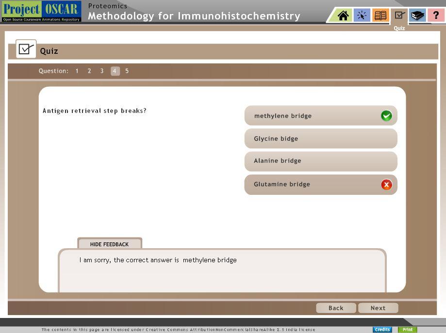
9 Antigen Retrieval Use sufficient volume of buffer in order to cover the slide. Keep a watch on the slides while boiling and avoid the slides from drying out. Less time may leave the antigens under-retrieved and more time may leave them over-retrieved. A series of experiments can help in better deciding the time for retrieving the antigen following which the user can proceed to immunostaining.


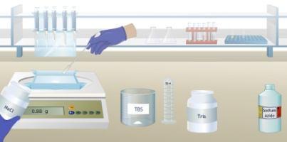
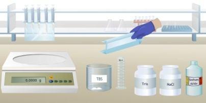
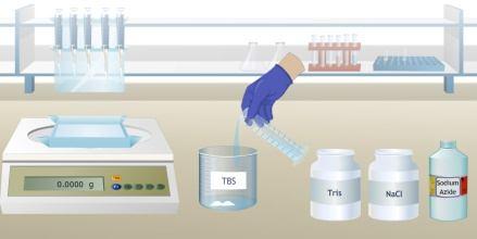
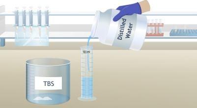
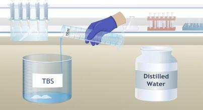
10 Staining of Sections Use sufficient volume of TBS buffer in order to cover the slide. The user can now proceed to immunostaining step.


11 Staining of Sections The concentration of working stock depends on the experiment. The working solution must be prepared in TBS depending on the concentration required for staining. The antibodies are very costly, so proper dilution for the working stock must be prepared by defining the experiment requirement. Store the working and the stock solution at -80 C.


12 Staining of Sections Each section must be treated with BSA to prevent nonspecific binding, later add primary antibody and place it for incubation overnight at 40 C. Incubation must be done in humidifier to avoid drying of sections.
13 Staining of Sections Mark the section boundary on the other side of the slide with a marker. Addition of the antibody must be sufficient enough to cover the complete section and must be within the marked boundary of the section. During primary antibody treatment, wash the slides with TBS to remove unbound primary antibodies. After primary antibody treatment, excess antibody solution must be cleaned out from the slide. Now add secondary antibody and incubate for 1hour inside a humidified chamber.


, nuclear fast red")

14 Staining of Sections The staining process depends on the user requirement, tissue used and the setup of microscope. Commonly used counterstains are hematoxylin (blue), nuclear fast red or methyl green. Once staining is over, the slide must go through dehydration step before viewing under microscope.


15 Dehydrating the Sections The dehydrating step is carried out to remove excess stain, if any, present in the slide. After the ethanol treatment, the slide with the section is placed in distilled water before proceeding to next step.


16 Microscopic Viewing The mounting solution like organic mounting media has better refractive index than aqueous mounting media. Now the slide is ready for viewing under microscope.


17 Microscopic Viewing For better image, fine adjust the microscope. Once confirmed, save the final image for use in publications and research articles.
18
19
20
21
22
23
24
Manufactured by. Zyagen Barnes Canyon Road San Diego, CA 92121, USA
 Alkaline Phosphatase Immunohistochemistry Detection kits For detection of mouse, rabbit, goat, rat, sheep, chicken, guinea pig, and human primary antibodies Size: 500 Tests Catalog #: AK-011, Mouse Kit
ab TripleStain IHC Kit: M&M&R on human tissue (DAB, Red/AP & DAB/Ni)
") ab183287 TripleStain IHC Kit: M&M&R on human tissue (DAB, Red/AP & DAB/Ni) Instructions for Use For the detection of Rabbit and Mouse Primary antibodies on Human tissue or cell samples. This product is
ab183287 TripleStain IHC Kit: M&M&R on human tissue (DAB, Red/AP & DAB/Ni) Instructions for Use For the detection of Rabbit and Mouse Primary antibodies on Human tissue or cell samples. This product is
Materials and Methods Materials Required for Fixing, Embedding and Sectioning. OCT embedding matrix (Thermo Scientific, LAMB/OCT)
") Page 1 Introduction Tissue freezing and sectioning is a rapid method of generating tissue samples (cryosections) for histological analysis, and obviates the need for wax embedding. The method is popular
Page 1 Introduction Tissue freezing and sectioning is a rapid method of generating tissue samples (cryosections) for histological analysis, and obviates the need for wax embedding. The method is popular
PREPARATION OF HISTOLOGICAL SPECIMENS
 PREPARATION OF HISTOLOGICAL SPECIMENS Histo-techniques Preparation of tissue for microscopic examination Series of processes Ultimate aim to make tissue visible as it is Pathology Vs Anatomy Steps vary
How To Optimize Your IMMUNOHISTOCHEMISTRY EXPERIMENT
 How To Optimize Your IMMUNOHISTOCHEMISTRY EXPERIMENT www.ptglab.com 2 How To Optimize Your IHC Experiment ptglab.com 3 CONTENTS 4 5 Introduction To Immunohistochemistry 6 9 General Protocols 10 11 Antigen
ab Mouse and Rabbit Specific HRP/DAB (ABC) Detection IHC Kit
 Detection IHC Kit") ab64264 - Mouse and Rabbit Specific HRP/DAB (ABC) Detection IHC Kit Instructions for Use For the detection of a specific antibody bound to an antigen in tissue sections. This product is for research use
ab64264 - Mouse and Rabbit Specific HRP/DAB (ABC) Detection IHC Kit Instructions for Use For the detection of a specific antibody bound to an antigen in tissue sections. This product is for research use
Anti-White Spot Syndrome Virus (WSSV) monoclonal antibody. Product no: P13
 monoclonal antibody. Product no: P13") Anti-White Spot Syndrome Virus (WSSV) monoclonal antibody Product no: P13 Product Description The monoclonal antibody (Mab) against the Vp28 protein of White Spot Syndrome Virus (WSSV) is specific for
Anti-White Spot Syndrome Virus (WSSV) monoclonal antibody Product no: P13 Product Description The monoclonal antibody (Mab) against the Vp28 protein of White Spot Syndrome Virus (WSSV) is specific for
Preparation of tissues for study
 Preparation of tissues for study HISTOLOGY : It is the branch of science which deals with the microscopic study of normal tissue HISTOPATHOLOGY : It is the branch of science which deals with the microscopic
BrdU IHC Kit. For the detection and localization of bromodeoxyuridine incorporated into newly synthesized DNA of actively proliferating cells
 K-ASSAY BrdU IHC Kit For the detection and localization of bromodeoxyuridine incorporated into newly synthesized DNA of actively proliferating cells Cat. No. KT-077 For Research Use Only. Not for Use in
ab BrdU Immunohistochemistry Kit
 ab125306 - BrdU Immunohistochemistry Kit Instructions for Use For the detection and localization of bromodeoxyuridine incorporated into newly synthesized DNA of actively proliferating cells. This product
ab Mouse and Rabbit AP/Fast-Red (ABC) Detection IHC Kit
 Detection IHC Kit") ab128967 - Mouse and Rabbit AP/Fast-Red (ABC) Detection IHC Kit Instructions for Use For the detection of a specific antibody bound to an antigen in tissue sections. This product is for research use only
ab128967 - Mouse and Rabbit AP/Fast-Red (ABC) Detection IHC Kit Instructions for Use For the detection of a specific antibody bound to an antigen in tissue sections. This product is for research use only
Pinpoint Slide DNA Isolation System Catalog No. D3001
 INSTRUCTIONS Pinpoint Slide DNA Isolation System Catalog No. D3001 Highlights Easily isolates genomic DNA in any targeted microscopic tissue area on a slide. The simple procedure combines Pinpoint tissue
Updated April 27, PRODUCT INSERT
 Updated April 27, 2009 1 PRODUCT INSERT Hypoxyprobe, Inc. 121 Middlesex Turnpike Burlington, MA 01803 USA www.hypoxyprobe.com Hypoxyprobe Gemini Kit Kit Contents: Solid pimonidazole HCl (Hypoxyprobe -1)
Updated PRODUCT INSERT. HPI Catalog # HP5-XXX
 Updated 2017 1 PRODUCT INSERT Hypoxyprobe, Inc. 121 Middlesex Turnpike Burlington, MA 01803 USA www.hypoxyprobe.com Hypoxyprobe Gemini Kit HPI Catalog # HP5-XXX Kit Contents: Solid pimonidazole HCl (Hypoxyprobe
Whole Mount IHC Protocol
 Whole Mount IHC Protocol Authors: Ruth Sullivan, Ryan Trevena and Kyle Wegner Creation Date: 03/17/2016 All steps should be conducted with gentle agitation on an orbital shaker, unless otherwise instructed.
Learning Objectives: Proteomics Isoelectric Focussing
 Proteomics Isoelectric Focussing Isoelectric Focussing Proteins exhibit unique iso-electric property, such unique property of the proteins are exploited for the separation into individual proteins from
PROTOCOL TO PREPARE PLANTAR FOOTSKIN FOR MORPHOMETRY. I. Removal and Fixation of Plantar Skin (see video)
") PROTOCOL TO PREPARE PLANTAR FOOTSKIN FOR MORPHOMETRY I. Removal and Fixation of Plantar Skin (see video) 1. Sacrifice the animal a. Anaesthetize the animal by placing in a closed chamber with isoflurane.
PROTOCOL TO PREPARE PLANTAR FOOTSKIN FOR MORPHOMETRY I. Removal and Fixation of Plantar Skin (see video) 1. Sacrifice the animal a. Anaesthetize the animal by placing in a closed chamber with isoflurane.
Overview of Immunohistochemistry. (with a focus on wax-embedded sections)
") Overview of Immunohistochemistry (with a focus on wax-embedded sections) Overview of Immunohistochemistry (with a focus on wax-embedded sections) Overview of Immunohistochemistry IHC is like cooking. There
Overview of Immunohistochemistry (with a focus on wax-embedded sections) Overview of Immunohistochemistry (with a focus on wax-embedded sections) Overview of Immunohistochemistry IHC is like cooking. There
ReliaPrep FFPE gdna Miniprep System
 TECHNICAL MANUAL ReliaPrep FFPE gdna Miniprep System Instructions for Use of Products A2351 and A2352 Revised 12/15 TM352 ReliaPrep FFPE gdna Miniprep System All technical literature is available at: www.promega.com/protocols/
RNAscope 2.5 LS Duplex Reagent Kit User Manual for BDZ 11. For use with Leica Biosystems BOND RX System
 RNAscope 2.5 LS Duplex Reagent Kit User Manual for BDZ 11 For use with Leica Biosystems BOND RX System For Research Use Only. Not for diagnostic use. 322440-USM Effective Date 03312017 For Research Use
Combined Digoxigenin-labeled in situ hybridization/ Immunohistochemistry protocol (for fixed frozen cryostat sections)
") Combined Digoxigenin-labeled in situ hybridization/ Immunohistochemistry protocol (for fixed frozen cryostat sections) A. Digoxigenin-UTP labeling of crna antisense probe Refer to laboratory protocol and
Combined Digoxigenin-labeled in situ hybridization/ Immunohistochemistry protocol (for fixed frozen cryostat sections) A. Digoxigenin-UTP labeling of crna antisense probe Refer to laboratory protocol and
For research use only. Not for use in diagnostic procedures.
 Dako LSAB 2 System-HRP for use on Rat Specimens Code K0609 Intended use For research use only. Not for use in diagnostic procedures. These instructions apply to the LSAB 2 System-HRP for use on RAT SPECIMENS,
ab In situ Apoptosis Detection Kit
 ab206386 In situ Apoptosis Detection Kit Instructions for Use For detection of apoptotic cells. This product is for research use only and is not intended for diagnostic use. Version 8 Last Updated 6 February
KCC Path-Core Page 1 of 5
 Instructions for Sample preparation for Paraffin embedding PLEASE NOTE: There is no one-size-fits-all method of tissue preparation for all experimental designs. Before harvesting tissue, you need to assess
TACS XL TM. In Situ Apoptosis Detection Kit. Catalog Number: TA200. DAB Kit. 30 tests FOR RESEARCH USE ONLY. NOT FOR USE IN DIAGNOSTIC PROCEDURES.
 TACS XL TM In Situ Apoptosis Detection Kit Catalog Number: TA200 DAB Kit 30 tests This package insert must be read in its entirety before using this product. FOR RESEARCH USE ONLY. NOT FOR USE IN DIAGNOSTIC
Phosphohistone-H3 (PHH3)
") Rev. 0.0 v3 Phosphohistone-H3 (PHH3) Rabbit Polyclonal Antibody PRODUCT AVAILABILITY Cat. No. Description 369A-14 0.1 ml, concentrate 369A-15 0.5 ml, concentrate 369A-16 1.0 ml, concentrate 369A-17 1.0
Rev. 0.0 v3 Phosphohistone-H3 (PHH3) Rabbit Polyclonal Antibody PRODUCT AVAILABILITY Cat. No. Description 369A-14 0.1 ml, concentrate 369A-15 0.5 ml, concentrate 369A-16 1.0 ml, concentrate 369A-17 1.0
Anti-8-hydroxyguanine antibody (Anti-8-oxo-dG)
") Anti-8-hydroxyguanine antibody (Anti-8-oxo-dG) Monoclonal Antibody for Detection of 8-oxo-dG Cat# 4359-MC-100 v.4359/0511a Anti-8-hydroxyguanine antibody (Anti-8-oxo-dG) Cat# 4359-MC-100 Monoclonal Antibody
Anti-8-hydroxyguanine antibody (Anti-8-oxo-dG) Monoclonal Antibody for Detection of 8-oxo-dG Cat# 4359-MC-100 v.4359/0511a Anti-8-hydroxyguanine antibody (Anti-8-oxo-dG) Cat# 4359-MC-100 Monoclonal Antibody
How to perform-control immunostaining experiment - microscopist subjective point of view. Pawel Pasierbek
 How to perform-control immunostaining experiment - microscopist subjective point of view. Pawel Pasierbek Immunolabeling and fluorescent detection became such a standard procedure in the biomedical research
The following materials, sufficient for processing at least 50 slides, are included in the ISH Detection System K0601. Stringent Wash Concentrate
 Dako In Situ Hybridization Detection System For Biotinylated Probes Code K0601 Intended use For In Vitro diagnostic use. The In situ Hybridization Detection System (K0601) is intended for the detection
!! PLEASE READ BEFORE USE!!
 In situ Proximity Ligation Assay protocols!! PLEASE READ BEFORE USE!! The test protocol is a guideline, user need to determine their optimal experimental condition for best performance. The following protocol
Fluorescent in-situ Hybridization
 Fluorescent in-situ Hybridization Presented for: Presented by: Date: 2 Definition In situ hybridization is the method of localizing/ detecting specific nucleotide sequences in morphologically preserved
Cell & Tissue Staining Kit
 Cell & Tissue Staining Kit For the detection of goat, mouse, rabbit, rat, or sheep primary IgG Antibodies Size: 50 Tests HRP-DAB System Goat Kit (Catalog Number CTS008) Mouse Kit (Catalog Number CTS002)
TheraLin. Universal Tissue Fixative Enabling Molecular Pathology
 TheraLin Universal Tissue Fixative Enabling Molecular Pathology TheraLin Universal Tissue Fixative Enabling Molecular Pathology Contents Page # TheraLin Universal Tissue Fixative 3 Introduction 5 Easy
Isolation, culture, and transfection of primary mammary epithelial organoids
 Supplementary Experimental Procedures Isolation, culture, and transfection of primary mammary epithelial organoids Primary mammary epithelial organoids were prepared from 8-week-old CD1 mice (Charles River)
RNAScope Multiplex Reagent Kit For Tissues
 RNAScope Multiplex Reagent Kit For Tissues Catalog no. R26963 Table 1 Contents and storage Component Amount* Storage** Pretreatment Module Pretreat 2 reagent, 10 4 70 ml Room temperature Pretreat 3 reagent
Immunohistochemistry. How does it look like? When do we need IHC? When do we need IHC? In clinic: In research:
 Introduction How does it look like? Immunohistochemistry Smooth muscle actin Parvalbumin Distrophyn Sandrine Bichet Head of Molecular Histology Platform Signal versus background 06.03.2012 IHC basics Introduction
Immunohistochemistry: Basics and Methods
 Immunohistochemistry: Basics and Methods Bearbeitet von Igor B Buchwalow, Werner Böcker 1st Edition. 2010. Buch. x, 153 S. Hardcover ISBN 978 3 642 04608 7 Format (B x L): 15,5 x 23,5 cm Gewicht: 445 g
Procedure for Immunohistochemical Examination
 Procedure for Immunohistochemical Examination 1. Preparation of Paraffin Block (Separate Attachment 2-2) Disposable bench protection sheet, cutting board, sectioning blade, tweezers, stainless
Strep-tag detection in Western blots
 Strep-tag detection in Western blots General protocol for the detection of Strep-tag fusion proteins Last date of revision April 2012 Version PR07-0010 www.strep-tag.com For research use only Important
USER MANUAL. Fluorescence
 USER MANUAL Fluorescence The protocols in this manual are compatible with all Duolink II PLA probes, Duolink II Detection Reagents Green (art no 92014), Orange (art no. 92007), Red (art no. 92008) and
Anti-8-hydroxyguanine antibody (Anti-8-oxo-dG) Cat #: 4355-MC-100 Monoclonal Antibody for Detection of 8-oxo-dG. Contents
 Cat #: 4355-MC-100 Monoclonal Antibody for Detection of 8-oxo-dG. Contents") Instructions for Use For Research Use Only Anti-8-hydroxyguanine antibody (Anti-8-oxo-dG) Cat #: Monoclonal Antibody for Detection of 8-oxo-dG Contents Section Page I Introduction 2 II Reagents Required
Instructions for Use For Research Use Only Anti-8-hydroxyguanine antibody (Anti-8-oxo-dG) Cat #: Monoclonal Antibody for Detection of 8-oxo-dG Contents Section Page I Introduction 2 II Reagents Required
HAM-56 antibody reacts with monocytes, but is unreactive with B and T-lymphocytes. Principles And Procedures
 Product Identification Cat. No. Description 44682 Macrophage Marker 0,1 M (HAM- 56) 44683 Macrophage Marker 1 M (HAM-56) 44330 Macrophage Marker RTU M (HAM- 56) Symbol Definitions P C A E S DIL DOC# DIS
Histological preparation of embryonic and adult zebrafish eyes
 Histological preparation of embryonic and adult zebrafish eyes Richard J. Nuckels 1 and Jeffrey M. Gross 1,2,3 1 Section of Molecular Cell and Developmental Biology 2 Institute of Cell and Molecular Biology
Segments of the obstructed intestinal loops were fixed in 4% paraformaldehyde
 Supplementary text Supplementary materials and methods Histopathological examination Segments of the obstructed intestinal loops were fixed in 4% paraformaldehyde (PFA) and embedded in paraffin wax with
Product # Specifications. Kit Specifications Maximum Column Binding Capacity (gdna) Maximum Column Loading Volume.
 Maximum Column Loading Volume.") 3430 Schmon Parkway Thorold, ON, Canada L2V 4Y6 Phone: 866-667-4362 (905) 227-8848 Fax: (905) 227-1061 Email: techsupport@norgenbiotek.com FFPE DNA Purification Kit Product # 47400 Product Insert Norgen
3430 Schmon Parkway Thorold, ON, Canada L2V 4Y6 Phone: 866-667-4362 (905) 227-8848 Fax: (905) 227-1061 Email: techsupport@norgenbiotek.com FFPE DNA Purification Kit Product # 47400 Product Insert Norgen
truxtrac FFPE DNA microtube Kit Column Purification (25)
") PROTOCOL truxtrac FFPE DNA microtube Kit Column Purification (25) Adaptive Focused Acoustics (AFA) -based DNA extraction and purification from Formalin-Fixed, Paraffin-Embedded Tissue using columns Product
PROTOCOL truxtrac FFPE DNA microtube Kit Column Purification (25) Adaptive Focused Acoustics (AFA) -based DNA extraction and purification from Formalin-Fixed, Paraffin-Embedded Tissue using columns Product
Qdot Conjugates Protocol Handbook
 Qdot Conjugates Protocol Handbook PN 90-0153, Rev 1.2 Table of Contents Immunocytochemistry with Qdot Conjugates in Cultured Cells 2 Immunolabeling Formalin-fixed Paraffin Embedded Tissue Sections.8 Immunolabeling
Immunofluorescence Staining Protocol for 3 Well Chamber, removable
 Immunofluorescence Staining Protocol for 3 Well Chamber, removable This Application Note presents a simple protocol for the cultivation, fixation, and staining of cells using the 3 Well Chamber, removable.
PreAnalytiX Supplementary Protocol
 PreAnalytiX Supplementary Protocol Purification of full-length proteins from sections of PAXgene Tissue fixed, paraffin-embedded (PFPE) tissue This protocol describes using the Qproteome FFPE Tissue kit
LAMININ. For Immunohistochemical Demonstration of Laminin in Paraffin-embedded and Frozen Human Tissue Sections Stock No. IMMH-7
 LAMININ For Immunohistochemical Demonstration of Laminin in Paraffin-embedded and Frozen Human Tissue Sections Stock No. IMMH-7 TABLE OF CONTENTS BACKGROUND AND PRINCIPLE... 4 REAGENTS AND EQUIPMENT PROVIDED...
(Spin Column) For purification of DNA and RNA from formalin-fixed, paraffin-embedded tissue sections. Instruction for Use
 For purification of DNA and RNA from formalin-fixed, paraffin-embedded tissue sections. Instruction for Use") AmoyDx FFPE DNA/RNA Kit (Spin Column) For purification of DNA and RNA from formalin-fixed, paraffin-embedded tissue sections Instruction for Use Instruction Version: B1.3 Revision Date: September 2015
AmoyDx FFPE DNA/RNA Kit (Spin Column) For purification of DNA and RNA from formalin-fixed, paraffin-embedded tissue sections Instruction for Use Instruction Version: B1.3 Revision Date: September 2015
Just SNAP and go! SNAP i.d. 2.0 system for Western blotting and IHC
 Just SNAP and go! SNAP i.d. 2.0 system for Western blotting and IHC EMD Millipore is a division of Merck KGaA, Darmstadt, Germany Multiple slides, multiple blots, multiple conditions. There s so much room
Anti-Human IDH1 R132H Astrocytoma, Oligodendroglioma Tumor Cell Marker Mouse Monoclonal Antibody Clone H09
 1 of 5 Technical Note 1 Procedure: Automated Immunostaining Ventana Benchmark XT XT ultraview DAB DIA--M (20µg) Reconstitution: DIA- (100µg), restore to 500µl Summary 1. Cut sections to 4 µm (Microm HM
Web Based Promotion! 10% Off any initial product order. Mention promo code 1204!
 Web Based Promotion! 10% Off any initial product order. Mention promo code 1204! Volume 2, Number 2 1998 FROM PATIENT TO EMBEDDING CENTER IN TWO HOURS OR LESS The single biggest factor in health care today
Orexin A (HUMAN, MOUSE, RAT, PORCINE, OVINE,
 Orexin A (HUMAN, MOUSE, RAT, PORCINE, OVINE, BOVINE) Western Blot Kit Protocol (Catalog #WBK-003-30) PHOENIX PHARMACEUTICALS, INC. TABLE OF CONTENTS 1. Kit Contents...2 2. Storage...2 3. Introduction...3
Comparing the Quality of Fixation for Gel-based Formalin (Formagel) versus Traditional Liquid-Based Formalin for Immunohistochemistry
 versus Traditional Liquid-Based Formalin for Immunohistochemistry") Comparing the Quality of Fixation for Gel-based Formalin (Formagel) versus Traditional Liquid-Based Formalin for Immunohistochemistry Brian H. Le, M.D., Reading Hospital Reviewed by Michael R. LaFrinere,
Comparing the Quality of Fixation for Gel-based Formalin (Formagel) versus Traditional Liquid-Based Formalin for Immunohistochemistry Brian H. Le, M.D., Reading Hospital Reviewed by Michael R. LaFrinere,
celldatasci.com/rnastorm RNAstorm RNA Isolation Kit for FFPE Tissue Samples Sample Kit (20 extractions)
") celldatasci.com/rnastorm info@celldatasci.com RNAstorm RNA Isolation Kit for FFPE Tissue Samples Sample Kit (20 extractions) Support: Email: support@celldatasci.com Phone: 650.285.2376 (option 2) Toll-free:
celldatasci.com/rnastorm info@celldatasci.com RNAstorm RNA Isolation Kit for FFPE Tissue Samples Sample Kit (20 extractions) Support: Email: support@celldatasci.com Phone: 650.285.2376 (option 2) Toll-free:
truxtrac FFPE DNA microtube Kit - Bead Purification (24)
") truxtrac FFPE DNA microtube Kit - Bead Purification (24) Adaptive Focused Acoustics (AFA) -based DNA extraction and purification from Formalin-Fixed, Paraffin-Embedded (FFPE) Tissue using Magnetic Beads
truxtrac FFPE DNA microtube Kit - Bead Purification (24) Adaptive Focused Acoustics (AFA) -based DNA extraction and purification from Formalin-Fixed, Paraffin-Embedded (FFPE) Tissue using Magnetic Beads
ab66110 In situ BrdU-Red DNA Fragmentation (TUNEL) Assay Kit
 Assay Kit") Version 9b Last updated 28 November 2017 ab66110 In situ BrdU-Red DNA Fragmentation (TUNEL) Assay Kit For the detection of DNA fragmentation in apoptosis by flow cytometry and fluorescence microscopy in
Version 9b Last updated 28 November 2017 ab66110 In situ BrdU-Red DNA Fragmentation (TUNEL) Assay Kit For the detection of DNA fragmentation in apoptosis by flow cytometry and fluorescence microscopy in
SANTA CRUZ BIOTECHNOLOGY, INC.
 TECHNICAL SERVICE GUIDE: Western Blotting 2. What size bands were expected and what size bands were detected? 3. Was the blot blank or was a dark background or non-specific bands seen? 4. Did this same
TrkB knockdown cell lines (i.e., BBM1-KD and 361-KD cells) were prepared by
 were prepared by") Supplemental Information Cell Culture TrkB knockdown cell lines (i.e., BBM1-KD and 361-KD cells) were prepared by transducing BBM1 or 361 cells with a lentivirus encoding shrna for TrkB. Transduction was
Supplemental Information Cell Culture TrkB knockdown cell lines (i.e., BBM1-KD and 361-KD cells) were prepared by transducing BBM1 or 361 cells with a lentivirus encoding shrna for TrkB. Transduction was
Whole Mount In Situ Hybridization Protocol
 Whole Mount In Situ Hybridization Protocol Purpose of in situs: To use an RNA probe to stain for the specific complementary mrna of an intact embryo, allowing visualization of mrna patterns. Note: When
Introduction to Fluorescent In Situ Hybridization (FISH)
") Robert Driscoll, M.F.S. Heather Cunningham, M.S. Introduction to Fluorescent In Situ Hybridization (FISH) Fluorescent In Situ Hybridization (FISH) FISH is a cytogenetic technique used to detect the presence
Robert Driscoll, M.F.S. Heather Cunningham, M.S. Introduction to Fluorescent In Situ Hybridization (FISH) Fluorescent In Situ Hybridization (FISH) FISH is a cytogenetic technique used to detect the presence
TACS 2 TdT-DAB In Situ Apoptosis Detection Kit
 Instructions For Research Use Only. Not For Use In Diagnostic Procedures TACS 2 TdT-DAB In Situ Apoptosis Detection Kit Reagent kit for in situ detection of apoptosis in tissue sections and cells. 30 samples
BCH 462. Western Blot
 BCH 462 Western Blot Blotting Immunoassay: A test that uses antibody and antigen complexes [immuno-complexes] as a means of generating measurable results. Antigens [Ag]: A substance that when introduced
Extraction of Nucleic Acids
 Maj Gen (R) Suhaib Ahmed, HI (M) DNA Extraction The first step in DNA analysis is to get a good quality sample; a process commonly known as extraction. Since DNA is a very long molecule it can easily get
UltraMap Alk Phos. UltraMap anti-ms Alk Phos
 UltraMap anti-rb Alk Phos UltraMap anti-ms Alk Phos Biotin-free Alkaline Phosphatase Detection Systems for the Ventana DISCOVERY Series of Instruments Catalog Number: s 760-4314 (UltraMap anti-rb Alk Phos)
NAG-1 (GDF-15, MIC-1) Prostate Cancer ELISA kit
 Prostate Cancer ELISA kit") NAG-1 (GDF-15, MIC-1) Prostate Cancer ELISA kit Catalog Number: NG1 Store at -0 C. FOR RESEARCH USE ONLY v. 1081 Introduction This sandwich ELISA kit is for determination of NAG-1 (GDF-15, MIC-1) levels
NAG-1 (GDF-15, MIC-1) Prostate Cancer ELISA kit Catalog Number: NG1 Store at -0 C. FOR RESEARCH USE ONLY v. 1081 Introduction This sandwich ELISA kit is for determination of NAG-1 (GDF-15, MIC-1) levels
Baraa Ayed AL-Odat. Israa Ayed. Heba kalbouneh
 1 Baraa Ayed AL-Odat Israa Ayed Heba kalbouneh Introduction: "lecture #1" The name " histology " is derived from the Greek words: "histo" means a tissue and "logos" means the study of. So, Histology mean
Human Pluripotent Stem Cell Functional Identification Kit
 Human Pluripotent Stem Cell Functional Identification Kit Catalog Number SC027B Reagents for the identification of human pluripotent stem cells by in vitro functional differentiation. This package insert
ab CFSE Fluorescent Cell Labeling Kit
 ab113853 CFSE Fluorescent Cell Labeling Kit Instructions for Use For the durable fluorescent labeling of live cells for fluorescent microscopy and flow cytometry, population growth studies and within sample
Department of Microbiology, Lab 016 instructions
 Protocol for standard FISH and DOPE-FISH for prokaryotes (slightly modified from Amann, 1995, note, for other modifications or other microorganisms like eukaryotes, consult special literature or check
The preparation of native chromatin from cultured human cells.
 Native chromatin immunoprecipitation protocol The preparation of native chromatin from cultured human cells. All solutions need to be ice cold. Sucrose containing solutions must be made up fresh on the
Anti-Nodavirus monoclonal antibody. Product no: P09
 Anti-Nodavirus monoclonal antibody Product no: P09 Product Description The monoclonal antibody (Mab) against Nodavirus is specific for this virus. The MAb has been tested by ELISA with Betanodavirus isolated
Histopathological techniques The adoption of routine fixation and paraffin wax embedding.
 CIHRT Exhibit P-3359 Page 1 Routine tissue preparation in modern diagnostic histopathology. Bryan R. Hewlett ART, MLT. Histopathological techniques The adoption of routine fixation and paraffin wax embedding.
High Pure PCR Template Preparation Kit for preparation of 100 nucleic acid samples Cat. No
 for preparation of 100 nucleic acid samples Cat. No. 1 796 88 Principle Cells are lysed during a short incubation with Proteinase K in the presence of a chaotropic salt (guanidine HCl), which immediately
RNAscope Sample Preparation and Pretreatment Guide for FFPE Tissue
 USER MANUAL RNAscope Sample Preparation and Pretreatment Guide for FFPE Tissue PART 1 Catalog Number 320511 Compatible with the following User Manuals: RNAscope 2.0 HD Detection Kit BROWN (Catalog No.
(Last update: 1 August 2000, Ver 1.4)
") In-situ hybridization using GeneDetect TM oligonucleotide probes DIG-labeled probe, paraffin tissue sections, Detection by AP (alkaline phosphatase), FITC or rhodamine. (Last update: 1 August 2000, Ver
In-situ hybridization using GeneDetect TM oligonucleotide probes DIG-labeled probe, paraffin tissue sections, Detection by AP (alkaline phosphatase), FITC or rhodamine. (Last update: 1 August 2000, Ver
Actin, Muscle Specific (HHF35)
") Product Identification Cat. No. Description 45235 IMPATH Actin Muscle Specific RTU M (HHF35) Symbol Definitions P A E S DOC# DIS ready-to-use ascites serum supernatant document number distributed by Intended
Product Identification Cat. No. Description 45235 IMPATH Actin Muscle Specific RTU M (HHF35) Symbol Definitions P A E S DOC# DIS ready-to-use ascites serum supernatant document number distributed by Intended
I n s t ru c t i o n s D AKO LSAB 2 System, P e r o x i d a s e
 I n s t ru c t i o n s D AKO LSAB 2 System, P e r o x i d a s e Universal Code No. K0672 K0673 K0675 Size 15 ml 15 ml 110 ml For Laboratory Use. 1. INTENDED USE FOR LABORATORY USE These instructions apply
#$% &%" ' " ( ) * % '-'
 * % '-'") !" #$% &%" ' " ( ) * '++, % '-' '"%!./,0012 DISPOSITION INSTRUCTIONS: Destroy this report when no longer needed. Do not return to the originator. DISCLAIMERS: The opinions or assertions contained herein
!" #$% &%" ' " ( ) * '++, % '-' '"%!./,0012 DISPOSITION INSTRUCTIONS: Destroy this report when no longer needed. Do not return to the originator. DISCLAIMERS: The opinions or assertions contained herein
Working Reagent Preparation 1:1,000 dilution in standard Alcoholic Ortho- Phosphoric acid staining solution
 www.smobio.com Product Information FluoroStain Protein Fluorescent Staining Dye (Red, 1,000X) PS1000 1 ml x 1 PS1001 1 ml x 5 Storage Protected from light -20 C for 24 months Working Reagent Preparation
Immunofluorescence of organoids embedded in Basement Membrane Matrix
 Immunofluorescence of organoids embedded in Basement Membrane Matrix Sol Degese 1, Gabe Benton 1 1 Organoid Resource Lab (ORL), Trevigen, Inc., 8405 Helgerman Court, Gaithersburg, MD 20877 Introduction
Stellaris RNA FISH Protocol for Simultaneous IF + FISH in Adherent Cells
 Stellaris RNA FISH Protocol for Simultaneous IF + FISH in Adherent Cells General Protocol & Storage Product Description A set of Stellaris RNA FISH Probes is comprised of up to 48 singly labeled oligonucleotides
Human Placental Lactogen (hpl) (Polyclonal)
 (Polyclonal)") Product Identification Cat. No. Description 45312 IMPATH Hpl RTU R (Poly) Symbol Definitions P A E S DOC# DIS ready-to-use ascites serum supernatant document number distributed by Intended Use This antibody
Product Identification Cat. No. Description 45312 IMPATH Hpl RTU R (Poly) Symbol Definitions P A E S DOC# DIS ready-to-use ascites serum supernatant document number distributed by Intended Use This antibody
Actin, Smooth Muscle (1A4)
") Product Identification Cat. No. Description 45121 IMPATH Actin Smooth Muscle RTU M (1A4) Symbol Definitions P A E S DOC# DIS ready-to-use ascites serum supernatant document number distributed by Intended
Product Identification Cat. No. Description 45121 IMPATH Actin Smooth Muscle RTU M (1A4) Symbol Definitions P A E S DOC# DIS ready-to-use ascites serum supernatant document number distributed by Intended
IMMUNOPRECIPITATION TROUBLESHOOTING TIPS
 IMMUNOPRECIPITATION TROUBLESHOOTING TIPS Creative Diagnostics Abstract Immunoprecipitation (IP) is the technique of precipitating a protein antigen out of solution using an antibody that specifically binds
EZ-10 SPIN COLUMN GENOMIC DNA MINIPREPS KIT HANDBOOK
 EZ-0 SPIN COLUMN GENOMIC DNA MINIPREPS KIT HANDBOOK (Bacteria, Plant, Animal, Blood) Version 8 Rev 05/0/03 EZ-0 Genomic DNA Kit Handbook Table of Contents Introduction Limitations of Use Features Applications
Western BLoT Ultra Sensitive HRP Substrate
 Cat. # T7104A For Research Use Western BLoT Ultra Sensitive Product Manual Table of Contents I. Description... 3 II. Components... 3 III. Storage... 3 IV. Materials Required but Not Provided... 3 V. Precautions...
In-Gel Western Detection Using Near-Infrared Fluorescence
 In-Gel Western Detection Using Near-Infrared Fluorescence Developed for: Aerius, and Odyssey Family of Imagers Please refer to your manual to confirm that this protocol is appropriate for the applications
Immunohistochemical Detection With Quantum Dots
 2 Immunohistochemical Detection With Quantum Dots Rizwan S. Akhtar, Cecelia B. Latham, Dario Siniscalco, Carlo Fuccio, and Kevin A. Roth Summary Quantum dot (QD) conjugates have many immunohistochemical
HiPer Sandwich ELISA Teaching Kit
 HiPer Sandwich ELISA Teaching Kit Product Code: HTI014 Number of experiments that can be performed: 4 Duration of Experiment: 2 days Day1-Coating of wells: 15 minutes Day2- protocol, observation and result:
The Effect of Antigen Retrieval on Cells Fixed in 10% Neutral Buffered Formalin Followed by Transfer to 70% Ethanol
 The Effect of Antigen Retrieval on Cells Fixed in 10% Neutral Buffered Formalin Followed by Transfer to 70% Ethanol Dennis Otali 1,2, Qinghua He 3, William E. Grizzle 1 * 1 Department of Pathology, University
Immunohistochemistry: Basics and Methods
 Immunohistochemistry: Basics and Methods Igor B. Buchwalow l Werner Böcker Immunohistochemistry: Basics and Methods Prof. Dr. Igor B. Buchwalow Prof. Dr. Werner Böcker Gerhard-Domagk-Institut für Pathologie
Lab 1 Microdissection 1. Microdissection
 Lab 1 Microdissection 1 Lab 1. Microdissection The goal of this techniques lab is to understand how to dissect and process tissues that have been incubated in bromodeoxyuridine (BrdU). BrdU is a synthetic
Immunohistochemical pitfalls in the demonstration of insulindegrading enzyme in normal and neoplastic human tissues
 Immunohistochemical pitfalls in the demonstration of insulindegrading enzyme in normal and neoplastic human tissues RAZVAN T. RADULESCU 1*, ANGELIKA JAHN 1, DANIELA HELLMANN 1, AND GREGOR WEIRICH 2 1 Clinical
Principles And Procedures
 Product Identification Cat. No. Description 44837 Vimentin 0,1 M (V9) 44838 Vimentin 1 M (V9) 44409 Vimentin RTU M (V9) Symbol Definitions P C A E S DIL DOC# DIS ready-to-use concentrate ascites serum
QuickExtract FFPE RNA Extraction Kit
 QuickExtract FFPE RNA Extraction Kit Cat. Nos. QFR82805 and QFR82050 Connect with Epicentre on our blog (epicentral.blogspot.com), Facebook (facebook.com/epicentrebio), and Twitter (@EpicentreBio). www.epicentre.com
Ion Exchange Chromatography. Learning Objectives:
 Proteomics Ion Exchange Chromatography Ion Exchange Chromatography Ion exchange chromatography is a purification technique, which involves the separation of the proteins based on the ion exchange property