Confocal Microscopy & Imaging Technology. Yan Wu
|
|
|
- Rhoda Bailey
- 6 years ago
- Views:
Transcription


1 Confocal Microscopy & Imaging Technology Yan Wu Dec. 05, 2014
2 Cells under the microscope What we use to see the details of the cell? Light and Electron Microscopy - Bright light / fluorescence microscopy - Confocal Laser Scanning microscopy - Transmission Electron Microscopy (TEM) - Scanning Electron Microscopy (SEM)
3 Light microcopy Light microscopes - Epi-light microscope - Inverted microscopes - Dissection microscopes Fluorescent microscope - incorporated in light microscopes


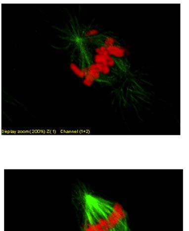
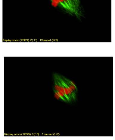
4 Confocal Laser Scanning Microscopy A fluorescence microscope with a laser as its source of illumination actin dynamic A guard cell development in Arabidopsis


5 Filter cube D.M. The excitation light reflects off the surface of the dichroic mirror into the objective. the fluorescence emission passes through the dichroic to the eyepiece or detection system. Dichroic mirror separates excitation and emission light paths.
6 Optical system of a fluorescence microscope Human eyes emitter v exciter only BLUE light is allowed to pass through Dic Figure 9-13 Molecular Biology of the Cell ( Garland Science 2008)

7 The most used fluorescence dyes DAPI CFP GFP YFP Rhodamine B Cy3 Alexa 568 RFP Cy5
8 Confocal Laser Scanning Microscopy (CLSM) Confocal Laser Scanning Microscopy (CLSM) one of a series of methods to generate slices from microscopic samples by means of optics. The sample stays intact, and the slicing may be repeated many times. The benefit of confocal imaging is a dramatically increased contrast by removal of out-of-focus haze. Z-sequences of optical slices (3D image stacks) are sources for subsequent rendering as anaglyphes, depth-coded maps or 3D movies.
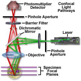
9 Confocal Microscope Inverted microscope Upright microscope Detailed info about FV1000: on/product.asp?product=1008&c=6 Imaging of complex 3D objects is possible with the Confocal Scanning Microscopy


10 光栅 Pinhole PMT 狭缝 Laser types: 1. Argon (gas) 2. LD

11 FV1000 ASW software


12 SIM - SIMultaneous scaner Evolved light stimulation The stimulation/imaging positions and laser wavelengths can be set separately with two independent beams.
13 Confocal Microscopy


14 光学切片 -3D 结构的准确定位 Projection - 3D
15 The confocal microscopes in College of Life Sciences FV 1000 linked with IX81 (inverted microscope) FV 1000 linked with BX61 (upright microscope) Leica TCS SP8 with inverted microscope Analyzing software, MetaMorph (Molecular Devices, USA)
16 Super-resolved fluorescence microscopy Surpassing the limitations of the light microscope The limitation of the light microscope, - ABBE S diffraction limit (0.2 µm)

17 Stimulated Emission Depletion, developed by Stefan Hell in 2000 Two laser beams utilized, one stimulates fluorescent molecules to glow, another cancels out all fluorescence except for that in a nanometresized volume. yields an image with a resolution better than Abbe s stipulated limit. Scanning over the sample, nanometre for nanometre
18 The diffraction limit PSF (point spread function): - The 3D intensity distribution of the image of a point object is called PSF The size of the PSF determines the resolution of the microscope: - Two points closer than the full width at half-maximum (FWHM) of the PSF will be difficult to resolve because their images overlap substantially. The diffraction limit of resolution in light microscopy does not affect most imaging at the organ or tissue level. However, when zooming into cells, where a large number of subcellular structures are smaller than the wavelength of the light, it becomes an obstacle for studying these structures in detail. Therefore, it is important to develop techniques that improve the spatial resolution of light microscopy without compromising its noninvasiveness and biomolecular specificity.
19 The Abbe diffraction limit for a microscope Ernst Abbe found in 1873 that light with wavelength λ, traveling in a medium with refractive index n and converging to a spot with angle θ will make a spot radius, d = λ/2n sin θ denominator n sin θ is called the numerical aperture (NA) can reach about in modern optics, hence the Abbe limit is d = λ/2x1.4 = λ/2.8 Considering green light around 500 nm and a NA of 1, the Abbe limit is roughly d = λ/2 = 250 nm (0.25 µm) To increase the resolution, shorter wavelengths can be used such as UV, X-ray microscopes Nowadays, Super-resolved microscopy is invented
20 Stimulated emission depletion microscopy The concept of STED microscopy was first proposed in 1994 and subsequently demonstrated experimentally it uses a second laser (STED laser) to suppress the fluorescence emission from the fluorophores located off the center of the excitation. This suppression is achieved through stimulated emission: When an excited-state fluorophore encounters a photon that matches the energy difference between the excited and the ground state, it can be brought back to the ground state through stimulated emission before spontaneous fluorescence emission occurs. This process effectively depletes excited-state fluorophores capable of fluorescence emission

21 single-molecule microscopy by Eric Betzig and William Moerner in 2006 Upon the possibility to turn the fluorescence of individual molecules on and off, scientists image the same area multiple times, letting just a few interspersed molecules glow each time. Superimposing these images yields a dense super-image resolved at the nanolevel. nanoscopy
22 Principles of super-resolution singlemolecule active control microscopy In conventional fluorescence microscopy, all molecules emit simultaneously, so their diffraction-limited images overlap on the detector (camera) and information about the underlying structure is irretrievably lost. Addition of on-off control, toggling any one singlemolecule emitter between a dark and a fluorescent state. If individual sparse subsets of single molecules that are spatially separated further than the diffraction limit are made to emit, their positions may be extracted in a time-sequential manner by finding the center position of a mathematical fit of the single-molecule images.
23 Exemplary imaging technology - to study gene function in the cell FRET Ratio imaging Bimolecular fluorescence complementation (BiFC)
24 Why FRET Dynamical detection of protein-protein interaction Visualization of protein-protein interaction in live It is important in study of special temporal activities of protein protein interactions Intramolecular association
25 Why Ratio Imaging Localization = activation Ratio imaging eliminates volume and concentration artifacts Overexpression of target not necessary direct excitation of dye (bright signal) flexible wavelengths Endogenous protein activation
26
Super-resolution Microscopy
 Semr oc kwhi t epaperser i es : 1. Introduction Super-resolution Microscopy Fluorescence microscopy has revolutionized the study of biological samples. Ever since the invention of fluorescence microscopy
Confocal Microscopes. Evolution of Imaging
 Confocal Microscopes and Evolution of Imaging Judi Reilly Hans Richter Massachusetts Institute of Technology Environment, Health & Safety Office Radiation Protection What is Confocal? Pinhole diaphragm
Partha Roy
 Fluorescence microscopy http://micro.magnet.fsu.edu/primer/index.html Partha Roy 1 Lecture Outline Definition of fluorescence Common fluorescent reagents Construction ti of a fluorescence microscope Optical
Simultaneous multi-color, multiphoton fluorophore excitation using dual-color fiber lasers
 Multiphoton Microscopy / Fiber Laser Simultaneous multi-color, multiphoton fluorophore excitation using dual-color fiber lasers Matthias Handloser, Tim Paasch-Colberg, Bernhard Wolfring TOPTICA Photonics
Practical light microscopy: an introduction
 Practical light microscopy: an introduction Dr. Mark Leake, Oxford University www.physics.ox.ac.uk/users/leake Aim of today s talk: Explanation of the very (very) basics of how a light microscope works
Resolution of Microscopes Visible light is nm Dry lens(0.5na), green(530nm light)=0.65µm=650nm for oil lens (1.4NA) UV light (300nm) = 0.13µm f
, green(530nm light)=0.65µm=650nm for oil lens (1.4NA) UV light (300nm) = 0.13µm f") Microscopes and Microscopy MCB 380 Good information sources: Alberts-Molecular Biology of the Cell http://micro.magnet.fsu.edu/primer/ http://www.microscopyu.com/ Approaches to Problems in Cell Biology
Microscopes and Microscopy MCB 380 Good information sources: Alberts-Molecular Biology of the Cell http://micro.magnet.fsu.edu/primer/ http://www.microscopyu.com/ Approaches to Problems in Cell Biology
Visualizing Cells Molecular Biology of the Cell - Chapter 9
 Visualizing Cells Molecular Biology of the Cell - Chapter 9 Resolution, Detection Magnification Interaction of Light with matter: Absorbtion, Refraction, Reflection, Fluorescence Light Microscopy Absorbtion
MICROSCOPY. "micro" (small) "scopeo" (to watch)
 scopeo (to watch)") MICROSCOPY "micro" (small) "scopeo" (to watch) THE RELATIVE SIZES OF MOLECULES, CELLS AND ORGANISMS THE RELATIVE SIZES OF MOLECULES, CELLS AND ORGANISMS MICROSCOPY 1590 2012 MICROSCOPY THE LIGHT Light:
MICROSCOPY "micro" (small) "scopeo" (to watch) THE RELATIVE SIZES OF MOLECULES, CELLS AND ORGANISMS THE RELATIVE SIZES OF MOLECULES, CELLS AND ORGANISMS MICROSCOPY 1590 2012 MICROSCOPY THE LIGHT Light:
Nodes of regulation in cellular systems
 Nodes of regulation in cellular systems cell membrane signal transduction ligands receptors oligomerization transport signal transduction modified protein Golgi transcription factor transport ER transport
Confocal Microscopy Analyzes Cells
 Choosing Filters for Fluorescence A Laurin Publication Photonic Solutions for Biotechnology and Medicine November 2002 Confocal Microscopy Analyzes Cells Reprinted from the November 2002 issue of Biophotonics
Fluorescence Nanoscopy
 Fluorescence Nanoscopy Keith A. Lidke University of New Mexico panda3.phys.unm.edu/~klidke/index.html Optical Microscopy http://en.wikipedia.org/wiki/k%c3%b6hler_illumination 30 µm Fluorescent Probes Michalet
Rice/TCU REU on Computational Neuroscience. Fundamentals of Molecular Imaging
 Rice/TCU REU on Computational Neuroscience Fundamentals of Molecular Imaging June 2, 2009 Neal Waxham 713-500-5621 m.n.waxham@uth.tmc.edu Objectives Introduction to resolution in light microscopy Brief
Stochastic Optical Reconstruction Microscopy (STORM): A Method for Superresolution Fluorescence Imaging
: A Method for Superresolution Fluorescence Imaging") Topic Introduction Stochastic Optical Reconstruction Microscopy (STORM): A Method for Superresolution Fluorescence Imaging Mark Bates, Sara A. Jones, and Xiaowei Zhuang The relatively low spatial resolution
Topic Introduction Stochastic Optical Reconstruction Microscopy (STORM): A Method for Superresolution Fluorescence Imaging Mark Bates, Sara A. Jones, and Xiaowei Zhuang The relatively low spatial resolution
Spectral Separation of Multifluorescence Labels with the LSM 510 META
 Microscopy from Carl Zeiss Spectral Separation of Multifluorescence Labels with the LSM 510 META Indians living in the South American rain forest can distinguish between almost 200 hues of green in their
Performance of the Micro Photon Devices PDM 50CT SPAD detector with PicoQuant TCSPC systems
 Technical Note Performance of the Micro Photon Devices PDM 5CT SPAD detector with PicoQuant TCSPC systems Rolf Krahl, Andreas Bülter, Felix Koberling, PicoQuant GmbH These measurements were performed to
Imagerie et spectroscopie de fluorescence par excitation non radiative
 Imagerie et spectroscopie de fluorescence par excitation non radiative comment s affranchir de la limite de diffraction Rodolphe Jaffiol, Cyrille Vézy, Marcelina Cardoso Dos Santos LNIO, UTT, Troyes NanoBioPhotonics
Confocal Microscopy of Electronic Devices. James Saczuk. Consumer Optical Electronics EE594 02/22/2000
 Confocal Microscopy of Electronic Devices James Saczuk Consumer Optical Electronics EE594 02/22/2000 Introduction! Review of confocal principles! Why is CM used to examine electronics?! Several methods
The analysis of fluorescence microscopy images for FRET detection
 The analysis of fluorescence microscopy images for FRET detection Ela Claridge, Dale J. Powner and Michael J.O. Wakelam School of Computer Science, The University of Birmingham B5 2TT Institute for Cancer
Next Level of Super Resolution Fluorescence Microscopy
 Work in your familiar GFP/YFP/RFP system from the first experiment to the nanoimage Bwcon business award winner: Inventor Prof Christoph Cremer Next Level of Super Resolution Fluorescence Microscopy Resolution:
a) JOURNAL OF BIOLOGICAL CHEMISTRY b) PNAS c) NATURE
 JOURNAL OF BIOLOGICAL CHEMISTRY b) PNAS c) NATURE") a) JOURNAL OF BIOLOGICAL CHEMISTRY b) c) d) ........................ JOURNAL OF BIOLOGICAL CHEMISTRY MOLECULAR PHARMACOLOGY TRENDS IN PHARMACOLOGICAL S AMERICAN JOURNAL OF PHYSIOLOGY-HEART AND CIRCULATORY
a) JOURNAL OF BIOLOGICAL CHEMISTRY b) c) d) ........................ JOURNAL OF BIOLOGICAL CHEMISTRY MOLECULAR PHARMACOLOGY TRENDS IN PHARMACOLOGICAL S AMERICAN JOURNAL OF PHYSIOLOGY-HEART AND CIRCULATORY
Advanced fluorescence microscopy techniques
 Practice-oriented, student-friendly modernization of the biomedical education for strengthening the international competitiveness of the rural Hungarian universities TÁMOP-4.1.1.C-13/1/KONV-2014-0001 Advanced
Genetically targeted all-optical electrophysiology with a transgenic Credependent
 Genetically targeted all-optical electrophysiology with a transgenic Credependent Optopatch mouse Short title: Transgenic Optopatch mouse Shan Lou 1, Yoav Adam 1, Eli N. Weinstein 1,4, Erika Williams 2,
Sapphire. Biomolecular Imager THE NEXT GENERATION OF LASER-BASED IMAGING
 Sapphire Biomolecular Imager THE NEXT GENERATION OF LASER-BASED IMAGING Breakthrough image capture and analysis The Sapphire Biomolecular Imager is a next generation laser scanning system that provides
BIO 315 Lab Exam I. Section #: Name:
 Section #: Name: Also provide this information on the computer grid sheet given to you. (Section # in special code box) BIO 315 Lab Exam I 1. In labeling the parts of a standard compound light microscope
SIL-based confocal fluorescence microscope for investigating individual nanostructures
 Cent. Eur. J. Phys. 9(2) 2011 293-299 DOI: 10.2478/s11534-010-0098-5 Central European Journal of Physics SIL-based confocal fluorescence microscope for investigating individual nanostructures Research
 Page 1 of 9 Fundamentals and Applications in Multiphoton Excitation Microscopy Two-photon excitation microscopy (also referred to as non-linear, multiphoton, or two-photon laser scanning microscopy) is
Size Estimation of Protein Clusters in the Nanometer Range by Using Spatially Modulated Illumination Microscopy
 FORMATEX 2007 A. Méndez-Vilas and J. Díaz (Eds.) Size Estimation of Protein Clusters in the Nanometer Range by Using Spatially Modulated Illumination Microscopy U. J. Birk 1,2, I. Upmann 1, D. Toomre 3,
SUPPLEMENTARY FIGURES
 SYNERGISTIC STRATEGY FOR MULTICOLOR TWO-PHOTON MICROSCOPY: APPLICATION TO THE ANALYSIS OF GERMINAL CENTER REACTIONS IN VIVO ASYLKHAN RAKHYMZHAN, RUTH LEBEN, HANNA ZIMMERMANN, ROBERT GÜNTHER, PEGGY MEX,
Digital resolution enhancement in surface plasmon microscopy
 Digital resolution enhancement in surface plasmon microscopy I.I. Smolyaninov 1) *, J. Elliott 2), G. Wurtz 2), A.V. Zayats 2), C.C. Davis 1) 1) Department of Electrical and Computer Engineering, University
Methods of Characterizing Neural Networks
 Methods of Characterizing Neural Networks Ashley Nord University of Minnesota Minneapolis, MN 55414 Advisors: Katsushi Arisaka, Adrian Cheng University of California Los Angeles Los Angeles, CA 90024 September
Lasers for Microscopy: Major Trends
 Lasers for Microscopy: Major Trends Marco Arrigoni, Nigel Gallaher, Darryl McCoy, Volker Pfeufer and Matthias Schulze, Coherent Inc. Laser development for the microscopy market continues to be driven by
Cellular imaging using Nano- Materials. A Case-Study based approach Arun Murali, Srivats V
 Cellular imaging using Nano- Materials A Case-Study based approach Arun Murali, Srivats V Agenda Discuss a few papers Explain a couple of new imaging techniques and their benefits over conventional imaging
A quantitative protocol for intensity-based live cell FRET imaging.
 A quantitative protocol for intensity-based live cell FRET imaging. Kaminski CF, Rees EJ, Schierle GS. Methods Mol Biol. 2014; 1076:445-454. Department of Chemical Engineering and Biotechnology, Pembroke
Fluorescent probes for superresolution imaging in living cells
 Fluorescent probes for superresolution imaging in living cells Marta Fernández-Suárez* and Alice Y. Ting* Abstract In 1873, Ernst Abbe discovered that features closer than ~200 nm cannot be resolved by
Welcome! openmicberkeley.wordpress.com. Open Berkeley
 Welcome! openmicberkeley.wordpress.com Agenda Jen Lee: Introduction to FRET Marla Feller: Using FRET sensors to look at time resolved measurements Becky Lamason: Using FRET to determine if a bacterial
Con-focal and Multi-photon Microscope Experiment Fundamental. Qian Hu, Lab of Laser Scanning Confocal & Two-Photon Microscopy, ION, CAS
 Con-focal and Multi-photon Microscope Experiment Fundamental Qian Hu, Lab of Laser Scanning Confocal & Two-Photon Microscopy, ION, CAS 1. Light is Electromagnetic Wave ν = c / λ 2. Image of a Point Source
Interferometric optical biosensor. Xingwei Wang
 Interferometric optical biosensor Xingwei Wang 1 Light Transverse electromagnetic wave Reflection Refraction Diffraction Interference 2 Fabry-Perot interferometer 3 Interferometer Two waves that coincide
SAPIENZA Università di Roma Laurea magistrale in Ingegneria delle Nanotecnologie A.A Biophotonics Laboratory Course
 SAPIENZA Università di Roma Laurea magistrale in Ingegneria delle Nanotecnologie A.A. 2016-2017 Biophotonics Laboratory Course Prof. Francesco Michelotti SAPIENZA Università di Roma Facoltà di Ingegneria
Introduction to N-STORM
 Introduction to N-STORM Dan Metcalf Advanced Imaging Manager Outline Introduction Principles of STORM Applications N-STORM overview Biological Scale Mitochondrion Microtubule Amino Acid 1Å Kinesin 1nm
Experts in Femtosecond Laser Technology. DermaInspect. Non-invasive multiphoton tomography of human skin
 Experts in Femtosecond Laser Technology DermaInspect Non-invasive multiphoton tomography of human skin In vivo optical biopsies with subcellular spatial resolution based on near infrared femtosecond laser
Nature Methods: doi: /nmeth Supplementary Figure 1. Retention of RNA with LabelX.
 Supplementary Figure 1 Retention of RNA with LabelX. (a) Epi-fluorescence image of single molecule FISH (smfish) against GAPDH on HeLa cells expanded without LabelX treatment. (b) Epi-fluorescence image
Bioinstrumentation Light Sources Lasers or LEDs?
 Bioinstrumentation Light Sources Lasers or LEDs? A comprehensive analysis of all the factors involved in designing and building life sciences instrumentation reveals that lasers provide superior performance
Development and Application of Two-Photon Excitation Stimulated Emission Depletion Microscopy for Superresolution Fluorescence Imaging in Thick Tissue
 Development and Application of Two-Photon Excitation Stimulated Emission Depletion Microscopy for Superresolution Fluorescence Imaging in Thick Tissue The Harvard community has made this article openly
MULTI-PHOTON MICROSCOPY: APPLICATIONS AND THEORY PART II
 MULTI-PHOTON MICROSCOPY: APPLICATIONS AND THEORY PART II JAMES LOPEZ PH.D. NATIONAL SALES APPLICATIONS SPECIALIST FLUOVIEW LASER SCANNING CONFOCAL AND MULTIPHOTON SINGLE PHOTON VERSUS TWO PHOTON IMAGING
Methods of Culturing Microorganisms. Chapter 3. Five Basic Techniques of Culturing Bacteria. Topics
 Chapter 3 Topics Methods of Culturing Microorganisms Microscope (History, Types, Definitions) Staining (Gram s) Methods of Culturing Microorganisms Five basic techniques of culturing Media Microbial growth
Electron microscopy II
 Electron microscopy II Nanomaterials characterization I RNDr. Věra Vodičková, PhD. Interaction ction: electrons solid matter Signal types SE.secondary e - AE Auger s e - BSE back scattered e - X-ray photons,
Photon Upconversion Sensitized Nanoprobes for
 Electronic Supplementary Material (ESI) for Nanoscale. This journal is The Royal Society of Chemistry 2014 Supporting Information Photon Upconversion Sensitized Nanoprobes for Sensing and Imaging of ph
In spite of its long history, optical
 Major Trends Laser development for the microscopy market continues to be driven by key trends in applications, which currently include superresolution techniques, multiphoton applications in optogenetics
ab CytoPainter ER Staining Kit Red Fluorescence
 ab139482 CytoPainter ER Staining Kit Red Fluorescence Instructions for Use Designed to detect Human endoplasmic reticulum by microscopy. This product is for research use only and is not intended for diagnostic
Imaging & analysis with the LSM780 NLO Discover the secrets beyond the twilight zone
 Imaging & analysis with the LSM780 NLO Discover the secrets beyond the twilight zone Sven Terclavers LSM780 System overview The Scan Module - Core of the LSM 780 1 V/tunable PTC laser ports (405/440, cw/ps;
CENTER FOR BRAIN EXPERIMENT
 CENTER FOR BRAIN EXPERIMENT Section of Brain Structure Associate Professor: ARII, Tatsuo, PhD 1967 Graduated from Tohoku University, Faculty of Science. Completed the doctoral course in Engineering, Nagoya
Supplementary Figure 1 Scanning electron micrograph (SEM) of a groove-structured silicon substrate. The micropillars are ca. 10 μm wide, 20 μm high
 of a groove-structured silicon substrate. The micropillars are ca. 10 μm wide, 20 μm high") Supplementary Figure 1 Scanning electron micrograph (SEM) of a groove-structured silicon substrate. The micropillars are ca. 10 μm wide, 20 μm high and own the gap of 10 μm. Supplementary Figure 2 Strictly
Supplementary Figure 1 Scanning electron micrograph (SEM) of a groove-structured silicon substrate. The micropillars are ca. 10 μm wide, 20 μm high and own the gap of 10 μm. Supplementary Figure 2 Strictly
Experiment 2b X-Ray Diffraction* Optical Diffraction Experiments
 * Experiment 2b X-Ray Diffraction* Adapted from Teaching General Chemistry: A Materials Science Companion by A. B. Ellis et al.: ACS, Washington, DC (1993). Introduction Inorganic chemists, physicists,
A simple introduction to multiphoton microscopy
 Journal of Microscopy, Vol. 243, Pt 3 2011, pp. 221 226 Received 29 April 2011; accepted 28 June 2011 doi: 10.1111/j.1365-2818.2011.03532.x A simple introduction to multiphoton microscopy A. USTIONE &
Breaking the Diffraction Barrier: Super-Resolution Imaging of Cells
 Leading Edge Primer Breaking the Diffraction Barrier: Super-Resolution Imaging of Cells Bo Huang, 1 Hazen Babcock, 2 and Xiaowei Zhuang 2,3, * 1 Department of Pharmaceutical Chemistry and Department of
It is instructive however for you to do a simple structure by hand. Rocksalt Structure. Quite common in nature. KCl, NaCl, MgO
 Today the structure determinations etc are all computer -assisted It is instructive however for you to do a simple structure by hand Rocksalt Structure Quite common in nature KCl, NaCl, MgO 9-1 Typical
Automated Digital Microscopy
 A p p l i c a t i o n G u i d e Peter Banks, Ph.D. and Peter J. Brescia, Applications Department, BioTek Instruments, Inc., Winooski, VT Table of Contents Introduction ----------------------------------------------------------------------------------------------------------------------
CyFlow Cube series Appealing from every angle
 CyFlow Cube series Appealing from every angle www.sysmex-partec.com CyFlow Cube 6 and Cube 8: compact, economic flow cytometers with a great performance Panta rhei a flexible solution for demands in flow
High-throughput three-dimensional (3D) lithographic microfabrication in biomedical applications
 lithographic microfabrication in biomedical applications") High-throughput three-dimensional (3D) lithographic microfabrication in biomedical applications The MIT Faculty has made this article openly available. Please share how this access benefits you. Your story
High-throughput three-dimensional (3D) lithographic microfabrication in biomedical applications The MIT Faculty has made this article openly available. Please share how this access benefits you. Your story
Q&A: Single-molecule localization microscopy for biological imaging
 Q U E S T I O N & ANSWER Q&A: Single-molecule localization microscopy for biological imaging Ann L McEvoy 1, Derek Greenfield 1,2,5, Mark Bates 3 and Jan Liphardt 1,2,4 * Open Access Why is it important
Transmission Electron Microscopy (TEM) Prof.Dr.Figen KAYA
 Prof.Dr.Figen KAYA") Transmission Electron Microscopy (TEM) Prof.Dr.Figen KAYA Transmission Electron Microscope A transmission electron microscope, similar to a transmission light microscope, has the following components along
Transmission Electron Microscopy (TEM) Prof.Dr.Figen KAYA Transmission Electron Microscope A transmission electron microscope, similar to a transmission light microscope, has the following components along
Flow Cytometry - The Essentials
 Flow Cytometry - The Essentials Pocket Guide to Flow Cytometry: 1. Know your Cytometer 2. Understanding Fluorescence and Fluorophores 3. Gating Process 4. Controls 5. Optimization 6. Panel Building 7.
Fs- Using Ultrafast Lasers to Add New Functionality to Glass
 An IMI Video Reproduction of Invited Lectures from the 17th University Glass Conference Fs- Using Ultrafast Lasers to Add New Functionality to Glass Denise M. Krol University of California, Davis 17th
Applicability of Hyperspectral Fluorescence Imaging to Mineral Sorting
 Institute of Industrial Information Technology Applicability of Hyperspectral Fluorescence Imaging to Mineral Sorting Optical Characterization of Materials, March 19, 2015 Sebastian Bauer, M.Sc. (Head:
A subclass of HSP70s regulate development and abiotic stress responses in Arabidopsis thaliana
 1 2 3 4 5 6 7 8 9 10 11 12 13 14 15 16 17 18 19 20 21 Journal of Plant Research A subclass of HSP70s regulate development and abiotic stress responses in Arabidopsis thaliana Linna Leng 1 Qianqian Liang
Attune TM Acoustic Focusing Cytometer Training. Manik Punj Attune Training
 Attune TM Acoustic Focusing Cytometer Training Manik Punj Attune Training Attune Training Agenda Section 1 An Introduction to Flow Cytometry Section 2 An Introduction to Acoustic Focusing Hydrodynamic
Live Specimen Microscopy
 Jens Jens Rietdorf: Rietdorf: This This presentation presentation is is meant meant give give some some general general hints hints for for live live specimen specimen microscopy microscopy Live Specimen
Flow Cytometry. Flow Cytometry Basics Guide
 Flow Cytometry Flow Cytometry Basics Guide Table of Contents Chapter 1 Chapter 2 Chapter 3 Chapter 4 Chapter 5 Principles of the Flow Cytometer Fluidics System.... 3 Optics and Detection.... 4 Signal and
Innovations To Meet Your Needs
 Innovations To Meet Your Needs Cooled CCD Camera 1340 x 1037 pixel resolution for greatest image quality 12-bit precision provides 3 orders of linear dynamic range Windows and Power Macintosh Software
FLUORESCENT PEPTIDES. Outstanding Performance and Wide Application Range
 FLUORESCENT PEPTIDES Peptides and amino acids labeled with and Tide Quencher TM We offer peptides and amino acids tagged with fluorescent dyes. They meet highest demands in fluorescence intensity and photo-stability,
AURORA AIRY BEAM LIGHT SHEET IMAGING SYSTEM THE CUSTOM DEVELOPMENT PROGRAMME
 AURORA AIRY BEAM LIGHT SHEET IMAGING SYSTEM THE CUSTOM DEVELOPMENT PROGRAMME The Custom Development Programme Collaboration breeds innovation Our aim at M Squared Life, a new Biophotonics division within
Chapter 10: Classification of Microorganisms
 Chapter 10: Classification of Microorganisms 1. The Taxonomic Hierarchy 2. Methods of Identification 1. The Taxonomic Hierarchy Phylogenetic Tree of the 3 Domains Taxonomic Hierarchy 8 successive taxa
Towards Probing Skin Cancer using Endogenous Melanin Fluorescence
 8 Towards Probing Skin Cancer using Endogenous Melanin Fluorescence Andra Colbert, McNair Scholar, Virginia State University Faculty Research Advisor: Dr. Ahmed A. Heikal Associate Professor of Bioengineering
Azure cseries. A new way to see the light. c600 c500 c400 c300
 Azure cseries A new way to see the light. c600 c500 c400 c300 Infrared Laser Excitation for Quantitative Western Blot Imaging in the NIR Improve Your Data Quality Imaging with infrared dyes offers signal
Supplementary Figure S1. Immunodetection of full-length XA21 and the XA21 C-terminal cleavage product.
 Supplementary Information Supplementary Figure S1. Immunodetection of full-length XA21 and the XA21 C-terminal cleavage product. Total protein extracted from Kitaake wild type and rice plants carrying
Far-Field Optical Microscopy Based on Stimulated Emission Depletion
 University of South Carolina Scholar Commons Theses and Dissertations 2017 Far-Field Optical Microscopy Based on Stimulated Emission Depletion Yunxia Wang University of South Carolina Follow this and additional
The importance of the photon arrival times in STED microscopy
 he importance of the photon arrival times in SED microscopy Marco Castello a,b, Luca Lanzanó a, Iván Coto Hernández a,d, Christian Eggeling c, Alberto Diaspro a,d,e and Giuseppe Vicidomini a a Nanophysics,
average diameter = 3 nm, from PlasmaChem) was mixed in NLCs to produce QDembedded
 was mixed in NLCs to produce QDembedded") Electronic Supplementary Material (ESI) for RSC Advances. This journal is The Royal Society of Chemistry 2014 Supporting information Experimental Section The blended CLC-monomer materials used to fabricate
Electronic Supplementary Material (ESI) for RSC Advances. This journal is The Royal Society of Chemistry 2014 Supporting information Experimental Section The blended CLC-monomer materials used to fabricate
How to perform-control immunostaining experiment - microscopist subjective point of view. Pawel Pasierbek
 How to perform-control immunostaining experiment - microscopist subjective point of view. Pawel Pasierbek Immunolabeling and fluorescent detection became such a standard procedure in the biomedical research
Measurement of surface concentration of fluorophores using. fluorescence fluctuation spectroscopy
 Measurement of surface concentration of fluorophores using fluorescence fluctuation spectroscopy A. Delon 1, J. Derouard 1, G. Delapierre and R. Jaffiol 1 (1) Laboratoire de Spectrométrie Physique (UMR
Ultrasound and Photoacoustics
 Biomedical Imaging: From Drug Target Discovery to Medical Diagnostics FRAP and FLIP Fluorescence recovery after photobleaching (FRAP) and fluorescence loss in photobleaching (FLIP) exploit the principle
ONLINE DATA SUPPLEMENT SUPEROXIDE MODULATES MYOGENIC CONTRACTIONS OF MOUSE AFFERENT ARTERIOLES
 ONLINE DATA SUPPLEMENT SUPEROXIDE MODULATES MYOGENIC CONTRACTIONS OF MOUSE AFFERENT ARTERIOLES En Yin Lai 1,3, Anton Wellstein 2, William J. Welch 1,3, Christopher S. Wilcox 1,3 1 Division of Nephrology
CytoPainter Golgi Staining Kit Green Fluorescence
 ab139483 CytoPainter Golgi Staining Kit Green Fluorescence Instructions for Use Designed for the detection of Golgi bodies by microscopy This product is for research use only and is not intended for diagnostic
Direct visualization, sizing and concentration measurement of fluorescently labeled nanoparticles using NTA
 Direct visualization, sizing and concentration measurement of fluorescently labeled nanoparticles using NTA NANOSIGHT RANGE Visualize and Measure Nanoparticle Size and Concentration PARTICLE SIZE PARTICLE
Introduction to Fluorescence Jablonski Diagram
 ntroduction to Fluorescence Jablonski Diagram Excited Singlet Manifold S1 internal conversion S2 k -isc k isc Excited riplet Manifold 1 S0 k nr k k' f nr fluorescence k p phosphorescence Singlet round
Monitoring and Optimizing the Lipopolysaccharides-plasmid DNA interaction by FLIM-FRET
 Transactions on Science and Technology Vol. 4, No. 3-3, 342-347, 2017 Monitoring and Optimizing the Lipopolysaccharides-plasmid DNA interaction by FLIM-FRET Nur Syahadatain Abdul Razak 1#, Clarence M.
arxiv: v3 [physics.med-ph] 10 Aug 2015
![arxiv: v3 [physics.med-ph] 10 Aug 2015](/thumbs/75/71994741.jpg "arxiv: v3 [physics.med-ph] 10 Aug 2015") 3D super-resolved in vitro multiphoton microscopy by saturation of excitation arxiv:1507.01408v3 [physics.med-ph] 10 Aug 2015 Anh Dung Nguyen, 1, Franois Duport, 1 Arno Bouwens, 1 Frédérique Vanholsbeeck,
3D super-resolved in vitro multiphoton microscopy by saturation of excitation arxiv:1507.01408v3 [physics.med-ph] 10 Aug 2015 Anh Dung Nguyen, 1, Franois Duport, 1 Arno Bouwens, 1 Frédérique Vanholsbeeck,
Fluorescence Imaging with One Nanometer Accuracy Lab
 I. Introduction. Fluorescence Imaging with One Nanometer Accuracy Lab Traditional light microscope is limited by the diffraction limit of light, typically around 250 nm. However, many biological processes
CyFlow Space Your flexible flow cytometer
 CyFlow Space Your flexible flow cytometer www.sysmex-partec.com CyFlow Space its flexibility gives you the space you need for your work Analysing cells and particles, be it from blood, plasma, tissue,
Strong electromagnetic confinement near dielectric microspheres to enhance single-molecule fluorescence
 Strong electromagnetic confinement near dielectric microspheres to enhance single-molecule fluorescence Davy Gérard, Jérôme Wenger, Alexis Devilez, David Gachet, Brian Stout, Nicolas Bonod, Evgeny Popov,
AFM-Raman Characterization of Pharmaceutical Tablets
 AFM-Raman Characterization of Pharmaceutical Tablets Compound Distribution Studies in Pharmaceutical Tablets by Integrated AFM-Raman Instrument 1,2 1 Sergey Shashkov and Pavel Dorozhkin, 1 NT-MDT Co.,
The analysis of fluorescence microscopy images for FRET detection
 The analysis of fluorescence microscopy images for FRET detection Ela Claridge 1, Dale J. Powner 2 and Michael J.O. Wakelam 2 1 School of Computer Science, The University of Birmingham B15 2TT, U.K. 2
Chapter 03 - Tools of the Laboratory: Methods for the Culturing of Microscopic Analysis of microorganisms
 Microbiology: A Systems Approach 4th Edition Cowan Test Bank Completed download: https://testbankreal.com/download/microbiology-systems-approach-4thedition-test-bank-cowan/ (Downloadable package TEST BANK
A legacy of innovation and discovery
 A legacy of innovation and discovery CellInsight CX7 LZR High Content Analysis Platform Quantifiably brilliant data Since the introduction of Thermo Scientific ArrayScan High Content Analysis (HCA) Readers
Chemiluminescence Detection. Using UVP s ChemiDoc-It System
 Chemiluminescence Detection Using UVP s ChemiDoc-It System Protein Detection Agents Chemiluminescence Fluorescence Colorimetric Detection Radioisotopic Detection Chemiluminescence Generation of light through
Small-angle X-ray scattering (SAXS) with synchrotron radiation
 with synchrotron radiation") Small-angle X-ray scattering (SAXS) with synchrotron radiation Martin Müller Institut für Experimentelle und Angewandte Physik der Christian-Albrechts-Universität zu Kiel Introduction to small-angle scattering
Small-angle X-ray scattering (SAXS) with synchrotron radiation Martin Müller Institut für Experimentelle und Angewandte Physik der Christian-Albrechts-Universität zu Kiel Introduction to small-angle scattering
Digital Image Analysis of Cells
 Digital Comprehensive Summaries of Uppsala Dissertations from the Faculty of Science and Technology 596 Digital Image Analysis of Cells Applications in 2D, 3D and Time AMALKA PINIDIYAARACHCHI ACTA UNIVERSITATIS
Super-resolution microscopy at a glance
 ARTICLE SERIES: Imaging Cell Science at a Glance 1607 Super-resolution microscopy at a glance Catherine G. Galbraith 1, * and James A. Galbraith 2, * 1 National Institute of Health, 1 NICHD and 2 NINDS,
Optical Filters for Clinical Fluorescence Microscopy
 clpmag.com http://www.clpmag.com/2007/01/optical-filters-for-clinical-fluorescence-microscopy/ Optical Filters for Clinical Fluorescence Microscopy Introduction The use of fluorescence in clinical microscopy
Post-expansion antibody delivery, after epitope-preserving homogenization.
 Supplementary Figure 1 Post-expansion antibody delivery, after epitope-preserving homogenization. (a, b) Wide-field fluorescence images of Thy1-YFP-expressing mouse brain hemisphere slice before expansion