Single cell molecular profiling using Quantum Dots. Technical Journal Club Rahel Gerosa
|
|
|
- Cora Ward
- 6 years ago
- Views:
Transcription

1 Single cell molecular profiling using Quantum Dots Technical Journal Club Rahel Gerosa
2 Molecular Profiling Powerful technique to study complex molecular networks underlying physiological and pathological processes Useful in biomedical research, clinical diagnostics and targeted therapy Fluorescence microscopy widely used for evaluation of phenotypes of healthy cells as well as for detection of molecular signatures of diseases Organic fluorophores widely used in these applications, highlighting cell structures or for specific labeling biomarkers Use of organic fluorophores for molecular profiling is limited by the quick photobleaching, spectral overlap between probes, and the need to excite fluorophores at unique wavelengths Other tools for molecular imaging visualization, characterization and quantification of biological processs at the molecular level????
3 Quantum Dots Quantum Dots are nanocrystals made of semiconductor materials Nanocrystals are nanoparticals with a crystalline structure A semiconductor is a material which has electrical conductivity to a degree between that of a metal and that of an insulator movement of electrons inside a crystalline structure of atoms In certain semiconductors, excited electrons can relax by emitting light instead of producing heat these semiconductors are used in the construction of fluorescent quantum dots (QDs) QD probes have become an essential tool for quantitative multiplexed studies enabling simultaneous detection and analysis of multiple targets within a single specimen QD photostability is essential for robust image acquisition and accurate quantitative analysis of staining intensity Multiplexed quantitative analysis of cellular phenotypes, real-time monitoring of intracellular processes, and in vivo molecular imaging are possible using QDs

4 Quantum Dots - Functionality Valence band: Ground state Electrons are localized to individual atoms Conduction band Excited state Electrons move freely throuought the material Zrazhevskiy P et al., Chem. Soc. Rev., 2009 Band gap Energy range in a solid where no electron states can exist Energy difference between the top of the valence band and the bottom of the conduction band Equivalent to the energy required to free an electron from its orbit to become a mobile charge carrier, able to move freely within the solid material So the band gap is a major factor determining the electrical conductivity of a solid Electrons can be promoted to higher energy levels by supplying an amount of energy (e.g. a photon) that exceeds the band gap One way for the system to relax is to emit a photon, thus losing its energy (fluorescence) So fluorescence is the re-emission of longer wavelength photons (lower frequency or energy) by a molecule that has absorbed photons of shorter wavelengths (higher frequency or energy)
5 Organic Fluoresent Probes Narrow excitation spectrum simultaneous excitation difficult Broad emission spectrum long tail at red wavelengths Fluorescein Quantum Dots Broad, continuous excitation spectrum simultaneous excitation possible Narrower emission no red tail Typical water-soluble nanocrystal Photobleaching Photochemical stable Excitation and emission are tunable Bruchez M. Jr. et al., Science, 1998
 QDs are crystalline particles that range from 2 to 10 nanometers in diameter Preparation of QD-based probes represents a multi-step process each step aimed at controlling optical, physical and")
6 Engineering of QD probes QDs are semiconductor nanoparticles often made from hundreds to thousands of atoms of group II and VI elements (e.g. CdSe and CdTe) or group III and V elements (e.g. InP and InAs) QDs are crystalline particles that range from 2 to 10 nanometers in diameter Preparation of QD-based probes represents a multi-step process each step aimed at controlling optical, physical and chemical properties of the final probe Inorganic nanoparticle core manipulation of the core chemical composition, size, and structure controls the photo-physical properties of the probe Zrazhevskiy P et al., Chem. Soc. Rev., 2009


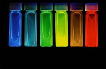
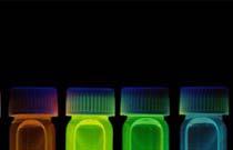
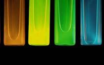
7 Design of the QD core Nanocrystal probes in aqueous buffer, all illuminated simultaneously with a handheld ultraviolet lamp Zrazhevskiy P et al., Chem. Soc. Rev., 2009 The QD core defines optical properties of the probe The color of emitted light can be fine-tuned by adjusting the QD size QDs emitting light from the UV, throughout the visible, and into the infrared spectra ( nm) Little or no cross-talk between adjacent colors enables simultaneous detection and quantification of multiple fluorescence signals Blue series represents different sizes of CdSe nanocrystals with diameters of 2.1, 2.4, 3.1, 3.6, and 4.6 nm Green series is of InP nanocrystals with diameters of 3.0, 3.5, and 4.6 nm Red series is of InAs nanocrystals with diameters of 2.8, 3.6, 4.6, and 6.0 nm. Bruchez M. Jr. et al., Science, 1998
 QDs are crystalline particles that range from 2 to 10 nanometers in diameter Preparation of QD-based probes represents a multi-step process each step aimed at controlling optical, physical and")
8 Engineering of QD probes QDs are semiconductor nanoparticles often made from hundreds to thousands of atoms of group II and VI elements (e.g. CdSe and CdTe) or group III and V elements (e.g. InP and InAs) QDs are crystalline particles that range from 2 to 10 nanometers in diameter Preparation of QD-based probes represents a multi-step process each step aimed at controlling optical, physical and chemical properties of the final probe Inorganic nanoparticle core manipulation of the core chemical composition, size, and structure controls the photo-physical properties of the probe 2. Design of coating materials that can encapsulate the QD core and shield it from the environment water soluble biocompatible Zrazhevskiy P et al., Chem. Soc. Rev., 2009
9 Design of an organic shell Synthesized QDs are hydrophobic and soluble only in nonpolar organic solvents, such as chloroform and hexane To be useful for biological applications QDs must be made water-soluble QDs should be soluble and stable in biological buffers, preserve the original photo-physical properties, retain relatively small particle size, and provide reactive groups for subsequent conjugation to biomolecules Methods used are: Ligand-exchange Encapsulation
 or phospholipids (H) Ligand-exchange approaches often yield compact probes at an expense of reduced stability and fluorescence efficiency Encapsulation")
10 Design of an organic shell Ligand exchange: replacing hydrophobic surface ligands with hydrophilic ones by direct anchoring of ligands to the QD surface Encapsulation: encapsulate the hydrophobic QDs with amphiphilic molecules such as polymers (G) or phospholipids (H) Ligand-exchange approaches often yield compact probes at an expense of reduced stability and fluorescence efficiency Encapsulation produces exceptionally stable and bright particles at an expense of increased size Zrazhevskiy P et al., Chem. Soc. Rev., 2009
 QDs are crystalline particles that range from 2 to 10 nanometers in diameter Preparation of QD-based probes represents a multi-step process each step aimed at controlling optical, physical and")
11 Engineering of QD probes QDs are semiconductor nanoparticles often made from hundreds to thousands of atoms of group II and VI elements (e.g. CdSe and CdTe) or group III and V elements (e.g. InP and InAs) QDs are crystalline particles that range from 2 to 10 nanometers in diameter Preparation of QD-based probes represents a multi-step process each step aimed at controlling optical, physical and chemical properties of the final probe 1. Inorganic nanoparticle core manipulation of the core chemical composition, size, and structure controls the photo-physical properties of the probe 2. Design of coating materials that can encapsulate the QD core and shield it from the environment water soluble biocompatible 3. Further decoration of the QDs with biomolecules give biofunctionality and enables probe interaction with biological systems Zrazhevskiy P et al., Chem. Soc. Rev., 2009
, non-covalent coordination of thiol groups or polyhistidine")
12 Design of surface ligands Bio-functionality has to be added to otherwise inert nanoparticles Usually achieved by decorating QDs with proteins, peptides, nucleic acids, or other biomolecules that mediate specific interactions with living systems Decoration of QD surface with bio-ligands can be achieved via covalent conjugation (A, B), non-covalent coordination of thiol groups or polyhistidine tags with the QD surface metal atoms (C), or electrostatic deposition of charged molecules on the QD organic shell (D). Zrazhevskiy P et al., Chem. Soc. Rev., 2009
13 Biological applications require water-soluble nanocrystals First time it was demonstrated that semiconductor nanoparticles could be made watersoluble and used as biological imaging probes Extended the chemistry of the core-shell systems by adding a third layer of silica that makes the core-shell water soluble The core-shell nanocrystals prepared in this manner are soluble and stable in water or buffered solution
 red fluorescence Nanocrystals coated with trimethoxysilylpropyl urea and acetate groups were found to bind with high affinity in the cell")


14 Fluorescently labeled 3T3 mouse fibroblast cells using two different size CdSe-CdS core-shell nanocrystals enclosed in a silica shell The smaller nanocrystals (2-nm core) emitted green fluorescence the larger (4-nm core) red fluorescence Nanocrystals coated with trimethoxysilylpropyl urea and acetate groups were found to bind with high affinity in the cell nucleus property was used to stain the nucleus with the green-colored nanocrystals Avidin-biotin interaction, a model system for ligand-receptor binding, was used here to specifically label the F-actin filaments with red nanocrystal probes Biotin was covalently bound to the nanocrystal surface fibroblasts had been incubated in phalloidin-biotin and streptavidin Samples were imaged with both conventional wide-field and laser-scanning confocal fluorescence microscopes Green and red labels were clearly spectrally resolved to the eye and to a color Polaroid camera Nonspecific labeling of the nuclear membrane by both the red and the green probes resulted in a yellow color Red actin filaments, however, were specifically stained

15
16
17 Introduction The state of the art is imaging of 5 6 molecular targets with custom-designed QD probes Use the extensive multiplexing potential of QDs Development of a multicolour multicycle molecular profiling (M3P) technology potentially capable of examining over 100 molecular targets in single cells at subcellular resolution
18 The M3P Technology The M3P Technology consists of three main steps
19 1. Staining The M3P Technology Universal QD SpA bioconjugates are used to capture intact Abs in solution during a pre-staining step to form functional QD-SpA-Ab fluorescent probes Different probes are pooled in a single cocktail Cocktail is incubated with cells for parallel multiplexed staining
20 1. Staining 2. Spectral imaging The M3P Technology Fluorescence microscopy with HSI capability is utilized to acquire and unmix signals from each QD color to generate quantitative antigen expression profiles in separate channels HSI=Hyperspectral imaging divides the spectrum into many bands. This technique of dividing images into bands can be extended beyond the visible
21 1. Staining 2. Spectral imaging 3. De-staining The M3P Technology Complete de-staining of the specimen is done by brief washing with a regeneration buffer, enabling the next full cycle of IF staining for a different subset of targets
22 Used QDs In general Ten spectrally distinct QDs with at least 20nm separation between fluorescence maxima are commercially available currently and can be linked to ten Abs This multiplexing capability is complemented by the spectral unmixing power of HSI (also ten channels at this time) for separation and quantification of individual colors from multicolor images In this study As a platform for M3P technology, use of highly stable and bright QDs coated with a nonfouling layer of polyethylene glycol (PEG) water-soluble Protein A from Staphylococcus aureus (SpA) is covalently linked to QDs as an adaptor for orientation-controlled IgG immobilization via binding to the Fc region of IgG Engineering a universal QD-protein A (QD-SpA) platform for flexible and fast preparation of a library of functional QD-antibody (QD-SpA-Ab) probes and utilizing such probes in a multicycle staining procedure QD-based multicolour multicycle technology heavily depends on several key challenges: 1. Each QD probe must be uniquely matched to a specific target, exhibiting no crosstalk between different probes within a staining cocktail 2. QD fluorescence must be completely removed after each imaging cycle to prevent signal carry-over to the next staining cycle 3. Target antigenicity and specimen integrity must be retained throughout multiple staining/imaging/destaining cycles

23 QD-Ab probe specificity Staining 5 molecular targets separately in formalin-fixed HeLa cells The five model targets used represent a spectrum of cell compartment localizations and expression levels Comparing the relative target distribution patterns with those obtained by conventional two-step IF using commercially available QD-labelled 2 Ab and Alexa Fluor 568-labelled 2 Ab Ki-67 HSP90 Lamin A Cox-4 β-tubulin a) QD585-Spa-Ab labelled in a single step procedure b) QD585 2 Ab labelled in a conventional two-step staining c) Alexa Fluor 568-labelled 2 Ab in a conventional two-step staining Staining patterns are consistent throughout all three procedures, indicating preserved specificity and affinity of antibodies in the QD SpA Ab complex.

 into individual QD545 (middle panels) and QD585 (right")
24 Lamin A QD-SpA-Ab probe Stability True color image QD545 QD585 QD545 + QD585 QD545 QD585 QD545-SpA and QD585-SpA probes mixed together with Ab and incubated with cells efficiently captured free Ab from solution and produced mixed-color Lamin A staining with nearly 50% contribution each Mixed fully assembled QDSpA- Ab probes with counterpart non-complexed QD-SpA (for example, QD545-SpA-Ab mixed with QD585-SpA, or QD585- SpA-Ab with QD545-SpA), and incubated cells with this solution Did not observe any crosstalk or interference with either QD545 or QD585 probes HSI used to unmix true-colour images (left panels) into individual QD545 (middle panels) and QD585 (right panels) channels
 Ki-67 Nucleus 525 nm HSP90")
25 Parallel multiplex staining with five preassambled QD-SpA-Ab probes Demonstrate the utility of the new probes for multicolour imaging: parallel staining of five model molecular targets is performed Ki-67, HSP90, Lamin A, Cox-4 and β-tubulin are simultaneously stained with QD-SpA-Ab probes emitting at 525, 545, 565, 585 and 605 nm, respectively(false color image) Ki-67 Nucleus 525 nm HSP90 Cytoplasm 545 nm Lamin A Nuclear membrane 565 nm Cox-4 Mitochondria 585 nm β-tubulin Microtubules 605 nm
 HSI and spectral unmixing are used to extract individual QD c) Single-color")

26 Parallel multiplex staining with five preassambled QD-SpA-Ab probes Ki-67 HSP90 Lamin A Cox-4 β-tubulin b) HSI and spectral unmixing are used to extract individual QD c) Single-color staining performed with colour-matched QD-SpA-Ab probes Relative expression levels Singleplexed staining performed with same QD585-SpA probe for all targets Quantitative analysis of staining intensity

27 1. Staining 2. Spectral imaging 3. De-staining The M3P Technology Complete de-staining of the specimen is done by brief washing with a regeneration buffer, enabling the next full cycle of IF staining for a different subset of targets
 Characteristic nuclear envelope staining is obtained with QD545-SpA probes pre-assembled with anti- Lamin A antibody b) Brief incubation with regeneration buffer removes QD-SpA-Ab probes,")
28 Specimen regeneration and target restaining with multicycle staining procedure New QD-SpA-Ab probes, quick and efficient de-staining by brief exposure to low-ph/detergent-based regeneration buffer because a) Characteristic nuclear envelope staining is obtained with QD545-SpA probes pre-assembled with anti- Lamin A antibody b) Brief incubation with regeneration buffer removes QD-SpA-Ab probes, achieving specimen restoration to pre-staining condition and enabling nearly complete target re-staining c) Incubation of regenerated specimen with blank QD545-SpA probes (lacking target-specific Abs) fails to produce any residual staining confirming complete removal of 1 Abs during de-staining
 and HSP90")
 and Lamin A pattern in QD585 channel (k) (e) and (i) are true-colour images and (h) and (l) are false-colour")
29 Specimen regeneration and target restaining with multicycle staining procedure Similarly, in a dual colour staining of Lamin A and HSP90 with QD545 and QD585 probes complete exchange of fluorescent reporters during the second cycle HSI reveals distinct Lamin A staining pattern in QD545 channel (f) and HSP90 pattern in QD585 channel (g) Following de-staining, during the second cycle the same targets are stained with the counterpart QD probes (i l), yielding clear HSP90 pattern in QD545 channel (j) and Lamin A pattern in QD585 channel (k) (e) and (i) are true-colour images and (h) and (l) are false-colour composite images.
 or HSP90 (n) HSI was used for quantitative analysis of average staining intensities Measured average")
30 Specimen regeneration and target restaining with multicycle staining procedure Lamin A HSP90 To evaluate effect of repeated regeneration procedure on specimen stability and staining intensity, up to 10 degradation cycles are performed on separate specimens before cells are stained for Lamin A (m) or HSP90 (n) HSI was used for quantitative analysis of average staining intensities Measured average fluorescence staining intensity does not vary significantly regardless of the number of degradation cycles performed Demonstrating sufficient preservation of specimen morphology and target antigenicity required for staining
 and in reverse (d f) Matching relative profile obtained")
31 Evaluation of the robustness of sequential staining procedure Complete specimen regeneration and lack of specimen degradation enables staining of the molecular targets Correct staining pattern and target expression profile can be obtained for staining sequence from highly condensed Lamin A down to diffusely distributed β-tubulin (a c) and in reverse (d f) Matching relative profile obtained from reference single-cycle staining with QD585-SpA (g). No carry-over or residual staining is detectable throughout the five cycles

32 Validation of M3P technology with 25-target staining Ki-67 HSP90 Lamin A Cox-4 β-tubulin Merge 5 colors x 5 cycles Re-stain the same model 5-target panel with multicolor QD-SpA-Ab probes for five cycles and perform quantitative analysis of staining intensity after each cycle All five targets are consistently stained through five cycles yielding accurate staining patterns and identical average staining intensity profiles (no carry over)
33 Summary M3P of single cells and tissue specimens is based on production of QD-Ab libraries via simple, fast and inexpensive methods The universal QD-SpA platform features on-demand, flexible, single-step, purification-free QD-Ab assembly, along with high probe specificity Demonstrated that the SpA Ab bond is sufficiently stable to avoid QD-SpA-Ab probe crosstalk Used QDs coated with a non-fouling layer of PEG, which resists nonspecific binding even after bioconjugation with SpA and Ab With this probe design, fully assembled functional QD-SpA-Ab allows direct target labelling while the high brightness of QD probes makes imaging without signal amplification possible Demonstrated that at least ten staining cycles can be performed without loss of specimen antigenicity and cell morphology Brief treatment with regeneration buffer leads to effective removal of QD SpA Ab complex and thus completely regenerates specimens for next-cycle staining enabling re-staining of the same or probing of a different panel of molecular targets
34 Conclusion Developed a technically straightforward, yet analytically powerful, imaging platform for comprehensive in situ single-cell molecular profiling Overcoming the fundamental limitations of conventional and nanoparticle-based methods, the functionality of this technology comes from the unique combination of favorable optical properties of QDs, flexible QD-Ab probe preparation and the ability to perform multiple cycles of staining/de-staining without affecting specimen antigenicity and cell morphology Further development of M3P technology, especially by integrating it with automated staining imaging instruments, will offer exciting opportunities in systems biology, signalling pathway analysis, gene expression studies, molecular diagnostics and drug discovery


35 T H A N K S F O R YO UR A T T E N T I O N
TARGETED IMAGING. Maureen Chan and Ruwani Mahathantila
 TARGETED IMAGING Maureen Chan and Ruwani Mahathantila Overview 2 Introduction to fluorescent imaging Fluorescent agents Quantum Dots Physical properties How QDs work In Vivo QD imaging Future Video What
Spectra Chacracterizations of Optical Nanoparticles
 THAI NGUYEN UNIVERSITY OF EDUCATION Spectra Chacracterizations of Optical Nanoparticles Chu Viet Ha Department of Physics 18/2018 1 THAI NGUYEN UNIVERSITY OF EDUCATION Address 20 Luong Ngoc Quyen Street,
Engineering Quantum Dots for Live-Cell Single-Molecule Imaging
 Engineering Quantum Dots for Live-Cell Single-Molecule Imaging Andrew M. Smith and Shuming Nie Georgia Tech and Emory University Department of Biomedical Engineering 2011 NSF Nanoscale Science and Engineering
2004 Debye Lecture 4 C. B. Murray. Quantum Dot Applications: Sun Screen. Solar Cells. Bio-tagging. Solid State Lighting?
 2004 Debye Lecture 4 C. B. Murray Quantum Dot Applications: Sun Screen Solar Cells Bio-tagging Solid State Lighting? Quantum Dot Solar cells Nanocrystal Solar Cells Double-labeling of mitochondria
Visualisation, Sizing and Counting of Fluorescent and Fluorescently-Labelled Nanoparticles
 Visualisation, Sizing and Counting of Fluorescent and Fluorescently-Labelled Nanoparticles Introduction Fluorescent molecules have long been used to specifically label particular structures and features
Qdot nanocrystal. wide range of biological investigations, Qdot nanocrystals are powerful complements
 Feature nanocrystal conjugates for flow cytometry Take the easy route to multicolor flow cytometry. With applications across a wide range of biological investigations, nanocrystals are powerful complements
Fluorescence Microscopy. Terms and concepts to know: 10/11/2011. Visible spectrum (of light) and energy
 and energy") Fluorescence Microscopy Louisiana Tech University Ruston, Louisiana Microscopy Workshop Dr. Mark DeCoster Associate Professor Biomedical Engineering 1 Terms and concepts to know: Signal to Noise Excitation
Fluorescence Microscopy Louisiana Tech University Ruston, Louisiana Microscopy Workshop Dr. Mark DeCoster Associate Professor Biomedical Engineering 1 Terms and concepts to know: Signal to Noise Excitation
Widefield Microscopy Bleed-Through
 In widefield microscopy the excitation wavelengths which illuminate the sample, and the emission wavelengths which reach the CCD camera are selected throughout a filter cube. A filter cube consists of
More on fluorescence
 More on fluorescence Last class Fluorescence Absorption emission Jablonski diagrams This class More on fluorescence Common fluorophores Jablonski diagrams to spectra Properties of fluorophores Excitation
Imaging Quantum Dots using FUJIFILM LAS 4000
 Imaging Quantum Dots using FUJIFILM LAS 4000 Application Note John Pizzonia, Ph.D. 9-28-07 Quantum dots (also known as nanocrystals) are a special class of materials known as semiconductors, which are
August 15, 2018 $ " % ' & OMe. NEt2 MeO
 Taking a deep look: a near infrared fluorescent dye for long term bioimaging ~ Promising photostable tool for single molecule tracking and multicolor imaging ~ August 15, 2018 A team of researchers at
Visualizing Cells Molecular Biology of the Cell - Chapter 9
 Visualizing Cells Molecular Biology of the Cell - Chapter 9 Resolution, Detection Magnification Interaction of Light with matter: Absorbtion, Refraction, Reflection, Fluorescence Light Microscopy Absorbtion
SURFACE ENHANCED RAMAN SCATTERING NANOPARTICLES AS AN ALTERNATIVE TO FLUORESCENT PROBES AN EVALUATION
 APPLICATION NOTE SURFACE ENHANCED RAMAN SCATTERING NANOPARTICLES AS AN ALTERNATIVE TO FLUORESCENT PROBES AN EVALUATION Summary: Interest in using nanoparticles specifically, Surface Enhanced Raman Scattering
Design for Manufacturability (DFM) in the Life Sciences
 in the Life Sciences") T E C H N I C A L N O T E Design for Manufacturability (DFM) in the Life Sciences Fluorescence Spectroscopy Product Platform Realized with TracePro TM Suite of Opto-Mechanical Design Software Tools Authors:
T E C H N I C A L N O T E Design for Manufacturability (DFM) in the Life Sciences Fluorescence Spectroscopy Product Platform Realized with TracePro TM Suite of Opto-Mechanical Design Software Tools Authors:
HYPERSPECTRAL MICROSCOPE PLATFORM FOR HIGHLY MULTIPLEX BIOLOGICAL IMAGING. Marc Verhaegen
 HYPERSPECTRAL MICROSCOPE PLATFORM FOR HIGHLY MULTIPLEX BIOLOGICAL IMAGING Marc Verhaegen CMCS, MONTREAL, MAY 11 th, 2017 OVERVIEW Hyperspectral Imaging Multiplex Biological Imaging Multiplex Single Particle
Fluorescence Light Microscopy for Cell Biology
 Fluorescence Light Microscopy for Cell Biology Why use light microscopy? Traditional questions that light microscopy has addressed: Structure within a cell Locations of specific molecules within a cell
Dino-Lite knowledge & education. Fluorescence Microscopes
 Dino-Lite knowledge & education Fluorescence Microscopes Dino-Lite Fluorescence models Smallest fluorescence microscope in the world Revolution to biomedical and educational applications Flexible Easy
QImaging Camera Application Notes Multicolor Immunofluorescence Imaging
 QImaging Camera Application Notes Multicolor Immunofluorescence Imaging In order to image localization of intracellular proteins with high specificity, it is frequently necessary to multiplex antibody
Five steps for publication-quality cell imaging the first time
 Five steps for publication-quality cell imaging the first time Follow this proven guide to capture the best possible fixed-cell images Introduction We are all driven by great scientific innovation and
What is an Aptamer? smallest unit of repeating structure
 What is an Aptamer? apto: mer: to fit smallest unit of repeating structure Aptamers are single stranded folded oligonucleotides that bind to molecular (protein) targets with high affinity and specificity
Flow Cytometry - The Essentials
 Flow Cytometry - The Essentials Pocket Guide to Flow Cytometry: 1. Know your Cytometer 2. Understanding Fluorescence and Fluorophores 3. Gating Process 4. Controls 5. Optimization 6. Panel Building 7.
2.3 Quantum Dots (QDs)
") 2.3 Quantum Dots (QDs) QDs are inorganic nanocrystals, approximately 1 10 nm in size, with unique optical properties of broad excitation, narrow size-tunable emission spectra, high photochemical stability,
2.3 Quantum Dots (QDs) QDs are inorganic nanocrystals, approximately 1 10 nm in size, with unique optical properties of broad excitation, narrow size-tunable emission spectra, high photochemical stability,
ACS Nano, Article ASAP DOI: /nn800632j
 Synthesis, Characterization, and Bioconjugation of Fluorescent Gold Nanoclusters toward Biological Labeling Applications Walter H. Chang et. al. Department of Biomedical Engineering, Chung Yuan Christian
A legacy of innovation and discovery
 A legacy of innovation and discovery CellInsight CX7 LZR High Content Analysis Platform Quantifiably brilliant data Since the introduction of Thermo Scientific ArrayScan High Content Analysis (HCA) Readers
Contents. 11 The Use of Epitope Tags in Histochemistry References... 98
 Contents 1 Antibodies for Immunohistochemistry... 1 1.1 Structure of Antibodies... 2 1.2 Polyclonal Antibodies... 4 1.3 Mouse Monoclonal Antibodies... 4 1.4 Rabbit Monoclonal Antibodies... 5 1.5 Protein
F* techniques: FRAP, FLIP, FRET, FLIM,
 F* techniques: FRAP, FLIP, FRET, FLIM, FCS Antonia Göhler March 2015 Fluorescence explained in the Bohr model Absorption of light (blue) causes an electron to move to a higher energy orbit. After a particular
Quantum Dots and Carbon Nanotubes in Cancer diagnose EE453 Project Report submitted by Makram Abd El Qader
 Quantum Dots and Carbon Nanotubes in Cancer diagnose EE453 Project Report submitted by Makram Abd El Qader abdelqad@unlv.nevada.edu, Fall 2008 Abstract On the basis of research and cancer medical treatment,
Tracking Cellular Protein Localization and Movement in Cells with a Flexible Fluorescent Labeling Technology. Chad Zimprich January 2015
 Tracking Cellular Protein Localization and Movement in Cells with a Flexible Fluorescent Labeling Technology Chad Zimprich January 2015 Presentation verview HaloTag Fusion Technology Design Functionality
Spectral Separation of Multifluorescence Labels with the LSM 510 META
 Microscopy from Carl Zeiss Spectral Separation of Multifluorescence Labels with the LSM 510 META Indians living in the South American rain forest can distinguish between almost 200 hues of green in their
Quantum Dots for Live Cell and In Vivo Imaging
 Int. J. Mol. Sci. 2009, 10, 441-491; doi:10.3390/ijms10020441 OPEN ACCESS International Journal of Molecular Sciences ISSN 1422-0067 www.mdpi.com/journal/ijms Review Quantum Dots for Live Cell and In Vivo
Contents. SCHOOL of FLUORESCENCE. For more information, go to lifetechnologies.com/imagingbasics
 MPSF educator packet This packet contains illustrations and figures from the Molecular Probes School of Fluorescence website. They illustrate concepts from the basic physical properties that underlie fluorescence
Using Quantum Dots in Fluorescence Resonance Energy Transfer Studies
 p.1/31 Using Quantum Dots in Fluorescence Resonance Energy Transfer Studies Rajarshi Guha Pennsylvania State University p.2/31 Introduction Using organic fluorophores as labels A brief overview of fluorescence
Contact Details. Dr Alexander Galkin. Office: MBC Room 186. Tel: (028) Frequency and wavelength.
 Frequency and wavelength.") Contact Details The electromagnetic spectrum Biological Spectroscopy Dr Alexander Galkin Email: a.galkin@qub.ac.uk Dr Alexander Galkin MSc Biomolecular Function - BBC8045 Office: MBC Room 186 Tel: (028)
Contact Details The electromagnetic spectrum Biological Spectroscopy Dr Alexander Galkin Email: a.galkin@qub.ac.uk Dr Alexander Galkin MSc Biomolecular Function - BBC8045 Office: MBC Room 186 Tel: (028)
CHAPTER 4. DEVOLEPMENT OF IMMUNO-DOT BLOT ASSAY USING DUAL LABELED GOLD NANOPARTICLE PROBE TO DETECT Cryptosporidum parvum
 51 CHAPTER 4 DEVOLEPMENT OF IMMUNO-DOT BLOT ASSAY USING DUAL LABELED GOLD NANOPARTICLE PROBE TO DETECT Cryptosporidum parvum 4.1 INDRODUCTION Cryptosporidium parvum is a significant diarrhoea causing parasitic
BIOSENOSRS BIO 580. Nanobiosensors WEEK-13 Fall Semester. Faculty: Dr. Javed H. Niazi KM Faculty of Engineering & Natural Sciences Sabanci University
 BIOSENOSRS BIO 580 Nanobiosensors WEEK-13 Fall Semester Faculty: Dr. Javed H. Niazi KM Faculty of Engineering & Natural Sciences Sabanci University Topics that will be covered in the course History of
Quality Matters: Bethyl DyLight Antibody Conjugates
 Quality Matters: Bethyl DyLight Antibody Conjugates Criteria for Fluorescent Probe Selection The use of fluorescent probes has become a mainstream technique to detect speci ic molecular targets in both
Imaging facilities at WUR
 Imaging facilities at WUR Advanced light microscopy facilities at Wageningen UR Programme Thursday 13 June 2013 Lunch meeting organized by Cat-Agro Food 12.00 Welcome and sandwich lunch 12.10 Introduction
BIO 315 Lab Exam I. Section #: Name:
 Section #: Name: Also provide this information on the computer grid sheet given to you. (Section # in special code box) BIO 315 Lab Exam I 1. In labeling the parts of a standard compound light microscope
Biochemistry. Biochemical Techniques. 18 Spectrofluorimetry
 Description of Module Subject Name Paper Name 12 Module Name/Title 1. Objectives 1.1 To understand technique of Spectrofluorimetry. 1.2 To explain instrumentation design 1.3 What are applications of Spectrofluorimetry?
BIO 315 Lab Exam I. Section #: Name:
 Section #: Name: Also provide this information on the computer grid sheet given to you. (Section # in special code box) BIO 315 Lab Exam I 1. In labeling the parts of a standard compound light microscope
Confocal Microscopy Analyzes Cells
 Choosing Filters for Fluorescence A Laurin Publication Photonic Solutions for Biotechnology and Medicine November 2002 Confocal Microscopy Analyzes Cells Reprinted from the November 2002 issue of Biophotonics
DYNAMICBIOSENSORS. Chip functionalization DNA-encoded addressing and chip configuration. Technology Information #120
 Technology Information #120 Chip functionalization DNA-encoded addressing and chip configuration The switchsense biochip features 24 detection spots, which are arranged in 2 or 4 seperate flow channels.
Fluorescence spectroscopy
 Fluorescence spectroscopy The light: electromagnetic wave Zoltán Ujfalusi Biophysics seminar Dept. of Biophysics, University of Pécs 14-16 February 2011 Luminescence: light is not generated by high temperatures!!!
Odyssey Fc Imaging System Infrared fluorescent and chemiluminescent imaging in one system!
 Odyssey Infrared Imaging System A fundamental change in western blot analysis Odyssey Fc Imaging System Infrared fluorescent and chemiluminescent imaging in one system! Accurate Quantification Wide linear
Color-Rich Fluoro-Max Dyed Microparticles March 2008
 Fluoro-Max Dyed Microparticles March 2008 Introduction Dyed Microparticles Thermo Scientific Seradyn dyed and fluorescent microparticles are monodisperse particles prepared by unique and proprietary emulsion
Absorption of an electromagnetic wave
 In vivo optical imaging?? Absorption of an electromagnetic wave Tissue absorption spectrum Extinction = Absorption + Scattering Absorption of an electromagnetic wave Scattering of an electromagnetic wave
UNIVERSITY OF ROME LA SAPIENZA NANOTECHNOLOGIES ENGINEERING NANOPARTICLES IN BIOMEDICINE
 UNIVERSITY OF ROME LA SAPIENZA NANOTECHNOLOGIES ENGINEERING NANOPARTICLES IN BIOMEDICINE Challenges The challenges are: in-situ analysis and in vivo at micro level recognize biomolecules functionality
DNA Microarray Technology
 2 DNA Microarray Technology 2.1 Overview DNA microarrays are assays for quantifying the types and amounts of mrna transcripts present in a collection of cells. The number of mrna molecules derived from
Goat Anti Rabbit IgG Antibodies
 Goat Anti Rabbit IgG Antibodies Table 1. Contents and storage information. Material Amount Concentration Storage Upon Receipt Stability Whole antibodies 0.5 ml F(ab ) 2 fragments 250 µl 2 mg/ml in 0.1
Fluorescence spectroscopy
 Fluorescence spectroscopy The light: electromagnetic wave Tamás Huber Biophysics seminar Dept. of Biophysics, University of Pécs 05-07. February 2013. Luminescence: light emission of an excited system.
The future of fluorescence
 Nanotechnology The future of fluorescence Qdot nanocrystal technology Nanotechnology Qdot nanocrystals: The vision of nanotechnology Qdot nanocrystals offer revolutionary fluorescence performance: Long-term
nanoprecipitation mpeg-pla 2 nd Emulsion mpeg-pla in DCM
 THERAPEUTIC NANOTECHNOLOGY LAB MODULE Location: BioNano Lab, 3119 Micro and Nanotechnology Laboratory (MNTL) Instructor: Jianjun Cheng, Assistant Professor of Materials Science and Engineering Lab Assistants:
Supporting Information for
 Supporting Information for Building Electromagnetic Hot Spots in Living Cells via Target-Triggered Nanoparticle Dimerization Wen Zhou, 1,2 Qiang Li, 1 Huiqiao Liu, 1 Jie Yang, 1 Dingbin Liu 1,2 * 1. College
A Brief History of Light Microscopy And How It Transformed Biomedical Research
 A Brief History of Light Microscopy And How It Transformed Biomedical Research Suewei Lin Office: Interdisciplinary Research Building 8A08 Email: sueweilin@gate.sinica.edu.tw TEL: 2789-9315 Microscope
THE BASICS OF IMMUNOHISTOCHEMISTRY
 THE BASICS OF IMMUNOHISTOCHEMISTRY Introduction Immunohistochemistry (IHC) identifies specific tissue components by means of a specific antigen/antibody reaction tagged with a visible label. IHC makes
Overview of Immunohistochemistry
 Overview of Immunohistochemistry Immunohistochemistry (IHC) combines anatomical, immunological and biochemical techniques to identify discrete tissue components by the interaction of target antigens with
MagSi Beads. Magnetic Silica Beads for Life Science and Biotechnology study
 MagSi Beads Magnetic Silica Beads for Life Science and Biotechnology study MagnaMedics Diagnostics B.V. / Rev. 9.2 / 2012 Wide range of products for numerous applications MagnaMedics separation solutions
Azure Biosystems Western Blotting Workflow
 Azure Biosystems Western Blotting Workflow PROBE PLAN SEPARATE ANALYZE VISUALIZE PLAN Plan your experiment and choose your detection method Chemiluminescent Western Blotting The most common method for
Qdots technology for biodetection available at LLNL
 Qdots technology for biodetection available at LLNL Daniele Gerion Physics and Advanced Technology, L-415 gerion1@llnl.gov In the following note, I will present the qdots technology available at LLNL with
CBI Toolbox Tour 2015
 CBI Toolbox Tour 2015 Thermophoresis (NanoTemper) NT.115 & NT.LabelFree Images: NanoTemper Circular Dichroism Jasco J-1500 Spectrometer Six Position Turreted Peltier Temperature Control System Automated
7/24/2012. DNA Probes. Hybridization and Probes. CLS 420 Immunology & Molecular Diagnostics. Target Sequences. Target Sequences. Nucleic Acid Probes
 Hybridization and Probes CLS 420 Immunology & Molecular Diagnostics Molecular Diagnostics Techniques: Hybridization and Probes Nucleic acid probes: A short, known sequence of DNA or RNA Used to detect
Comparing MSD Electrochemiluminescent Detection Technology to Traditional ELISA for Clinical Applications
 Enzyme- linked immunosorbent assays (s) are widely used in clinical settings for detecting substances such as antibodies, peptides, or hormones. However, assays have been gaining in popularity due to advantages
Tunable band structure in coreshell quantum dots through alloying of the core
 Tunable band structure in coreshell quantum dots through alloying of the core A. Guille ǀ D. Mourad ǀ T. Aubert ǀ A. Houtepen ǀ R. Van Deun ǀ E. Brainis ǀ Z. Hens 1. Introduction Semiconductor nanocrystals
TECHNICAL NOTE. microrna Profiling with FirePlex mirselect: A Technical Overview and Cross-Cytometer Comparison
 TECHNICAL NOTE microrna Profiling with FirePlex mirselect: A Technical Overview and Cross-Cytometer Comparison FirePlex mirselect is a high-throughput, customizable solution for microrna profiling across
APPLICATION NOTE Rev. 7/2017, v4.0 Fluorescent Nanodiamonds: Bio-applications. Physical and Fluorescence Properties
 APPLICATION NOTE Rev. 7/2017, v4.0 Fluorescent Nanodiamonds: Bio-applications Fluorescent nanodiamonds (FNDs) offer a unique alternative to currently existing fluorescent biomarkers. With exceptional photo
ab CytoPainter ER Staining Kit Red Fluorescence
 ab139482 CytoPainter ER Staining Kit Red Fluorescence Instructions for Use Designed to detect Human endoplasmic reticulum by microscopy. This product is for research use only and is not intended for diagnostic
Microarray Industry Products
 Via Nicaragua, 12-14 00040 Pomezia (Roma) Phone: +39 06 91601628 Fax: +39 06 91612477 info@lifelinelab.com www.lifelinelab.com Microarray Industry Products Page 10 NBT / BCPIP Chromogenic phosphatase
Biophotonics?? Biophotonics. technology in biomedical engineering. Advantages of the lightwave
 Biophotonics - Imaging: X-ray, OCT, polarimetry, DOT, TIRF, photon migration, endoscopy, confocal microscopy, multiphoton microscopy, multispectral imaging - Biosensing: IR spectroscopy, fluorescence,
Electron microscopy technology of reticulocytes after sorting with
 Electron microscopy technology of reticulocytes after sorting with magnetic beads The Cell Analysis Center Scientific Bulletin Part 2 For efficient analysis of cells, sorting of the target cells is crucial.
Optical Observation - Hyperspectral Characterization of Nano-scale Materials In-situ
 Optical Observation - Hyperspectral Characterization of Nano-scale Materials In-situ Research at the nanoscale is more effective, when research teams can quickly and easily observe and characterize a wide
More choices, better detection.
 3 Protein Detection More choices, better detection. Perform Western blotting experiments faster and easier with our new Thermo Scientific Pierce G2 Fast Blotter and accessories, and push your microscopy
AMREP FLOW CYTOMETRY CORE FACILITY. Flow Cytometry Data Analysis Workshop Friday 24 th October
 AMREP FLOW CYTOMETRY CORE FACILITY Flow Cytometry Data Analysis Workshop Friday 24 th October Polychromatic Flow Cytometry Data Analysis: Immunophenotyping Lymphocyte sub-populations and targeting their
Quantum Dot Labeling of Stem Cells during Proliferation and Differentiation
 Copyright 2006 Tech Science Press MCB, vol.3, no.4, pp.153-155, 2006 Quantum Dot Labeling of Stem Cells during Proliferation and Differentiation E. K. Moioli 1, B. Shah 1, P. A. Clark 1, M. Stroscio 1
Enhance Your Protein Interaction Research with A New Level of Bench-Top Productivity
 LAMDAGEN C O R P O R A T I O N Enhance your research with real-time monitoring and quantitation of biomolecular binding with highly sensitive detection in a label-free format 9 Enhance Your Protein Interaction
Concept review: Fluorescence
 16 Concept review: Fluorescence Some definitions: Chromophore. The structural feature of a molecule responsible for the absorption of UV or visible light. Fluorophore. A chromophore that remits an absorbed
FRAUNHOFER IME SCREENINGPORT
 FRAUNHOFER IME SCREENINGPORT Detection technologies used in drug discovery Introduction Detection technologies in drug discovery is unlimited only a biased snapshot can be presented Differences can presented
Direct visualization, sizing and concentration measurement of fluorescently labeled nanoparticles using NTA
 Direct visualization, sizing and concentration measurement of fluorescently labeled nanoparticles using NTA NANOSIGHT RANGE Visualize and Measure Nanoparticle Size and Concentration PARTICLE SIZE PARTICLE
Site-specific targeting of enterovirus capsid by functionalized monodisperse gold nanoclusters
 Site-specific targeting of enterovirus capsid by functionalized monodisperse gold nanoclusters Varpu Marjomäki a,b, Tanja Lahtinen b,c, Mari Martikainen a,b, Jaakko Koivisto b,c, Sami Malola b,d, Kirsi
Partha Roy
 Fluorescence microscopy http://micro.magnet.fsu.edu/primer/index.html Partha Roy 1 Lecture Outline Definition of fluorescence Common fluorescent reagents Construction ti of a fluorescence microscope Optical
Supplementary Figure 1: (a) 3D illustration of the mobile phone microscopy attachment, containing a 3D movable sample holder (red piece for z
 3D illustration of the mobile phone microscopy attachment, containing a 3D movable sample holder (red piece for z") Supplementary Figure 1: (a) 3D illustration of the mobile phone microscopy attachment, containing a 3D movable sample holder (red piece for z movement and blue piece for x-y movement). Illumination sources
Supplementary Figure 1: (a) 3D illustration of the mobile phone microscopy attachment, containing a 3D movable sample holder (red piece for z movement and blue piece for x-y movement). Illumination sources
The strategy. using Atomic Force Microscope; Biomolecules and Neutraceuticals examples
 The strategy for Bionanomolecules Characterizations using Atomic Force Microscope; Biomolecules and Neutraceuticals examples Dr. NagibAli Elmarzugi, PhD Head of Nanotechnology Research gp., Biotechnology
SUPPLEMENTARY INFORMATION
 doi:10.1038/nature10016 Supplementary discussion on binding site density for protein complexes on the surface: The density of biotin sites on the chip is ~10 3 biotin-peg per µm 2. The biotin sites are
Fluorescence spectroscopy
 Fluorescence spectroscopy The light: electromagnetic wave Tamás Huber Biophysics seminar Dept. of Biophysics, University of Pécs 05-06. February 2014. 1 Luminescence: light emission of an excited system.
Supplementary Data. In-Vivo Tumor Targeting and Spectroscopic Detection with Surface- Enhanced Raman Nanoparticle Tags
 Supplementary Data In-Vivo Tumor Targeting and Spectroscopic Detection with Surface- Enhanced Raman Nanoparticle Tags X.-M. Qian, 1 Xiang-Hong Peng, 2 Dominic O. Ansari, 1 Qiqin Yin-Goen, 3 Georgia Z.
SUPPLEMENTAL MATERIALS. FIGURE S1. CdTe quantum dot (QD) preparation displays photostability and symmetric emission spectra.
 preparation displays photostability and symmetric emission spectra.") UPPLMNAL MARIAL Wang et al. Relative luorescence Intensity 535 nm mission wavelength (nm) IUR 1. de quantum dot (Q) preparation displays photostability and symmetric emission spectra. A preparation of
UPPLMNAL MARIAL Wang et al. Relative luorescence Intensity 535 nm mission wavelength (nm) IUR 1. de quantum dot (Q) preparation displays photostability and symmetric emission spectra. A preparation of
Chapter 3. Clonal selection
 Chapter 3. Clonal selection I have called this principle, by which each slight variation, if useful, is preserved, by the term of Natural Selection -Charles Darwin, On the Origin of Species, 1859 4 The
THE BEACON OPTOFLUIDIC PLATFORM. 1000s of cells 100X the insights 10X faster 1/X total cost BERKELEY LIGHTS. DIGITAL CELL BIOLOGY AT LIGHT SPEED.
 THE BEACON OPTOFLUIDIC PLATFORM 1000s of cells 100X the insights 10X faster 1/X total cost BERKELEY LIGHTS. DIGITAL BIOLOGY AT LIGHT SPEED. FINDING THE MOST IMPORTANT INDIVIDUAL S REQUIRES THE MOST TIME,
FLUORESCENT PEPTIDES. Outstanding Performance and Wide Application Range
 FLUORESCENT PEPTIDES Peptides and amino acids labeled with and Tide Quencher TM We offer peptides and amino acids tagged with fluorescent dyes. They meet highest demands in fluorescence intensity and photo-stability,
Multiplex Fluorescent Western Blot Starter Kit for the Bio- Rad ChemiDoc MP
 Page 1 of 7 INSTRUCTIONS: Z-310 Multiplex Fluorescent Western Blot Starter Kit for the Bio- Rad ChemiDoc MP Rockland Immunochemicals and Bio-Rad Laboratories have jointly developed an easy to use multiplex
Anti-MOUSE IgG (H&L) (GOAT) Antibody DyLight 488 Conjugated (Min X Bv Ch Gt GP Ham Hs Hu Rb Rt & Sh Serum Proteins)
 (GOAT) Antibody DyLight 488 Conjugated (Min X Bv Ch Gt GP Ham Hs Hu Rb Rt & Sh Serum Proteins)") Anti-MOUSE IgG (H&L) (GOAT) Antibody DyLight 488 Conjugated (Min X Bv Ch Gt GP Ham Hs Hu Rb Rt & Sh Serum Proteins) - 610-141-121 Code: 610-141-121 Size: 100 µg Product Description: Anti-MOUSE IgG (H&L)
Anti-MOUSE IgG (H&L) (GOAT) Antibody DyLight 488 Conjugated (Min X Bv Ch Gt GP Ham Hs Hu Rb Rt & Sh Serum Proteins) - 610-141-121 Code: 610-141-121 Size: 100 µg Product Description: Anti-MOUSE IgG (H&L)
Lab 5: Optical trapping and single molecule fluorescence
 Lab 5: Optical trapping and single molecule fluorescence PI: Matt Lang Lab Instructor: Jorge Ferrer Summary Optical tweezers are an excellent experimental tool to study the biophysics of single molecule
STORM/PALM. Super Resolution Microscopy 10/31/2011. Looking into microscopic world of life
 Super Resolution Microscopy STORM/PALM Bo Huang Department of Pharmaceutical Chemistry, UCSF CSHL Quantitative Microscopy, 1/31/211 Looking into microscopic world of life 1 µm 1 µm 1 nm 1 nm 1 nm 1 Å Naked
PRODUCT DATA SHEET. Carboxylated Fluorescent Gold Nanoparticles. Description. Characteristics
 PRODUCT DATA SHEET Carboxylated Fluorescent Gold Nanoparticles Description Cytodiagnostics carboxylated fluorescent gold nanoparticles is a unique product that combines our Cyto fluorescent dyes and gold
Cytomics in Action: Cytokine Network Cytometry
 Cytomics in Action: Cytokine Network Cytometry Jonni S. Moore, Ph.D. Director, Clinical and Research Flow Cytometry and PathBioResource Associate Professor of Pathology & Laboratory Medicine University
Streamline Your Antibody Enrichment Using Scalable Magnetic Bead-Based Chemistries
 Streamline Your Antibody Enrichment Using Scalable Magnetic Bead-Based Chemistries Samantha Lewis, Ph.D. 2013. Webinar Outline Introduction Magnetic Bead-Based Method Overview Scalability Chemistries o
LUPAS Luminescent Polymers for in vivo Imaging of Amyloid Signatures
 LUPAS Luminescent Polymers for in vivo Imaging of Amyloid Signatures A research project for innovative diagnostics for neurodegenerative disorders Funded by the European Union under the 7 th Framework
Cellular imaging using Nano- Materials. A Case-Study based approach Arun Murali, Srivats V
 Cellular imaging using Nano- Materials A Case-Study based approach Arun Murali, Srivats V Agenda Discuss a few papers Explain a couple of new imaging techniques and their benefits over conventional imaging
AD (Leave blank) TITLE: Molecular profiling of prostate cancer specimens using Multicolor Quantum Dots. PRINCIPAL INVESTIGATOR: Xiaohu Gao
 TITLE: Molecular profiling of prostate cancer specimens using Multicolor Quantum Dots. PRINCIPAL INVESTIGATOR: Xiaohu Gao") AD (Leave blank) Award Number: W81XWH-07-1-0117 TITLE: Molecular profiling of prostate cancer specimens using Multicolor Quantum Dots PRINCIPAL INVESTIGATOR: Xiaohu Gao CONTRACTING ORGANIZATION: University
AD (Leave blank) Award Number: W81XWH-07-1-0117 TITLE: Molecular profiling of prostate cancer specimens using Multicolor Quantum Dots PRINCIPAL INVESTIGATOR: Xiaohu Gao CONTRACTING ORGANIZATION: University
Supplementary Information. Facile fabrication of microsphere-polymer brush hierarchically. three-dimensional (3D) substrates for immunoassays
 substrates for immunoassays") Electronic Supplementary Material (ESI) for ChemComm. This journal is The Royal Society of Chemistry 2015 Supplementary Information Facile fabrication of microsphere-polymer brush hierarchically three-dimensional
Electronic Supplementary Material (ESI) for ChemComm. This journal is The Royal Society of Chemistry 2015 Supplementary Information Facile fabrication of microsphere-polymer brush hierarchically three-dimensional
What is Nano-Bio? Non-Covalent Interactions
 - - What is Nano-Bio? Physicist: Biotech: Biologists: -study of molecular interactions -application of nano-tools to study biological systems. -application of nano-tools to detect, treat, and prevent disease