Fluorescence Microscopy. Terms and concepts to know: 10/11/2011. Visible spectrum (of light) and energy
|
|
|
- Lydia Beasley
- 6 years ago
- Views:
Transcription
 Emission Wavelength Photon Spectrum Barrier filter Beam splitting mirror")
1 Fluorescence Microscopy Louisiana Tech University Ruston, Louisiana Microscopy Workshop Dr. Mark DeCoster Associate Professor Biomedical Engineering 1 Terms and concepts to know: Signal to Noise Excitation (Absorption) Emission Wavelength Photon Spectrum Barrier filter Beam splitting mirror 2 Visible spectrum (of light) and energy Image modification from: spectrum/ 3 1
2 Visible spectrum (of light): the colors High Energy (Excitation) Low Energy (Emission) VIB G. YOR ROY G. BIV 4 Fluorescence: Excitation and Emission Emission Excitation Image modification from: 5 Specific molecules can be located in cells by fluorescence microscopy Fluorescent molecules absorb light at one wavelength and emit it at another, longer wavelength. If such a compound is illuminated at its absorbing wavelength and then viewed through a filter that allows only light of the emitted wavelength to pass, it is seen to glow against a dark background. Because the background is dark, the signal to noise ratio is increased over most ordinary, white light stains. The same number of molecules of an ordinary stain viewed conventionally would be practically invisible because they would give only the faintest tinge of color to the light transmitted through this stained portion of the cell. 6 2

3 Specific molecules can be located in cells by fluorescence microscopy 2 The fluorescent dyes used for staining cells are detected by a fluorescence microscope. The microscope is similar to a conventional scope, except that the light source is passed through two filters, one to filter the light before it hits the sample, and one to filter the light obtained from the sample. The first filter is chosen so that passes only wavelengths that excite the particular fluorescent probe, while the second blocks out this light and passes only wavelengths emitted when the dye fluoresces. (Figure 9 12)

4 Specific molecules can be located in cells by fluorescence microscopy 3 Fluorescence microscopy is most often used to detect specific proteins of other molecules in cells and tissues. By coupling fluorescent dyes to antibodies, highly specific staining reagents can be obtained. Two commonly used fluorescent dyes used for cell staining are fluorescein, which emits green light when excited with blue light, and rhodamine, which emits red light when excited with green yellow light. By coupling one type of antibody to the fluorescein and one to rhodamine, the distribution of different molecules can be compared in the same cell, because of the distinct color differences the two molecules are visualized separately in the microscope by switching back and forth between the appropriate filter sets which excite and collect light from fluorescein or rhodamine. As more fluorescent dyes are synthesized, the excitation and emission and spectrum (figure 9 13) can be utilized to visualize 3 or more dyes in the same sample (Figure 9 14). 10 The maximum excitation and emission wavelengths of several commonly used fluorescent dyes are shown in relation to the corresponding colors of the spectrum. The photon emitted by a dye molecule is necessarily of lower energy (higher wavelength) than the photon absorbed and this accounts for the difference between the excitation and emission peaks. GFP is a green fluorescent protein, not a dye. DAPI is widely used as a general fluorescent DNA dye that absorbs UV light and fluoresces bright blue. The other dyes shown are all commonly used to fluorescently label antibodies and other proteins. 11 Example Flurophore (Dye): DAPI Modified Image from: J.R. Lakowicz: Principles of Fluorescence Spectroscopy, 3 rd Edition. 12 4
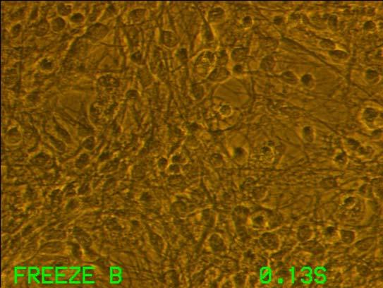
, and fluorescent staining of nuclei using DAPI (Panel B).")

5 Dapi staining: improved signal to noise Figure 3. Rat brain microvascular endothelial cells (RBMVECs) remodeling into tubular orientations in culture. Endothelial cells shown using phase microscopy (Panel A), and fluorescent staining of nuclei using DAPI (Panel B). Note curved growth patterns, as indicated by arrows in 3A and circled by ovals in 3B, demonstrating potential tube formation in culture. ****Note: MONOCHROME CAMERA***** 13 Multiple fluorescent probe microscopy. In this composite micrograph of a cell in mitosis, three different fluorescent probes have been used to stain three different cellular components. The spindle microtubules are revealed with a green fluorescent antibody, centromeres with a red fluorescent antibody and the DNA of the condensed chromosomes with the blue fluorescent dye DAPI. 14 Fluorescence Microscopy and Antibodies: Revealing structure of the cell 5
 Acidic")
 Culture= Neurons and")
6 Astrocytes: Glial Fibrillary Acidic Protein (GFAP) Astrocytes: Glial Fibrillary Acidic Protein (GFAP) Astrocytes: Glial Fibrillary Acidic Protein (GFAP) Culture= Neurons and Astrocytes 6
 Culture= Neurons and")
 Culture=")

7 Astrocytes: Glial Fibrillary Acidic Protein (GFAP) Culture= Neurons and Astrocytes Neuronal Markers: Microtubule Associated Protein 2 (MAP 2) Culture= Retinal Neurons Neuronal Markers: Microtubule Associated Protein 2 (MAP 2) Culture= Retinal Neurons 7


8 New Fluorescent Microscopy Tools: Green Fluorescent Protein and Quantum Dots 22 New Fluorescent Microscopy Tools: GFP 2 ****Note: Other colors are available now*** 23 Nanotech imaging quantum dots Organic dyes have the disadvantage of sometimes fading quickly when continuously illuminated. More stable inorganic fluorochromes have recently been developed composed of tiny crystals of semiconductor material, called quantum dots. These nanoparticles, when coupled to other probes such as antibodies, are ideal for tracking molecules over time. 24 8

, but they all absorb across a broad spectrum.")

9 Figure 9-16a Molecular Biology of the Cell ( Garland Science 2008) Quantum dots: quantum dots are typically composed of semiconductor cores such as cadmium selenide, with a coating to make them water soluble 25 Quantum dots can be coupled to protein probes such as antibodies or streptavidin and, when introduced into a cell, will bind to a protein of interest. Different sized quantum dots emit light of different colors (larger dots emit at longer wavelength), but they all absorb across a broad spectrum. Quantum dots can keep shining for weeks, unlike most fluorescent organic dyes. In this cell, a nuclear protein is labelled green, with the organic dye alexa 488, while microtubules are stained red with quantum dots bound to streptavidin. On continuous exposure to blue light the fluorescent dye fades quickly while the quantum dots continue to fluoresce. Figure 9-16b Molecular Biology of the Cell ( Garland Science 2008) 26 Videos!
Dino-Lite knowledge & education. Fluorescence Microscopes
 Dino-Lite knowledge & education Fluorescence Microscopes Dino-Lite Fluorescence models Smallest fluorescence microscope in the world Revolution to biomedical and educational applications Flexible Easy
More on fluorescence
 More on fluorescence Last class Fluorescence Absorption emission Jablonski diagrams This class More on fluorescence Common fluorophores Jablonski diagrams to spectra Properties of fluorophores Excitation
The Nuclear Area Factor (NAF): a measure for cell apoptosis using microscopy and image analysis
: a measure for cell apoptosis using microscopy and image analysis") The Nuclear Area Factor (NAF): a measure for cell apoptosis using microscopy and image analysis Mark A. DeCoster Department of Biomedical Engineering and Institute for Micromanufacturing, Louisiana Tech
The Nuclear Area Factor (NAF): a measure for cell apoptosis using microscopy and image analysis Mark A. DeCoster Department of Biomedical Engineering and Institute for Micromanufacturing, Louisiana Tech
Visualizing Cells Molecular Biology of the Cell - Chapter 9
 Visualizing Cells Molecular Biology of the Cell - Chapter 9 Resolution, Detection Magnification Interaction of Light with matter: Absorbtion, Refraction, Reflection, Fluorescence Light Microscopy Absorbtion
Concept review: Fluorescence
 16 Concept review: Fluorescence Some definitions: Chromophore. The structural feature of a molecule responsible for the absorption of UV or visible light. Fluorophore. A chromophore that remits an absorbed
Lesson Plan: Fluorescence
 Lesson Plan: Fluorescence Background Fluorescence is produced when a material or substance absorbs light of a given color and then gives off light of another color. The light that is given off, or emitted,
Partha Roy
 Fluorescence microscopy http://micro.magnet.fsu.edu/primer/index.html Partha Roy 1 Lecture Outline Definition of fluorescence Common fluorescent reagents Construction ti of a fluorescence microscope Optical
Fluorescence spectroscopy
 Fluorescence spectroscopy The light: electromagnetic wave Zoltán Ujfalusi Biophysics seminar Dept. of Biophysics, University of Pécs 14-16 February 2011 Luminescence: light is not generated by high temperatures!!!
What to look for in a fluorophore. What to do with a fluorophore. Types of fluorochromes
 What to do with a fluorophore Intracellular localization (ER, Golgi, PM, nuclear, lysosome, MT, actin,...) Dynamic processes (protein synthesis, trafficking, turnover, DNA replication, cytoskeletal remodeling,
Fluorescence Light Microscopy for Cell Biology
 Fluorescence Light Microscopy for Cell Biology Why use light microscopy? Traditional questions that light microscopy has addressed: Structure within a cell Locations of specific molecules within a cell
Imaging Quantum Dots using FUJIFILM LAS 4000
 Imaging Quantum Dots using FUJIFILM LAS 4000 Application Note John Pizzonia, Ph.D. 9-28-07 Quantum dots (also known as nanocrystals) are a special class of materials known as semiconductors, which are
TARGETED IMAGING. Maureen Chan and Ruwani Mahathantila
 TARGETED IMAGING Maureen Chan and Ruwani Mahathantila Overview 2 Introduction to fluorescent imaging Fluorescent agents Quantum Dots Physical properties How QDs work In Vivo QD imaging Future Video What
Quantum Dot applications in Fluorescence Imaging for Calibration and Molecular Imaging
 Quantum Dot applications in Fluorescence Imaging for Calibration and Molecular Imaging Introduction In this application note, we will discuss the application of quantum dots in fluorescence imaging, both
Fluorescence spectroscopy
 Fluorescence spectroscopy The light: electromagnetic wave Tamás Huber Biophysics seminar Dept. of Biophysics, University of Pécs 05-07. February 2013. Luminescence: light emission of an excited system.
QImaging Camera Application Notes Multicolor Immunofluorescence Imaging
 QImaging Camera Application Notes Multicolor Immunofluorescence Imaging In order to image localization of intracellular proteins with high specificity, it is frequently necessary to multiplex antibody
BIO 315 Lab Exam I. Section #: Name:
 Section #: Name: Also provide this information on the computer grid sheet given to you. (Section # in special code box) BIO 315 Lab Exam I 1. In labeling the parts of a standard compound light microscope
Selected Topics in Electrical Engineering: Flow Cytometry Data Analysis
 Selected Topics in Electrical Engineering: Flow Cytometry Data Analysis Bilge Karaçalı, PhD Department of Electrical and Electronics Engineering Izmir Institute of Technology Outline Experimental design
Visualisation, Sizing and Counting of Fluorescent and Fluorescently-Labelled Nanoparticles
 Visualisation, Sizing and Counting of Fluorescent and Fluorescently-Labelled Nanoparticles Introduction Fluorescent molecules have long been used to specifically label particular structures and features
BIO 315 Lab Exam I. Section #: Name:
 Section #: Name: Also provide this information on the computer grid sheet given to you. (Section # in special code box) BIO 315 Lab Exam I 1. In labeling the parts of a standard compound light microscope
Fluorescence spectroscopy
 Fluorescence spectroscopy The light: electromagnetic wave Tamás Huber Biophysics seminar Dept. of Biophysics, University of Pécs 05-06. February 2014. 1 Luminescence: light emission of an excited system.
BASICS OF FLOW CYTOMETRY
 BASICS OF FLOW CYTOMETRY AUTHOR: Ana Isabel Vieira APPROVAL: Henrique Veiga Fernandes Ana Sílvia Gonçalves SOP.UCF.002 03-09-2015 Pag. 1/9 Overview Flow: Fluid Cyto: Cell Metry: Measurement Flow cytometry
Widefield Microscopy Bleed-Through
 In widefield microscopy the excitation wavelengths which illuminate the sample, and the emission wavelengths which reach the CCD camera are selected throughout a filter cube. A filter cube consists of
Principles of flow cytometry: overview of flow cytometry and its uses for cell analysis and sorting. Shoreline Community College BIOL 288
 Principles of flow cytometry: overview of flow cytometry and its uses for cell analysis and sorting Shoreline Community College BIOL 288 Flow Cytometry What is Flow Cytometry? Measurement of cells or particles
Illumatool ΤΜ Tunable Light System: A Non-Destructive Light Source For Molecular And Cellular Biology Applications. John Fox, Lightools Research.
 Illumatool ΤΜ Tunable Light System: A Non-Destructive Light Source For Molecular And Cellular Biology Applications. John Fox, Lightools Research. Fluorescent dyes and proteins are basic analytical tools
Characterizing Phenotypes of Bacteria by Staining Method
 Experiment 3 Laboratory to Biology III Diversity of Microorganisms / Wintersemester / page 1 Experiment 3 Characterizing Phenotypes of Bacteria by Staining Method Advisor NN Reading Chapters in BBOM 9
Characterizing Phenotypes of Bacteria by Staining Method
 Experiment 3 Laboratory to Biology III Diversity of Microorganisms / Wintersemester / page 1 Experiment Characterizing Phenotypes of Bacteria by Staining Method Advisor Reading NN Chapters 3.1, 3.7, 3.8,
Color-Rich Fluoro-Max Dyed Microparticles March 2008
 Fluoro-Max Dyed Microparticles March 2008 Introduction Dyed Microparticles Thermo Scientific Seradyn dyed and fluorescent microparticles are monodisperse particles prepared by unique and proprietary emulsion
Absorption of an electromagnetic wave
 In vivo optical imaging?? Absorption of an electromagnetic wave Tissue absorption spectrum Extinction = Absorption + Scattering Absorption of an electromagnetic wave Scattering of an electromagnetic wave
2004 Debye Lecture 4 C. B. Murray. Quantum Dot Applications: Sun Screen. Solar Cells. Bio-tagging. Solid State Lighting?
 2004 Debye Lecture 4 C. B. Murray Quantum Dot Applications: Sun Screen Solar Cells Bio-tagging Solid State Lighting? Quantum Dot Solar cells Nanocrystal Solar Cells Double-labeling of mitochondria
Quality Matters: Bethyl DyLight Antibody Conjugates
 Quality Matters: Bethyl DyLight Antibody Conjugates Criteria for Fluorescent Probe Selection The use of fluorescent probes has become a mainstream technique to detect speci ic molecular targets in both
Imaging facilities at WUR
 Imaging facilities at WUR Advanced light microscopy facilities at Wageningen UR Programme Thursday 13 June 2013 Lunch meeting organized by Cat-Agro Food 12.00 Welcome and sandwich lunch 12.10 Introduction
August 15, 2018 $ " % ' & OMe. NEt2 MeO
 Taking a deep look: a near infrared fluorescent dye for long term bioimaging ~ Promising photostable tool for single molecule tracking and multicolor imaging ~ August 15, 2018 A team of researchers at
FLUORESCENCE. Matyas Molnar and Dirk Pacholsky
 FLUORESCENCE Matyas Molnar and Dirk Pacholsky 1 Information This lecture contains images and information from the following internet homepages http://micro.magnet.fsu.edu/primer/index.html http://www.microscopyu.com/
11/19/2013. Janine Zankl FACS Core Facility 13. November Cellular Parameters. Cellular Parameters. Monocytes. Granulocytes.
 DEPARTEMENT BIOZENTRUM Janine Zankl FACS Core Facility 13. November 2013 Cellular Parameters Granulocytes Monocytes Basophils Neutrophils Lymphocytes Eosinophils Cellular Parameters 1 What Is Flow Cytometry?
Contents. SCHOOL of FLUORESCENCE. For more information, go to lifetechnologies.com/imagingbasics
 MPSF educator packet This packet contains illustrations and figures from the Molecular Probes School of Fluorescence website. They illustrate concepts from the basic physical properties that underlie fluorescence
Sapphire. Biomolecular Imager THE NEXT GENERATION OF LASER-BASED IMAGING
 Sapphire Biomolecular Imager THE NEXT GENERATION OF LASER-BASED IMAGING Breakthrough image capture and analysis The Sapphire Biomolecular Imager is a next generation laser scanning system that provides
A Brief History of Light Microscopy And How It Transformed Biomedical Research
 A Brief History of Light Microscopy And How It Transformed Biomedical Research Suewei Lin Office: Interdisciplinary Research Building 8A08 Email: sueweilin@gate.sinica.edu.tw TEL: 2789-9315 Microscope
Confocal Microscopy & Imaging Technology. Yan Wu
 Confocal Microscopy & Imaging Technology Yan Wu Dec. 05, 2014 Cells under the microscope What we use to see the details of the cell? Light and Electron Microscopy - Bright light / fluorescence microscopy
ab CytoPainter ER Staining Kit Red Fluorescence
 ab139482 CytoPainter ER Staining Kit Red Fluorescence Instructions for Use Designed to detect Human endoplasmic reticulum by microscopy. This product is for research use only and is not intended for diagnostic
F* techniques: FRAP, FLIP, FRET, FLIM,
 F* techniques: FRAP, FLIP, FRET, FLIM, FCS Antonia Göhler March 2015 Fluorescence explained in the Bohr model Absorption of light (blue) causes an electron to move to a higher energy orbit. After a particular
Time-resolved Measurements Using the Agilent Cary Eclipse Fluorescence Spectrophotometer A Versatile Instrument for Accurate Measurements
 Time-resolved Measurements Using the Agilent Cary Eclipse Fluorescence Spectrophotometer A Versatile Instrument for Accurate Measurements Technical Overview Authors Dr. Fabian Zieschang, Katherine MacNamara,
Single cell molecular profiling using Quantum Dots. Technical Journal Club Rahel Gerosa
 Single cell molecular profiling using Quantum Dots Technical Journal Club 01.10.2013 Rahel Gerosa Molecular Profiling Powerful technique to study complex molecular networks underlying physiological and
Biochemistry. Biochemical Techniques. 18 Spectrofluorimetry
 Description of Module Subject Name Paper Name 12 Module Name/Title 1. Objectives 1.1 To understand technique of Spectrofluorimetry. 1.2 To explain instrumentation design 1.3 What are applications of Spectrofluorimetry?
Direct visualization, sizing and concentration measurement of fluorescently labeled nanoparticles using NTA
 Direct visualization, sizing and concentration measurement of fluorescently labeled nanoparticles using NTA NANOSIGHT RANGE Visualize and Measure Nanoparticle Size and Concentration PARTICLE SIZE PARTICLE
Sub-micron scale patterning of fluorescent. silver nanoclusters using low-power laser
 Sub-micron scale patterning of fluorescent silver nanoclusters using low-power laser Puskal Kunwar 1,*, Jukka Hassinen 2, Godofredo Bautista 1, Robin H. A. Ras 2, and Juha Toivonen 1 1 Tampere University
INTRODUCTION TO FLOW CYTOMETRY
 DEPARTEMENT BIOZENTRUM INTRODUCTION TO FLOW CYTOMETRY F ACS C ore F acility Janine Zankl FACS Core Facility 3. Dezember 2015, 4pm Cellular Parameters Granulocytes Monocytes Basophils Lymphocytes Neutrophils
1st Faculty of Medicine, Charles University in Prague Center for Advanced Preclinical Imaging (CAPI)
") ADVANTAGES Optical Imaging OI Optical Imaging is based on the detection of weak light by a highly sensitive and high resolution CCD camera DISADVANTAGES High sensitivity Limited penetration depth Easy
ADVANTAGES Optical Imaging OI Optical Imaging is based on the detection of weak light by a highly sensitive and high resolution CCD camera DISADVANTAGES High sensitivity Limited penetration depth Easy
Contact Details. Dr Alexander Galkin. Office: MBC Room 186. Tel: (028) Frequency and wavelength.
 Frequency and wavelength.") Contact Details The electromagnetic spectrum Biological Spectroscopy Dr Alexander Galkin Email: a.galkin@qub.ac.uk Dr Alexander Galkin MSc Biomolecular Function - BBC8045 Office: MBC Room 186 Tel: (028)
Contact Details The electromagnetic spectrum Biological Spectroscopy Dr Alexander Galkin Email: a.galkin@qub.ac.uk Dr Alexander Galkin MSc Biomolecular Function - BBC8045 Office: MBC Room 186 Tel: (028)
Classroom Tested Lesson
 Classroom Tested Lesson Video Description Secrets of the Sequence, Show 124, Episode 2 A Green Light for Biology approximately 10 minutes viewing time This discovery known as Green Fluorescent Protein
A Thin Layer Imaging with the Total Internal Reflection Fluorescence Microscopy
 Journal of Optoelectronical Nanostructures Islamic Azad University Summer 2017 / Vol. 2, No. 2 A Thin Layer Imaging with the Total Internal Reflection Fluorescence Microscopy Neda Roostaie 1, Elham Sheykhi
Fuel Fluorescence Logging using the Optical Image Profiler (OIP)
") Fuel Fluorescence Logging using the Optical Image Profiler (OIP) Note: A Patent is Pending for this System. Daniel Pipp Chemist, Geoprobe Systems Presented May 2017 at the Battelle Bioremediation Symposium
Fuel Fluorescence Logging using the Optical Image Profiler (OIP) Note: A Patent is Pending for this System. Daniel Pipp Chemist, Geoprobe Systems Presented May 2017 at the Battelle Bioremediation Symposium
Biophotonics?? Biophotonics. technology in biomedical engineering. Advantages of the lightwave
 Biophotonics - Imaging: X-ray, OCT, polarimetry, DOT, TIRF, photon migration, endoscopy, confocal microscopy, multiphoton microscopy, multispectral imaging - Biosensing: IR spectroscopy, fluorescence,
Hydroxystilbamidine Protocol
 ab138870 Hydroxystilbamidine Protocol Instructions for Use Staining of DNA and RNA in cells. This product is for research use only and is not intended for diagnostic use. 1 Table of Contents 1. Biological
Fluorescence Microscopy
 Fluorescence Microscopy Dr. Arne Seitz Swiss Institute of Technology (EPFL) Faculty of Life Sciences Head of BIOIMAGING AND OPTICS BIOP arne.seitz@epfl.ch Fluorescence Microscopy Why do we need fluorescence
Immunofluorescence microscopy for Lyme borreliosis
 Vector-Borne-Infection Research, Analysis, Strategy Immunofluorescence microscopy for Lyme borreliosis At present, UK NHS testing fails to detect Lyme in an unknown number of patients which could amount
[6.2] Rename Scheme 1 as Figure 1 : renamed as requested
![[6.2] Rename Scheme 1 as Figure 1 : renamed as requested](/thumbs/89/98097923.jpg "[6.2] Rename Scheme 1 as Figure 1 : renamed as requested") David Casto; Thanh Le Department of Chemistry, University of Missouri 205 Schlundt Hall 600 S. College Avenue Columbia, MO 65211 Telephone: (816) 332-0299 E-mail: ptld49@mail.missouri.edu Professor Rainer
David Casto; Thanh Le Department of Chemistry, University of Missouri 205 Schlundt Hall 600 S. College Avenue Columbia, MO 65211 Telephone: (816) 332-0299 E-mail: ptld49@mail.missouri.edu Professor Rainer
Drexel-SDP GK-12 ACTIVITY
 Drexel-SDP GK-12 ACTIVITY Subject Areas: Chemistry, Physical Science, Numbers & Operations, Problem Solving Activity Title: Color Switching Molecules the Magic of Fluorescence The green plastic tops of
Each question may have MULTIPLE correct answers. Select all that are correct.
 Knowledge Assessment Flow Cytometry Workshop, Part 1 April 20, 2015 Each question may have MULTIPLE correct answers. Select all that are correct. 1. Tandem dyes are a. highly stable fluorophores after
Introduction to Fluorescence Jablonski Diagram
 ntroduction to Fluorescence Jablonski Diagram Excited Singlet Manifold S1 internal conversion S2 k -isc k isc Excited riplet Manifold 1 S0 k nr k k' f nr fluorescence k p phosphorescence Singlet round
Simultaneous multi-color, multiphoton fluorophore excitation using dual-color fiber lasers
 Multiphoton Microscopy / Fiber Laser Simultaneous multi-color, multiphoton fluorophore excitation using dual-color fiber lasers Matthias Handloser, Tim Paasch-Colberg, Bernhard Wolfring TOPTICA Photonics
A Survey of Laser Types. Gas Lasers
 Mihail Pivtoraiko Andrei Rozhkov Applied Optics Winter 2003 A Survey of Laser Types Laser technology is available to us since 1960 s, and since then has been quite well developed. Currently, there is a
ab CytoPainter ER Staining Kit Red Fluorescence
 ab139482 CytoPainter ER Staining Kit Red Fluorescence Instructions for Use Designed to detect Human endoplasmic reticulum by microscopy. This product is for research use only and is not intended for diagnostic
Cellular imaging using Nano- Materials. A Case-Study based approach Arun Murali, Srivats V
 Cellular imaging using Nano- Materials A Case-Study based approach Arun Murali, Srivats V Agenda Discuss a few papers Explain a couple of new imaging techniques and their benefits over conventional imaging
Contents Preface xiii Introduction Fabrication and manufacturing technology for optical MEMS
 Contents Preface xiii 1 Introduction 1 1.1 Optical MEMS and optofluidics 1 1.2 History 1 1.2.1 Processes and materials 1 1.2.2 Early devices and systems 2 1.3 Progress in optical MEMS and optofluidics
Imaging of Cells using fluorescents dyes. By: Josué A. Benjamín Rivera September 27, 2018
 Imaging of Cells using fluorescents dyes By: Josué A. Benjamín Rivera September 27, 2018 1 History Sir William Henry Perkin BRITISH CHEMIST In 1856, at the age of 18, William Henry Perkin set out with
SURFACE ENHANCED RAMAN SCATTERING NANOPARTICLES AS AN ALTERNATIVE TO FLUORESCENT PROBES AN EVALUATION
 APPLICATION NOTE SURFACE ENHANCED RAMAN SCATTERING NANOPARTICLES AS AN ALTERNATIVE TO FLUORESCENT PROBES AN EVALUATION Summary: Interest in using nanoparticles specifically, Surface Enhanced Raman Scattering
Flow Cytometry - The Essentials
 Flow Cytometry - The Essentials Pocket Guide to Flow Cytometry: 1. Know your Cytometer 2. Understanding Fluorescence and Fluorophores 3. Gating Process 4. Controls 5. Optimization 6. Panel Building 7.
Fluorescence Microscopy
 Fluorescence Microscopy Dr. Arne Seitz Swiss Institute of Technology (EPFL) Faculty of Life Sciences Head of BIOIMAGING AND OPTICS BIOP arne.seitz@epfl.ch Fluorescence Microscopy Why do we need fluorescence
Two-Photon Microscopy for Deep Tissue Imaging of Living Specimens
 for Deep Tissue Imaging of Living Specimens Tilman Franke* and Sebastian Rhode TILL Photonics GmbH, an FEI company, Lochhamer Schlag 21, D-82166 Gräfelfing, Germany *tilman.franke@fei.com Introduction
Total Internal Reflection Fluorescence Microscopy
 Total Internal Reflection Microscopy Nicole O Neil Indiana University October 24, 2005 Agenda Why use TIRFM? Theory behind TIR Snell s Law Instrumentation Evanescent Wave Excitation of Fluorophores Advantages/Disadvantages
Microscopy from Carl Zeiss
 Microscopy from Carl Zeiss LSM 710 In Tune with Your Application Enjoy new freedom in selecting fluorescent dyes with In Tune, the new laser system for the LSM 710. Whatever the wavelength, you can match
Immunohistochemistry guide
 Immunohistochemistry guide overview immunohistochemistry Overview Immunohistochemistry is a laboratory technique utilized for the visual detection of antigens in tissue. When working with cells this technique
luminometer.committed
 luminometer.committed The Modulus is a multifunctional single tube instrument designed to give you the utmost flexibility for measuring fluorescence, luminescence, and absorbance. The Modulus operates
Spectra Chacracterizations of Optical Nanoparticles
 THAI NGUYEN UNIVERSITY OF EDUCATION Spectra Chacracterizations of Optical Nanoparticles Chu Viet Ha Department of Physics 18/2018 1 THAI NGUYEN UNIVERSITY OF EDUCATION Address 20 Luong Ngoc Quyen Street,
(A) Antigen is in excess. (B) Antibody is in excess. (C) Antibody is added to the antigen. (D) Antigen and antibody are at optimal concentrations.
 Antigen is in excess. (B) Antibody is in excess. (C) Antibody is added to the antigen. (D) Antigen and antibody are at optimal concentrations.") Amount of Antibody Precipitated Immunology - Problem Drill 21: Antigen-Antibody Interactions Question No. 1 of 10 1. When antigen and antibodies bind, maximal precipitation occurs when? Question #1 (A)
Amount of Antibody Precipitated Immunology - Problem Drill 21: Antigen-Antibody Interactions Question No. 1 of 10 1. When antigen and antibodies bind, maximal precipitation occurs when? Question #1 (A)
BIOSENOSRS BIO 580. Nanobiosensors WEEK-13 Fall Semester. Faculty: Dr. Javed H. Niazi KM Faculty of Engineering & Natural Sciences Sabanci University
 BIOSENOSRS BIO 580 Nanobiosensors WEEK-13 Fall Semester Faculty: Dr. Javed H. Niazi KM Faculty of Engineering & Natural Sciences Sabanci University Topics that will be covered in the course History of
Confocal Microscopes. Evolution of Imaging
 Confocal Microscopes and Evolution of Imaging Judi Reilly Hans Richter Massachusetts Institute of Technology Environment, Health & Safety Office Radiation Protection What is Confocal? Pinhole diaphragm
Cell Imaging. Cell Imaging 48
 Cell Imaging 48 bio-rad.com/zoe Cell Imaging Bio-Rad s suite of tools for fluorescence microscopy and cell imaging includes the ZOE fluorescent cell imager and nuclear dyes. See Also PureBlu Hoechst 33342
Resolution of Microscopes Visible light is nm Dry lens(0.5na), green(530nm light)=0.65µm=650nm for oil lens (1.4NA) UV light (300nm) = 0.13µm f
, green(530nm light)=0.65µm=650nm for oil lens (1.4NA) UV light (300nm) = 0.13µm f") Microscopes and Microscopy MCB 380 Good information sources: Alberts-Molecular Biology of the Cell http://micro.magnet.fsu.edu/primer/ http://www.microscopyu.com/ Approaches to Problems in Cell Biology
Microscopes and Microscopy MCB 380 Good information sources: Alberts-Molecular Biology of the Cell http://micro.magnet.fsu.edu/primer/ http://www.microscopyu.com/ Approaches to Problems in Cell Biology
Elecrtonic Supplementary Information. Application of quantum dot barcodes prepared using biological self-assembly to multiplexed immunoassays
 Elecrtonic Supplementary Information Application of quantum dot barcodes prepared using biological self-assembly to multiplexed immunoassays Sakandar Rauf, Andrew Glidle and Jonathan M Cooper Department
SOLAR ENERGY. Approximately 120,000 TW of solar energy strikes the earth s surface, capturing only a fraction could supply all of our energy needs.
 SOLAR ENERGY Approximately 120,000 TW of solar energy strikes the earth s surface, capturing only a fraction could supply all of our energy needs. What is Photovoltaics? Photovoltaics is a high-technology
Post-expansion antibody delivery, after epitope-preserving homogenization.
 Supplementary Figure 1 Post-expansion antibody delivery, after epitope-preserving homogenization. (a, b) Wide-field fluorescence images of Thy1-YFP-expressing mouse brain hemisphere slice before expansion
CF Dyes Next Generation Fluorescent Dyes Secondary antibody
 CF Dyes Next Generation Fluorescent Dyes Secondary antibody OZYME 10 AVENUE AMPÈRE - CS 30268-78053 ST QUENTIN EN YVELINES CEDEX Tél. : 01 34 60 24 24 - Fax : 01 34 60 92 12 - www.ozyme.fr/info CF Dyes
MICROSCOPY. "micro" (small) "scopeo" (to watch)
 scopeo (to watch)") MICROSCOPY "micro" (small) "scopeo" (to watch) THE RELATIVE SIZES OF MOLECULES, CELLS AND ORGANISMS THE RELATIVE SIZES OF MOLECULES, CELLS AND ORGANISMS MICROSCOPY 1590 2012 MICROSCOPY THE LIGHT Light:
MICROSCOPY "micro" (small) "scopeo" (to watch) THE RELATIVE SIZES OF MOLECULES, CELLS AND ORGANISMS THE RELATIVE SIZES OF MOLECULES, CELLS AND ORGANISMS MICROSCOPY 1590 2012 MICROSCOPY THE LIGHT Light:
Rice/TCU REU on Computational Neuroscience. Fundamentals of Molecular Imaging
 Rice/TCU REU on Computational Neuroscience Fundamentals of Molecular Imaging June 2, 2009 Neal Waxham 713-500-5621 m.n.waxham@uth.tmc.edu Objectives Introduction to resolution in light microscopy Brief
Live cell microscopy
 Live cell microscopy 1. Why do live cell microscopy? 2. Maintaining living cells on a microscope stage. 3. Considerations for imaging living cells. 4. Fluorescence labeling of living cells. 5. Imaging
Real-Time PCR Principles and Applications
 Real-Time PCR Principles and Applications Dr Esam Ibraheem Azhar (BSc, MSc, Ph.D Molecular Medical Virology) Asst. Prof. Medical Laboratory Technology Department Objectives Real-Time PCR Principles and
seminar
 seminar 02.26.-28.2013. Flow cytometry Process or measurement method can measure discrete properties physical, chemical, biochemical, biological parameters of separate particles, e.g. biological cells
PALM/STORM, BALM, STED
 PALM/STORM, BALM, STED Last class 2-photon Intro to PALM/STORM Cyanine dyes/dronpa This class Finish localization super-res BALM STED Localization microscopy Intensity Bins = pixels xx 2 = ss2 + aa 2 /12
Design for Manufacturability (DFM) in the Life Sciences
 in the Life Sciences") T E C H N I C A L N O T E Design for Manufacturability (DFM) in the Life Sciences Fluorescence Spectroscopy Product Platform Realized with TracePro TM Suite of Opto-Mechanical Design Software Tools Authors:
T E C H N I C A L N O T E Design for Manufacturability (DFM) in the Life Sciences Fluorescence Spectroscopy Product Platform Realized with TracePro TM Suite of Opto-Mechanical Design Software Tools Authors:
Theremino DNA Meter. Fluorometer for DNA Measurements. Ana Rodriguez (MUSE) - Lodovico Lappetito (Theremino)
 - Lodovico Lappetito (Theremino)") Theremino DNA Meter Fluorometer for DNA Measurements Ana Rodriguez (MUSE) - Lodovico Lappetito (Theremino) Theremino_DNAMeter_Hardware_ENG - 11/01/2016 Pag. 1 Table of Contents Principle of Operation...
Theremino DNA Meter Fluorometer for DNA Measurements Ana Rodriguez (MUSE) - Lodovico Lappetito (Theremino) Theremino_DNAMeter_Hardware_ENG - 11/01/2016 Pag. 1 Table of Contents Principle of Operation...
Localization Microscopy
 Localization Microscopy Theory, Sample Prep & Practical Considerations Patrina Pellett & Ann McEvoy Applications Scientist GE Healthcare, Cell Technologies May 27 th, 2015 Localization Microscopy Talk
Fluorescent In Situ Hybridization (FISH) Assay
 Assay") Fluorescent In Situ Hybridization (FISH) Assay 1 What is FISH 2 Probes 3 FISH Procedure 4 Application Definition, Principle and Sample Types The core of FISH technology A quick and simple FISH protocol
Fluorescent In Situ Hybridization (FISH) Assay 1 What is FISH 2 Probes 3 FISH Procedure 4 Application Definition, Principle and Sample Types The core of FISH technology A quick and simple FISH protocol
Supplementary Data. In-Vivo Tumor Targeting and Spectroscopic Detection with Surface- Enhanced Raman Nanoparticle Tags
 Supplementary Data In-Vivo Tumor Targeting and Spectroscopic Detection with Surface- Enhanced Raman Nanoparticle Tags X.-M. Qian, 1 Xiang-Hong Peng, 2 Dominic O. Ansari, 1 Qiqin Yin-Goen, 3 Georgia Z.
Definitions. What functions does a flow cytometer be able to do? The build of the Flow cytometry and sorting. Flow cytometry
 The build of the Flow cytometry and sorting Flow cytometry Seminar Mónika Tóth 11-12-13.04.2011 Definitions Flow cytometry Process or measurement method can measure discrete properties physical, chemical,
Beginner s guide to Real-time PCR
 Beginner s guide to Real-time PCR Beginner s guide to Real-time PCR PCR or the Polymerase Chain Reaction has become the cornerstone of modern molecular biology the world over. Real-time PCR is a bespoke
Studies on Structural and Optical Properties of Iron doped Cds Nanoparticles ABSTRACT
 Studies on Structural and Optical Properties of Iron doped Cds Nanoparticles Atheek P., Vidhya M. and Balasundaram O. N Department of Physics, PSG College of Arts & Science, Coimbatore, Tamil Nadu, India
ALP (alkaline phosphatase) calibrators were analyzed manually in microtiter plates to find the linearity range by following this protocol:
 calibrators were analyzed manually in microtiter plates to find the linearity range by following this protocol:") Exam Mol 3008 May 2009 Subject 1 (15p) ALP (alkaline phosphatase) calibrators were analyzed manually in microtiter plates to find the linearity range by following this protocol: Reaction solutions: 50
Exam Mol 3008 May 2009 Subject 1 (15p) ALP (alkaline phosphatase) calibrators were analyzed manually in microtiter plates to find the linearity range by following this protocol: Reaction solutions: 50
Microelectromechanical Drug Delivery Systems. Sarah Smith & Jurek Smolen
 Microelectromechanical Drug Delivery Systems Sarah Smith & Jurek Smolen Current Drug Delivery Systems Common Administration Methods Problems Oral Intravenous Intramuscular Transdermal Difficult to control
Skull optical clearing window for in vivo imaging of the mouse cortex at synaptic resolution
 SUPPLEMENTARY INFORMATION for Skull optical clearing window for in vivo imaging of the mouse cortex at synaptic resolution Yanjie Zhao 1,2, Tingting Yu 1,2, Chao Zhang 1,2, Zhao Li 1,2, Qingming Luo 1,2,