Correspondence should be addressed to Anders Bruun Mathiasen;
|
|
|
- Lawrence Richardson
- 6 years ago
- Views:
Transcription
1 Stem Cells International Volume 2013, Article ID , 10 pages Research Article Optimal Labeling Dose, Labeling Time, and Magnetic Resonance Imaging Detection Limits of Ultrasmall Superparamagnetic Iron-Oxide Nanoparticle Labeled Mesenchymal Stromal Cells Anders Bruun Mathiasen, 1 Louise Hansen, 1 Tina Friis, 1 Carsten Thomsen, 2 Kishore Bhakoo, 3 and Jens Kastrup 1 1 Cardiac Stem Cell Laboratory and Catheterization Laboratory, Rigshospitalet, Copenhagen University Hospital, Blegdamsvej 9, 2100 Copenhagen, Denmark 2 Department of Radiology, Rigshospitalet, Copenhagen University Hospital, Copenhagen, Denmark 3 Translational Molecular Imaging Group, Singapore Bioimaging Consortium, Agency for Science, Technology and Research (A -STAR), Singapore Correspondence should be addressed to Anders Bruun Mathiasen; abbe@dadlnet.dk Received 30 December 2012; Revised 9 February 2013; Accepted 11 February 2013 Academic Editor: Weian Zhao Copyright 2013 Anders Bruun Mathiasen et al. This is an open access article distributed under the Creative Commons Attribution License, which permits unrestricted use, distribution, and reproduction in any medium, provided the original work is properly cited. Background. Regenerative therapy is an emerging treatment modality. To determine migration and retention of implanted cells, it is crucial to develop noninvasive tracking methods. The aim was to determine ex vivo magnetic resonance imaging (MRI) detection limits of ultrasmall superparamagnetic iron-oxide (USPIO) labeled mesenchymal stromal cells (MSCs). Materials and Methods. 248 gel-phantoms were constructed and scanned on a 1.5T MRI-scanner. Phantoms contained human MSCs preincubated with USPIO nanoparticles for 2, 6, or 21 hours using 5 or 10 μguspio/10 5 MSCs. In addition, porcine hearts were scanned after injection of USPIO labeled MSCs. Results. Using 21 h incubation time and 10 μguspio/10 5 MSCs, labeled cells were clearly separated from unlabeled cells on MRI using (P < 0.001), (P = 0.007), and MSCs (P = 0.008). At lower incubation times and doses, neither labeled nor unlabeled cells could be separated. In porcine hearts labeled, but not unlabeled, MSCs were identified on MRI. Conclusions. As few as MSCs can be detected on MRI using 21 h incubation time and 10 μguspio/10 5 MSCs. At lower incubation times and doses, several million cells are needed for MRI detection. USPIO labeled cells can be visualized by MRI in porcine myocardial tissue. 1. Introduction Stem cell therapy with potential to regenerate damaged myocardium is an emerging treatment modality for ischemic heart disease [1 3]. For future success of cardiac stem cell therapy, it is crucial to develop noninvasive tracking methods for determining the biodistribution and fate of the stem cells after delivery. Thus far, tracking of cardiovascular delivered stem cells in a clinical setting has been limited to direct cell labeling with radioisotopes and tracking with gamma-cameras, single-photon emission computed tomography, or positron emission tomography [4]. Although providing highly sensitive visualization, these methods are limited by low spatial resolution and short half-lives of radioisotopes from minutes to hours, thus only permitting short-term tracking of the cells. Other drawbacks are exposure to ionizing radiation and nontarget signal leakage. Tracking of cells labeled with superparamagnetic ironoxide (SPIO) or ultrasmall superparamagnetic iron-oxide (USPIO) nanoparticles using magnetic resonance imaging (MRI) offers high spatial resolution in combination with high
2 2 Stem Cells International soft tissue detail, without exposing the patient to ionizing radiation. Furthermore, the cells can be tracked for months. Cellular labeling methods with SPIO or USPIO are relatively simple, fast and inexpensive. Iron-oxide is nontoxic, since iron is a naturally occurring metal in the human body, and the iron oxide core is coated with biocompatible shell, allowing its eventual assimilation via endogenous metabolic iron cycles. The use of SPIO and USPIO labeling is clinically safe and does not influence cell function [5]. MRI tracking of SPIO and USPIO labeled cells has been utilized in vivo in rat, canine, and porcine models of myocardial infarction (MI) using a variety of delivery methods [6 13]. The labeled cells were tracked for up to 8 weeks after delivery. In vivo tracking of SPIO and USPIO labeled cells has not yet been utilized in a clinical cardiovascular setting, but both SPIO and USPIO have been used successfully in a number of noncardiovascular clinical studies [14 19]. There has been some concern that MRI signals from SPIO and USPIO labeled cells may originate from macrophages that have engulfed the labeled cells. This was seen in a fewratstudies[6, 20], but the majority of animal studies have shown the opposite, that the MRI does in fact originate from the labeled cells and not macrophages [7 9, 11 13, 21, 22]. A general concern for cardiovascular cell therapy has been that the number of cells that remain in the heart after treatment may be limited to only a few percent. However, it has recently been demonstrated that these studies may be severely biased, as there is considerable spontaneous leaking of the radioisotopes used in these studies [23]. Therefore, the number of cells remaining in the heart after treatment may be as high as 60% one week after treatment. For tracking of nonphagocytic cells, USPIO particles are probably more suitable than SPIO particles, due to higher cellular uptake [24] and longer plasmatic half-life [25]. The USPIO particles used in the present study (IODEX) have an additional cross-linking of the dextran coating compared to traditionally used SPIO and USPIO particles [26]. This stabilizes the iron core of the particles allowing for longer cell tracking periods. The aim of the present study was to determine ex vivo MRI detection limits of IODEX labeled human MSCs with respect to cell numbers and USPIO concentration and incubation period for future clinical application. 2. Materials and Methods 2.1. Isolation and Culture Expansion of MSCs. Bone marrow was obtained from the iliac crest by needle aspiration from healthy donors. The studies were conducted under local ethical approval. Mononuclear cells were then isolated by gradient centrifugation and cultured in complete medium consisting of Dulbecco s modified Eagle medium supplemented with HEPES and L-glutamine, (PAA Laboratories, Austria), 10% fetal bovine serum (PAA Laboratories, Austria), and 1% penicillin/streptomycin (Invitrogen, Austria). Cells were incubated at 37 Cinhumidairwith5%CO 2.Medium was changed twice a week. The cells were grown to confluence before each passage. After two passages, the cells were washed with PBS (Invitrogen, Austria) and harvested with TrypLE Select (Invitrogen, Austria). Cells from each donor were characterized by flow cytometry for CD90, CD73, CD105, CD13, CD45, and CD34, in accordance with the minimal criteria for defining multipotent mesenchymal stromal cells [27] USPIO Preparation. Tat-peptide derivatized USPIO nanoparticles coated with dextran (IODEX-TAT-FITC; nm) were prepared in our laboratory using the method described by Josephson et al. [28]. Briefly, the dextrancoated USPIO nanoparticles were synthesized and subsequently conjugated with TAT-fluorescein isothiocyanate (FITC) peptide [GRKKRRQRRR GYK(FITC)C-NH2]. TAT- FITC was synthesized using FMOC-protected amino acid (2- (1-H-benzotriazol-2-yl)-1,1,3,3-tetramethyluronium hexafluorophosphate; HBTU) activation chemistry. The final iron concentration was 2.5 mg/ml, and the solution was sterilized by gamma-irradiation prior to use USPIO Labeling of MSCs. Dose titrating evaluation of iron concentrations added to cells and resulting amounts of iron bound to cells by Josephson et al. [28] revealed that a plateau phase was reached at 100 μg ironper10 6 cells (10 μg ironper10 5 cells). In the present study, we wanted to evaluate both this maximum dose of 10 μg ironper10 5 cells and also the half of this dose, 5 μg ironper10 5 cells, as this dose reached near optimum iron binding in the original titration study [28]. In the mentioned titrating study, cells were incubated overnight (18 21 hours), whereas animal studies using IODEX-TAT-FITC for labeling MSCs have used only 4 6 hours of incubation [13, 29]. In the present study, we evaluate the mentioned iron doses at 2, 6, and 21 hours of incubation. MSCs were labeled by incubation with USPIO nanoparticles at a concentration of either 5 μg ironper10 5 cells (half dose) or 10 μg ironper10 5 cells (full dose) in complete medium for 2, 6, or 21 hours at 37 Cinhumidairwith 5% CO 2.Then,thecellswerewashed3timesinPBSand harvested with TrypLE Select and centrifuged 5 min at 300 g. After centrifugation, the cells were resuspended in PBS, and thenumberofcellsandcellviabilitywasdeterminedby propidium iodide staining using a NucleoCounter NC-100 (Chemometec, Denmark) USPIO Iron Concentration. MSCs in a volume corresponding to cells were transferred to microfuge tubes andcentrifugedfor5minat500g.cellpelletwasfrozen andstoredat 20 C until date of quantification. Then, the cells were resuspended in 50 μl PBS,hydrolysedfor30min with 100 μl 6 M HCl, and ph neutralized by addition of 60 μl 10 M NaOH. The cells were centrifuged for 2 min at 1300 g, and 100 μl supernatant was used for automatic iron quantitation by use of a Konelab 60i robot (Therma Electron, Finland).
 MRI image of 2 phantoms with an ellipsoid region of interest placed in the upper phantom. USPIO: ultrasmall superparamagnetic iron-oxide. Table 1: Number of MRI phantoms.")

3 Stem Cells International 3 (a) (b) Figure 1: MRI phantoms. (a) Two phantoms containing USPIO labeled cells. (b) MRI image of 2 phantoms with an ellipsoid region of interest placed in the upper phantom. USPIO: ultrasmall superparamagnetic iron-oxide. Table 1: Number of MRI phantoms. Number of MSCs USPIO dose USPIO incubation time 2 hours 6 hours 21 hours full full full half half half MSC: mesenchymal stromal cell; USPIO: ultrasmall super-paramagnetic iron-oxide. USPIO dose full = 10 μgper10 5 cells; half = 5 μgper10 5 cells MRI-Phantoms. Labeled and unlabeled MSCs were transferred to microfuge tubes with ,5 10 5,or MSCs per tube. Tubes were centrifuged at 500 g for 5 minutes. The cells were then suspended in 500 μl 1% agarosegel. In total, 248 phantoms were constructed containing either unlabeled MSCs or MSCs labeled with half or full USPIO dose,incubatedfor2,6,or21hours.twophantomsareshown in Figure 1(a).AnoverviewofallMRIphantomsisprovided in Table 1. A number of reference phantoms containing only agarose-gel were constructed as reference controls MRI Phantom Scanning Protocol and Image Analysis. Phantoms were scanned using a 1.5T GE Signa Excite HD MRI scanner with a 4-channel receive-transmit brain coil (GE Healthcare). Two phantoms and one reference phantom with no cells were scanned concurrently. Phantoms were placed in an Eppendorf tube rack, with the reference phantom in the center and a randomly selected MSC phantom on eachsidewith4cmdistancetothereferencephantom.the rack was placed and fixated with tape on top of 4 other racks inside the coil to achieve a central position within the coil. Images were acquired using a brain-hemorrhage T2 - weighted gradient-echo (GRE) sequence with repetition-time (TR) = 620 ms, echo-time (TE) = 15.7 ms, flip-angle = 35, matrix = , field of view (FOV) = mm, and slice thickness = 7 mm. Image analysis was performed using an Advantage Workstation AW (GE Healthcare). An ellipsoid region of interest (ROI) of 20 mm 2 was placed on the images in the center of each phantom, avoiding the edges. The postprocessing tool produces mean intensity values for each ROI. Each pixel in the ROI is given an intensity value between 0 and The mean intensity value is the mean of these values for all the pixels in the ROI (Operators manual, GE Healthcare). For comparative analysis, the difference in mean intensity values between reference and cell phantom was used. Figure 1(b) shows MRI image of 2 phantoms with an ellipsoid ROI placed in the upper phantom.
4 4 Stem Cells International Table 2: Cellular iron content. Group Iron content per cell n Multiple comparisons (Bonferroni corrected) Group A Group B Group C Group D Group E Group F Group G (A) Unlabeled 0.48 ± 0.17 pg 44 ns P = 0.02 P < P < P < P < (B) Half dose, 2 hours incubation 1.22 ± 0.52 pg 17 ns ns P = P < P < (C) Full dose, 2 hours incubation 1.54 ± 0.83 pg 17 ns P = 0.03 P < P < (D) Half dose, 6 hours incubation 2.36 ± 0.65 pg 17 ns P < P < (E) Full dose, 6 hours incubation 2.71 ± 0.86 pg 21 P < P < (F) Half dose, 21 hours incubation 4.26 ± 1.59 pg 37 P = (G)Fulldose,21hoursincubation 5.24± 1.50 pg 44 Values are shown ± SD. USPIO: ultrasmall super-paramagnetic iron-oxide. USPIO dose full = 10 μgper10 5 cells. half = 5 μgper10 5 cells, ns: nonsignificant Porcine Hearts. Two hearts from freshly slaughtered pigs were placed and fixated with small wooden sticks in a polystyrene box. The hearts were MRI scanned before and after injection of MSCs. One heart was injected with 4 injections of USPIO labeled MSCs (full dose 21 hours incubation), each injection with approximately MSCs in 0.4 ml. The other heart received 4 injections with unlabeled cells. Care was taken that the hearts remained in the exact same position before and after injections. Cellular iron content (pg) MRI Scanning of Porcine Hearts. Hearts were scanned using a 1.5T Siemens Magnetom Avanto MRI scanner and a body matrix coil (Siemens AG, Germany). The scanning protocol was a thalassemia T2 weighted GRE sequence with TR = 200 ms, flip angle = 20,matrix=96 256, FOV = mm, and slice thickness of 5 mm. The entire left ventricle was scanned with concurrent slice thickness of 5 mm with no gaps. The protocol produces 8 images for each slice, with different TE times (3.05, 5.89, 8.73, 11.57, 14.41, 17.25, 20.09, and ms) Statistical Analysis. Statistical analysis was carried out using SPSS 20 (SPSS Inc., USA). One-way ANOVA tests were used for comparing cellular iron content and MRI intensity differences between groups. A P value < 0.05 was considered significant. If the ANOVA test of the groups was significant, a multiple group versus group comparison was made within the ANOVA procedure, to determine which of the groups differed. All P values in these tests were adjusted using the Bonferroni method to counteract the issue of multiple comparisons. Normality was determined for each group with Kolmogorov-Smirnov and Shapiro-Wilk tests. Equal variances were determined with Levene s test for homogeneity of variances. 3. Results 3.1.IronContentinMSCs. Determination of the cellular iron load showed a positive correlation between iron content per cell and the length of the USPIO incubation period. The results are illustrated in Figure 2, and iron values and statistics areprovidedintable2. 0 Unlabeled Unlabeled Half dose Full dose 2 hours USPIO incubation 6 hours USPIO incubation 21 hours USPIO incubation Figure 2: Cellular iron content. The iron content per cell was determined in unlabeled MSC and MSC incubated with half or full dose USPIO for 2, 6, and 21 hours. MSC: mesenchymal stromal cells. USPIO: ultrasmall superparamagnetic iron-oxide. USPIO dose full: 10 μgper10 5 cells; half: 5 μgper10 5 cells. After 2 hours USPIO incubation time, the cellular iron content was only slightly higher than that of the unlabeled cells. This increase was only significant for the full USPIO dose compared to the unlabeled cells. After 6 hours USPIO incubation time, there was a highly significant increase in cellular iron content compared to unlabeled cells. When comparing to 2-hour incubation times, only the full USPIO dose was significantly higher after 6 hours. After 21 hours, the increase in cellular iron content was highly significant compared to both unlabeled and labeled cells for 2 and 6 hours at both USPIO doses. The cells labeled for 21 hours with the full USPIO dose also had significantly higher iron content than the cells labeled for 21 hours with only half USPIO dose MRI of USPIO Incubated Phantoms. Overall MRI intensity diminished with increasing cell numbers and USPIO dosage. A graphical illustration of the absolute MRI intensities of phantoms incubated with USPIO for 21 hours is provided in Figure 3, and an illustration of the numeric
5 Stem Cells International 5 Table 3: MRI intensity differences after 21-hour USPIO incubation. USPIO dose cells cells cells Full 249 ± ± ± 64 Half 134 ± ± ± 92 Unlabeled 46 ± ± 47 8 ± 53 Multiple comparisons P < P = P = Full versus unlabeled P < P = P = Full versus half P = ns ns Half versus unlabeled ns ns ns The MRI intensities are mean pixel intensities (values between 0 and 4095) of a 20 mm 2 region of interest in the center area of each phantom, supplied by the imaging software. Values are shown ± SD. USPIO: ultrasmall super-paramagnetic iron-oxide. USPIO dose full = 10 μgper10 5 cells. half = 5 μgper10 5 cells, ns: non-significant MRI intensity difference 200 MRI intensity Number of cells ( 1000) 1000 Full Half None USPIO dose Number of cells ( 1000) Full Half None USPIO dose Figure 3: Absolute phantom MRI intensities after 21 hour USPIO incubation. MRI intensities are absolute mean values. USPIO: ultrasmall superparamagnetic iron-oxide. USPIO dose: full = 10 μg per 10 5 cells; half = 5 μgper10 5 cells. Figure 4: Phantoms intensity differences after 21-hour USPIO incubation. MRI intensity differences are the mean numeric difference between absolute MRI intensities of phantoms and reference gels. USPIO = ultrasmall superparamagnetic iron-oxide. USPIO dose: full = 10 μgper10 5 cells; half = 5 μgper10 5 cells. differences in MRI intensity compared to the reference gels is provided in Figure4. The differences and statistics are shown in Table 3. USPIO labeled MSCs in amounts of , , and could all be significantly separated on MRI fromthesamenumberofunlabeledcells,whenusinguspio incubation time of 21 hours and full USPIO dosage. MSCs labeled with half USPIO dosage could not be separated from unlabeled MSCs at any concentration on MRI. With 2 and 6 hours of incubation time, it was not possible to differentiate between labeled and unlabeled cells at any dose or concentration on MRI (see Tables 4 and 5). Therefore, the MRI detection limits are as low as cells when using full USPIO dose and 21 hours of incubation time. For cells labeled with lower USPIO dose and lower incubation times, no significant difference was detected on MRI compared to unlabeled cells, and the detection limits for cells labeled using these conditions will therefore be at least several million cells MRI of Porcine Hearts. There are distinct differences in the before and after images when looking at MRI images from porcine hearts receiving USPIO labeled MSCs (Figure 5). Hypointense areas can be identified in the after images which are equivalent to the USPIO labeled MSC injection areas. The figure images are with TE = ms, which was the TE that gave the best visualization of the differences. MRI images from the heart receiving unlabeled cells were without visual differences; thus, unlabeled MSCs are undetectable on MRI (Figure 6).
 (a2) (b2) (a3) (b3) (a3) (b3) (a4) (b4) (a4) (b4)")

 and after injection (b1 b5) of USPIO-labeled MSCs.")
. MSCs: mesenchymal stromal cells.")

 and after injection (b1 b5) of unlabeled MSCs.")


 of a 20 mm 2 region of")


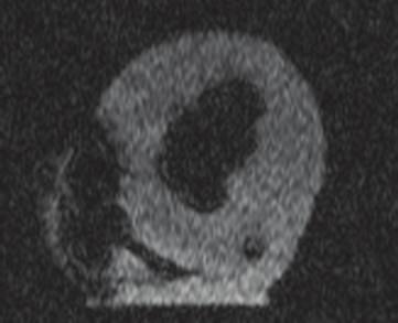
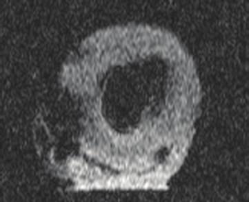
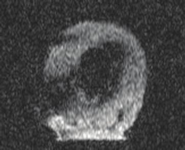
6 6 Stem Cells International (a1) (b1) (a1) (b1) (a2) (b2) (a2) (b2) (a3) (b3) (a3) (b3) (a4) (b4) (a4) (b4) (a5) (b5) (a5) (b5) Figure 5: MRI images of porcine myocardium before and after USPIO labeled MSC injection. T2 -images of porcine myocardium before injection (a1 a5) and after injection (b1 b5) of USPIO-labeled MSCs. USPIO labeled MSCs are identified as hypointense areas (arrows). MSCs: mesenchymal stromal cells. USPIO: ultrasmall superparamagnetic iron-oxide. Figure 6: MRI images of porcine myocardium before and after unlabeled MSC injection. T2 -images of porcine myocardium before injection (a1 a5) and after injection (b1 b5) of unlabeled MSCs. Unlabeled cells cannot be identified. MSCs: mesenchymal stromal cells. USPIO: ultrasmall superparamagnetic iron-oxide. Table 4: MRI intensity differences after 6-hour USPIO incubation. USPIO dose cells cells cells Full 74 ± ± ± 30 Half 57 ± ± ± 36 Unlabeled 47 ± ± 20 6 ± 50 ns ns ns The MRI intensities are mean pixel intensities (values between 0 and 4095) of a 20 mm 2 region of interest in the center area of each phantom, supplied by the imaging software. Values are shown ± SD. USPIO: ultrasmall super-paramagnetic iron-oxide. USPIO dose full = 10 μgper10 5 cells. half = 5 μgper10 5 cells. ns: non-significant. Table 5: MRI intensity differences after 2-hour USPIO incubation. USPIO dose cells cells cells Full 59 ± ± ± 68 Half 39 ± ± ± 70 Unlabeled 71 ± ± ± 39 ns ns ns The MRI intensities are mean pixel intensities (values between 0 and 4095) of a 20 mm 2 region of interest in the center area of each phantom, supplied by the imaging software. Values are shown ± SD. USPIO: ultrasmall super-paramagnetic iron-oxide. USPIO dose full = 10 μgper10 5 cells; half = 5 μgper10 5 cells. ns: non-significant.
7 Stem Cells International 7 4. Discussion In the present study, we have examined MRI detection limits of USPIO labeled MSCs in vitro with regard to cell numbers and USPIO incubation dosage and incubation time. The study demonstrated that an incubation period of 21 hours with USPIO is superior to 2 and 6 hours incubation times and that a USPIO incubation dose of 10 μg per10 5 cells is preferable over 5 μgper10 5 cells. In addition, USPIO labeled MSCs could be distinguished by MRI when injected into myocardial tissue. The hypointense MRI injection-regions were due to USPIO labeling, as unlabeled cells were not visibleonmriscans. MRI tracking of cells labeled with iron-oxide based nanoparticles in cardiovascular disease has been utilized in a variety of animal studies. In one study, rats were subjected to MI and intramyocardial injection of SPIO labeled allogeneic MSCs [21]. The MSCs were injected in the border zone of the infarct area. Hypointense regions were visible on MRI in the entire 16-week followup period. In non-mi control rats injected with labeled cells, the hypointense regions were only visibleonmrifor1week.thiswasalsothecaseformi controlratsreceivingspioparticlesalone.thisindicatedthat cell retention is dependent on the presence of inflammation inthetargettissueandalsothatspioparticlesfromdeadcells will be cleared from the area, and therefore the hypointense regions on MRI corresponded to SPIO particles within live cells. This was confirmed in histologic analysis done after 1, 16, and 20 weeks. SPIO-containing cells were identified at the injection site. Macrophage specific CD68 staining showed that macrophages were only present after 1 week and not after 16and20weeks.ThemajorityofCD68positivecellsdidnot contain iron, and most of the iron-containing cells did not express CD68. Thus, the originally labeled cells were present andnotwithinmacrophages.inacaninemimodel,spio labeledmscswereinjectedintramyocardiallyintotheborder zone of the infarct [7].InjectionsiteswerevisibleonMRIas hypointense regions for the entire 8 week followup period. Histology with Prussian Blue (PB) staining showed presence of SPIO containing cells well integrated within the tissue. Interesting results were found in a study using a rat MI model, where SPIO labeled MSCs were injected intramyocardially directly into the infarct lesion [6]. Hypointense regions were visible on MRI in the entire followup period of 4 weeks. In this study, postmortem tissue staining revealed that the delivery sites for both labeled and nonlabeled cells were infiltrated with inflammatory cells and that most MSCs did not survive. Similar results, where iron particles were engulfed in macrophages, were found in another rat study, where rats received intramyocardial injections of either xenogeneic human cells or allogeneic rat cells [20]. In both studies, the cells were injected directly into infarct lesion, whereas most other studies have injected their cells into the border zone of the infarct area. Perhaps the alternative injection site could explain the diverse results in these studies. Moreover, five other studies have also histologically evaluated intramyocardial injection [8] and intracoronary infusion of SPIO and USPIO labeled autologous MSCs [9, 12, 22] orendothelialprogenitorcells[11] inporcinemi settings. In all these studies, hypointense regions were visible on MRI in the infarct region in the entire followup periods of 3 8 weeks. Interestingly and in strong contrast to the aforementioned rat study, post mortem analysis of sections of the infarct regions showed that SPIO and USPIO particles remained within the originally labeled cells and that these cells corresponded with hypointense MRI signals. Only one of the studies using SPIO labeling detected sparse amounts of macrophages in the tissue, and these were clearly separated from labeled cells [11]. These findings are supported by another study using a rat MI model, where USPIO labeled MSCs were intramyocardially injected. Histology in this study confirmed that the original labeled MSCs remained in the infarcted area up to 6 weeks after implantation using both MRI and histology [13]. A major concern with MRI tracking of SPIO and USPIO labeled cells has been that the obtained MRI signals could originate from macrophages that consumed the SPIO particles after cell death of the original labeled cells. The vast majority of animal studies have found the labeled cells to live at the injection sites with no signs of macrophages or other phagocytic cells. Therefore, concern for phagocytic engulfment of injected cells seems overrated and should not hinder future studies in this area. Another concern has been that the number of cells that remain in the heart for a prolonged period of time may be limited. A number of animal studies have attempted to assess the number of cells that remain in the heart at different time points after intramyocardial injection. The study by Tran et al. [23] used 111 In-oxine labeled culture expanded autologous MSCs from rats injected intramyocardially 4 weeks after MI. In an initial in vitro experiment, the spontaneous leaking rate of 111 In-oxine from labeled MSCs was 28% per hour during the first 2 hours, and hereafter decreased rapidly. As a consequence of this, only 44% of 111 In-oxine was retained within the MSCs at 2 hours, 27% at day 1 and 20% at day 7. Using a gamma-camera, 111 In-oxine activity in the hearts after 2 hours was 27.1%, 17.4% at day 1, and 11.5% at day 7. Once these values were corrected for the 111 In-oxine leakage measured at the same time-points, a mean constant value of 60% of injected MSCs could be estimated to be retained within the hearts over a period of 7 days. Similar studies in porcine using radioisotope labeled cells to determine long time cell retention have reported low long-term cell numbers similar to the uncorrected observations reported by Tran et al. [23]. These studies did not take into account the spontaneous radioisotope leakage from labeled cells, and long-term cell numbers would probably have been considerably higher if this had been taken into consideration [30, 31]. Long-term cell retention has also been evaluated in an allogeneic rat MI model, where female rats received intramyocardial injections with male MSCs [32]. Rats were sacrificed at different time intervals, and samples from the hearts were used to quantify male cell retention. At the initial time point, the cell retention varied from 9% to 80%, and after 6 weeks, the cell retention was down to 2% and 3.5%. However, the study has limitations as cell retention numbers were based on analysis ofsmallmyocardialsamplesthatmaynotrepresentthewhole
8 8 Stem Cells International myocardium. Moreover, the number of rats (only 2 to 5 in each group) was very small. That cells which may present in the heart for much longer were recently demonstrated using reporter genes in a mouse study where human CD34 + cells were detected up to 52 weeks after intramyocardial injection in the heart after MI [33]. MRI tracking of iron-oxide-based nanoparticles labeled cells has yet to be carried out in a clinical cardiovascular setting, but both SPIO and USPIO have been used successfully in different clinical studies. In one study, 10 patients with spinal cord injury received spinal injections of autologous CD34 + cells labeled with magnetic beads [14]. Treatment was safe and the labeled cells were tracked with MRI as hypointense signals in five patients up to 35 days after injection. In another study, SPIO labeled autologous dendritic cells were injected in lymph nodes in 11 melanoma patients [15]. The treatment was safeandlabeledcellscouldbetrackedonmri.histologyof resected lymph nodes confirmed the presence of the original labeledcells.thespiolabeledcellswerenegativeforthe macrophage marker CD68, indicating that the SPIO positive cells were not macrophages. SPIO labeled cells were also used in 15 patients with multiple sclerosis and 19 patients with amyotrophic lateral sclerosis [16]. Patients received spinal injections of SPIO labeled MSCs. Treatment was safe, and the labeled MSCs were visualized as hypointense signals with MRI. SPIO labeled pancreatic islets were transplanted into the livers of 4 patients with type 1 diabetes [18]. Treatment was safe, and labeled islets were identified as hypointense spots in 3 of 4 patients with MRI. In yet another study, one patient with brain trauma was transplanted with SPIO labeled neural cells [19]. Treatment was safe, and the labeled cells were tracked for 3 weeks. USPIO particles have been used with success in clinical settings as an MRI contrast in patients with stroke [17] and for sentinel node identification in cancer patients [34, 35]. For clinical use, commercial SPIO and USPIO products were available a few years ago, but these products have been taken of the market. The USPIO particles used in the present study (IODEX) were developed for clinical use and can be used as such, when produced under Good Manufacturing Practice (GMP) conditions. The IODEX particles are designed to remain stable for months without releasing the iron core. This is achieved with additional cross-linking of the dextran coating with epichlorohydrin [26]. In addition, the highly cationic HIV-tat peptide was used for internalization of intracellular MRI contrast agents [28]. This has a much higher cellular uptake than traditionally used such as poly- L-lysine or protamine sulfate. As an intracellular contrast agent for nonphagocytic cells, we find that USPIO particles might be more suitable than SPIO particles for clinical use. In a comparison study, USPIO particles exhibited significantly higher uptake in nonphagocyticcellscomparedtospioparticles[24]. These data suggest that smaller particles are internalized more efficiently into nonphagocytic cells. In contrast, another study found that uptake of SPIO was better than USPIO for nonphagocytic cells [36]. In this study, however, cells were only incubated for 4 hours, which may explain the lower USPIO uptake. As demonstrated in the present study, a longer incubation time is needed for optimal labeling with USPIO particles. Forlabelingofphagocyticcellsthough,SPIOparticlesmight be more suitable, as SPIO particles are easily recognized and internalized into monocytes and macrophages [37]. Another advantage of USPIO particles is that they have longer plasmatic half-life (>36 hours) and this allows for longer cell tracking observation periods [25]. 5. Conclusions The present study demonstrated that to label MSCs for MRI tracking, the preferable USPIO incubation time and dosage were 21 hours and 10 μg USPIOper10 5 MSCs, respectively. In porcine myocardial tissue, the USPIO labeled MSC could be visualized on MRI, whereas unlabeled cells could not. Clinical studies should be conducted, with MRI tracking of myocardial injected USPIO labeled MSCs in patients with heart disease to obtain important information on retention, migration, and efficacy of the cells after implantation. Conflict of Interests The authors declare that they have no conflict of interests. Acknowledgment The authors would like to thank chief physician Niels Fogh- Andersen, Department of Clinical Biochemistry, Herlev Hospital, Denmark for assistance with quantification of cellular iron content and lending of equipment. References [1] T. Friis, M. Haack-Sørensen, A. B. Mathiasen et al., Mesenchymal stromal cell derived endothelial progenitor treatment in patients with refractory angina, Scandinavian Cardiovascular Journal,vol.45,no.3,pp ,2011. [2] A. B. Mathiasen, M. Haack-Sørensen, and J. Kastrup, Mesenchymal stromal cells for cardiovascular repair: current status and future challenges, Future Cardiology,vol.5,no.6,pp , [3] J. Kastrup, Stem cells therapy for cardiovascular repair in ischemic heart disease: how to predict and secure optimal outcome? EPMA Journal,vol.2,no.1,pp ,2011. [4] W.Y.Zhang,A.D.Ebert,J.Narula,andJ.C.Wu, Imagingcardiac stem cell therapy: translations to human clinical studies, Journal of Cardiovascular Translational Research, vol.4,no.4, pp ,2011. [5] J. W. M. Bulte, In vivo MRI cell tracking: clinical studies, American Journal of Roentgenology,vol.193,no.2,pp , [6] Y. Amsalem, Y. Mardor, M. S. Feinberg et al., Iron-oxide labeling and outcome of transplanted mesenchymal stem cells in the infarcted myocardium, Circulation, vol.116,no.11,pp. I38 I45, [7]J.W.M.Bulte,L.Kostura,A.Mackayetal., Feridex-labeled mesenchymal stem cells: cellular differentiation and MR assessment in a canine myocardial infarction model, Academic Radiology, vol. 12, no. 5, pp. S2 S6, 2005.
9 Stem Cells International 9 [8] D. L. Kraitchman, A. W. Heldman, E. Atalar et al., In vivo magnetic resonance imaging of mesenchymal stem cells in myocardial infarction, Circulation, vol.107,no.18,pp , [9] G. S. Ma, C. M. Qi, N. F. Liu et al., Efficiently tracking of stem cells in vivo using different kinds of uperparamagnetic iron oxide in swine with myocardial infarction, Chinese Medical Journal,vol.124,no.8,pp ,2011. [10] C.Y.Yang,J.K.Hsiao,M.F.Taietal., DirectlabelingofhMSC with SPIO: the long-term influence on toxicity, chondrogenic differentiation capacity, and intracellular distribution, Molecular Imaging and Biology, vol. 13, no. 3, pp , [11] J. J. Graham, W. D. Foltz, A. K. Vaags et al., Long-term tracking of bone marrow progenitor cells following intracoronary injection post-myocardial infarction in swine using MRI, American Journal of Physiology,vol.299,no.1,pp.H125 H133,2010. [12] C. Peng, K. Yang, P. Xiang et al., Effect of transplantation with autologous bone marrow stem cells on acute myocardial infarction, International Journal of Cardiology, vol. 162, no. 3, pp ,2013. [13] C. Chapon, J. S. Jackson, E. O. Aboagye, A. H. Herlihy, W. A. Jones,andK.K.Bhakoo, Aninvivomultimodalimagingstudy using MRI and pet of stem cell transplantation after myocardial infarction in rats, Molecular Imaging and Biology, vol. 11, no. 1, pp.31 38,2009. [14] F. Callera and C. M. T. P. De Melo, Magnetic resonance tracking of magnetically labeled autologous bone marrow CD34 + cells transplanted into the spinal cord via lumbar puncture technique in patients with chronic spinal cord injury: CD34 + cells migration into the injured site, Stem Cells and Development, vol.16,no.3,pp ,2007. [15] I.J.M.DeVries,W.J.Lesterhuis,J.O.Barentszetal., Magnetic resonance tracking of dendritic cells in melanoma patients for monitoring of cellular therapy, Nature Biotechnology, vol.23, no. 11, pp , [16] D. Karussis, C. Karageorgiou, A. Vaknin-Dembinsky et al., Safety and immunological effects of mesenchymal stem cell transplantationinpatientswithmultiplesclerosisandamyotrophic lateral sclerosis, Archives of Neurology, vol.67,no.10, pp , [17] N. Nighoghossian, M. Wiart, S. Cakmak et al., Inflammatory response after ischemic stroke: a USPIO-enhanced MRI study in patients, Stroke,vol.38,no.2,pp ,2007. [18] C. Toso, J. P. Vallee, P. Morel et al., Clinical magnetic resonance imaging of pancreatic islet grafts after iron nanoparticle labeling, American Journal of Transplantation, vol. 8, no. 3, pp , [19] J. Zhu, L. Zhou, and F. XingWu, Tracking neural stem cells in patients with brain trauma, The New England Journal of Medicine,vol.355,no.22,pp ,2006. [20] J. Terrovitis, M. Stuber, A. Youssef et al., Magnetic resonance imaging overestimates ferumoxide-labeled stem cell survival after transplantation in the heart, Circulation, vol. 117, no. 12, pp , [21] D. J. Stuckey, C. A. Carr, E. Martin-Rendon et al., Iron particles for noninvasive monitoring of bone marrow stromal cell engraftment into, and isolation of viable engrafted donor cells from, the heart, Stem Cells, vol. 24, no. 8, pp , [22] K. Yang, P. Xiang, C. Zhang et al., Magnetic resonance evaluation of transplanted mesenchymal stem cells after myocardial infarction in swine, Canadian Journal of Cardiology,vol.27,no. 6, pp , [23] N.Tran,Y.Li,F.Maskalietal., Short-termheartretentionand distribution of intramyocardial delivered mesenchymal cells within necrotic or intact myocardium, Cell Transplantation, vol. 15, no. 4, pp , [24] M. Song, K. M. Woo, Y. Kim, D. Lim, I. C. Song, and B. W. Yoon, Labeling efficacy of superparamagnetic iron oxide nanoparticles to human neural stem cells: comparison of ferumoxides, monocrystalline iron oxide, cross-linked iron oxide (CLIO)- NH 2 and tat-clio, Korean Journal of Radiology, vol.8,no.5, pp ,2007. [25] B. Bonnemain, Nanoparticles: the industrial viewpoint. Applications in diagnostic imaging, Annales Pharmaceutiques Francaises,vol.66,no.5-6,pp ,2008. [26] P. Wunderbaldinger, L. Josephson, and R. Weissleder, Crosslinked iron oxides (CLIO): a new platform for the development of targeted MR contrast agents, Academic Radiology,vol.9,no. 2, pp. S304 S306, [27] M. Dominici, K. Le Blanc, I. Mueller et al., Minimal criteria for defining multipotent mesenchymal stromal cells. The International Society for Cellular Therapy position statement, Cytotherapy,vol.8,no.4,pp ,2006. [28] L.Josephson,C.H.Tung,A.Moore,andR.Weissleder, Highefficiency intracellular magnetic labeling with novel superparamagnetic-tat peptide conjugates, Bioconjugate Chemistry, vol.10,no.2,pp ,1999. [29] J. Jackson, C. Chapon, W. Jones, E. Hirani, A. Qassim, and K. Bhakoo, In vivo multimodal imaging of stem cell transplantation in a rodent model of Parkinson s disease, Journal of Neuroscience Methods,vol.183,no.2,pp ,2009. [30] T. Freyman, G. Polin, H. Osman et al., A quantitative, randomized study evaluating three methods of mesenchymal stem cell delivery following myocardial infarction, European Heart Journal,vol.27,no.9,pp ,2006. [31] D. Hou, E. A. S. Youssef, T. J. Brinton et al., Radiolabeled cell distribution after intramyocardial, intracoronary, and interstitial retrograde coronary venous delivery: implications for current clinical trials, Circulation, vol. 112, no. 9, pp. I150 I156, [32] J. Müller-Ehmsen, B. Krausgrill, V. Burst et al., Effective engraftment but poor mid-term persistence of mononuclear and mesenchymal bone marrow cells in acute and chronic rat myocardial infarction, Journal of Molecular and Cellular Cardiology, vol. 41, no. 5, pp , [33] J. Wang, S. Zhang, B. Rabinovich et al., Human CD34 + cells in experimental myocardial infarction: long-term survival, sustained functional improvement, and mechanism of action, Circulation Research,vol.106,no.12,pp ,2010. [34] R. Dinniwell, P. Chan, G. Czarnota et al., Pelvic lymph node topography for radiotherapy treatment planning from ferumoxtran-10 contrast-enhanced magnetic resonance imaging, International Journal of Radiation Oncology Biology Physics,vol.74,no.3,pp ,2009. [35] J. M. Froehlich, M. Triantafyllou, A. Fleischmann, P. Vermathen, G. N. Thalmann, and H. C. Thoeny, Does quantification of USPIO uptake-related signal loss allow differentiation of benign and malignant normal-sized pelvic lymph nodes? Contrast Media and Molecular Imaging, vol.7,no.3,pp , 2012.
10 10 Stem Cells International [36] D. L. J. Thorek and A. Tsourkas, Size, charge and concentration dependent uptake of iron oxide particles by non-phagocytic cells, Biomaterials, vol. 29, no. 26, pp , [37] R. D. Oude Engberink, S. M. A. Van Der Pol, E. A. Döpp, H. E. De Vries, and E. L. A. Blezer, Comparison of SPIO and USPIO for in vitro labeling of human monocytes: MR detection and cell function, Radiology,vol.243,no.2,pp ,2007.


11 International Journal of Peptides BioMed Research International Advances in Stem Cells International Virolog y International Journal of Genomics Journal of Nucleic Acids Zoology International Journal of Submit your manuscripts at The Scientific World Journal Journal of Signal Transduction Genetics Research International Anatomy Research International Enzyme Research Archaea Biochemistry Research International International Journal of Microbiology International Journal of Evolutionary Biology Molecular Biology International Advances in Bioinformatics Journal of Marine Biology
Des cellules-souches dans le poumon : pourquoi faire?
 Des cellules-souches dans le poumon : pourquoi faire? Karl-Heinz Krause Dept. of Pathology and Immunology, Medical Faculty Dept. of Genetic and Laboratory Medicine, University Hospitals Geneva, Switzerland
Overview: Unmet Need: 1 Cell Sense: Enabling in vivo cell tracking
 1 Cell Sense: Enabling in vivo cell tracking Overview: A challenge in the development and translation of new cellular therapies is effective tracking of cells post-transfer in both animal and human subjects.
Miyeoun Song, PhD 1 Woo Kyung Moon, MD 2 Yunhee Kim, MS 1 Dongyeol Lim, PhD 3 In-Chan Song, PhD 2 Byung-Woo Yoon, MD 1
 Labeling Efficacy of Superparamagnetic Iron Oxide Nanoparticles to Human Neural Stem Cells: Comparison of Ferumoxides, Monocrystalline Iron Oxide, Cross-linked Iron Oxide (CLIO)-NH 2 and tat-clio Miyeoun
4006: Cellular Therapy Infusion
 4006: Cellular Therapy Infusion Registry Use Only Sequence Number: Date Received: Key Fields CIBMTR Center Number: Event date: / / CIBMTR Form 4006 revision 1 (page 1 of 9). Last Updated July, 2016. If
A Comparative Study of Upconverting Nanoparticles Versus Lentiviral GFP Transduction for Labeling Mesenchymal Stem Cells
 A Comparative Study of Upconverting Nanoparticles Versus Lentiviral GFP Transduction for Labeling Mesenchymal Stem Cells Artem Kutikov, B.S., Liang Zhao, Gang Han, Ph.D., Jie Song, Ph.D.. University of
UNIT CELL PROCESSES UNDERLYING TISSUE ENGINEERING AND REGENERATIVE MEDICINE
 Massachusetts Institute of Technology Harvard Medical School Brigham and Women s Hospital VA Boston Healthcare System 2.79J/3.96J/20.441/HST522J UNIT CELL PROCESSES UNDERLYING TISSUE ENGINEERING AND REGENERATIVE
Magnelle Cell Tracking Solutions
 Magnelle Cell Tracking Solutions Bell Biosystems, Inc. www.bellbiosystems.com Recent advances in regenerative medicine demonstrate the potential of cell therapies to provide new, and in some cases curative
Development of non-substantially manipulated cell-based ATMPs 1 : flexibility introduced via the application of the risk-based approach
 3 July 2017 EMA/CAT/216556/2017 Inspections, Human Medicines, Pharmacovigilance and Committees Division Development of non-substantially manipulated cell-based ATMPs 1 : flexibility introduced via the
REMEDI. Regenerative Medicine Institute (REMEDI) NUI Galway, Ireland GENERAL PRESENTATION. Director: Prof. Frank Barry
 NUI Galway, Ireland GENERAL PRESENTATION. Director: Prof. Frank Barry") Regenerative Medicine Institute (REMEDI) NUI Galway, Ireland Director: Prof. Frank Barry GENERAL PRESENTATION Contact person in NEWGEN: Dr. Jessica Hayes Working Group Involvement: Member of Working Group
Regenerative Medicine Institute (REMEDI) NUI Galway, Ireland Director: Prof. Frank Barry GENERAL PRESENTATION Contact person in NEWGEN: Dr. Jessica Hayes Working Group Involvement: Member of Working Group
Background. Background. Background. Background. Background 3/4/2013
 3/4/213 Human Very Small Embryonic-like Stem Cells (s) Capacity to Regenerate Bone Tissue: A Potential Cell-based Therapy for Autogenous Bone Grafting Aaron M. Havens, DMD Need to discover an improved
Outcomes in Mesenchymal Stem Cell Manufacturing. Athena Russell, MT(AAB) Human Cellular Therapy Laboratory Mayo Clinic Jacksonville, FL
 Human Cellular Therapy Laboratory Mayo Clinic Jacksonville, FL") Outcomes in Mesenchymal Stem Cell Manufacturing Athena Russell, MT(AAB) Human Cellular Therapy Laboratory Mayo Clinic Jacksonville, FL Background HCTL established in 1992 to support BMT programs of Mayo
Outcomes in Mesenchymal Stem Cell Manufacturing Athena Russell, MT(AAB) Human Cellular Therapy Laboratory Mayo Clinic Jacksonville, FL Background HCTL established in 1992 to support BMT programs of Mayo
In vitro Human Umbilical Vein Endothelial Cells (HUVEC) Tube-formation Assay. Josephine MY Ko and Maria Li Lung *
 Tube-formation Assay. Josephine MY Ko and Maria Li Lung *") In vitro Human Umbilical Vein Endothelial Cells (HUVEC) Tube-formation Assay Josephine MY Ko and Maria Li Lung * Clinical Oncology Department, The Univerisity of Hong Kong, Hong Kong, Hong Kong SAR *For
In vitro Human Umbilical Vein Endothelial Cells (HUVEC) Tube-formation Assay Josephine MY Ko and Maria Li Lung * Clinical Oncology Department, The Univerisity of Hong Kong, Hong Kong, Hong Kong SAR *For
Stem Cells Canadian Perspective
 Stem Cells Canadian Perspective John Laffey Critical Illness and Injury Research Centre, Keenan Research Centre, St Michael s Hospital. Departments of Anesthesia, Physiology and Interdepartmental Division
Regenerative medicine approaches to the treatment of cardiovascular disease
 Regenerative medicine approaches to the treatment of cardiovascular disease Author: Noah Weisleder, Ph.D. Abstract: Ischemic heart disease related myocardial infarction (MI) remains the leading cause of
InVivo Therapeutics. Developing Innovative Products for Spinal Cord Injury
 1 Developing Innovative Products for Spinal Cord Injury 2 Forward-Looking Statements Before we begin, we would like to remind everyone that during our presentation, we will be making forward-looking statements
All quality control test results are reported on a lot specific Certificate of Analysis which is available at or upon request.
 PRIME-XV MSC Expansion XSFM PRIME-XV MSC Expansion XSFM is a serum-free and xeno-free complete medium optimized for the maintenance and expansion of purified human MSCs. This product does not contain antibiotics.
Simple, intuitive and accessible MRI solution for preclinical research. M-Series Compact MRI Systems
 Simple, intuitive and accessible MRI solution for preclinical research M-Series Compact MRI Systems Application Oriented Imaging Anatomy and Morphology In vivo soft tissue imaging for morphological characterization.
Molecular Imaging: Definition, Overview and Goals
 This tutorial will define what is currently considered molecular imaging. It will provide history and an overview, discuss the goals and the advantages of molecular imaging. It will clarify what is and
Cell Processing at CAGT. Overview. Adrian P. Gee Center for Cell & Gene Therapy. CAGT Introduction
 Cell Processing at CAGT Adrian P. Center for Cell & Gene Therapy CAGT Introduction Overview Facility Description Products prepared Flow cytometry Quality Control Quality Assurance 1 Center for Cell & Gene
Functional Assessment and Clinical Outcome
 Tissue Engineering Functional Assessment and Clinical Outcome STEVEN A. GOLDSTEIN Orthopaedic Research Laboratories, Department of Orthopaedic Surgery, University of Michigan, Ann Arbor, Michigan 48109,
Adipose rabbit mesenchymal stem cells for the treatment of the chronic scar tissue of the vocal cords
 Adipose rabbit mesenchymal stem cells for the treatment of the chronic scar tissue of the vocal cords Dr. Vasiliki E Kalodimou, Head of Flow Cytometry-Research and Regenerative Medicine Department, IASO-Maternity
New Hope for Thalassemia Patients
 New Hope for Thalassemia Patients Until only a few years ago, the outlook for babies born with thalassemia major was bleak. But new MRI techniques and drugs are increasing life expectancy and quality of
Proteomics And Cancer Biomarker Discovery. Dr. Zahid Khan Institute of chemical Sciences (ICS) University of Peshawar. Overview. Cancer.
 University of Peshawar. Overview. Cancer.") Proteomics And Cancer Biomarker Discovery Dr. Zahid Khan Institute of chemical Sciences (ICS) University of Peshawar Overview Proteomics Cancer Aims Tools Data Base search Challenges Summary 1 Overview
Proteomics And Cancer Biomarker Discovery Dr. Zahid Khan Institute of chemical Sciences (ICS) University of Peshawar Overview Proteomics Cancer Aims Tools Data Base search Challenges Summary 1 Overview
2. The Principles of Dynabeads
 2. The Principles of What Are? 9 How Are Used? 9 Different Types of 10 Surface Activated, Primary and Secondary-Coated 10 Small and Large 10 Different Separation Strategies 11 Positive Isolation: Binding
PRIME-XV Cell Therapy Products by
 PRIME-XV Cell Therapy Products by Product List 2017 TRINOVA BIOCHEM now distributes the PRIME-XV Cell Therapy line by Irvine Scientific in Germany, Austria and Switzerland. Irvine Scientific is a worldwide
The New News in Stem Cell Research Andrés Bratt-Leal, PhD 12/1/2017
 The New News in Stem Cell Research Andrés Bratt-Leal, PhD 12/1/2017 Cell Therapy and Parkinson s Disease Very specific neural degeneration >50% of DA neurons are gone by diagnosis 1 million in the USA,
PRIME-XV Cell Therapy Products by
 PRIME-XV Cell Therapy Products by Product List 2017 TRINOVA BIOCHEM now distributes the PRIME-XV Cell Therapy line by Irvine Scientific in Germany, Austria and Switzerland. Irvine Scientific is a worldwide
TITLE: LIVE/DEAD VIABILITY FOR CLINICAL SAMPLES
 Paul K. Wallace, Ph.D. Director Roswell Park Cancer Institute Elm & Carlton Streets Voice:(716) 845-8471 Buffalo, NY 14263 Fax:(716) 845-8806 FILE NAME FL-SRP-2090.00 Live/Dead Viability for Clinical Samples
Radiography Curriculum Analysis
 Program Number Program Name Date / /20 Radiography Curriculum Analysis DIRECTIONS: Determine the course(s) in which each of the following content area is covered and enter the course number(s) and/or title(s).
Xeno-Free Systems for hesc & hipsc. Facilitating the shift from Stem Cell Research to Clinical Applications
 Xeno-Free Systems for hesc & hipsc Facilitating the shift from Stem Cell Research to Clinical Applications NutriStem Defined, xeno-free (XF), serum-free media (SFM) specially formulated for growth and
New PET/CT from Siemens helps more patients benefit from premium technologies
 Press Healthcare Erlangen, October 9, 2015 EANM 2015, Congress Center Hamburg (CCH) New PET/CT from Siemens helps more patients benefit from premium technologies Versatile new PET/CT system addresses a
tel: IRON OXIDE-BASED SUPERPARAMAGNETIC CONTRAST AGENTS
 tel:4006551678 email: fige007@163.com IRON OXIDE-BASED SUPERPARAMAGNETIC CONTRAST AGENTS Molday ION TM product line is a family of iron oxide-based superparamagnetic contrast reagents designed to label
Flow Cytometry - The Essentials
 Flow Cytometry - The Essentials Pocket Guide to Flow Cytometry: 1. Know your Cytometer 2. Understanding Fluorescence and Fluorophores 3. Gating Process 4. Controls 5. Optimization 6. Panel Building 7.
INUED DISCONTINUED DISCONTINUED DISCON MAKING THE IMPOSSIBLE POSSIBLE CENTER FOR REGENERATIVE MEDICINE
 INUED DISCONTINUED DISCONTINUED DISCON MAKING THE IMPOSSIBLE POSSIBLE CENTER FOR > SOLUTIONS AND HOPE Millions of people worldwide suffer from deadly diseases, chronic conditions and congenital disorders
HOST DEFENSE SMALL GROUP PROBLEM SOLVING SESSION CLINICAL IMMUNOLOGIC ASSAYS-II
 HOST DEFENSE SMALL GROUP PROBLEM SOLVING SESSION CLINICAL IMMUNOLOGIC ASSAYS-II Monday, March 24, 2008 2:00 PM 4:00 PM Small Group Classrooms LEARNING GOAL Understanding in vitro assessment of immunologic
NucleoCounter NC-3000
 Application note No. 3002. Rev. 1.5 NucleoCounter NC-3000 Cell cycle analysis of fixed cells Product description The NucleoCounter NC-3000 system enables the user to perform automated cell counting and
Immunophenotyping of Peripheral Blood and Bone Marrow Cells by Flow Cytometry *Akanni EO and # Palini A.
 Immunophenotyping of Peripheral Blood and Bone Marrow Cells by Flow Cytometry *Akanni EO and # Palini A. * Department of Haematology & Blood Transfusion,College of Health Science, Ladoke Akintola University
Midori Green DNA Stain Safety Test Reports
 Midori Green DNA Stain Safety Test Reports IDENTIFICATION OF THE PRODUCT AND OF THE COMPANY Product name Catalog number Supplier Information in case of emergency Midori Green DNA Stain MG01 MG02 Nippon
Blue Marble University Biology Course Handbook
 Blue Marble University Biology Course Handbook A Summary of Course Descriptions Courses and Content Subject to Change Without Notice BlueMarbleUniversity.com Info@bluemarbleuniversity.com Stem Cell Biology
UNC s Biomedical Imaging Research Center (BRIC) Roger Sit, Ph.D., CHP NCHPS Spring 2013 Meeting February 28, 2013
 Roger Sit, Ph.D., CHP NCHPS Spring 2013 Meeting February 28, 2013") UNC s Biomedical Imaging Research Center (BRIC) Roger Sit, Ph.D., CHP NCHPS Spring 2013 Meeting February 28, 2013 Outline Introduction History Describe capabilities of the Center developed over last few
UNC s Biomedical Imaging Research Center (BRIC) Roger Sit, Ph.D., CHP NCHPS Spring 2013 Meeting February 28, 2013 Outline Introduction History Describe capabilities of the Center developed over last few
Supplementary information for. An Ultrasensitive Biosensor for DNA Detection Based on. Hybridization Chain Reaction Coupled with the Efficient
 Supplementary information for An Ultrasensitive Biosensor for DNA Detection Based on Hybridization Chain Reaction Coupled with the Efficient Quenching of Ruthenium Complex to CdTe Quantum Dot Yufei Liu,
The time has come. Philips GEMINI TF PET/CT with TruFlight technology
 The time has come Philips GEMINI TF PET/CT with TruFlight technology TruFlight has arrived Time-of-flight technology has always held the promise of better PET imaging. But it took Philips to harness its
In vivo BrdU Incorporation Assay for Murine Hematopioetic Stem Cells Ningfei An, Yubin Kang *
 In vivo BrdU Incorporation Assay for Murine Hematopioetic Stem Cells Ningfei An, Yubin Kang * Division of Hematology-Oncology, Department of Medicine, Medical University of South Carolina, Charleston,
Osteogenic Differentiation and Analysis of MSC
 Osteogenic Differentiation and Analysis of MSC Application Note Background Mesenchymal Stem Cells (MSC) are fibroblastoid multipotent adult stem cells with a high capacity for self-renewal. So far, these
ReproRNA -OKSGM is a non-integrating, self-replicating RNA-based reprogramming vector for generating induced pluripotent stem (ips)
") Kit for generating ips cells using ReproRNA -OKSGM, a non-integrating, self-replicating RNA reprogramming vector Product Description ReproRNA -OKSGM is a non-integrating, self-replicating RNA-based reprogramming
Kit for generating ips cells using ReproRNA -OKSGM, a non-integrating, self-replicating RNA reprogramming vector Product Description ReproRNA -OKSGM is a non-integrating, self-replicating RNA-based reprogramming
PACT. PACT Program. Production Assistance for Cellular Therapies
 PACT Production Assistance for Cellular Therapies University of Wisconsin PACT-sponsored Workshop Developing Cellular Therapies: From Preclinical Safety To Clinical Evaluation Tuesday, April 09, 2013 Robert
Publication for the Philips MRI Community Issue 40 May 2010
 FieldStrength Publication for the Philips MRI Community Issue 40 May 2010 MRI scanner at Manipal Hospital gets a new lease on life New coils and upgrade to release 12 bring Manipal Hospital s aging Intera
Corning PureCoat rlaminin-521 (Human) for Expansion and Differentiation of Human Neural Stem Cells
 for Expansion and Differentiation of Human Neural Stem Cells") PureCoat rlaminin-521 (Human) for Expansion and Differentiation of Human Neural Stem Cells Application Note Audrey Bergeron 1, Hilary Sherman 1, Pilar Pardo 1, Hannah Gitschier 1, Himabindu Nandivada 2,
PureCoat rlaminin-521 (Human) for Expansion and Differentiation of Human Neural Stem Cells Application Note Audrey Bergeron 1, Hilary Sherman 1, Pilar Pardo 1, Hannah Gitschier 1, Himabindu Nandivada 2,
LacZ beta Galactosidase Intracellular Detection Kit
 ab189816 LacZ beta Galactosidase Intracellular Detection Kit Instructions for Use For the detection of beta-galactosidase using Microplate or FACS Assay This product is for research use only and is not
LUPAS Luminescent Polymers for in vivo Imaging of Amyloid Signatures
 LUPAS Luminescent Polymers for in vivo Imaging of Amyloid Signatures A research project for innovative diagnostics for neurodegenerative disorders Funded by the European Union under the 7 th Framework
Current situation on nonclinical safety evaluation of regenerative medical products in Japan
 Current situation on nonclinical safety evaluation of regenerative medical products in Japan Takuya Nishimura Office of Cellular and Tissue based Products PMDA Disclaimers The views expressed in this presentation
AssayMax Human Transferrin ELISA Kit
 AssayMax Human Transferrin ELISA Kit Assaypro LLC 3400 Harry S Truman Blvd St. Charles, MO 63301 T (636) 447-9175 F (636) 395-7419 www.assaypro.com For any questions regarding troubleshooting or performing
COMPONENT NAME COMPONENT # QUANTITY STORAGE SHELF LIFE FORMAT. Do not freeze. Store at 2-8 C. Do not freeze. Store at 2-8 C.
 This document is available at www.stemcell.com/pis Positive Selection Catalog #7896 EasySep Human Cord Blood CD3 Positive For processing 000 ml of cord blood Description Isolate highly purified CD3+ cells
Calcein AM Cell Viability Kit
 Instructions For Research Use Only. Not For Use In Diagnostic Procedures Calcein AM Cell Viability Kit Catalog# 4892-010-K 1000 Tests* * Calculated based on using 1 μm final concentration of Calcein AM;
Modeling Cardiac Hypertrophy: Endothelin-1 Induction with qrt-pcr Analysis
 icell Cardiomyocytes Application Protocol Modeling Cardiac Hypertrophy: Endothelin-1 Induction with qrt-pcr Analysis Introduction Cardiac hypertrophy is characterized by several different cellular changes,
Stem Cell Principle -
 Effective Date: 31.10.2017 Doc ID: 20290214 Version: 1.0 Status: Approved Planned Effective Date: 31-Oct-2017 00:00 CET (Server Date) Stem Cell Principle - Rationale Research on human stem cells and their
ab Ran Activation Assay Kit
 ab173247 Ran Activation Assay Kit Instructions for Use For the simple and fast measurement of Ran activation. This product is for research use only and is not intended for diagnostic use. Version 1 Last
Expansion of Human Mesenchymal Stem Cells Using Corning HYPER Technology Cell Culture Vessels
 Expansion of Human Mesenchymal Stem Cells Using Corning HYPER Technology Cell Culture Vessels SnAPPshots A brief technical report from the Corning Development Group Introduction Ana Maria P. Pardo and
PTH Effects Bone Healing and Vasculogenesis in Calvaria Bone Allograft Model
 PTH Effects Bone Healing and Vasculogenesis in Calvaria Bone Allograft Model Dmitriy Sheyn, PhD 1, Doron Cohn-Yakubovich, BSc 2, Ilan Kallai, MSc 2, Susan Su, MD 1, Xiaoyu Da, MSc 1, Gadi Pelled, DMD,
Electron microscopy technology of reticulocytes after sorting with
 Electron microscopy technology of reticulocytes after sorting with magnetic beads The Cell Analysis Center Scientific Bulletin Part 2 For efficient analysis of cells, sorting of the target cells is crucial.
Myers Lab ChIP-seq Protocol v Modified January 10, 2014
 Myers Lab ChIP-seq Protocol V011014 1 Contact information: Dr. Florencia Pauli Behn HudsonAlpha Institute for Biotechnology 601 Genome Way Huntsville, AL 35806 Telephone: 256-327-5229 Email: fpauli@hudsonalpha.org
Press Presse Press Presse
 Press Presse Press Presse Healthcare Sector Imaging & IT Division Erlangen,, March 4, 2010 European Congress of Radiology 2010: Siemens introduces innovations for imaging and diagnostics One of the most
The science behind Betalutin : why is it unique? Roy H. Larsen PhD Sciencons AS, Oslo, Norway
 The science behind Betalutin : why is it unique? Roy H. Larsen PhD Sciencons AS, Oslo, Norway Speaker credentials Roy H. Larsen, PhD >25 years of experience in research on targeted radionuclide therapy
PluriQ TM Serum Replacement (PluriQ TM SR)
") PluriQ TM Serum Replacement (PluriQ TM SR) (PluriQ SR) is a defined, serum-free supplement formulated as a direct replacement for FBS (fetal bovine serum) used in the growth and maintenance of undifferentiated
PluriQ TM Serum Replacement (PluriQ TM SR) (PluriQ SR) is a defined, serum-free supplement formulated as a direct replacement for FBS (fetal bovine serum) used in the growth and maintenance of undifferentiated
Stem cell transfection guide
 APPLICATION NOTE Stem cell transfection guide Gene delivery solutions Introduction Stem cells continue to show immense promise for the future of regenerative medicine and personalized therapeutic treatments.
ab Complex IV Human Enzyme Activity Microplate Assay Kit
 ab109909 Complex IV Human Enzyme Activity Microplate Assay Kit Instructions for Use For the quantitative measurement of Complex IV activity in samples from Human and Cow. This product is for research use
384-Well BiO Assay TM Kit Instruction Manual
 384-Well BiO Assay TM Kit Instruction Manual Introduction 1. Introduction... 2 2. Materials and Supplies... 3 3. Instructions a. Treating Cells with NanoShuttle TM -PL... 4 b. Cell Detachment... 5 c. Magnetic
Titration of Fluorochrome-Conjugated Antibodies for Labeling Cell Surface Markers on Live Cells
 Titration of Fluorochrome-Conjugated Antibodies for Labeling Cell Surface Markers on Live Cells Ruud Hulspas 1 UNIT 6.29 1 Cytonome/ST, Boston, Massachusetts ABSTRACT Nonspecific antibody binding is best
Stephen Strom (Cell Transplantation and Regenerative Medicine, KI) Stem Cell Therapy of Liver Disease
 Stem Cell Therapy of Liver Disease") Final Programme Tuesday 13 th October Location: HERM Seminar Room, NOVUM, 4 th Floor (entrance through HERM) 9.15-9.20 Welcome and Introduction to the Sessions Chair: Hong Qian (HERM, KI) 9.20-10.15 Hong
Final Programme Tuesday 13 th October Location: HERM Seminar Room, NOVUM, 4 th Floor (entrance through HERM) 9.15-9.20 Welcome and Introduction to the Sessions Chair: Hong Qian (HERM, KI) 9.20-10.15 Hong
Propagation of H7 hesc From: UW (John Stamatoyannopoulos) ENCODE group Date: 12/17/2009 Prepared By: S. Paige/S. Hansen (UW)
 ENCODE group Date: 12/17/2009 Prepared By: S. Paige/S. Hansen (UW)") Propagation of H7 hesc From: UW (John Stamatoyannopoulos) ENCODE group Date: 12/17/2009 Prepared By: S. Paige/S. Hansen (UW) Growth and Harvest Modifications Addendum to: Propagation of H7 hesc from UW
Propagation of H7 hesc From: UW (John Stamatoyannopoulos) ENCODE group Date: 12/17/2009 Prepared By: S. Paige/S. Hansen (UW) Growth and Harvest Modifications Addendum to: Propagation of H7 hesc from UW
Corning BioCoat Matrigel Matrix 6-well Plates for Embryonic Stem (ES) Cell Culture. Catalog Number Guidelines for Use
 Cell Culture. Catalog Number Guidelines for Use") Corning BioCoat Matrigel Matrix 6-well Plates for Embryonic Stem (ES) Cell Culture Catalog Number 354671 Guidelines for Use Discovery Labware, Inc., Two Oak Park, Bedford, MA 01730, Tel: 1.978.442.2200
Corning BioCoat Matrigel Matrix 6-well Plates for Embryonic Stem (ES) Cell Culture Catalog Number 354671 Guidelines for Use Discovery Labware, Inc., Two Oak Park, Bedford, MA 01730, Tel: 1.978.442.2200
Neutrophil/Monocyte Respiratory Burst Assay Kit
 Neutrophil/Monocyte Respiratory Burst Assay Kit Item No. 601130 www.caymanchem.com Customer Service 800.364.9897 Technical Support 888.526.5351 1180 E. Ellsworth Rd Ann Arbor, MI USA TABLE OF CONTENTS
At the conclusion of this lesson you should be able to:
 Learning Objectives At the conclusion of this lesson you should be able to: Understand the key terms and definitions regarding stem cells Differentiate between the adult and embryonic stem cells Differentiate
leading the way in research & development
 leading the way in research & development people. passion. possibilities. ABBVIE 2 immunology AbbVie Immunology has a demonstrated record of success in identifying and developing both small molecule and
ab CFSE Fluorescent Cell Labeling Kit
 ab113853 CFSE Fluorescent Cell Labeling Kit Instructions for Use For the durable fluorescent labeling of live cells for fluorescent microscopy and flow cytometry, population growth studies and within sample
Antibody-based HLA (Human Leucocyte Antigen ) tissue-typing technologies
 tissue-typing technologies") Human AB Serum Product Description Human AB Serum is a vital cell culture reagent for some human cell types providing growth factors, vitamins, nutrients as well as trace elements and transport factors,
Human AB Serum Product Description Human AB Serum is a vital cell culture reagent for some human cell types providing growth factors, vitamins, nutrients as well as trace elements and transport factors,
The One Year Fate of Iron Oxide Coated Gold. Nanoparticles in Mice
 Supporting Information The One Year Fate of Iron Oxide Coated Gold Nanoparticles in Mice Jelena Kolosnjaj-Tabi, 1,2+ Yasir Javed, 3+ Lénaic Lartigue, 1,3 Jeanne Volatron, 1 Dan Elgrabli, 1 Iris Marangon,
Systems & Biomedical Engineering Department. Systems and Biomedical Engineering Department Faculty of Engineering Cairo University
 Systems & Biomedical Engineering Department Agenda Students' Mindsets and Careers Biomedical Engineering Biomedical Engineering Careers in Egypt Non-Biomedical Careers in Egypt Courses Agenda Students'
Test Report. The Report of Acute Oral Toxicity Test
 Test Report Sample No.: 2011KF0435 Test Item: Safe Nucleic Acid Stain Sponsor: Yekta Tajhiz Azma The Report of Acute Oral Toxicity Test Test item: Safe Nucleic Acid Stain Sample No.: 2011KF0435 Subject
EZ-10 SPIN COLUMN GENOMIC DNA MINIPREPS KIT HANDBOOK
 EZ-0 SPIN COLUMN GENOMIC DNA MINIPREPS KIT HANDBOOK (Bacteria, Plant, Animal, Blood) Version 8 Rev 05/0/03 EZ-0 Genomic DNA Kit Handbook Table of Contents Introduction Limitations of Use Features Applications
Table of Contents 1.0 Introduction Thawing Cells, Plating and Colony Enumeration...2
 i Table of Contents 1.0 Introduction...1 2.0 Thawing Cells, Plating and Colony Enumeration...2 2.1 Supplies and Reagents Included in the QC Kit...2 2.2 Additional Reagents and Equipment Necessary to Perform
Amaxa Basic Neuron SCN Nucleofector Kit
 Amaxa Basic Neuron SCN Nucleofector Kit For Primary Neural Cells (Small Cell Number) SCN Nucleofector Kits are compatible with Nucleofector ll Devices of serial version S with software version S4 4 or
ALL THE ESSENTIALS OF HEALING, NATURALLY
 ALL THE ESSENTIALS OF HEALING, NATURALLY Harvest Technologies Developing Cellular platforms that may help optimize conditions for healing orthopedic sites Unlocking the Biologic Potential What is the Optimal
NPTEL Biotechnology Tissue Engineering. Stem cells
 Stem cells S. Swaminathan Director Centre for Nanotechnology & Advanced Biomaterials School of Chemical & Biotechnology SASTRA University Thanjavur 613 401 Tamil Nadu Joint Initiative of IITs and IISc
T ECHNICAL MANUAL. Culture of Human Mesenchymal Stem Cells Using MesenCult -XF Medium
 T ECHNICAL MANUAL Culture of Human Mesenchymal Stem Cells Using MesenCult -XF Medium i Table of Contents 1.0 Materials... 1 1.1 MesenCult -XF Medium and Required Products... 1 1.2 Additional Required
Intelligent and Automatic In Vivo Detection and Quantification of Transplanted Cells in MRI
 FULL PAPER Magnetic Resonance in Medicine 00:000 000 (2016) Intelligent and Automatic In Vivo Detection and Quantification of Transplanted Cells in MRI Muhammad Jamal Afridi, 1 Arun Ross, 1 Xiaoming Liu,
Convoy TM Transfection Reagent
 Convoy TM Transfection Reagent Catalog No.11103 0.25ml (40-80 transfections in 35mm dishes) Catalog No.11105 0.5 ml (80-165 transfections in 35mm dishes) Catalog No.11110 1.0 ml (165-330 transfections
EZ-DNA. Instructions for Use. Genomic DNA Isolation Reagent. Product Description. Kit Reagent. Reagent Required But Not Supplied.
 EZ-DNA Genomic DNA Isolation Reagent Cat. No.: 20-600-50 Store at: Room Temperature Instructions for Use Protocol for Genomic DNA Isolation Tissue Specific Recommendations for the Use of EZ-DNA Assessing
Part 2 Topics. Experimental Design Considerations Creating Images & Archiving Imaging Display & Analysis
 Part 2 Topics Experimental Design Considerations Creating Images & Archiving Imaging Display & Analysis Tracer Detection Optical: Fluorescence Bioluminescence PET, SPECT CT MR Pros Multiple photons/molecule
BD MOSAIC h MSC CELL CULTURE ENVIRONMENT DEFINED SERUM FREE
 BD MOSAIC h MSC CELL CULTURE ENVIRONMENT DEFINED SERUM FREE As the promise of stem cell therapy grows, so does the need for reliable tools for cell expansion in both research and clinical applications.
Catalog # Product Size PRIME-XV Hematopoietic Cell Basal XSFM 500 ml (liquid) Additional package sizes are available at request
 Additional package sizes are available at request") PRIME-XV HEMATOPOIETIC CELL BASAL XSFM PRIME-XV Hematopoietic Cell Basal XSFM is an optimized xeno- and serum free media recommended for use in the expansion of human hematopoietic cells, including hematopoietic
PRIME-XV HEMATOPOIETIC CELL BASAL XSFM PRIME-XV Hematopoietic Cell Basal XSFM is an optimized xeno- and serum free media recommended for use in the expansion of human hematopoietic cells, including hematopoietic
Phagocytosis Assay Kit (IgG FITC)
") Phagocytosis Assay Kit (IgG FITC) Item No. 500290 Customer Service 800.364.9897 * Technical Support 888.526.5351 www.caymanchem.com TABLE OF CONTENTS GENERAL INFORMATION 3 Materials Supplied 4 Precautions
Phagocytosis Assay Kit (IgG FITC) Item No. 500290 Customer Service 800.364.9897 * Technical Support 888.526.5351 www.caymanchem.com TABLE OF CONTENTS GENERAL INFORMATION 3 Materials Supplied 4 Precautions