Poly (glycerol sebacate) nanofibers and nanofilms for tissue engineering application
|
|
|
- Sydney Barton
- 5 years ago
- Views:
Transcription
1 University of Arkansas, Fayetteville Biomedical Engineering Undergraduate Honors Theses Biomedical Engineering Poly (glycerol sebacate) nanofibers and nanofilms for tissue engineering application Stephanie Grace Cone University of Arkansas, Fayetteville Follow this and additional works at: Recommended Citation Cone, Stephanie Grace, "Poly (glycerol sebacate) nanofibers and nanofilms for tissue engineering application" (2014). Biomedical Engineering Undergraduate Honors Theses This Thesis is brought to you for free and open access by the Biomedical Engineering at It has been accepted for inclusion in Biomedical Engineering Undergraduate Honors Theses by an authorized administrator of For more information, please contact
2 Poly (Glycerol-Sebacate) Nanofibers and Nanofilms for Tissue Engineering Applications An Undergraduate Honors College Thesis in the Department of Biomedical Engineering College of Engineering University of Arkansas Fayetteville, AR By Stephanie Grace Cone
3 Cone 2
4 Cone 3 Acknowledgments I would like to thank my advisor, Dr Kartik Balachandran, for the opportunities and guidance he has provided throughout my years in his lab. I would also like to thank all of the members of the Mechanobiology and Soft Materials Laboratory for all of their help both with research techniques and presentation skills. I would especially like to thank Patrick Anderson for his assistance with the centrifugal jet spinning work. Finally, I would like to thank the University of Arkansas Honors College Research Grant, and the Student Undergraduate Research Fellowship for their assistance in funding this research project.
5 Cone 4 List of Tables and Figures Table 1: Young s modulus for nanofiber scaffolds. Figure 1: Size scale of biological features. Figure 2: Apparatus designs. Figure 3: Super aligned nanofibers. Figure 4: Cell interactions with nanomaterials. Figure 5: Synthesis mechanism and structure of PGS. Figure 6: Process of PGS production. Figure 7: Process of nanofiber scaffold production. Figure 8: Nanofiber scaffolds at different size scales. Figure 9: Nanofibers created at different rotational speeds. Figure 10: Average nanofiber diameters. Figure 11: Orientation order parameters of nanofiber scaffolds. Figure 12: Stress versus strain plot. Figure 13: Water contact angles from nanofilms.
6 Cone 5 Table of Contents Acknowledgments 2 Signatures 3 List of Tables and Figures 4 Table of Contents 5 Abstract 6 Section 1: Introduction 7 Section 2: Materials and Methods 16 Section 3: Results 3.1 Nanofibers Nanofilms 25 Section 4: Discussion 27 Section 5: Future Work 29 Appendix A: Experimental Protocol 30 Appendix B: Analysis Protocols 32 References 45
7 Cone 6 Abstract In the field of tissue engineering, the development of biodegradable scaffolds that provide both structure and functionality is a major challenge. The use of biological implants, such as decellularized tissues, provides a complete matrix with full tissue functionality, however, problems with immunogenicity often arise when implants from other organisms are used in treatments. As such, there are many benefits to the use of polymeric constructs in tissue engineering. The ability to customize the material, design, and size of an implant provides opportunities for increased structural support and controlled rates of biodegradation. The selection of polymers for tissue engineering applications requires a strong definition of desired material properties. When designing a biodegradable scaffold, a material may be designed to mimic the properties of the extracellular matrix. This design suggests that an elastomer which is flexible, strong, and entirely biodegradable would be fitting for scaffold applications. Poly (glycerol-sebacate) (PGS) is an inexpensive elastomer which exhibits many of these desirable characteristics. Within this project, PGS was synthesized by previously published methods. The polymer was combined with PCL in order to make a mechanically robust solution, and nanofibers were fabricated using the PGS/PCL compound solution. Nanofilms were created with both the compound and pure PGS in order to study the difference in surface characteristics between the two. Key words: Biomedical Engineering, Materials Science Engineering
8 Cone 7 Section 1: Introduction Nanomaterials The advent of nanotechnology has brought many opportunities to the field of biomedical engineering. New imaging, characterization, and assembly techniques have been introduced, along with an entire branch of nanomaterials. Nanomaterials can be defined as materials which have basic components such as grains, particles, or fibers which are less than one hundred nanometers long in one or more dimensions 14. Nanomaterials have the potential to improve current methods in all levels of medicine, from prevention to treatment. Nanomaterials provide many new opportunities in the medical field primarily because they exhibit a range of surface properties which is not seen in traditional microscale materials. This is partially due to the increased surface area to volume ratios in nanomaterials and nanostructures 14. Increased surface area leads to both heightened physiochemical properties and increased surface energetics. With these changes, nanomaterials interact with cells and other nanoscale features with greater strength than their microscale counterparts. The increased available surface area seen with nanomaterials leads to an increase in binding sites to proteins and other organic components, which can result in greater biocompatibility in devices and implants which have a nanomaterial surface. Nanomaterials can be designed with patterns which encourage the growth and development of native cells in optimal directions. This is due to the similarity in size scale of nanofeatures and biomolecules 14. The scale of human biological components can be seen below in figure 1. Within natural tissues, an extracellular matrix is created with nanoscale features and

9 Cone 8 properties 14. By patterning biomedical constructs with nanoscale features, implants can be designed in a manner which encourages the directional growth of new cells. Figure 1: Size scale of biological components and the appropriate equipment for studying features 8. Despite the previously mentioned benefits to using nanomaterials in biomedical applications, there are still some issues with the field. Nanotechnology is a relatively young field, as it was only introduced on a public stage in With this young age, there is little information on the long-term impact of nanoscale features in human applications. Some studies have shown nanoparticle interaction to be potentially hazardous to human health, as nanoparticle debris may be taken up by native cells in the body, sometimes leading to issues with
10 Cone 9 cytotoxicity 14. Until these effects can be better understood and controlled, there will be major limits to the applications of nanomaterials in human medicine. Nanofibers In order to develop tissue engineering scaffolds, biomaterials must be formed into primary structures. One type of base structure which can be created is composed of nanofibers. Nanofibers are strands of material with a diameter which measures in the nanometer scale. Nanofibers have traditionally been created through the process of electrospinning, but the field of centrifugal jet spinning provides a means of creating large amounts of highly aligned nanofibers with much greater efficiency than electrospinning. Nanofibers are good base structures for tissue scaffolds for a variety of reasons. When nanofibers are compiled into a scaffold, the amount of material packing is relatively low and as such the structure has a high level of porosity 4. This is beneficial in tissue engineering because with greater levels of porosity, there is a greater level of host cell colonization. Once the host cells have become integrated into the scaffold structure, the porous nature of the nanofibers also allows for greater interaction with the host extracellular matrix, and nutrients and waste can be exchanged within the scaffold with more efficiency than a tightly packed scaffold would permit 4. The method of electrospinning is widely used as the current standard method of nanofiber production. Electrospinning uses an electric field to direct the flow of a solution through a nozzle, creating nanofibers which are then directed onto a collection vessel 4. While this method is well understood, there are potential ways to improve the standards of nanofiber production. The advent of centrifugal jet spinning provides a way to produce nanofiber scaffold more efficiently, with a wider range of materials, and with much greater degrees of alignment 2.

11 Cone 10 Centrifugal jet spinning is a method of nanofiber production which uses the physical forces of rapid rotation to extrude a stream of polymer nanofiber from a central nozzle 2. An example of a centrifugal jet spinner design and an electrospinner can be seen in figure 2B below. By using high speeds, in the order of 20,000 to 30,000 RPM, centrifugal jet spinners are capable of producing fibers from ten milliliters of solution in less than a minute, whereas electrospinners produce fibers from around one milliliter of solution in an hour 2. This increase in production speed is a major step in the process of commercializing nanofiber products, as centrifugal jet spun fibers are efficient enough to use in large scale productions. A B Figure 2. Apparatus designs for centrifugal jet spinning (A) and electrospinning(b) 2,11. Another benefit to using centrifugal jet spinning is the alignment of the resulting nanofibers. With electrospinning, nanofibers are simply deposited onto a plate, becoming a tangle of fibers. While there are some versions of electrospinners which deposit the fibers onto a rotating mandrel, resulting in some alignment, the fibers are never tightly packed with consistent alignment. Through the use of centrifugal jet spinning, nanofibers are produced in a single scaffold with a high degree of alignment. A sample of super aligned nanofibers can be seen in the macro-, micro-, and nano-scales can be seen below in figure 3. A major benefit of having

12 Cone 11 highly aligned nanofiber scaffolds is that the relative strength of a scaffold in the direction of alignment can be very high due to the anisotropic alignment of the fibers. Figure 3. Super aligned nanofibers as produced by the Disease Biophysics Group at Harvard University 2. Scale bars for the second and third images are 100 microns and 10 microns, respectively. When composed of biocompatible materials, nanofiber scaffolds provide new opportunities in many fields within biomedical engineering. While current planned applications include drug delivery and tissue engineering applications, the high levels of cytocompatibility and wide variety of material options associated with centrifugal jet spinning could lead to the involvement of nanofibers in any branch of medicine. Nanofilms Nanofilms are an area of interest for many reasons. In biomedical implants, an important aspect of material selection involves the surface characteristics of a device. This is due to the importance of surface interactions between the native tissue and the engineered implant. As such, the addition of a coating to a device can completely change the way it interacts with tissue. In many cases, the addition of coatings which contain either adhesive proteins or growth factors can influence native cells to behave in specific manners 4. This can lead to native tissue covering a device, which may be beneficial to the acceptance of a therapeutic implant in a patient. If designed properly, these coatings can also encourage specific types of cells to grow in patterns,

13 Cone 12 which may aid in the development of new tissue with components such as vasculature or natural grain boundaries. In studying the development of nanomaterials, nanofilms are a valuable topic. Nanofilms can provide a consistent surface with well-characterizable surface properties. Due to the similar size features of nanomaterials and the organic components mentioned earlier in figure 1, nanofilms provide a means of integrating greater biocompatibility to devices, while adding minimal excess volume. The interaction between nanomaterials and natural cells can be seen below in figure 4. A B C Figure 4. Protein adsorption (A), osteoblast attachment (B), and osteoblast differentiation (C) with nanomaterials and conventional materials 14. Poly (Glycerol-Sebacate) In selecting materials for biomedical applications, there are many important characteristics for optimal performance. The chemical environments, temperature gradients, and mechanical stresses found within the human body require implants which can react to many situations in a manner similar to native tissues. Poly (glycerol-sebacate) (PGS) is a robust elastomer with tunable biodegradation rates and controllable mechanical properties. These features qualify PGS as a candidate material for tissue engineering scaffolds.
14 Cone 13 PGS is an elastomer which is strong, flexible, biocompatible, capable of reversible deformations, and inexpensive 12. The properties of this material are similar to those found in collagen and elastin, which are two of the major fibrous components of the human extracellular matrix 12. This similarity leads to the opportunity for utilizing PGS in scaffolds which will be absorbed into native tissue over time. PGS is synthesized through a process of hydrolysis between glycerol and sebacic acid 12. Both of these materials are biocompatible and nontoxic, and the Food and Drug Administration has previously approved the use of glycerol as well as polymers which contain sebacic acid in biomedical implants and applications 12. As such, PGS is a valid material for use in tissue engineering and medical scaffolds. PGS was designed to contain an ester bond, which provides a hydrolysable bond within the polymer 12. This is beneficial in having the polymer degrade easily within tissue environments. The synthesis and final chemical structure of PGS can be seen below in figure 5. Figure 5. Synthesis mechanism and structure of PGS 12. One of the major characteristics of PGS is its elastomeric behavior. This is partially caused by the low amounts of crosslinking found in the polymer. This behavior can be altered by either a change in the curing time or the molar ratios used in the synthesis process, resulting in a rigid polymer which contains a much higher degree of crosslinking 12. PGS has been used to
15 Cone 14 develop sheets, foams, tubes, and discs through methods including plate molding and salt fusion molding in previous research projects 6,12. In cell culture studies, PGS has been shown to be a viable substrate for cell types such as fibroblasts, hepatocytes, cardiac muscle, smooth muscle tissue, and Schwann cells 6. In vivo studies have also been done with PGS, in which implants were determined to dissolve completely within 60 days, with tissue sites returning completely to their natural state 12. The mechanism by which PGS degrades in tissue samples has been determined to be surface erosion 10. PGS implants degrade at a rate which has been experimentally found as a function of the degree of crosslinking exhibited by the polymer, which can be controlled by the length and heat intensity of the curing stage in the synthesis reaction 10. This information is very valuable, as some biomedical scaffolds may be required to last longer than others, depending on the amount of support required by the local tissue. Different degradation rates may also be desired in PGS depending on the purpose of a specific scaffold. While some implants may be primarily used as a method of drug delivery, where a rapid degradation rate may be needed, other implants may be intended to provide long-term support to tissue while the natural architecture of the muscle heals. With PGS, these varied characteristics can be achieved by altering the synthesis protocol. PGS Nanofibers When selecting the materials and base structures for tissue engineering scaffolds, there are many factors to consider. The biomaterials must be biocompatible, have similar mechanical properties to natural tissue, and must not create any toxic byproducts. Base structures should
16 Cone 15 provide optimized mechanical properties, provide a means for controlling the surface area of the scaffold, and be produced efficiently. The fabrication of PGS nanofibers through centrifugal jet spinning is an efficient process that results in a strong option for tissue engineering scaffolds. The scaffold consists of a tough, biodegradable elastomeric biomaterial with a high ratio of surface area to volume.


17 Cone 16 Section 2: Methods and Materials PGS Synthesis PGS was synthesized through a polycondesation reaction of glycerol and sebacic acid. Glycerol and sebacic acid were added to a three neck flask in a one-to-one molar ratio. The polymer was reacted under argon at 120 C for 24 hours. The flask was then moved to a vacuum oven and the prepolymer was cured for 48 hours at 120 C and 40 mtorr. This experimental protocol was developed following methods from previously published work 12. The experimental setup for the synthesis reaction can be seen below in figure 6. Figure 6. Experimental setups for PGS synthesis reaction.


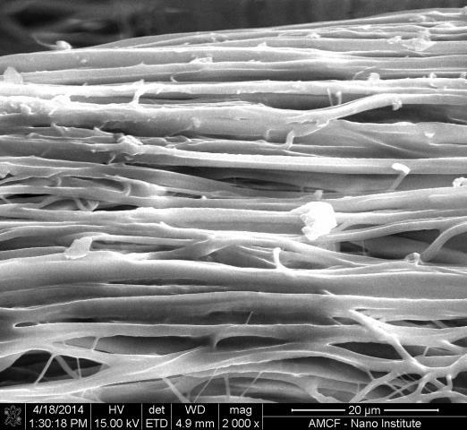
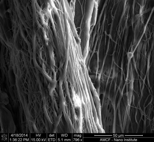
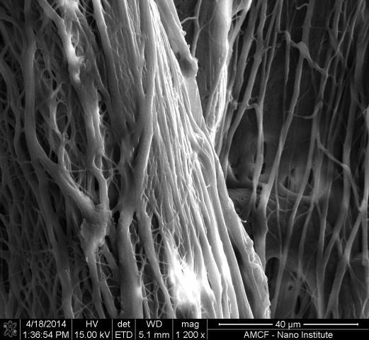
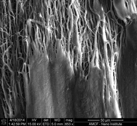
18 Cone 17 Centrifugal Jet Spinning Nanofiber scaffolds were fabricated by using a centrifugal jet spinner with polymeric solutions. PGS was combined with PCL and dissolved in HFIP. This solution was injected into a centrifugal jet spinner nozzle, and then run at rotational speeds of 20,000, 25,000, and 30,000 RPM. Highly aligned nanofibers would then but cut down the side with a razor blade, and were carefully removed in a single ribbon from the nozzle using tweezers. The ribbons were stored in petri dishes labeled with pertinent information such as date, chemical composition, and rotational speeds. Nanofiber scaffolds were then collected from the nozzle with tweezers. Through centrifugal jet spinning, super aligned scaffolds of polymer nanofibers were produced. Once spinning was complete, the ribbon was removed from the nozzle without disturbing the relative orientations of the fibers. This process can be seen in figure 7A and 7B below. Following the removal of the scaffold from the nozzle, the fibers were studied at different size scales. This can be seen in figure 8 in the next section. A B Figure 7. The process of fiber scaffold creation can be seen in (A) the design of the centrifugal jet spinner, and (B) the removal of a scaffold from a nozzle.
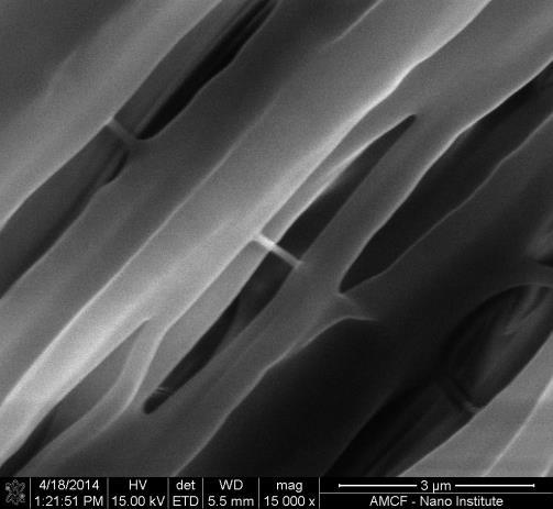
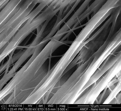
19 Cone 18 Spincoating In order to develop nanofilms from polymer solutions, 7% solutions of PCL in HFIP, PGS in chloroform, PGS in HFIP, and a 50/50 combination of PGS and PCL were deposited of glass coverslips. A spincoater was used with two different recipes which are described in Appendix A to create nanofilms of two thicknesses from each solution. The recipes used in the spincoater differed by their maximum rotational speeds, with recipe one reaching 4000 RPM and recipe 15 reaching 6000 RPM. Mechanical Testing Mechanical strain testing was performed on the nanofiber scaffolds in order to determine the Young s modulus of PGS/PCL nanofiber scaffold created at rotational speeds of 20,000, 25,000, and 30,000 RPM. For this test, ribbons of nanofiber scaffolds were mounted within one inch squares of paper using double sided tape. The mounts were placed in an Instron strain tester, and a strain test was performed. Data was recorded on the mechanical force and change in length throughout the test. Calculations were then performed to find the stress vs. strain plots of the nanofiber scaffolds, and then to find the Young s modulus for scaffolds created at each rotational speed. In order to complete these calculations, the dimensions of each specimen were input into a custom Matlab code. Scanning Electron Microscopy Scanning electron microscopy was used to study the features of individual PGS/PCL nanofibers, as well as the general alignment of the scaffolds. Pieces of nanofiber ribbon were mounted on studs and sputtercoated with gold in preparation for the imaging. Images were gathered from scaffolds created at 20,000, 25,000, and 30,000 RPM. Images were taken at three
20 Cone 19 different length scales in order to compare the alignment as well as the individual fiber properties. Water Contact Angle Testing Sessile drop water contact angle testing was performed on nanofilms containing all of the nanofilms created in the spincoating step. This test was conducted in order to study the surface properties of the polymer films. Three drops were tested on each nanofilms sample, resulting in 6 data points for every solution. Water contact angles were studied for both the nanofilms created with spincoater recipe 1 and 15, however, the films created with recipe 15 did not provide ample coverage of the coverslips, and the water contact angles were affected by the contact of water with the glass coverslips. Water contact angle testing was also performed on PGS/PCL nanofiber scaffolds for comparison of the polymer properties in nanofibers and nanofilms.
, or a compound of both.")

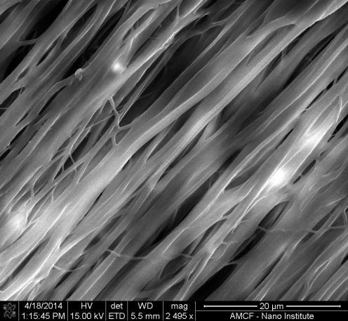
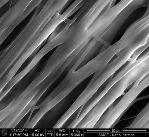
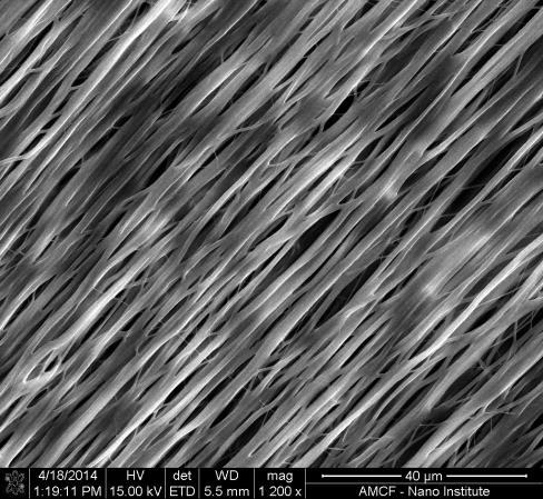
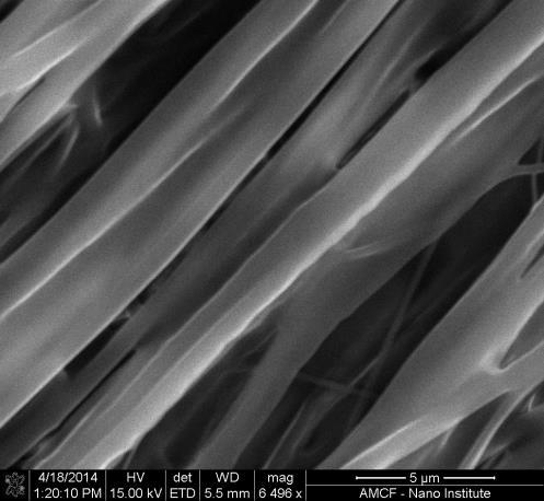
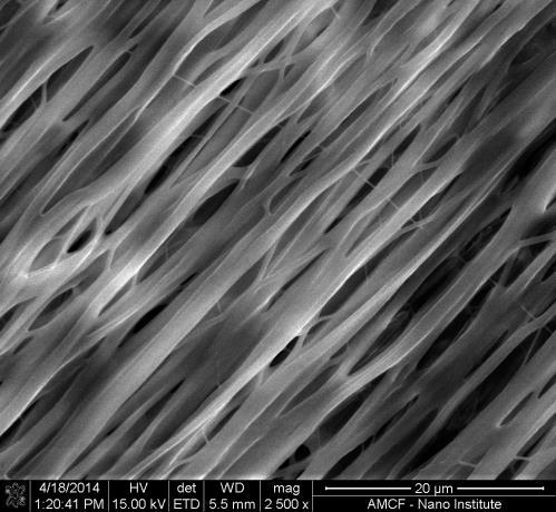
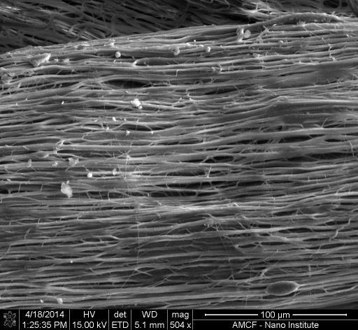
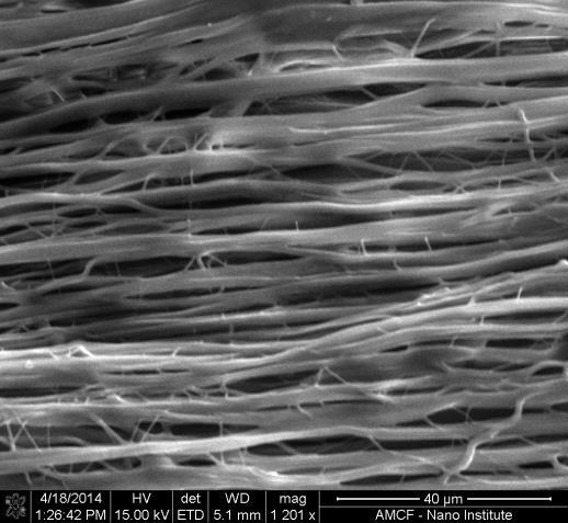
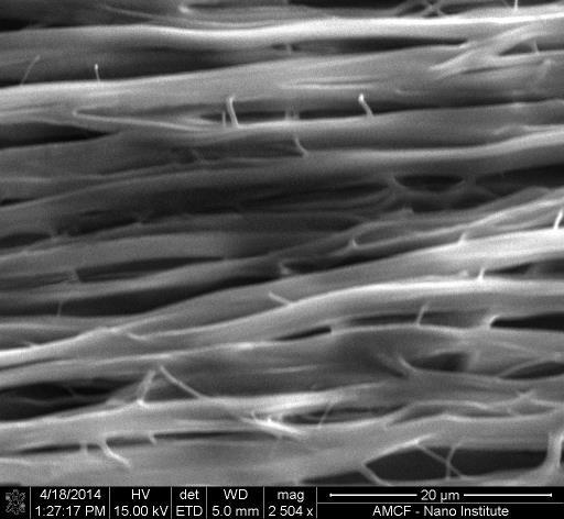
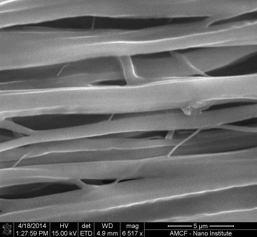
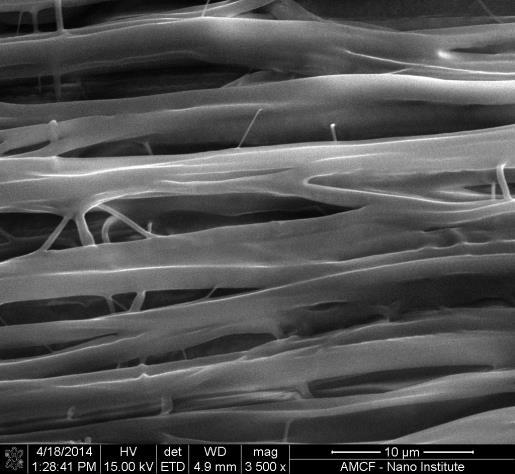
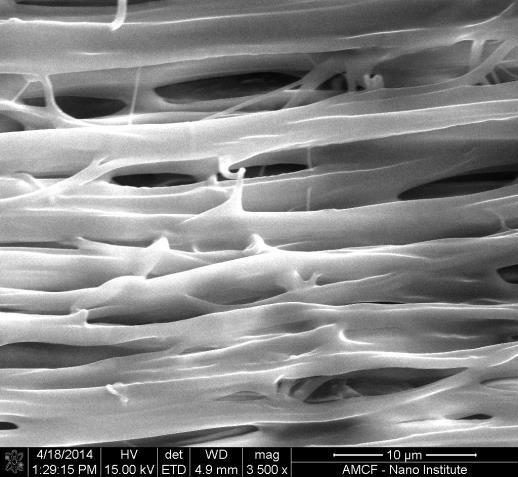
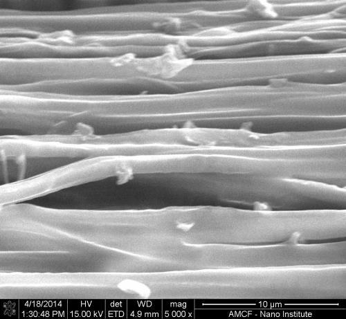
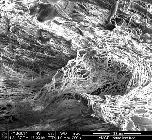
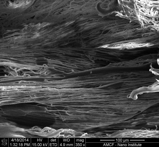
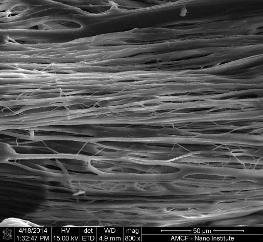
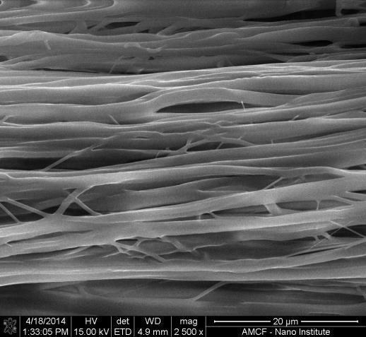
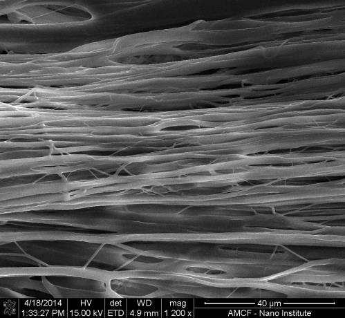
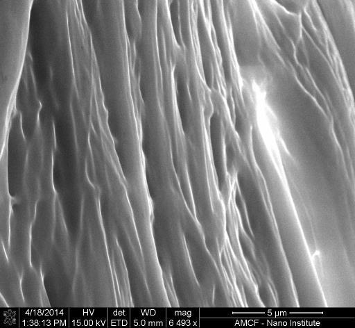
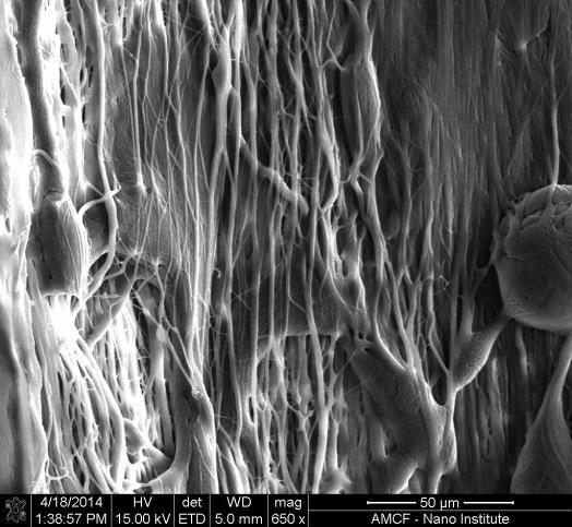
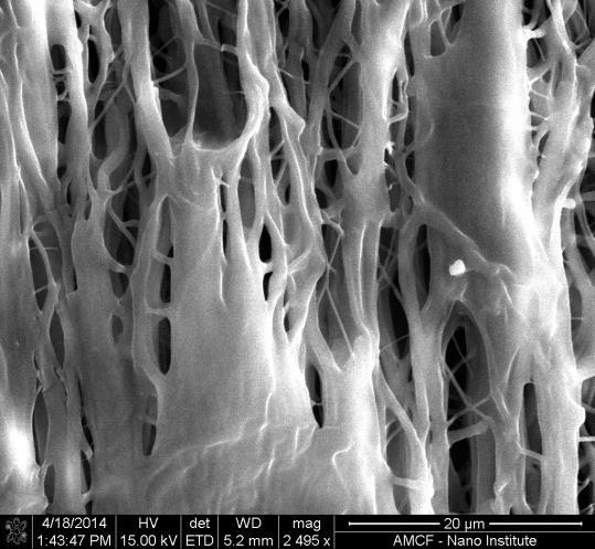
21 Cone 20 Section 3: Results The results of this project can be separated into the categories of nanofilms and nanofibers, both of which utilized materials consisting of poly (glycerol-sebacate) (PGS), polycaprolactone (PCL), or a compound of both. Nanofiber Results Nanofibers can be used as a scaffold for many tissue engineering applications as they exhibit many beneficial physical and mechanical properties. In order to characterize these properties, analytical tests were performed on PGS/PCL nanofiber ribbons which were made at a variety of rotational speeds. The fibers were tested for their water contact angle, Young s modulus, orientation order parameter, and average fiber diameter. Figure 8. PGS/PCL compound nanofibers created at 20,000 RPM seen at different length scales. Scale bars are 50, 10, and 5 microns, respectively. PGS/PCL nanofibers were created at 20,000 RPM, 25,000 RPM, and 30,000 RPM. The resulting ribbons were studied with scanning electron microscopy in order to analyze the effect of rotational speed on the nanofiber characteristics. Images of scaffolds created at these speeds can be seen in figure 9.


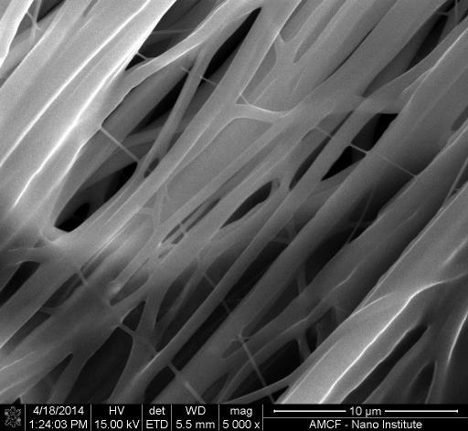
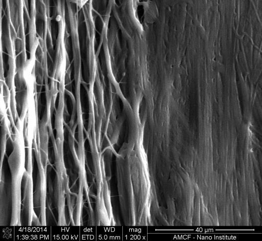
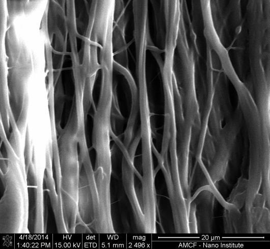
22 Cone 21 Figure 9. PGS/PCL compound nanofibers which were created by centrifugal jet spinning at speeds of (A) 20,000 RPM (B) 25,000 RPM and (C) 30,000 RPM. Scale bars are all 10 microns. Through analysis of the scanning electron microscopy images such as those in figure 9, an average fiber diameter was calculated for the scaffolds created at 20,000, 25,000, and 30,000 RPM. The average diameters can be seen below in figure 10. With the increasing rotational speeds, decreasing average fiber diameters were observed. When the average values were plotted and a line of best fit was applied, an R 2 value of was retuned. As seen in the caption in figure 10, a one way ANOVA test showed that the differences in fiber diameters were statistically significant between all of the rotational speeds, with p values less than 0.05 in each case. This correlation confirmed the anticipated pattern of greater speeds leading to lower average fiber diameters.
23 Average Fiber Diameters (microns) Cone Average Fiber Diameters * * * ,000 RPM 25,000 RPM 30,000 RPM Figure 10. Average fiber diameters measured for PGS/PCL compound nanofibers which were spun at three different rotational speeds. *=p value<0.05. In order to study the levels of alignment found within nanofiber scaffolds, the orientation order parameter (OOP) was calculated. In a perfectly aligned sample, the OOP would be 1, while in a sample with perfectly random alignment, the OOP would be 0. The anticipated correlation is that with greater rotational speeds, nanofibers should be better aligned, and as such will have higher OOP values. Following analysis of fibers created at three speeds, the OOP values were shown to increase with increasing speeds in PGS/PCL compound nanofibers. The values were plotted in figure 11 below, and when a line of best fit was applied to the graph, it returned an R 2 value of
24 Order Orientation Parameter Cone 23 Order Orientation Parameter Rotational Speed (RPM) Figure 11. Orientation order parameter (OOP) values for PGS/PCL compound nanofiber scaffolds created at various rotational speeds. The final testing performed on nanofiber scaffolds consisted of determining the Young s modulus of each ribbon. This was calculated by plotting the stress versus strain plots of each scaffold. The plots from each scaffold can be seen below in figure 12, while the values of the Young s modulus of each can be seen in table 1. The stress vs strain plots for the 20,000 and 30,000 RPM scaffolds both follow an expected form, with a toe region, linear region, and a plateau at the peak of stress. The linear regions of the stress versus strain plots can be seen below in figure 12. The Young s modulus values were found for these samples, and can be seen in table 1. The values decreased with the increasing speeds, which correlates with the decreasing fiber diameters. The smaller fibers from the 30,000 RPM tests were weaker than the other fibers, which is reflected by its lower Young s modulus.
25 Cone 24 A B
26 Cone 25 C Figure 12. Stress versus strain curves for nanofiber scaffolds created at (A) 20,000 RPM, (B) 25,000 RPM, and (C) 30,000 RPM. 20,000 RPM 25,000 RPM 30,000 RPM Young s Modulus Table 1. Young s modulus values for nanofiber scaffolds. Nanofilm Results In order to study the surface properties related to PGS nanofilms, solutions consisting of PGS and PCL were made using both chloroform and HFIP as solvents. Nanofilms were created on glass coverslips using a spincoater. The hydrophobicity aspects of the nanofilms were studied by taking the water contact angles with sessile drop goniometry. In order to understand the impact of combining PCL and PGS into a polymer compound, nanofilms of both pure substances and a 50:50 compound solution were characterized by their water contact angles. The water contact angle study also provided information on the effect of different solvents on the polymers. As seen in figure 13, the nanofilms made of the same solution
27 Water Contact Angle Cone 26 produced similar water contact angles to one another. Overall, the water contact angles of both the PCL and PGS films were lower than the angle reported for PDMS, meaning that both of the polymers were more hydrophilic than PDMS. The compound nanofilms produced water contact angles between those of pure PCL and pure PGS solutions, which suggest that they are exhibiting a combination of surface properties from their constituents Water Contact Angles of Nanofilms PCL PGS chloroform PGS HFIP PGS PCL PDMS Figure 13. Average water contact angles from nanofilms of various solutions.
28 Cone 27 Section 4: Discussion In this research, nanofibers and nanofilms were fabricated containing a compound of PGS and PCL. These structures were characterized through studying their surface properties, mechanical properties, and topographies. The nanostructures were studied in with specific consideration to their potential as tissue engineering scaffolds. Nanofibers were fabricated with a compound solution of PGS and PCL through a process of centrifugal jet spinning. These fibers followed many trends commonly found in centrifugal jet spun fibers, such as a decrease in average fiber diameter with increasing rotational speeds. The fiber scaffolds were imaged with scanning electron microscopy, and scaffolds produced at 20,000, 25,000, and 30,000 RPM were shown to contain a network of highly aligned polymeric fibers. As seen in the results section, as rotational speeds were increased in scaffold fabrication, the resulting nanofibers became smaller in diameter and more highly aligned. Both of these trends were anticipated, as they were also reported in previously published articles with other polymer solutions 2. Following mechanical testing of PGS/PCL nanofiber scaffolds, it seems that the Young s modulus decreased with increasing rotational speeds. This trend was expected, as higher speeds produced thinner nanofibers whose properties were not as elastic as the thinker fibers produced at lower speeds. Nanofilms consisting of pure PGS, pure PCL, and a combination of the two were tested for hydrophobicity with a sessile drop goniometer, and the compound films exhibited water contact angles that were between the angles found with PGS nanofilms and PCL nanofilms. The water contact angles for all of the nanofilms were relatively similar to one another, and there was
29 Cone 28 a difference seen in the PGS which had been dissolved in chloroform and the PGS in HFIP, so the data may not have been completely representative of the compound properties of PGS and PCL. All of the water contact angles measured were lower than the water contact angle of PDMS, and as such all of the nanofilms were more hydrophilic than PDMS. All of the nanofilms resulted in water contact angles less than 90 degrees, which means that all of the materials were hydrophilic. This is beneficial for tissue engineering constructs, as proteins are more likely to bind to a hydrophilic material than a hydrophobic one. The water contact angles were reported from a spincoating method which only reached a maximum speed of 4,000 RPM, as the method which reached 6,000 RPM resulted in streaky films that may not have provided accurate water contact angle values. Overall, the fabrication of centrifugal jet spun PGS nanofibers resulted in many of the desired properties for tissue engineering scaffolds. The scaffolds consisted of a network of aligned fibers, with diameters that could be controlled by the rotational speeds used in the experimental protocol. The use of PGS in nanofiber scaffolds resulted in a scaffold with surface properties which mimic many of the natural properties of collagen and elastin. These features suggest that PGS nanofibers may be a useful base material for many tissue engineering applications.
30 Cone 29 Section 5: Future Work The development of PGS nanofilms and nanofibers provides the opportunity for many tissue engineering innovations. In the future, further experimentation on the viability of these films and fibers may be done through cell cultures, and eventually mouse models. In the future, test for in vitro cell attachment with PGS nanofiber scaffolds may be completed in order to study the viability of the nanofibers as a tissue engineering scaffold. Comparisons of the growth rates of cells on PGS nanofibers compared to PCL nanofibers will provide information on the biocompatibility of PGS. A study of cells grown on PGS/PCL nanofilms compared to nanofibers will provide information on the influence of a nanofiber scaffold in cell alignment properties. Following the confirmation of PGS nanofibers and nanofilms as tissue engineering scaffolds, a project may be developed to study the ability to use these scaffolds as drug delivery systems. This could be accomplished either through the synthesis of a compound solution of PGS and various drugs, or through creating multi-layered nanostructures.
31 Cone 30 Appendix A: Experimental Protocol Spincoat Recipe 1 Protocol The following steps were completed by a spincoater for nanofilms produced by recipe 1. Step 1: Ramp 5, RPM 500, Dwell 5 Step 2: Ramp 5, RPM 1000, Dwell 5 Step 3: Ramp 10, RPM 1000, Dwell 10 Step 4: Ramp 10, RPM 4000, Dwell 60 Step 5: Ramp 10, RPM 2000, Dwell 15 Step 6: Ramp 10, RPM 1000, Dwell 10 Step 7: Ramp 5, RPM 500, Dwell 5 Spincoat Recipe 15 Protocol The following steps were completed by a spincoater for nanofilms produced by recipe 15. Step 1: Ramp 10, RPM 3000, Dwell 5 Step 2: Ramp 10, RPM 6000, Dwell 60 Step 3: Ramp 10, PRM 3000, Dwell 5
32 Cone 31 Centrifugal Jet Spinning Protocol 1. Attach nozzle to motor base using set screws 2. Set desired rotational speed through computer interface 3. Supply power to motor to initiate nozzle rotations 4. Fill pipet with 5 ml of polymer solution 5. Inject solution into nozzle while it is spinning 6. Wait for polymer to stop spraying out of nozzle 7. Disconnect power from motor 8. Remove nozzle from motor base 9. Slice nanofiber scaffold down the side of the nozzle 10. Peel scaffold from the nozzle using tweezers


33 Cone 32 Scanning Electron Microscopy PGS/PCL nanofibers spun at 20,000 RPM Appendix B: Analysis Protocol


34 Cone 33


35 Cone 34

36 PGS/PCL nanofibers spun at 25,000 RPM Cone 35
37 Cone 36

38 Cone 37

39 PGS/PCL nanofibers spun at 30,000 RPM Cone 38

40 Cone 39

41 Cone 40 Nanofilm Water Contact Angle Test Images PCL in HFIP Spincoat Recipe 1 PCL in HFIP Spincoat Recipe 15 PGS in chloroform Spincoat Recipe 1 PGS in chloroform Spincoat Recipe 15
42 Cone 41 PGS in HFIP Spincoat Recipe 1 PGS in HFIP Spincoat Recipe 15 PGS/PCL in HFIP Spincoat Recipe 1
43 Cone 42 Nanofilm Water Contact Angle Values Sample Left Angle Right Angle Drop Average Drop Standard Deviation PCL R PCL R PCL R PCL R PCL R PCL R PGS chloroform R PGS chloroform R PGS chloroform R PGS chloroform R PGS chloroform R PGS chloroform R PGS HFIP R PGS HFIP R PGS HFIP R PGS HFIP R PGS HFIP R PGS HFIP R PGS PCL R PGS PCL R PGS PCL R PGS PCL R PGS PCL R PGS PCL R
44 Cone 43 ANOVA Analysis of Average Fiber Diameters One Way Analysis of Variance Monday, April 21, 2014, 11:45:54 AM Data source: Data 1 in Notebook1 Normality Test (Shapiro-Wilk) Passed (P = 0.813) Equal Variance Test: Passed (P = 0.097) Group Name N Missing Mean Std Dev SEM 20k k k Source of Variation DF SS MS F P Between Groups <0.001 Residual Total The differences in the mean values among the treatment groups are greater than would be expected by chance; there is a statistically significant difference (P = <0.001). Power of performed test with alpha = 0.050: All Pairwise Multiple Comparison Procedures (Holm-Sidak method): Overall significance level = 0.05 Comparisons for factor: Comparison Diff of Means t P P< k vs. 30k <0.001 Yes 20k vs. 25k <0.001 Yes 25k vs. 30k Yes
45 Cone 44 Matlab Code for Stress Strain Plots % Balalab 4/23/2014 % This code is applied to plot the linear region of tensile stress curves % Make sure you delete all the headers in the *.CSV file % Rename the file so that there are no spaces or dots % The file should only start with the data % calculate stress in column 5 % calculate strain in column 4 % limit spreadsheet to data in the linear region close all %import data from spreadsheet [rawdata,pathname]=uigetfile('.csv','select the data file?') raw_data=open(rawdata); %truncate.csv extension stringsize=size(rawdata); filename=rawdata(1:stringsize(2)-4); % change this file name to match the data set data=raw_data.specimen_rawdata_125r1s; %defines data locations in the spreadsheet stress=(data(:,5)); strain=(data(:,4)); %plots linear region of stress/strain curve plot(stress,strain); xlabel('strain') ylabel('stress [MPa]') %change name for the sample title title('25k Stress vs Strain')
46 Cone 45 References 1 Amalorpava M L, Senthilram T, et al. Centrifugal spun ultrafine fibrous web as a potential drug delivery vehicle. Polymer Letters (2013) 7:3, Badrossamay M, Balachandran K, et al. Engineering hybrid polymer-protein super-aligned nanofibers via rotary jet spinning. Biomaterials (2014) 35, Badrossamay M, McIlwee H, Goss J, Parker K. Nanofiber assembly by rotary jet-spinning. Nano Lett (2010) 10, Engel E, Michiardi A, et al. Nanotechnology in regenerative medicine: the materials side. Trends in Biotechnology (2007) 26:1, Gandaglia A, Bagno A, et al. Cells, scaffolds and bioreactors for tissue-engineered heart valves: a journey from basic concepts to contemporary developmental innovations. Eur J Cardio-Thorac (2011) 39, Gao J, Capro P, and Wang Y. Macroporous elastomeric scaffolds with extensive micropores for soft tissue engineering. Tissue Engineering (2006) 12:4, Goldberg M, Langer R, and Jia X. Nanostructured materials for applications in drug deliver and tissue engineering. J Biomater Sci Polym Ed (2007) 18: Liu H, Webster T. Nanomedicine for implants: a review of studies and necessary experimental tools. Biomaterials (2007) 28, Marsano A, et al. Scaffold stiffness affects the contractile function of three -dimensional engineered cardiac constructs. Biotechnol Prog (2010) 26:5,
47 Cone Pomerantseva I, et al. Degredation behavior of poly(glycerol sebacate). J Biomed Mater Res A (2009) 91, Ramakrishna S, et al. Electrospun nanofibers: solving global issues. Materials Today (2006) 9:3, Wang Y, et al. A tough biodegradable elastomer. Nat Biotechnol (2002) 20, Wu W, Allen R, and Wang Y. Fast-degrading elastomer enables rapid remodeling of a cell-free synthetic graft into a neoartery. Nat Med, advance online publication. 14 Zhang L, Webster T. Nanotechnology and nanomaterials: promises for improved tissue regeneration. Nano Today (2009) 4,
Artificial blood vessels
 Artificial blood vessels S. Swaminathan Director Centre for Nanotechnology & Advanced Biomaterials School of Chemical & Biotechnology SASTRA University Thanjavur 613 401 Tamil Nadu Joint Initiative of
Regenerez : A Next-Generation Material for Functional Bioresorbable Coatings. by Carissa Smoot and Jeremy Harris, Ph.D, Secant Medical, Inc.
 Regenerez : A Next-Generation Material for Functional Bioresorbable Coatings by Carissa Smoot and Jeremy Harris, Ph.D, Secant Medical, Inc. Coatings consisting of natural and synthetic origin play an important
Prof. Steven S. Saliterman. Department of Biomedical Engineering, University of Minnesota
 Department of Biomedical Engineering, University of Minnesota http://saliterman.umn.edu/ Mimicking the fibrillar structure of the extracellular matrix is important for scaffolds. Clinical trails to date
Regenerez : A Next-Generation Material for Bioresorbable Coatings
 R Regenerez : A Next-Generation Material for Bioresorbable Coatings A White Paper by Carissa Smoot, Scientist, Secant Group and Jeremy Harris, Ph.D, Secant Group Coatings consisting of natural and synthetic
Regenerez Degradation and Release Kinetics White Paper
 R Regenerez Degradation and Release Kinetics White Paper A White Paper by Carissa Smoot, Scientist, Secant Group, Jeremy Harris, Ph.D, Secant Group and Stephanie Reed, Ph.D, Secant Group Introduction Implantable
Pittsburgh Tissue Engineering Initiative Annual Progress Report: 2011 Formula Grant
 Pittsburgh Tissue Engineering Initiative Annual Progress Report: 2011 Formula Grant Reporting Period July 1, 2012 December 31, 2012 Formula Grant Overview The Pittsburgh Tissue Engineering Initiative received
Investigating the Mechanical Behaviors of Organic/Inorganic Composite Bone Scaffolds
 University of Arkansas, Fayetteville ScholarWorks@UARK Biomedical Engineering Undergraduate Honors Theses Biomedical Engineering 5-2016 Investigating the Mechanical Behaviors of Organic/Inorganic Composite
Supporting Information
 Supporting Information Novel Interwoven Polymer Composites via Dual- Electrospinning with Shape Memory/Self-healing Properties Jaimee M. Robertson, Hossein Birjandi Nejad, Patrick T. Mather* Syracuse Biomaterials
Articular Cartilage Engineering Using Human Mesenchymal Stem Cells and Nanostructured Biomaterials
 Articular Cartilage Engineering Using Human Mesenchymal Stem Cells and Nanostructured Biomaterials REU Participant: Nicole Green 1 Advisors: Joel Wise 2, Dr. Michael Cho 2, Dr. Constantine Megaridis 3
Pittsburgh Tissue Engineering Initiative Annual Progress Report: 2010 Formula Grant
 Pittsburgh Tissue Engineering Initiative Annual Progress Report: 2010 Formula Grant Reporting Period July 1, 2011 December 31, 2011 Formula Grant Overview The Pittsburgh Tissue Engineering Initiative,
Mechano-dependent biosynthetic response of microintegrated cells in elastomeric scaffolds.
 Mechano-dependent biosynthetic response of microintegrated cells in elastomeric scaffolds. Lauren Anderson, Department of Bioengineering, The Pennsylvania State University Dr. Michael Sacks, Mentor, Department
2. Precision Extrusion Deposition Previous research has focused on Fuse Deposition Modeling (FDM) for the fabrication of
 for the fabrication of") PRECISION EXTRUSION DEPOSITION OF POLYCAPROLACTONE/ HYDROXYAPATITE TISSUE SCAFFOLDS L. Shor, S. Güçeri, W. Sun Laboratory for Computer-Aided Tissue Engineering Department of Mechanical Engineering and
PRECISION EXTRUSION DEPOSITION OF POLYCAPROLACTONE/ HYDROXYAPATITE TISSUE SCAFFOLDS L. Shor, S. Güçeri, W. Sun Laboratory for Computer-Aided Tissue Engineering Department of Mechanical Engineering and
Bioreactors in tissue engineering
 Bioreactors in tissue engineering S. Swaminathan Director Centre for Nanotechnology & Advanced Biomaterials School of Chemical & Biotechnology SASTRA University Thanjavur 613 401 Tamil Nadu Joint Initiative
Supporting Information
 Copyright WILEY-VCH Verlag GmbH & Co. KGaA, 69469 Weinheim, Germany, 2013. Supporting Information for Adv. Mater., DOI: 10.1002/adma.201300794 Highly Stretchable Patterned Gold Electrodes Made of Au Nanosheets
Heart-on-Chip Technologies for in vitro Cardiomyocyte Assessment Studies
 Heart-on-Chip Technologies for in vitro Cardiomyocyte Assessment Studies May 15 th, 2018 Herdeline Ann M. Ardoña, Ph.D. Disease Biophysics Group, Wyss Institute for Biologically-Inspired Engineering, John
Nanospherical Ceramics: Resisting Bacteria Infection. While increasing bone cell functions, nanomaterials reduce bacteria functions. S.
 Nanospherical Ceramics: Resisting Bacteria Infection While increasing bone cell functions, nanomaterials reduce bacteria functions. S. Epidermis Conventional ZnO Positive Control (Wrought Ti) Nanophase
FLUIDNATEK CUTTING EDGE ELECTROSPINNING & ELECTROSPRAYING DEVICES
 FLUIDNATEK CUTTING EDGE ELECTROSPINNING & ELECTROSPRAYING DEVICES by THE FLUIDNATEK TOOLS we create the future USE CASES FLUIDNATEK Lab Tools are research instruments designed for the fabrication of small
Versatile Core-Sheath Biofibers using Coaxial Electrospinning
 Mater. Res. Soc. Symp. Proc. Vol. 1094 2008 Materials Research Society 1094-DD06-02 Versatile Core-Sheath Biofibers using Coaxial Electrospinning Daewoo Han 1, Steven T. Boyce 2, and Andrew J. Steckl 1
Thermoresponsive Membranes from Electrospun. Mats with Switchable Wettability for Efficient
 Thermoresponsive Membranes from Electrospun Mats with Switchable Wettability for Efficient Oil/Water Separations Yan Liu, a,b Sinem Tas, b Kaihuan Zhang, b, Wiebe M. de Vos, c Jinghong Ma, a,* and G. Julius
Mechanical Properties and Solubility Characteristics of. PES-PCL Core-Shell Fibers Used as Nanofiber Substrates. in Cell Migration Studies
 Mechanical Properties and Solubility Characteristics of PES-PCL Core-Shell Fibers Used as Nanofiber Substrates in Cell Migration Studies Undergraduate Honors Research Thesis Presented in Partial Fulfillment
Biomaterials in Medical device design
 Biomaterials in Medical device design 1 Dr Joseph Buhagiar B. E n g ( H o n s. ) P h D ( B h a m ) D e p a r t m e n t o f M e t a l l u r g y a n d M a t e r i a l s E n g i n e e r i n g U n i v e r
Active biomaterials/scaffolds for stem cell-based soft tissue engineering (in a nutshell )
") Active biomaterials/scaffolds for stem cell-based soft tissue engineering (in a nutshell ) Emanuele Giordano Responsabile Lab ICM BioEngLab DEI CIRI SdV-TS Università di Bologna emanuele.giordano@unibo.it
Active biomaterials/scaffolds for stem cell-based soft tissue engineering (in a nutshell ) Emanuele Giordano Responsabile Lab ICM BioEngLab DEI CIRI SdV-TS Università di Bologna emanuele.giordano@unibo.it
High-Throughput Method for Microfluidic Placement of Cells in Micropatterned Tissues
 High-Throughput Method for Microfluidic Placement of Cells in Micropatterned Tissues Emily N. Sevcik Faculty Mentor: Patrick W. Alford Undergraduate Research Opportunities Program Project Final Report
Regenerative and Immune Engineering Focus Area Upper-Level Engineering Courses updated October, 2018 For BME Class of 2021 and beyond
 Regenerative and Immune Engineering Focus Area Upper-Level Engineering Courses updated October, 2018 For BME Class of 2021 and beyond EN.510.311 Structure of Materials 3 EN.510.312 Thermodynamics/Materials
Regenerative and Immune Engineering Focus Area - Upper-Level Engineering Courses updated June, 2018 For BME Class of 2021 and beyond
 Regenerative and Immune Engineering Focus Area - Upper-Level Engineering Courses updated June, 2018 For BME Class of 2021 and beyond EN.510.311 Structure of Materials 3 EN.510.312 Thermodynamics/Materials
Quantitative analysis of human mesenchymal stem cell alignment by electrospun polymer nanofibrous scaffolds
 Quantitative analysis of human mesenchymal stem cell alignment by electrospun polymer nanofibrous scaffolds Nicole Green 1, Joel Wise 2, Dr. Michael Cho 2, Dr. Constantine Megaridis 3 1 Department of Chemical
Comparative Fatigue Analysis of Metals and Polymers for Engineering Applications
 University of Arkansas, Fayetteville ScholarWorks@UARK Mechanical Engineering Undergraduate Honors Theses Mechanical Engineering 5-2012 Comparative Fatigue Analysis of Metals and Polymers for Engineering
Affinity. A Paradigm Shift in Skeletal Reconstruction
 Affinity A Paradigm Shift in Skeletal Reconstruction INTRODUCING TRS AFFINITY SKELETAL RECONSTRUCTION BREAKTHROUGH Tissue Regeneration Systems (TRS ) is a start-up medical device company commercializing
Characterizing Interfacial Bonds in Hybrid Metal AM Structures John Linn, Jason M. Weaver, Michael P. Miles, Yuri Hovanski Brigham Young University
 Solid Freeform Fabrication 218: Proceedings of the 29th Annual International Solid Freeform Fabrication Symposium An Additive Manufacturing Conference Reviewed Paper Characterizing Interfacial Bonds in
Applications in Cardiology Hollow Fiber Membranes and Applications
 Applications in Cardiology Want is meant by the term silicones?; Describe in general terms a typical synthetic scheme for a silicone consisting of half PDMS and half polysiloxane; Describe three cross-linking
Design, Fabrication, and Testing of an Electrospinning Apparatus for the Deposition of PMMA Polymer for Biomedical Applications
 Inquiry: The University of Arkansas Undergraduate Research Journal Volume 12 Article 11 Fall 2011 Design, Fabrication, and Testing of an Electrospinning Apparatus for the Deposition of PMMA Polymer for
PRINCIPLES AND PRACTICE OF TISSUE ENGNEERING:
 Harvard-MIT Division of Health Sciences and Technology HST.535: Principles and Practice of Tissue Engineering Instructor: Myron Spector Massachusetts Institute of Technology Harvard Medical School Brigham
ELECTROSPUN NANOFIBER PROCESS CONTROL
 CELLULOSE CHEMISTRY AND TECHNOLOGY Received April 26, 2010 ELECTROSPUN NANOFIBER PROCESS CONTROL University of Guilan, P.O. Box 3756, Rasht, Iran Fiber diameter is an important structural characteristic
Polymer Nanocomposites for Medical Applications
 Department of Bioengineering Liu Research Group www. Liugroup.org Polymer Nanocomposites for Medical Applications Huinan Liu, Ph.D. Department of Bioengineering Interdisciplinary Materials Science and
Nanodiamond-Polymer Composite Fibers and Coatings
 Nanodiamond-Polymer Composite Fibers and Coatings Yury Gogotsi et al. A.J. Drexel Nanotechnology Institute and Department of Materials Science and Engineering Drexel University, Philadelphia, Pennsylvania
Development of Class A Surface Polyurethane LFI Composites by Usama Younes, Bayer Material Science
 American Composites Manufacturers Association January 15-17, 2009 Tampa, FL USA Development of Class A Surface Polyurethane LFI Composites by Usama Younes, Bayer Material Science Abstract Recent advancements
Directed Osteoblast Adhesion at Metal Particle Boundaries
 Directed Osteoblast Adhesion at Metal Particle Boundaries Ti6Al4V (nanophase) Bar = 1 m. Ti6Al4V (conventional) Ti6Al4V (nanophase) Bar = 1 m. Ti6Al4V (conventional) Directed Osteoblast Adhesion at Metal
A Belt-like superfine film fabricated by bubble-electrospinning Hao Dou 1,a, Bao-qi Zuo 1
 Advanced Materials Research Online: 2013-11-21 ISSN: 1662-8985, Vol. 843, pp 82-85 doi:10.4028/www.scientific.net/amr.843.82 2014 Trans Tech Publications, Switzerland A Belt-like superfine film fabricated
The Effect of Hydrophobic Patterning on Micromolding of Aqueous-Derived Silk Structures
 The Effect of Hydrophobic Patterning on Micromolding of Aqueous-Derived Silk Structures Konstantinos Tsioris 1, Robert D White 1, David L Kaplan 2, and Peter Y Wong 1 1 Mechanical Engineering, Tufts University,
Microelectromechanical Drug Delivery Systems. Sarah Smith & Jurek Smolen
 Microelectromechanical Drug Delivery Systems Sarah Smith & Jurek Smolen Current Drug Delivery Systems Common Administration Methods Problems Oral Intravenous Intramuscular Transdermal Difficult to control
White Paper: Textile Engineered Tissue Scaffolds Offer Advances in Hollow Organ Regenerations. Peter D. Gabriele Director, Emerging Technology
 White Paper: Textile Engineered Tissue Scaffolds Offer Advances in Hollow Organ Regenerations Peter D. Gabriele Director, Emerging Technology Regenerative medicine (RM) holds the potential to address some
Centrifugal spinning of nanofiber webs - A parameter study of a novel spinning process
 Centrifugal spinning of nanofiber webs - A parameter study of a novel spinning process Jonas Engström Senior scientist at Swerea IVF. Finished his PhD in 2006 with a thesis titled Functional compolymers
Department of Polymer and Fiber Engineering
 Department of Polymer and Fiber Engineering Educational programs 70 undergraduate students 25 graduate students Several post-docs 8 faculty members B.Sc., M.Sc., M.E. and Ph.D. Strong foreign exchange
Alternative MicroFabrication and Applications in Medicine and Biology
 Alternative MicroFabrication and Applications in Medicine and Biology Massachusetts Institute of Technology 6.152 - Lecture 15 Fall 2003 These slides prepared by Dr. Hang Lu Outline of Today s Materials
Prof. Oded Shoseyov The Hebrew University of Jerusalem NANO BIO MIMETICS: MATERIALS FOR THE FUTURE.
 Prof. Oded Shoseyov The Hebrew University of Jerusalem Shoseyov@agri.huji.ac.il NANO BIO MIMETICS: MATERIALS FOR THE FUTURE Oded Shoseyov Our current materials, structures and machines Our current materials
Effect of CNTs on Shape memory properties of PLLA/PCL blends
 Effect of CNTs on Shape memory properties of PLLA/PCL blends Maryam Amirian 1, Ali Nabipour Chakoli 2, Hossein Afarideh 3, t=0s t=2s t=5s t=10s t=15s t=20s 1 Dep. of Physics, Teachers Uni., Tehran, Iran,
The formulation currently used for this kind of dissolvable strips can be seen below:
 Hydrophilic and Hydrophobic Properties of Dissolvable Thin Films Developed by: Mike Evangelista, Nathan Haden, Alex Jannini, Rowan University, Department of Chemical Engineering Edited by: C. Stewart Slater
3D MICRO-NANO FIBROUS SCAFFOLD PREPARED BY MELTBLOWN IN COMBINATION WITH ELECTROSPINNING FOR THE BONE TISSUE ENGENEERING
 3D MICRO-NANO FIBROUS SCAFFOLD PREPARED BY MELTBLOWN IN COMBINATION WITH ELECTROSPINNING FOR THE BONE TISSUE ENGENEERING Jakub ERBEN a, Kateřina PILAŘOVÁ a, Filip SANETRNÍK a, Jiří CHVOJKA a, Věra JENČOVÁ
Electret Polyvinylidene Fluoride Nanofibers Hybridized by Polytetrafluoroethylene Nanoparticles for High-Efficiency Air Filtration
 Supporting Information Electret Polyvinylidene Fluoride Nanofibers Hybridized by Polytetrafluoroethylene Nanoparticles for High-Efficiency Air Filtration Shan Wang,,, Xinglei Zhao,,, Xia Yin,*,, Jianyong
Comparison between Electrospun and Bubbfil-spun Polyether Sulfone Fibers
 ISSN 1517-7076 artigo 11564 pp.363-369, 2014 Comparison between Electrospun and Bubbfil-spun Polyether Sulfone Fibers Ya Li 1,2, Rou-xi Chen 1,2, Fu-Juan Liu 1,2 1 National Engineering Laboratory for Modern
The Effects of Aggregate Size on Shear Dynamic Modulus from Torsion Bar
 University of Arkansas, Fayetteville ScholarWorks@UARK Civil Engineering Undergraduate Honors Theses Civil Engineering 5-217 The Effects of Aggregate Size on Shear Dynamic Modulus from Torsion Bar Farida
Cell and Tissue Engineering, Nanotechnology
 Cell and Tissue Engineering, Nanotechnology Cells Tissue Engineering Scaffolds BIOE 506 Muqeem Qayyum Biorectors Signals Why? In-vitro In-vivo Because we can no longer to view a cell as self contained
Biomedical Engineering 3D BIOPRINTING OF SILK FOR CELLULAR APPLICATIONS. Martina Ravizza
 Biomedical Engineering 3D BIOPRINTING OF SILK FOR CELLULAR APPLICATIONS Martina Ravizza Supervisor: Prof. Ferdinando Auricchio Correlator: Dott. Michele Conti Sericina Fibroina Academic year 2014/2015
Surface Characterization of Biomaterials
 Surface Characterization of Biomaterials Biomaterials are determined The desire for well-being and long life expectancy goes hand in-hand with the use of non-viable materials to conserve health. 2000 years
3D In Vitro Living Systems for Biological Application
 3D In Vitro Living Systems for Biological Application Hossein Hosseinkhani Graduate Institute of Biomedical Engineering, National Taiwan University of Science and Technology (TAIWAN TECH), Taipei, Taiwan
Potential Graft Materials for ACL Reconstruction Surgery
 Washington University in St. Louis Washington University Open Scholarship Mechanical Engineering and Materials Science Independent Study Mechanical Engineering & Materials Science 12-23-2017 Potential
Ultrasensitive and Highly Stable Resistive Pressure Sensors with. Biomaterial-Incorporated Interfacial Layers for Wearable
 Supporting Information Ultrasensitive and Highly Stable Resistive Pressure Sensors with Biomaterial-Incorporated Interfacial Layers for Wearable Health-Monitoring and Human-Machine Interfaces Hochan Chang,,
W BIOENGINEERING. Nanomaterials Applications in Biology and Medicine. Topics:
 Nanomaterials Applications in Biology and Medicine Obejctive: Develop a firm understanding of the fundamental materials science and engineering principles underlying synthetic/engineered materials used
Lecture Outline. History. Purpose? Func:on of Bioscaffolds. Extracellular Matrix (ECM) 12/08/15
 12/08/15") Associate Professor Rod Dilley Dr Rob Marano Ear Sciences Centre School of Surgery Harry Perkins Research Building 4 th Floor Lecture Outline History Purpose Functions Properties Approaches to bioscaffold
Associate Professor Rod Dilley Dr Rob Marano Ear Sciences Centre School of Surgery Harry Perkins Research Building 4 th Floor Lecture Outline History Purpose Functions Properties Approaches to bioscaffold
UConn BMES Meeting #2 (9/28) T R A C K P R E S E N T A T I O N S
 T R A C K P R E S E N T A T I O N S") UConn BMES Meeting #2 (9/28) T R A C K P R E S E N T A T I O N S Concept of Biomedical Engineering Biomedical Engineering is an interdisciplinary application of engineering principals The major is an application
UConn BMES Meeting #2 (9/28) T R A C K P R E S E N T A T I O N S Concept of Biomedical Engineering Biomedical Engineering is an interdisciplinary application of engineering principals The major is an application
STRENGTH OF METAL TO POLYMER ADHESIVE BONDED AND RIVETED JOINTS
 The 3rd International Conference on Computational Mechanics and Virtual Engineering COMEC 2009 29 30 OCTOBER 2009, Brasov, Romania STRENGTH OF METAL TO POLYMER ADHESIVE BONDED AND RIVETED JOINTS T. Sandu
Cell-Environment Interactions. Chieh-Chun Chen
 Cell-Environment Interactions Chieh-Chun Chen Part 1: Soft Lithography in Biology and Biochemistry Chieh-Chun Chen Outlines Introduction Key features of soft lithography Applications In microscopic biochemical
Synthesis and Characterization of Biodegradable Hemostat Gelatin Sponge for Surgery Application
 R Iranian Journal of Pharmaceutical Sciences Summer 2008: 4(3): 193-200 www.ijps.ir Original Article Synthesis and Characterization of Biodegradable Hemostat Gelatin Sponge for Surgery Application Rana
Biocompatibility and War and Peace
 Professor David Williams, D.Sc.,F.R.Eng., Professor and Director of International Affairs, Wake Forest Institute of Regenerative Medicine, North Carolina, USA Editor-in-Chief, Biomaterials President-elect,
Supporting information for: Microfluidic single cell mrna isolation and analysis
 Supporting information for: Microfluidic single cell mrna isolation and analysis Joshua S. Marcus 1,2, W. French Anderson 2,3 & Stephen R. Quake 2,4,5 1 Biochemistry and Molecular Biophysics, 2 Applied
UNIT CELL PROCESSES UNDERLYING TISSUE ENGINEERING AND REGENERATIVE MEDICINE
 Massachusetts Institute of Technology Harvard Medical School Brigham and Women s Hospital VA Boston Healthcare System 2.79J/3.96J/20.441/HST522J UNIT CELL PROCESSES UNDERLYING TISSUE ENGINEERING AND REGENERATIVE
CHALLENGES OF 3D BIOPRINTING IN CLINICAL PRACTICE
 CENTRE DE THÉRAPIE TISSULAIRE & CELLULAIRE CHALLENGES OF 3D BIOPRINTING IN CLINICAL PRACTICE Pr. D. Dufrane MD, PhD 3D-BIOPRINTING: MYTH OR REALITY? 2 REGENERATIVE MEDICINE FOR ORGAN AND TISSUE A LARGE
STUDIES ON DRY-JET-WET SPINNING OF POLY(LACTIC ACID) FILAMENT AND ITS KNITTING. Doctor of Philosophy
 FILAMENT AND ITS KNITTING. Doctor of Philosophy") STUDIES ON DRY-JET-WET SPINNING OF POLY(LACTIC ACID) FILAMENT AND ITS KNITTING By NILESH S. REVAGADE Department of Textile Technology Submitted In fulfillment of the requirements of the degree of Doctor
STUDIES ON DRY-JET-WET SPINNING OF POLY(LACTIC ACID) FILAMENT AND ITS KNITTING By NILESH S. REVAGADE Department of Textile Technology Submitted In fulfillment of the requirements of the degree of Doctor
Fig. 1 A Tensile Test. Fig. 2 Data from Test CellScale Biomaterials Testing I
 Many biological materials from skin to ligaments to blood vessel walls carry tensile loads. To function properly, these natural structures and their engineered replacements must have suitable tensile strength
3D Cell Culture Product Intro. Bio-Byblos Biomedical
 3D Cell Culture Product Intro Bio-Byblos Biomedical Rundown Product Intro Cellusponge Series Go Matrix Applications Upcoming Products Degradable series Cellusponge CB series Cell Alignment plate Vivoalign
The Electrospinning Company
 The Electrospinning Company September 2015 Contact: info@electrospinning.co.uk +44 1235 567276 UK SME Design, develop and manufacture advanced biomaterial scaffolds Sales of product and service R&D collaborations
terials with at least one dimension in the nanoscale)
") CONTRIBUTED ORIGINAL ARTICLE INTEGRATING nanotechnology into MEDICINE: Past, PRESENT, AND FUTURE Thomas J. Webster, Northeastern University, Boston An acute shortage of organs is one of the most urgent
CONTRIBUTED ORIGINAL ARTICLE INTEGRATING nanotechnology into MEDICINE: Past, PRESENT, AND FUTURE Thomas J. Webster, Northeastern University, Boston An acute shortage of organs is one of the most urgent
Overview. Research area Most recent projects highlights. Dr. Elaheh Ghassemieh Mechanical Engineering Department University of Sheffield
 Overview Research area Most recent projects highlights Dr. Elaheh Ghassemieh Mechanical Engineering Department University of Sheffield Research areas Nonwovens Novel materials and their processes Bio-composites
Surface Modification of Electrospun PCL Fibers for Enhanced Cell Adhesion and. Proliferation
 LAWRENCE TECHNOLOGICAL UNIVERSITY, DECEMBER 2012 1 Surface Modification of Electrospun PCL Fibers for Enhanced Cell Adhesion and Proliferation Ahmad Arabi, Emily Boggs, Manan Patel, LTU Biomedical Engineering
Disclosure-Yaszemski
 Current and Future Uses of Additive Manufacturing in Neurologic, Musculoskeletal, Spinal, and Oncologic Surgery Michael J. Yaszemski, M.D., Ph.D. John & Posy Krehbiel Endowed Professor of Orthopedic Surgery
Progress on Cellulose Nanofiber-filled Thermoplastic Composites
 Progress on Cellulose Nanofiber-filled Thermoplastic Composites Douglas J. Gardner, Yousoo Han, Alper Kiziltas, and Yucheng Peng University of Maine Advanced Structures and Composites Center Orono, Maine
3D printed Nanocellulosic materials and their composite
 3D printed Nanocellulosic materials and their composite By Vincent Li 1, 2 Advised by Professor H.Qi 1,3, and Professor Y. Deng 1, 2 1 Renewable Bioproducts Institute 2 School of Chemical and Biomolecular
Mater. Res. Soc. Symp. Proc. Vol Materials Research Society
 Mater. Res. Soc. Symp. Proc. Vol. 940 2006 Materials Research Society 0940-P13-12 A Novel Fabrication Technique for Developing Metal Nanodroplet Arrays Christopher Edgar, Chad Johns, and M. Saif Islam
Monitoring the Effects of MMP Inhibitors on Extracellular Matrix Degradation for use in Implant Protection
 University of Arkansas, Fayetteville ScholarWorks@UARK Biomedical Engineering Undergraduate Honors Theses Biomedical Engineering 5-2015 Monitoring the Effects of MMP Inhibitors on Extracellular Matrix
J. Y. He, Z. L. Zhang, H. Kristiansen, Cracking and delamination of nanoscale metal coating on composite polymer particles. Proceedings of the 18 th
 J. Y. He, Z. L. Zhang, H. Kristiansen, Cracking and delamination of nanoscale metal coating on composite polymer particles. Proceedings of the 18 th European Conference on Fracture, Dresden, Germany, Aug.
INFLUENCE OF THE SURFACE MORPHOLOGY AT SPECIFIC SURFACE AREA OF MICROFIBRES MADE FROM POLY (L-LACTIDE) MACAJOVÁ Eva
 MACAJOVÁ Eva") INFLUENCE OF THE SURFACE MORPHOLOGY AT SPECIFIC SURFACE AREA OF MICROFIBRES MADE FROM POLY (L-LACTIDE) MACAJOVÁ Eva Department of Material Science, Technical University of Liberec, Liberec, Czech Republic,
INFLUENCE OF THE SURFACE MORPHOLOGY AT SPECIFIC SURFACE AREA OF MICROFIBRES MADE FROM POLY (L-LACTIDE) MACAJOVÁ Eva Department of Material Science, Technical University of Liberec, Liberec, Czech Republic,
Tissue Engineering: The art of growing body parts. Robby Bowles, Ph.D Cornell University
 Tissue Engineering: The art of growing body parts Robby Bowles, Ph.D Cornell University What is Tissue Engineering? What is Tissue Engineering? TE is an interdisciplinary field that applies the principles
IN-SITU POLYMERIZATION OF REINFORCED THERMOPLASTICS
 IN-SITU POLYMERIZATION OF REINFORCED THERMOPLASTICS Jim Mihalich Cyclics Corp Abstract Most reinforced thermoplastics are produced from fully polymerized resins which are then introduced to the reinforcement
STUDY ON HYDROXYAPATITE COATING ON BIOMATERIALS BY PLASMA SPRAY METHOD
 STUDY ON HYDROXYAPATITE COATING ON BIOMATERIALS BY PLASMA SPRAY METHOD Dr. Hanumantharaju H. G 1, Dr. H. K. Shivanand 2, Prashanth K. P 3, K. Suresh Kumar 4, S. P. Jagadish 5 1,2 UVCE, Department of Mechanical
Welcome to ENR116 Engineering Materials. This lecture summary is part of module 2, Material Properties.
 Welcome to ENR116 Engineering Materials. This lecture summary is part of module 2, Material Properties. 1 2 Mechanical properties. 3 The intended learning outcomes from this lecture summary are that you
CRYSTALLOGRAPHIC MATCHING EFFECT IN SELF-INDUCED NANOHYBRID SHISH-KEBAB STRUCTURE OF POLY E-CAPROLACTONE)
") CRYSTALLOGRAPHIC MATCHING EFFECT IN SELF-INDUCED NANOHYBRID SHISH-KEBAB STRUCTURE OF POLY E-CAPROLACTONE) Xiaofeng Wang, 1,2 Yanhong Gao, 2,3 Yiyang Xu, 1,2 Xuyan Li, 1,2 Lin Jiang, 2,3 Qian Li, 1,2* 1
CRYSTALLOGRAPHIC MATCHING EFFECT IN SELF-INDUCED NANOHYBRID SHISH-KEBAB STRUCTURE OF POLY E-CAPROLACTONE) Xiaofeng Wang, 1,2 Yanhong Gao, 2,3 Yiyang Xu, 1,2 Xuyan Li, 1,2 Lin Jiang, 2,3 Qian Li, 1,2* 1
Dense Aligned Copper Nanowire Composites as High Performance Thermal Interface Materials
 Supporting Information for Dense Aligned Copper Nanowire Composites as High Performance Thermal Interface Materials Michael T. Barako, 1,2* Scott G. Isaacson, 2 Feifei Lian, 1,2 Eric Pop, 2 Reinhold H.
1. Methacrylated poly(lactic acid) functionalized poly(ethylene glycol) (PEG) 2. Methacrylated sebacic acid
 functionalized poly(ethylene glycol) (PEG) 2. Methacrylated sebacic acid") LAB MODULE 2: Materials Degradation Surface vs. Bulk In this lab students will investigate how polymeric chemical structure can be used to control the degradation properties of the crosslinked network.
LAB MODULE 2: Materials Degradation Surface vs. Bulk In this lab students will investigate how polymeric chemical structure can be used to control the degradation properties of the crosslinked network.
INVESTIGATION OF HIGHLY POROUS POLY(Ε-CAPROLACTONE)
") szerzoi_jav_005_036.qxd 2008.06.20. 16:00 Page 30 INVESTIGATION OF HIGHLY POROUS POLY(Ε-CAPROLACTONE) SCAFFOLDS László Oláh 1, Lajos Borbás 2, Tibor Czigány 3 1 Polymer Competence Center Leoben GmbH 2
szerzoi_jav_005_036.qxd 2008.06.20. 16:00 Page 30 INVESTIGATION OF HIGHLY POROUS POLY(Ε-CAPROLACTONE) SCAFFOLDS László Oláh 1, Lajos Borbás 2, Tibor Czigány 3 1 Polymer Competence Center Leoben GmbH 2
Supplementary Figure S1. Swelling and mechanical properties as a function of the PS-b- PAA block copolymer composition. a, Swelling kinetics of PAA
 Supplementary Figure S1. Swelling and mechanical properties as a function of the PS-b- PAA block copolymer composition. a, Swelling kinetics of PAA homopolymer and PS-b-PAAs (4 mm disc with 1 mm thickness)
Stem cells and tissue engineering
 Stem cells and tissue engineering S. Swaminathan Director Centre for Nanotechnology & Advanced Biomaterials School of Chemical & Biotechnology SASTRA University Thanjavur 613 401 Tamil Nadu Joint Initiative
Micro/Nano Mechanical Systems Lab Class#16
 Microsystems Laboratory Micro/Nano Mechanical Systems Lab Class#16 Liwei Lin Professor, Dept. of Mechanical Engineering Co-Director, Berkeley Sensor and Actuator Center The University of California, Berkeley,
INFLUENCE OF INCORPORATED NANODIAMOND PARTICLES ON MECHANICAL PROPERTIES OF COMPOSITE MATERIAL BASED ON MULTI-LAYER PVA NANOFIBER TEXTILES
 INFLUENCE OF INCORPORATED NANODIAMOND PARTICLES ON MECHANICAL PROPERTIES OF COMPOSITE MATERIAL BASED ON MULTI-LAYER PVA NANOFIBER TEXTILES Zdeněk PROŠEK 1,2, Kateřina INDROVÁ 2,3, Zuzana RÁCOVÁ 2, Pavla
Low Strain Rate Testing Based on Drop Weight Impact Tester
 TECHNICAL ARTICLE Low Strain Rate Testing Based on Drop Weight Impact Tester Department of Mechanical Engineering, Michigan State University, East Lansing, MI Keywords Fiber Composite, Strain Rate, Drop
The Effects of Scaffold Rigidity on Retinal Pigment Epithelial Cells. Corina White Symposium on Biomaterials Science 24 October 2016
 The Effects of Scaffold Rigidity on Retinal Pigment Epithelial Cells Corina White Symposium on Biomaterials Science 24 October 2016 Background Physiology The retina is the light-responsive tissue layer
PROJECT STATEMENT AND SPECIFICATIONS THREE POINT BENDING DEVICE FOR FLEXURE TESTING OF SOFT TISSUES
 PROJECT STATEMENT AND SPECIFICATIONS THREE POINT BENDING DEVICE FOR FLEXURE TESTING OF SOFT TISSUES TEAM 4 Michael Harman Minh Xuan Nguyen Eric Sirois CLIENT CONTACT Wei Sun, Ph.D. Assistant Professor
LABORATORY MODULE UNCONFINED COMPRESSION OF HYDROGEL DISKS
 LABORATORY MODULE UNCONFNED The unconfined compression test is by far the most popular mechanical testing configuration to measure the mechanical properties of cylindrical samples. amples with non-planar
CHARACTERISTICS OF ELASTOMERIC NANOFIBER MEMBRANES PRODUCED BY ELECTROSPINNING
 CHARACTERISTICS OF ELASTOMERIC NANOFIBER MEMBRANES PRODUCED BY ELECTROSPINNING Yoshihiro Yamashita*, Akira Tanaka* and Frank Ko** *Department of Materials Science, The University of Shiga Prefecture 5
Methods in Bioengineering: 3D Tissue Engineering
 Methods in Bioengineering: 3D Tissue Engineering Berthiaume, Francois ISBN-13: 9781596934580 Table of Contents Preface Chapter 1. Chemical Modification of Porous Scaffolds Using Plasma Polymers 1.1. Introduction
Lab Module 7: Cell Adhesion
 Lab Module 7: Cell Adhesion Tissues are made of cells and materials secreted by cells that occupy the spaces between the individual cells. This material outside of cells is called the Extracellular Matrix