Introduction. Left at the Scene of the Crime. Background Information Excerpts from EDVO-Kits 191 & 130
|
|
|
- Annabel Campbell
- 5 years ago
- Views:
Transcription
1
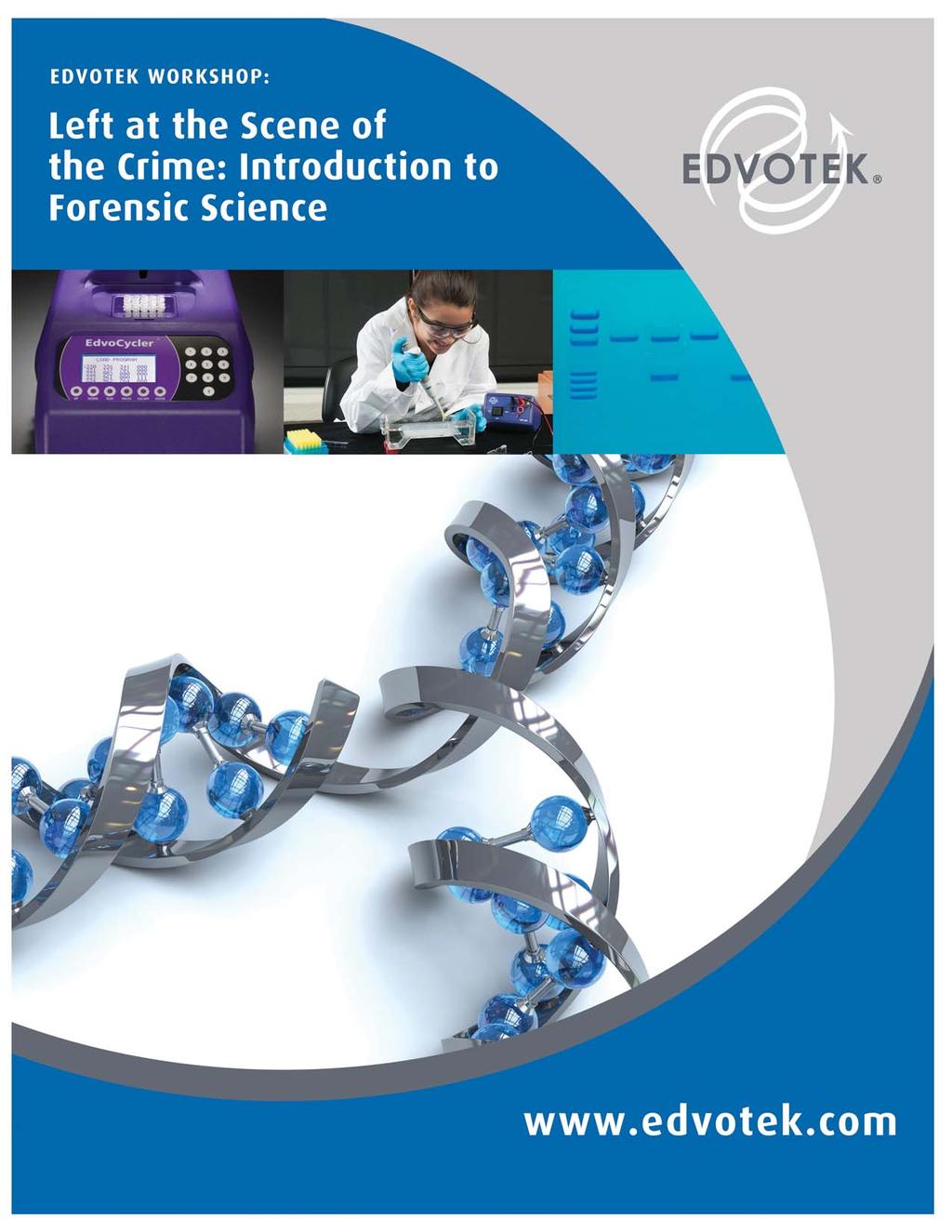
2 Left at the Scene of the Crime: Introduction to Forensic Science EDVOTEK WORKSHOP Introduction Explore genetic diversity using forensic science! In this lab, you become a crime scene investigator as you analyze biological evidence using both blood typing and DNA fingerprinting, a technique that identifies people via genetic differences. Gel electrophoresis is used to create DNA fingerprints from crime scene and suspect samples. A match between samples suggests which suspect committed the crime. Left at the Scene of the Crime Late one night, an intrepid scientist worked on an important biotechnology experiment in the laboratory. She was very close to creating a ground breaking vaccine that could save many lives. After working in the lab all day, she decided to go home to eat dinner and get a good night s rest. The next morning, the lab had been broken into and was a shambles. The scientist found many important pages had been ripped from her lab notebook. Furthermore, security footage showed that someone had stolen critical reagents from the laboratory. Upon investigating the crime scene, police found some blood spilled on the broken window. Detectives identified this window as a potential entry and collected the blood as evidence to be analyzed. As a forensic scientist, you will analyze these samples. The results will be presented as an report in the court of law. Background Information Excerpts from EDVO-Kits 191 & 13 WHAT FORENSICS INFORMATION DOES BLOOD TYPING PROVIDE? Although blood evidence is used primarily for DNA profiling, simpler blood tests are still widely used in the modern forensic laboratory. Blood group testing is one such test. Testing for blood groups relies on the possible precipitation of an antigen-antibody complex from mixing two blood samples together. This phenomenon, called agglutination is a confirmatory test for blood. Only blood will produce this agglutination, which is why it is classified as a confirmatory test. Group % Actual # in millions A B AB O Testing a sample for its blood group is a useful forensic test in a several ways. In addition to being a confirmatory test for the Total presence of blood, by knowing the blood group of a sample, we can use the test to screen potential suspects against an unknown sample. In this lab, students will simulate the detection of a suspect s ABO blood type. The ABO system of blood grouping is arguably the most famous and most important of all systems. If you ve ever donated blood or had blood drawn at a hospital, you ve probably heard of this group. What you may not know, is that the ABO group is just one of many blood groups that have been identified in the last century. In fact, the International Society of Blood Transfusions (ISBT) recognizes 3 blood groups systems! While we may not be able to use a particular piece of evidence, say a bloodstain for example, to say a unique person was the source of the evidence, we can say that it came from a single group of people, sharing the same blood type. How does this apply to blood? Take a look at the following table showing how many Americans have each type. Let s pretend that we found blood on a broken window at a crime scene. We test it and find that it is Type O. Who left it there? It s pretty easy to see from that table that just because a suspect has the same blood type as the blood found at the scene, he is not necessarily the person who left the blood. Because so many people have each blood type, it is easier to use the information to rule out a suspect. Using just the ABO group information, we can t say for sure that the person with Type O is also the one who left that bloodstain, but we can absolutely say that the person with Type AB did not leave the bloodstain on the broken window. This use of evidence is very important in Forensic Science. Individual pieces of evidence that may not identify someone on their own can be used to draw a much more specific picture when used together. Using serological evidence in conjunction with physical evidence and eyewitness accounts will always help narrow down the possibilities in a case. Precipitation reactions between soluble antigens and antibodies can be visible reactions if both components are in equivalence. Under this condition neither the antigen nor the antibody is in excess and antigen-antibody complexes 2 VISIT for complete experiment details & free student protocols.
3 EDVOTEK WORKSHOP Left at the Scene of the Crime: Introduction to Forensic Science form large networks that precipitate out of solution as diagrammed below (Figure 1). When an antigen is attached to a red blood cell, the reaction is called an agglutination and the lattice of antigen and antibody that is visible at equivalence is called an agglutinate. Agglutination is a routine and cost-effective serological procedure because the agglutinate is very easily detectable. Blood typing is an example of a clinical agglutination assay that is familiar to all of us. Since the specific blood antigens are on the surface of red blood cells (RBCs) they are termed hemagglutination reactions. Blood typing has various important forensic science applications and remains a powerful forensic tool in linking someone to a crime or accident scene. Blood Type A B AB O Antigen on Red Blood Cells A B both A and B neither A nor B Antigen in Serum anti-b anti-a neither anti-a nor anti-b both anti-a nor anti-b Antigen excess Equivalence Antibody excess DNA FINGERPRINTING In humans, DNA is packaged into 23 pairs of chromosomes that are inherited from an individual s biological parents. Although most of this genetic soluble antigen antibody material is identical in every person, small differences, or polymorphisms, in the DNA sequence occur Figure 1: Antigen and antibody reactions throughout the genome. For example, the simplest difference is a Single Nucleotide Polymorphism (or SNP). Changes in the number and location of restriction enzyme sites result in Restriction Fragment Length Polymorphisms (or RFLPs). Short repetitive stretches of DNA at specific locations in the genome can vary in number to produce STRs (Short Tandem Repeats) and VNTRs (Variable Number of Tandem Repeats). Although most polymorphisms occur in non-coding regions of DNA, those that disrupt a gene can result in disease. Medical diagnostic tests can identify specific polymorphisms associated with disease. Analyzing several different polymorphisms within a person s genome generates a unique DNA fingerprint. DNA fingerprints can allow us to distinguish one individual from another. Because polymorphisms are inherited, DNA fingerprints can also be used to determine paternity/maternity (and other familial relationships). The best-known application of DNA fingerprinting is in forensic science. DNA fingerprinting techniques are utilized to interpret blood, tissue, or fluid evidence collected at accidents and crime scenes. After DNA is extracted from these samples, forensic scientists can develop a DNA fingerprint. The DNA fingerprint from a crime scene can then be compared to the DNA fingerprints of different suspects. A match provides strong evidence that the suspect was present at the crime scene. Early fingerprinting analysis involved restriction digestion of the isolated DNA. Following electrophoresis of the digested sample, the DNA is transferred to a nylon membrane during a process known as Southern blotting. Sequence-specific DNA probes are used to visualize the membrane-bound DNA. If the DNA is not digested by the restriction enzyme, the probes will only hybridize to a single DNA segment. If a restriction site occurs within this sequence, the probe will hybridize with multiple bands of DNA. VNTRs are identified when a probe labels DNA at a dissimilar molecular weight. Although RFLP analysis is very precise, it is time-consuming and requires large amounts of DNA. To address these problems, forensic scientists use the polymerase chain reaction (PCR) to produce DNA fingerprints. PCR allows researchers to quickly create many copies of a specific region of DNA in vitro (summarized in Figure 2). This technique requires 5- fold less DNA than traditional RFLP analysis and it can be performed in an afternoon. To perform PCR, purified double-stranded DNA is mixed with primers (short synthetic DNA molecules that target DNA for amplification), a thermostable DNA polymerase (Taq) and nucleotides. The mixture is heated to 94 C to denature the DNA duplex (i.e., unzip it into single strands). Next, the sample is then cooled to 45 C - 6 C, allowing the primers to base pair with the target DNA sequence (called annealing ). Lastly, the temperature is raised to 72 C, the optimal temperature at which Taq polymerase will extend the primer to synthesize a new strand of DNA. Each PCR cycle (denaturation, annealing, extension) doubles the amount of the target DNA in less than five minutes. In order to produce enough DNA for analysis, twenty to forty cycles may be required. To simplify this process, a specialized machine, called a thermal cycler or a PCR machine, was created to rapidly heat and cool the samples. CONTACT US? CALL 1.8.EDVOTEK Fax info@edvotek.com 3
4 Left at the Scene of the Crime: Introduction to Forensic Science EDVOTEK WORKSHOP Figure 2: Polymerase Chain Reaction 4 VISIT for complete experiment details & free student protocols.

5 EDVOTEK WORKSHOP Left at the Scene of the Crime: Introduction to Forensic Science Experiment Procedures: What Forensics Information Does Blood Typing Provide? 1. Place a microtiter plate piece as shown below. Across the top of the plate, label the 8 wells A, B, AB, O, CS, S1, S2, and S3 respectively, using a laboratory marking pen. Label the 2 rows Anti-A and Anti-B respectively. The plate should look as pictured below. A B AB O CS S1 S2 S3 Anti A Anti B IMPORTANT: No actual blood or blood products are used in this experiment. IMPORTANT: Avoid cross-contamination by using a new disposable pipet or pipet tip when using an automatic micropipet for each blood sample. 2. Using a different pipet or pipet tip for each sample, transfer 3 drops (4 µl) of each blood type sample (see table) from the provided QuickStrip to the two corresponding wells. For example, control A blood type goes into the two wells under the letter A. Repeat the same procedure for crime scene collected blood and blood from each of the three suspects. 3. Use a new pipet to add one drop or 2 µl of Anti-A serum into each of the wells in row #1. 4. Use a new pipet to add one drop or 2 µl of Anti-B serum into each of the wells in row #2. 5. Let the plate sit undisturbed on the lab bench for 5-1 minutes. 6. Observe the wells for the presence or absence of agglutination. Agglutination has occurred if the mixture appears to be granular rather than smooth. Record your results in the diagram in the Results section. QuickStrip Label A B C D E F G H Sample Blood Type A Blood Type A Blood Type B Blood Type O Crime Scene Blood Suspect A Suspect B Suspect C - (no agglutination) + (agglutination) CONTACT US? CALL 1.8.EDVOTEK Fax info@edvotek.com 5
6 Left at the Scene of the Crime: Introduction to Forensic Science EDVOTEK WORKSHOP Experiment Procedures: Separation of DNA by Agarose Gel Electrophoresis x 1: Concentrated buffer Distilled water Agarose Wear gloves and safety goggles C 6 C Flask 6. WAIT 7. Pour 2 min. Caution! Flask will be HOT! NOTES: If casting 7 x 14 cm gels, each gel can be shared by 4 students. Place combs in the fi rst and third set of notches. If casting 7 x 7 cm gels, each gel can be shared by 2 students. Place comb in the fi rst set of notches. If you are unfamiliar with agarose gel prep and electrophoresis, detailed instructions and helpful resources are available at 1. DILUTE concentrated (5X) buffer with distilled water to create 1X buffer (see Table A). 2. MIX agarose powder with 1X buffer in a 25 ml flask (see Table A). 3. DISSOLVE agarose powder by boiling the solution. MICROWAVE the solution on high for 1 minute. Carefully REMOVE the flask from the microwave and MIX by swirling the flask. Continue to HEAT the solution in 15-second bursts until the agarose is completely dissolved (the solution should be clear like water). 4. COOL agarose to 6 C with careful swirling to promote even dissipation of heat. 5. While agarose is cooling, SEAL the ends of the gel-casting tray with the rubber end caps. PLACE the well template (comb) in the appropriate notch. 6. POUR the cooled agarose solution into the prepared gel-casting tray. The gel should thoroughly solidify within 2 minutes. The gel will stiffen and become less transparent as it solidifies. 7. REMOVE end caps and comb. Take particular care when removing the comb to prevent damage to the wells. Table A Size of Gel Casting tray 7 x 7 cm Individual.8% UltraSpec-Agarose Gel Concentrated Buffer (5x).6 ml + Distilled Water ml Amt of Agarose.23 g = TOTAL Volume 3 ml 7 x 1 cm 1. ml 49. ml.39 g 5 ml 7 x 14 cm 1.2 ml 58.8 ml.46 g 6 ml 6 VISIT for complete experiment details & free student protocols.
7 EDVOTEK WORKSHOP Left at the Scene of the Crime: Introduction to Forensic Science Experiment Procedures: Separation of DNA by Agarose Gel Electrophoresis Pour 1X Diluted Buffer Reminder: Before loading the samples, make sure the gel is properly oriented in the apparatus chamber Wear gloves and safety goggles 8. PLACE gel (on the tray) into electrophoresis chamber. COVER the gel with 1X electrophoresis buffer (See Table B for recommended volumes). The gel should be completely submerged. 9. LOAD the entire sample (35-38 µl) into the well. 1. PLACE safety cover. CHECK that the gel is properly oriented. Remember, the DNA samples will migrate toward the positive (red) electrode. 11. CONNECT leads to the power source and PERFORM electrophoresis (See Table C for time and voltage guidelines). 12. After electrophoresis is complete, REMOVE the gel and casting tray from the electrophoresis chamber and proceed to STAINING the agarose gel. Lane 1 2 Label A B Sample Name DNA Standard Marker Crime Scene PCR reaction 3 C Suspect 1 PCR reaction 4 D Suspect 2 PCR reaction 5 E Suspect 3 PCR reaction Table B 1x Electrophoresis Buffer (Chamber Buffer) EDVOTEK Model # M6+ & M12 (new) M12 (classic) M36 Total Volume Required 3 ml 4 ml 1 ml 5x Conc. Buffer 6 ml 8 ml 2 ml Dilution + Distilled Water 294 ml 392 ml 98 ml Table C Volts M6+ Min. / Max. 15/2 min. 2/3 min. 35 / 45 min. Time and Voltage Guidelines (.8% Agarose Gel) Electrophoresis Model M12 (new) Min. / Max. 2/3 min. 3/35 min. 55/7 min. M12 (classic) & M36 Min. / Max. 25 / 35 min. 35 / 45 min. 6 / 9 min. CONTACT US? CALL 1.8.EDVOTEK Fax info@edvotek.com 7

8 Left at the Scene of the Crime: Introduction to Forensic Science EDVOTEK WORKSHOP 8 VISIT for complete experiment details & free student protocols.

9 EDVOTEK WORKSHOP Left at the Scene of the Crime: Introduction to Forensic Science Experiment Results and Analysis BLOOD TYPING Anti A A B AB O CS S1 S2 S3 Anti B SEPARATION OF DNA BY AGAROSE GEL ELECTROPHORESIS Lane Tube 1 A DNA Standard marker 2 B Crime scene PCR reaction 3 C Suspect 1 PCR reaction 4 D Suspect 2 PCR reaction 5 E Suspect 3 PCR reaction The DNA standards in Lane 1 make it possible to measure the DNA bands obtained from the PCR reactions. The results of this analysis indicates an identical pattern in Lanes 2 and 4. This is strong evidence that the crime scene DNA and Suspect 2 match. In criminal investigations, several known variable regions in DNA are analyzed to match crime scene and suspect DNAs. CONTACT US? CALL 1.8.EDVOTEK Fax info@edvotek.com 9


10 Left at the Scene of the Crime: Introduction to Forensic Science EDVOTEK WORKSHOP Related Resources Here at EDVOTEK, we ve worked hard over the last year to bring you some new and exciting resources to make teaching biotechnology easier and more exciting than ever! We ve created Quick Guide manuals, FREE for you to download off our website. We have also filmed several Instructional Videos that show step-by-step procedures. We hope you take advantage of these resources and enjoy teaching and learning with EDVOTEK! Micropipetting Basics Dial µl µl µl 8 3 Agarose Gel Electrophoresis 6 C WAIT 2 min. Stains: Visualizing DNA 1 µl EDVOTEK EDVOTEK EDVOTEK 8 3 InstaStain Blue Tech Video youtube.com/edvotekinc Video: Preparing Agarose Gels Video: Staining with InstaStain Blue Video: Staining with InstaStain Ethidium Bromide Video: Performing Agarose Gel Electrophoresis Video: Staining with FlashBlue Video: Staining with SYBR Safe 1 VISIT for complete experiment details & free student protocols.
 to amplify human DNA obtained from crime scenes.")


11 EDVOTEK WORKSHOP Left at the Scene of the Crime: Introduction to Forensic Science Related Products Cat. #13 DNA Fingerprinting by PCR Amplification For 8 Gels. Forensic DNA fingerprinting has become a universally accepted crime-fighting tool. Recent advances use the polymerase chain reaction (PCR) to amplify human DNA obtained from crime scenes. This experiment, based on a crime scene scenario, has an inquiry-based component. Kit includes: instructions, Ready-to-Load QuickStrip DNA samples, UltraSpec-Agarose powder, practice gel loading solution, electrophoresis buffer, InstaStain Blue and FlashBlue stain, calibrated pipet, and microtipped transfer pipettes. All you need: electrophoresis apparatus, power supply, automatic micropipet and tips, balance, microwave or hot plate, visualization system Cat. #191 Forensics Blood Typing What Forensics Information Does Blood Typing Provide? For 1 Groups. Detectives, investigating a murder crime scene, have discovered possible blood residue on a gun dropped at the scene. After identifying three potential suspects, each with a probable motive, the detectives act to match a blood sample from each suspect to the residue found on the gun handle. This is completed while they await DNA testing. In this classroom experiment, students will first identify whether or not the recovered residue from the gun handle is blood and will then use a rapid blood type test to further the investigation. Kit includes: instructions, control ABO simulated blood samples, simulated crime scene, and suspect blood samples, anti-a and Anti-B serums, blood detection stock solutions, transfer pipets, microtiter plates, tubes. All you need: 95-1% Ethanol, marking pen, distilled water. Optional: automatic micropipet (5 5 µl). Cat. # 59 EDVOTEK Variable Micropipet 5-5 µl Micropipet Cat. #59 DuoSource 15 Power Supply 75 /15 V for 1 or 2 units Cat. #52 M12 Electrophoresis Apparatus For 2 Lab Groups CONTACT US? CALL 1.8.EDVOTEK Fax info@edvotek.com 11


12 Left at the Scene of the Crime: Introduction to Forensic Science EDVOTEK WORKSHOP Related Products Cat. #S-91 Whose Fingerprints Were Left Behind? For 1 Groups. After a crime has been committed, the evidence left behind can identify a potential culprit, although a single piece of evidence is not usually enough to convict someone. Even in this age of DNA, fingerprints and blood stains are still important at helping to identify a criminal. In this experiment your students will learn to detect and analyze fingerprints and then use these techniques to solve a classroom crime. Kit includes: instructions, brushes, magnifying lens, fingerprint cards, black dusting powder, grey dye dusting powder, fingerprint lifters. Cat. #S-51 Whose DNA Was Left Behind? For 1 Groups. DNA obtained from a single hair left behind at a crime scene can be used to identify a criminal. In this experiment, your students will compare simulated crime scene DNA with that of two suspects. Kit includes: instructions, Ready-to-Load dye samples, agarose powder, practice gel loading solution, electrophoresis buffer, microtipped transfer pipets. All you need: electrophoresis apparatus, power supply, microwave or hot plate. Cat. #195 Forensic Toxicology For 1 Groups. In this forensic science experiment, students will use the Enzyme Linked Immunosorbent Assay (ELISA) to analyze crime scene samples for the presence of drugs. Kit includes: instructions, victim and crime scene samples, control samples, solutions, microtiter plates, transfer pipets, conical tubes. All you need: distilled or deionized water, beakers, 37 C incubator, paper towels, automatic micropipets with tips (recommended), lab gloves and goggles. 12 VISIT for complete experiment details & free student protocols.