Skin Regeneration. Mark A. Carlson, MD. Department of Surgery University of Nebraska Medical Center Omaha VA Medical Center Omaha, Nebraska USA
|
|
|
- Darleen Hensley
- 5 years ago
- Views:
Transcription


1 Skin Regeneration Mark A. Carlson, MD Department of Surgery University of Nebraska Medical Center Omaha VA Medical Center Omaha, Nebraska USA Stem Cell Biology course, April 20, 2010

2 Disclosures: none Content available:
3 Overview Clinical problem Established treatments Research approaches Our strategy
4 Skin Loss: Clinical Problem Normal anatomy Healing v. Regeneration Sequelae of skin loss


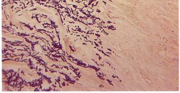
5 epidermis dermis 1 mm panniculus carnosus subcutis epidermis cf fb dermis 5 µm cf = collagen fibril fb = fibroblast 100 µm Normal skin anatomy (rat)
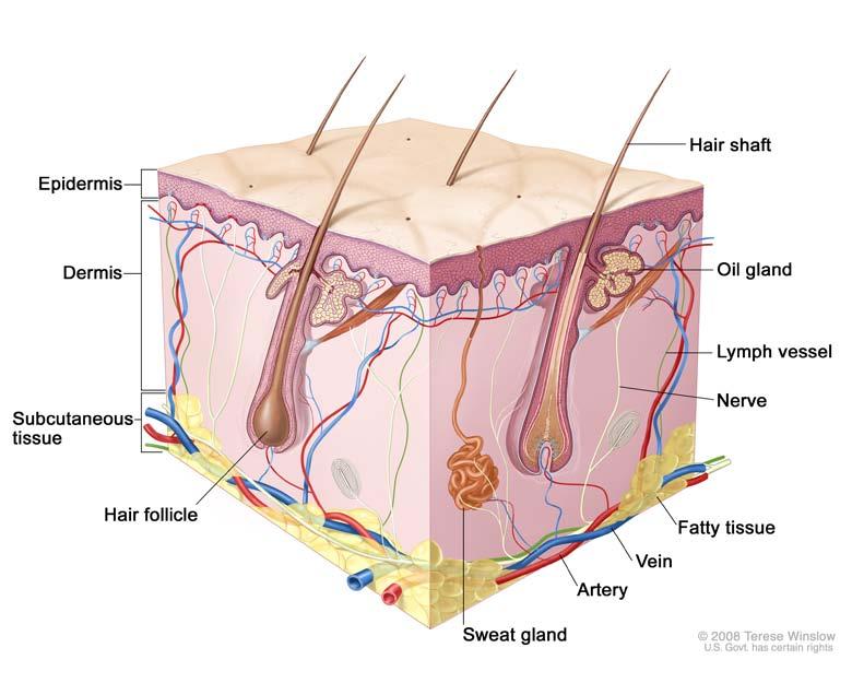
6 Normal skin anatomy structural components epidermis basal lamina dermis blood vessels hair follicles glands cutis (nail, claw, horn) (subcutaneous adipose) (skeletal muscle) functions barrier temperature regulation sensation coloration immune response synthetic specialized
7 Healing v. Regeneration Excisional wound: natural history 1 cm Dorsum of rat: 2 x 2 cm full-thickness excision, followed for 204 days

8 Growth (RJ Goss, 1992)

9 Healing v. Regeneration Amphibian limb amputation


")
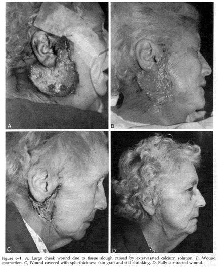
10 A type of growth: wound healing (corneal injury)
11 Regeneration vs. conventional healing

12 Elastin: scar vs. dermis
 4 mo (Anat Rec 2001;264:203) rat tibialis anterior")
13 control Negative growth: 1 mo atrophy (skeletal muscle 2 mo denervation) 4 mo (Anat Rec 2001;264:203) rat tibialis anterior (anti-laminin)


14 Paradigm tissue loss/injury response recovery inflammation regeneration death scar with major disability poor healing quality of life scale scar with minor disability good

15 Regeneration through the phyla Hydra H. simpson regenerative ability organism size organism complexity The larger and/or more advanced the animal, the less it can regenerate.
 1. Everything else")
16 Tissue-Specific Mammalian Regenerative Capacity HIGH 1. Epidermis 2. Liver 3. Endothelium 4. Epithelium LOW (but theoretically possible) 1. Everything else
17 So what is the problem with healing? In most cases, nothing. But

18 Sequela of skin loss: Burn wound contracture

19 Abnormal healing response: Keloid scarring
20 Tissue repair is designed to reconstruct morphological integrity; epimorphic regeneration is designed to restore function. Animals can live without epimorphic regeneration but not without tissue repair. Richard J. Goss, 1992 Wound healing = quick fix; poor function Regeneration = complicated; normal function
21 Skin Loss: Established Treatments Skin flaps/grafts Epidermal substitutes Dermal substitutes Combined (epiderm + derm)


22 Skin flaps: simple Excisional wound (rat dorsum)

23 Free flap (treatment of contracture)

")
24 Skin grafting Harvest ( µm thick) Forearm graft
25 Skin grafting: full- vs. split-thickness
26 Skin grafting: split-thickness
27 Skin grafting: Gold Standard for skin replacement Disadvantages Limited source of material Wound contraction Cosmesis Donor site morbidity


28 Skin grafting: Donor site morbidity
29 Skin loss treatments: Epidermal substitutes Strategy: cultured autologous keratinocytes/hair follicle cells Examples: Epicel, Laserskin, Epidex, MySkin
30 Epidermal replacement
31 Epidermal substitutes: Autologous keratinocytes Advantages: No rejection No large donor sites Disadvantages: Fragile, no supportive dermis Time requirement (culture)
32 Skin loss treatments: Dermal substitutes Strategy: natural, semisynthetic, or synthetic matrix, ± fibroblasts Examples: Alloderm, Biobrane, Dermagraft, Integra, Permacol, Transcyte
33 Dermal replacement
34 Dermal substitute: Alloderm Composition: processed cadaveric human dermis Advantages Minimal rejection Simplicity Disadvantages No cellular component Expensive
35 Dermal substitute: Biobrane Composition: nylon mesh + silicone membrane + porcine ECM Advantages Simplicity Large area coverage Disadvantages Not a permanent replacement
36 Dermal substitute: Dermagraft Composition: allogeneic neonatal fibroblasts in polyglactin mesh Advantages Minimal rejection Absorbable ECM Disadvantages Complexity Small area coverage
37 Dermal substitute: Integra Composition: bovine col + GAGs topped with silicone Advantages Encourage cellular ingrowth Integrates with host Disadvantages Needs autograft after silicone removal Bovine allergy risk
38 Dermal substitute: Permacol Composition: treated porcine dermal collagen Advantages Non-immunogenic Supports ingrowth from host Disadvantages Needs autograft after incorporation Expensive
39 Dermal substitute: Transcyte Composition: collagen-coated nylon seeded with allogeneic neonatal fibroblasts, topped with nylon Advantages Integrates with host, encourages ingrowth Disadvantages Nonabsorbable Not permanent
40 Skin loss treatments: Combined epidermal/dermal substitutes (composites) Strategy: cultured allogeneic cells populating a bilaminar structure Examples: Apligraf, Orcel
41 Composite substitute: Apligraf; Orcel Composition: allogeneic neonatal keratinocytes + fibroblasts, type I col matrix with cytokines Advantages Minimal rejection Rational design Disadvantages Complexity, time, expense Limited area coverage
42 Skin Regeneration: Research Approaches Endogenous regeneration Fetal paradigm Tissue engineering
43 Research approaches: Endogenous regeneration Enable inherent regenerative mechanism ( inside-out approach) Requires understanding of regeneration vs. healing Resident stem cell biology relevant
44 Mammals do not regenerate Exceptions: Antler growth Rabbit ear regeneration Distal fingertip amputations in mice and young children (Others?)

45 Dedifferentiation in nature? Blastema formation: central event of limb regeneration (Goss, 1992)

46 Dedifferentiation in nature?? Limb amputation blastema What s in there? A. Stem cells? B. Dediff cells? C. A & B?

47 Stem cells & dedifferentiation undifferentiated stem cell forward pathway: canonical (differentiation) reverse pathway = DEDIFFERENTIATION Does this pathway exist? If so, where, how, and how frequently? differentiated daughter cell (Der Alchemist, E. van Hove)
48 Research approaches: Fetal paradigm Mammalian fetus: regenerative response instead of inflammation/healing Transition point in utero, after which regeneration no longer occurs Phenomenology followed by mechanistic studies (no applications yet)


49 Scarless healing in the dermis of the mammalian fetus Excisional wounds in the fetal rat after 72 hr E16 E18 (Plast Reconstr Surg 2001;160:209)
: collagen")

50 Scarless fetal healing (cont d): collagen organization in the E16 fetus 72 hr after wounding on E16 unwounded
: collagen")

51 Scarless fetal healing (cont d): collagen organization in the E18 fetus 72 hr after wounding on E18 unwounded
52 Fetal paradigm: Comparison with adult Fetal ECM: enriched in type III collagen, hyaluronic acid, tenascin-c Elevated levels of TGF-β3, IL-10; decreased TGF-β1/2, IL-8, many others Differences in fibroblast phenotype
53 Research approaches: Tissue engineering Replace lost tissue with engineered construct ( outside-in approach) Requires understanding of how implants interact with host Pluripotent stem cell biology relevant
54 Tissue engineering: scaffolds for construction Materials include organic polymers (e.g., polyglactin, polylactide), gelatin, chitosan, PEG hydrogels A variety of synthetic techniques can control micro- and nano-architecture (e.g., fiber diameter, pore size, physical properties)
55 Tissue engineering: Smart biomaterials Older strategy of 100% synthetic matrix failed Newer strategy uses semisynthetic materials (e.g., polymer coated with ECM) Coating encourages cellular ingrowth
56 Tissue engineering: ECM coating Adsorbed or covalently bound to scaffold Examples include: type I/III collagens, elastin, HA, fibronectin, RGD peptides, other peptide mimetics
57 Tissue engineering: cellular embedding Embed vs. ingrowth (chemoattraction) Cell type: differentiated vs. stem If stem, what type (MSC, ipsc, ESC, etc.)
58 Skin Regeneration: Our Strategy Nanoengineered scaffold ECM surfacing Cells (MSC + fibroblast) Cytokine slow-release Multi-layer recapitulation
59 Objective: To develop a replacement therapy for epidermis/dermis (i.e., to engineer a complete skin equivalent for clinical use)


60 dermal replacement: strategy synthetic backbone Material can be manipulated at the nano-level for architectural and physical properties 20 µm 100 µm 20 µm Material can be engineered to contain nanoparticles for slowrelease of various cytokines/ growth factors


61 collagen matrix with cells: dermal fibroblasts mesenchymal stem 5 mm neodermis dermal replacement: strategy 50 µm 10 µm
62 basal lamina (type IV collagen, laminin) dermal replacement: strategy neodermis
63 dermal replacement: strategy keratinocytes (epidermis)
64 dermal replacement: strategy finished skin equivalent plug into animal model

65 1 cm PWD 0 PWD 0 Regeneration Assay (wound contraction) 4 PWD 7 PWD 14 wound area (cm 2) goal PWD PWD PWD 204 Additional endpoints Tensile strength Microscopic morphology
66 Looks easy, but potential problems: Co-culture conditions Blood supply to implant Infection (see next item) Inflammation at interface Strength & durability Nerves, glands, hair, etc. Translation from rodents to humans Cellular source

67 Transplanted tissue requires a blood supply (Donor kidney just prior to transplantation)


68 Inflammation (Excisional wound bed, postwounding day 3)
69 Conclusions 1. Proofs of concept are readily available, but 2. Translation into practical treatments have been rare 3. Road to practical treatments will be long and difficult

70 Take-home message: Healing bad, regeneration good. Current reality: When my liver fails, don't give me a bone marrow transplant [i.e., stem cells], give me a liver. Irving Weissman, 2004
71 (end)