Chapter 3 An Introduction to Gene Function 3.1 Storing Information 3.2 Replication 3.3 Mutation
|
|
|
- Cody Shepherd
- 5 years ago
- Views:
Transcription
1 Chapter 3 An Introduction to Gene Function 3.1 Storing Information 3.2 Replication 3.3 Mutation
![3.1 Storing Information [Central Dogma] Producing a protein from DNA => by transcription and translation codon : the 3 base sequence that](/docs-images/84/91030622/images/2-0.jpg "determines what amino acid is used Template strand : the DNA strand that is used to generate the mrna Nontemplate strand : not used in")
2 3.1 Storing Information [Central Dogma] Producing a protein from DNA => by transcription and translation codon : the 3 base sequence that determines what amino acid is used Template strand : the DNA strand that is used to generate the mrna Nontemplate strand : not used in transcription
 Proteins have 20")
 A specific side chain (R) : determines structure and")
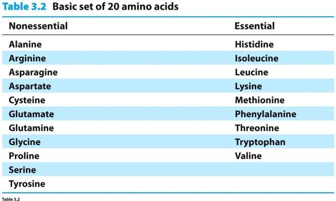
3 Protein Structure Proteins are chain-like polymers of small subunits, called amino acids DNA has 4 different nucleotides (A,G, C, T) Proteins have 20 different amino acids with: An amino group (NH 3 +) A carboxyl group (COO - ) A hydrogen atom (H) A specific side chain (R) : determines structure and function

4 Amino Acids
5 Polypeptides Amino acids are joined together via peptide bonds polypeptides Proteins are composed of 1 or more polypeptides Polypeptides have polarity (or directionality) Free amino group at one end is the amino- or N-terminus Free carboxyl group at the other end is the carboxyl- or C-terminus
6 Polypeptides
7 Types of Protein Structure (4)
8 Primary Structure The linear order (sequence) of amino acids Determined by genes
 Hydrogen bond Repeating patterns α-helices and")
 Not α helix or β")
9 Secondary Structure Chemical and physical interactions cause folding Ex) Hydrogen bond Repeating patterns α-helices and β-sheets Key determinants of a protein s characteristics Random coiled regions (or turn and loop) Not α helix or β pleated sheet

10 α-helix Secondary Structure

11 β-sheet Secondary Structure By forming H-bonds Polypeptide chains are packed side by side


12 Tertiary Structure The total threedimensional shape of a polypeptide is its tertiary structure A prominent aspect of this structure is the interaction of the amino acid side chains The globular form of a polypeptide is a roughly spherical structure Tertiary structure of myoglobin Tertiary structure of guanidinoacetate methyltransferase
13 Quaternary Structure Made up of 2 or more polypeptides Protein subunits : individual polypeptides
14 Protein Structure 5 factors promoting protein folding and stability 1. Hydrogen bonds 2. Ionic bonds and other polar interactions 3. Hydrophobic effects 4. Van der Waals forces 5. Disulfide bonds

15 Protein Domains Compact structural regions of a protein are referred to as domains Immunoglobulins provide an example of 4 globular domains Domains may contain common structural-functional motifs Zinc finger Hydrophobic pocket Quaternary structure is the interaction of 2 or more polypeptides 3-15

16 Protein-protein interactions Many cellular processes involve steps in which two or more different proteins interact with each other Specific binding at surface Use first 4 factors 1. Hydrogen bonds 2. Ionic bonds and other polar interactions 3. Hydrophobic effects 4. Van der Waals forces
17 Summary Proteins are polymers of amino acids linked through peptide bonds The sequence of amino acids in a polypeptide (primary structure) gives rise to that molecule s: Local shape (secondary structure) Overall shape (tertiary structure) Interaction with other polypeptides (quaternary structure)
18 Proteins: Protein Function Provide the structure that help give cells integrity and shape ex) cytoskeleton Serve as hormones carrying signals from one cell to another ex) Insulin Bind and carry substances ex) Carrier proteins Control the activities of genes ex) Transcription factors Serve as enzymes that catalyze hundreds of chemical reactions ex) Ribonuclease
19 One-Gene/One-Polypeptide Many enzymes contain more than one polypeptide chain and each polypeptide is usually encoded in one gene These observations have lead to the one-gene / onepolypeptide hypothesis Summary : Most genes contain the information for making one polypeptide
20 Information Carrier In the 1950s and 1960s, the concept that messenger RNA carries information from gene to ribosome was developed An intermediate carrier was needed as DNA is found in the nucleus, while proteins are made in the cytoplasm Therefore, some type of molecule must move the information from the DNA in the nucleus to the site of protein synthesis in the cytoplasm
21 Discovery of Messenger RNA Ribosomes are the cytoplasmic site of protein synthesis Jacob and colleagues proposed that an alternative of nonspecialized ribosomes, translate unstable RNAs called messengers. These messengers are independent RNAs that move information from genes to ribosomes
22 Experiment to Test the mrna Hypothesis Experimental test of the messenger hypothesis. Heavy E. coli ribosomes were made by labeling the bacterial cells with heavy isotopes of carbon and nitrogen. The bacteria were then infected with phage T2 and simultaneously shifted to light medium containing the normal isotopes of carbon and nitrogen, plus some 32 P to make the phage RNA radioactive. (a) Crick had proposed that ribosomal RNA carried the message for making proteins. If this were so, then whole new ribosomes with phage-specific ribosomal RNA would have been made after phage infection. In that case, the new 32 P-labeled RNA (green) should have moved together with the new, light ribosomes (pink). (b) Jacob and colleagues had proposed that a messenger RNA carried genetic information to the ribosomes. According to this hypothesis, phage infection would cause the synthesis of new, phage-specific messenger RNAs that would be 32 P-labeled (green). These would associate with old, heavy ribosomes (blue). The radioactive label would therefore move together with the old, heavy ribosomes in the density gradient. This was indeed what happened.
23 Crick and Jacob Experiments Radio-labeled phage RNA in experiments was found to be associated with old ribosomes whose rrna was made before infection rrna doesn t carry information from DNA A different class of unstable RNAs associate transiently with ribosomes Messenger RNAs (mrnas) carry the genetic information from the genes to the ribosomes, which then synthesize polypeptides
24 Transcription Transcription follows the same basepairing rules as DNA replication Remember U replaces T in RNA This base-pairing pattern ensures that the RNA transcript is a faithful copy of the gene For transcription to occur at a significant rate, its reaction is mediated by enzyme RNA polymerase
25 Synthesis of RNA Making RNA. (a) Phosphodiester bond formation in RNA synthesis. ATP and GTP are joined together to form a dinucleotide. Note that the phosphorus atom closest to the guanosine is retained in the phosphodiester bond. The other two phosphates are removed as a by-product called pyrophosphate. (b) Synthesis of RNA on a DNA template.
26 Phases of Transcription Transcription occurs in three phases: 1. Initiation 2. Elongation 3. Termination
27 Initiation RNA polymerase recognizes a specific region, the promoter, which lies just upstream of gene The polymerase binds tightly to the promoter causing localized separation of the two DNA strands The polymerase starts building the RNA chain by adding ribonucleotides After several ribonucleotides are joined together, the enzyme leaves the promoter and elongation begins
28 Elongation RNA polymerase directs the addition of ribonucleotides in the 5 to 3 direction Movement of the polymerase along the DNA template causes the bubble of separated DNA strands to move also As the RNA polymerase proceeds along the DNA, the two DNA strands that have opened for the bubble reform the double helix behind the transciptional machinery
29 Transcription and DNA Replication Two fundamental differences between transcription and DNA replication 1. RNA polymerase only makes one RNA strand during transcription, it copies only one DNA strand in a given gene This makes transcription asymmetrical Replication is semiconservative 2. DNA melting is limited and transient during transcription, but the separation is permanent in replication

30 Termination Analogous to the initiating activity of promoters, there are regions at the other end of genes that serve to terminate transcription These terminators work with the RNA polymerase to loosen the association between the RNA product and the DNA template As a result, the RNA dissociates from the RNA polymerase and the DNA and transcription stops
31 Important Note about Conventions RNA sequences are written 5 to 3, left to right Translation occurs 5 to 3 with ribosomes reading the message 5 to 3 Genes are written so that transcription proceeds in a left to right direction The gene s promoter area lies just before the start area, said to be upstream of transcription Genes are therefore said to lie downstream of their promoters
32 Summary Transcription takes place in three stages: Initiation Elongation Termination Initiation involves the binding of RNA polymerase to the promoter, local melting and forming the first few phosphodiester bonds During elongation, the RNA polymerase links together ribonucleotides in the 5 to 3 direction to make the rest of the RNA In termination, the polymerase and RNA product dissociate from the DNA template
33 Translation - Ribosomes Ribosomes are protein synthesizing machines Ribosome subunits are designated with numbers such as 50S or 30S Number is the sedimentation coefficient - a measure of speed with which the particles sediment through a solution spun in an ultracentrifuge based on mass and shape Each ribosomal subunit contains RNA and protein E. coli ribosome structure. (a) The 70S ribosome is shown from the side with the 30S particle (yellow) and the 50S particle (red) fitting together. (b) The 70S ribosome is shown rotated 90 degrees relative to the view in part (a). The 30S particle (yellow) is in front, with the 50S particle (red) behind. 3-33
34 Ribosome Ribosomes are the cell s protein factories Bacteria contain 70S ribosomes Each ribosome has 2 subunits 50 S 30 S Each subunit contains rrna and many proteins The 30S subunit includes one molecule of ribosomal RNA (rrna) with a sedimentation coeffi cient of 16S, plus 21 ribosomal proteins. The 50S subunit is composed of 2 rrnas (23S 1 5S) and 34 proteins.
35 Ribosomal RNA The two ribosomal subunits both contain ribosomal RNA (rrna) molecules and a variety of proteins rrnas participate in protein synthesis but do NOT code for proteins No translation of rrna occurs
36 Translation Bacterial mrna 5 ribosomal-binding site Start codon usually AUG Typical polypeptide is a few hundred amino acids in length 1 of 3 stop codons Termination or nonsense codons UAA, UAG or UGA

37 Translation Genetic code sequence of bases in an mrna molecule Read in groups of three nucleotide bases or codons Most codons specify a particular amino acid Also start and stop codons Degenerate- more than one codon can specify the same amino acid

38 Reading frame DNA sequence of gene transcribed into mrna mrna Codon set of 3 RNA nucleotides T of DNA substituted for U of RNA trna Anticodon 3 RNA nucleotide part of trna molecule Allows binding of trna to mrna codon

39 Overall ribosome shape determined by rrna Ribosomes Discrete sites for trna binding and polypeptide synthesis P site- peptidyl site A site- aminoacyl site E site- exit site
40 trna: Translation Adapter Molecule Generating protein from ribosomes requires change from the nucleic acid to amino acid This change is described as translation from the nucleic acid base pair language to the amino acid language Crick proposed that some type of adapter molecule was needed to provide the bridge for translation, perhaps a small RNA The physical interface between the mrna and the ribosome
41 Transfer RNA: Adapter Molecule Transfer RNA is a small RNA that recognizes both RNA and amino acids A cloverleaf model is used to illustrate trna structure The 3 end binds to a specific amino acid The anticodon loop contains a 3 base pair sequence that pairs with complementarity to a 3 base pair codon in mrna
42 Codons and Anticodons Enzymes that catalyze attachment of amino acid to trna are aminoacyl-trna synthetases A triplet in mrna is called a codon The complementary sequence to a codon found in a trna is the anticodon
43 Summary Two important sites on trnas allow them to recognize both amino acids and nucleic acids One site binds covalently to an amino acid The site contains an anticodon that base-pairs with a 3-bp codon in the mrna trnas are capable of serving the adapter role and are the key to the mechanism of translation

44 3 Stages of Translation 1. Initiation mrna, first trna and ribosomal subunits assemble 2. Elongation Synthesis from start codon to stop codon 3. Termination Complex disassembles at stop codon releasing completed polypeptide

45 Initiation of Protein Synthesis The initiation codon (AUG) interacts with a special aminoacyl-trna In eukaryotes this is methionyl-trna In bacteria it is a derivative called N- formylmethionyl-trna Position of the AUG codon: At start of message AUG is initiator In middle of message AUG is regular methionine Shine-Dalgarno sequence lies just upstream of the AUG, functions to attract ribosomes Unique to bacteria Eukaryotes have special cap on 5 -end of mrna

46 Translation Elongation During initiation the initiating aminoacyltrna binds within the P site of the ribosome Elongation adds amino acids one at a time to the initiating amino acid The first elongation step is binding second aminoacyl-trna to the A site on the ribosome This process requires: An elongation factor, EF-Tu Energy from GTP The formation of a peptide bond between the amino acids
 Elongation adds amino acids one at a time to the initiating amino acid The first elongation step is binding second aminoacyl-trna to the A site on the ribosome This process requires: (a) EF-Tu,")
47 Overview of Translation Elongation (E.coli) Elongation adds amino acids one at a time to the initiating amino acid The first elongation step is binding second aminoacyl-trna to the A site on the ribosome This process requires: (a) EF-Tu, with help from GTP, transfers the second aminoacyl-trna to the A site. (b) Peptidyl transferase, an integral part of the large rrna in the 50S subunit, forms a peptide bond between fmet and the second aminoacyltrna. This creates a dipeptidyl-trna in the A site. (c) EF-G, with help from GTP, translocates the mrna one codon s length through the ribosome. This brings codon 2, along with the peptidyl-trna, to the P site, and codon 3 to the A site. It also moves the deacylated trna out of the P site into the E site (not shown), from which it is ejected. The A site is now ready to accept another aminoacyl-trna to begin another round of elongation.

48 Overview of Translation Elongation

49 Termination of Translation Three different codons (UAG, UAA, UGA) cause translation termination Proteins called release factors (not trnas) recognize these stop codons causing Translation to stop The release of the polypeptide chain The initiation codon and termination codon at the ends of the mrna define an open reading frame (ORF)
50 Structural Relationship Between Genes, mrna and Protein Transcription of DNA does not begin or end at same places as translation Transcription begins at the transcription initiation site dependent upon the promoter upstream of the gene Translation begins at the start codon and ends at a stop codon Therefore mrna has a 5 -untranslated region/ 5 -UTR and a 3 -UTR or portions of each end of the transcript that are untranslated
51 Genes replicate faithfully 3.2 Replication The Watson-Crick model for DNA replication assumes that as new strands of DNA are made, they follow the usual base-pairing rules of A with T and G with C Newly made strands are daughter strands Original strands are parental strands
52 Types of Replication Three hypotheses for DNA replication: Semiconservative: each daughter has 1 parental and 1 new strand Conservative: 2 parental strands stay together Dispersive: DNA is fragmented, both new and old DNA coexist in the same strand
53 3.3 Mutations Genes accumulate changes or mutations Mutation is essential for evolution If a nucleotide in a gene changes, likely a corresponding change will occur in an amino acid of that gene s protein product If a mutation results in a different codon for the same amino acid it is a silent mutation Often a new amino acid is structurally similar to the old and the change is conservative
54 Sickle Cell Disease Sickle cell disease is a genetic disorder The disease results from a single base change in the gene for β-globin The altered base causes the insertion of an incorrect amino acid into the β-globin protein The altered protein results in distortion of red blood cells under lowoxygen conditions This disease illustrates that a change in a gene can cause a corresponding change in the protein product of the gene

55 Comparison of Sequences from Normal and Sickle-Cell β-globin The glutamate codon, GAG, is changed to a valine codon, GUG Changing the gene by one base pair leads to a disastrous change in the protein product
56 Fingerprints of Hemoglobin