Directed Osteoblast Adhesion at Metal Particle Boundaries
|
|
|
- Erica Mathews
- 5 years ago
- Views:
Transcription


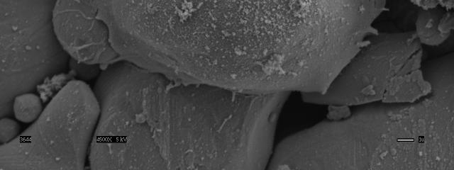
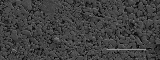
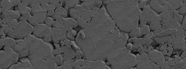
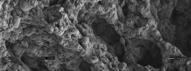
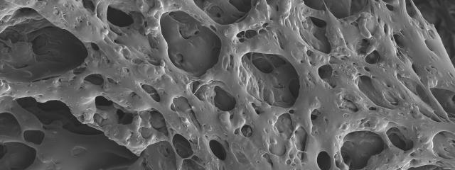
1 Directed Osteoblast Adhesion at Metal Particle Boundaries Ti6Al4V (nanophase) Bar = 1 m. Ti6Al4V (conventional) Ti6Al4V (nanophase) Bar = 1 m. Ti6Al4V (conventional)
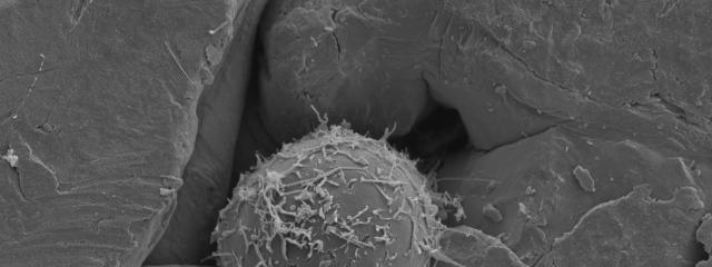
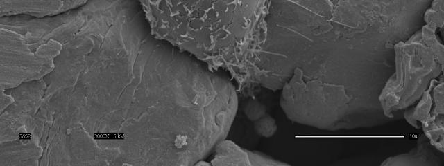

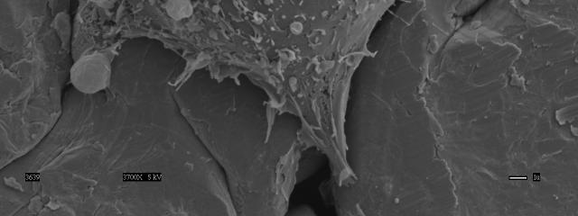
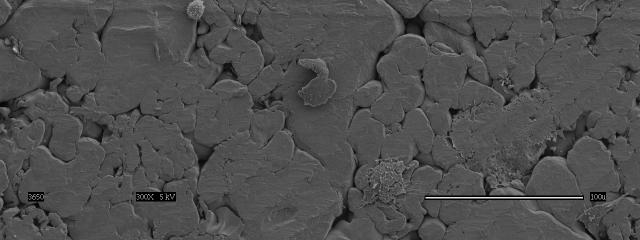

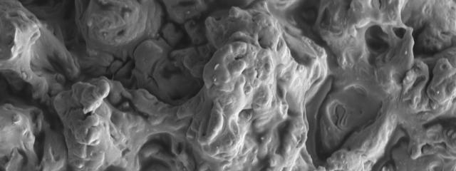
2 Directed Osteoblast Adhesion at Metal Particle Boundaries Ti (nanophase) Ti (nanophase) Bar = 1 m. Bar = 1 m. Ti (conventional) Ti (conventional)
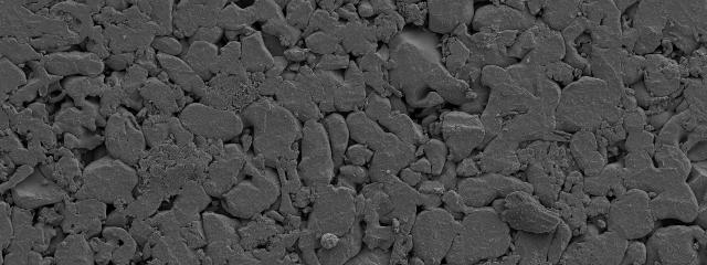
3 Enhanced Osteoblast Adhesion on Nanophase CoCrMo Cell Density (cells/sq.cm) * 5 Wrought Ti (reference) CoCrMo Nanophase CoCrMo Conventional Glass (reference) Time = 1 hour. Values are mean +/- SEM; n = 3; * p <.1 (compared to respective cell adhesion on conventional CoCrMo).
4 Enhanced Adhesion Translates into Increased Subsequent Functions Stages of Osteoblast Differentiation Proliferation and extracellular matrix synthesis OSTEOBLAST PROLIFERATION Extracellular matrix development and maturation OSTEOBLAST DIFFERENTIATION Synthesis of : Type I collagen Fibronectin Vitronectin Extracellular matrix mineralization Synthesis of : Osteopontin Alkaline phosphatase Collagenase 12 Synthesis of : Osteocalcin Bone sialoprotein 21 Days in Culture T. J. Webster, in Advances in Chemical Engineering Vol. 27, Academic Press, NY, pgs , 21.
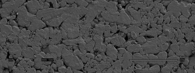
5 Decreased Fibroblast Adhesion on Nanophase Ti and Ti6Al4V 25 Cell Density (cells/sq.cm) 2 15 * 1 * 5 Wrought Ti (reference) Nanophase Ti Nanophase Ti6Al4V Conventional Conventional Ti Ti6Al4V Glass (reference) Time = 1 hour. Values are mean +/- SEM; n = 3; * p <.1 (compared to respective cell adhesion on conventional Ti or Ti6Al4V).
6 Decreased Fibroblast Adhesion on Nanophase CoCrMo Cell Density (cells/sq.cm) * 5 Wrought Ti (reference) CoCrMo Nanophase CoCrMo Conventional Glass (reference) Time = 1 hour. Values are mean +/- SEM; n = 3; * p <.1 (compared to respective cell adhesion on conventional CoCrMo).
7 Anodized Titanium PROCEDURES: Pretreatment: chemical polishing using HF/HNO3 mixture Anodization:.5 or 1.5%HF Voltage: 2V Time: 2 min Rinse and dry Clean: acetone and ethanol Sketch map of anodization system Sterilize


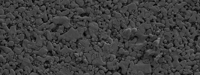
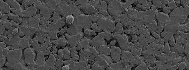
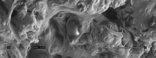
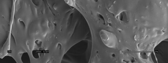
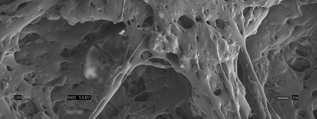
8 Increased Osteoblast Functions on Anodized Ti Unanodized Ti Bar = 1 micron Anodized Ti 1.5% HF treatment Nanotube Anodized Ti.5% HF treatment Nano-particles
9 PART I (cont.): BONE Anticancer Implants Se (Micron) Se (Nano) Se (Sub-Micron) Se (Nano) Bar = 1 micron
: Development of New Entheses (Collaboration")

 -Metal implant with")

10 PART I (cont.): Development of New Entheses (Collaboration with DePuy Orthopedics) - Cell modulating layer - Bioactive uncalcified zone Bioactive region - Calcified zone - Nanomaterial Mechanical Interlock to Implant (LPS) -Metal implant with porous surface UF T-Tidemark; CF B-Bone; CF-Calcified fibrocartilage; T Successful Regeneration of Entheses Implant UF-Uncalcified fibrocatrilage

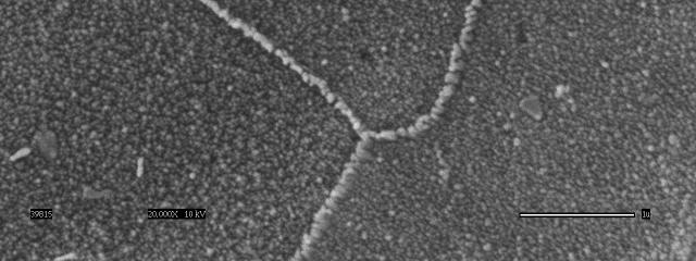
11 Summary for Bone Applications Compared to conventional formulations, nanophase: microns microns microns Conventional Grain Size microns microns microns spherical particle size ceramics, fiber particle size ceramics, carbon nanofibers/nanotubes, polymer composites containing nanophase particles, polymer molds of nanophase materials, organic nanotubes, chemically-treated metals, and spherical particle size metals, Nanophase Grain Size increase bone tissue regeneration. T. J. Webster, in Advances in Chemical Engineering Vol. 27, Academic Press, NY, pgs , 21.
12 PART II Cartilage: Nanostructured Polymers
1 Wear resistant Shock absorbent Distributes applied load throughout the bone 1 http://www.soarmedical.com/medical-library/knee/anatomy/ Armstrong, C.G. et al.")
13 The Problem: Current Cartilage Implant Failures 1 4 mm in thickness Provides a smooth joint lubrication (μ.2)1 Wear resistant Shock absorbent Distributes applied load throughout the bone 1 Armstrong, C.G. et al. Scientific Foundations of Orthopaedics and Traumatology. London: William Heinemann, 198:


14 The Problem: Current Cartilage Implant Failures Bone Articular cartilage Direct trauma Degeneration due to ACL tear Arthritis Bone Articular cartilage Degeneration osteochondral defect articular cartilage defect LaPrade R, Konowalchuk B, Fritts H, Wentorf F. The physician and sportsmedicine, 21;29: cular_cartilage.php


: ): Basic")

15 Cartilage Tissue: Another Nano-structured Tissue Fibrous Extracellular Matrix Water, collagen type II, proteoglycans, other proteins Chondrocytes Collagen fiber architecture Mow VC, Proctor CS, Kelly MA, in Nordin M, Frankel VH (eds (eds): ): Basic Biomechanics of the Musculoskeletal System, ed 2. Philadelphia, Lea & Febiger Febiger,, 1989, pp


16 Increased Functions of Chondrocytes on Nano-structured Polymers Bar = 1 microns Conventional PLGA Nano-structured PLGA Conventional PLGA Nano-structured PLGA
17 PART III Vascular: Nanostructured Polymers


18 The Problem: Current Vascular Implant Failures Vascular Disease One of the leading causes of death in the US Peripheral and coronary artery diseases (hardening of the arteries) Affects approximately 58 million people Source for pictures:
19 Vascular Tissue: Another Nano-structured Tissue
20 Attempts to Improve Vascular Tissue Engineering Scaffolds Reduction of PGA fiber size has been observed from A (more than 1 microns) to C (less than 1 microns) after treatment with 1N NaOH for 1 minutes*. Bar = 1 m hydrolysis R1COOR2 Ester group R1COOH + R2OH Carboxylic acid * Source: Gao J et al., J. Biomed. Mat. Res. 42(3): , Alkanol