Applications of self-assembling peptides in controlled drug delivery
|
|
|
- Berenice Hood
- 5 years ago
- Views:
Transcription
1 Applications of self-assembling peptides in controlled drug delivery Sotirios Koutsopoulos, Ph.D. Problems associated with drug administration Drug concentration in blood C toxic C effective Time 1
2 The importance of Control: Bio-compatible Non-toxic, non-immunogenic Bio-degradable Easy to handle Protect the cargo Deliver small, large, hydrophilic and hydrophobic molecules Programmed dose and time release for as long as it is required Flexibility & patient compliance Reduced risk of side effects from overdose The importance of Control: Most common systems 1. Electronic devices e.g., insulin pumps 2. Particle- and matrix-based systems e.g. polymers or inorganic materials 3. Liposomes World drug delivery market: $17-29 billion for 26 $33-67 billion for 29 $65-97 billion for 213 2
 Controller (computer")
 Poly-ethylene glycol (PEG)")

3 The importance of Control: 1. Devices First controlled drug delivery systems were built in the 196s Insulin reservoir (like a regular syringe) Controller (computer chip) Glucose sensor Infusion device (pump) to deliver insulin to the body through a needle The importance of Control: 2. Polymer micro- & nano-particles, matrices, and hydrogels Poly-lactic-co-glycolic acid (PLGA) Poly-ethylene glycol (PEG) Poly-phosphoesters Poly-vinyl alcohol (PVA) Poly-ethylene oxide (PEO). Clinical studies show: Immune reaction to polymer surfaces Degradation products may be toxic Often toxic initiators are used Not very efficient for protein delivery Nanoparticles microparticles Composite polymer materials Micelles Colloids Nanogels Emulsions Pluronic acid micelles (PEO/PPO) Thin films Polymer brushes Hydrogels 3


")
")

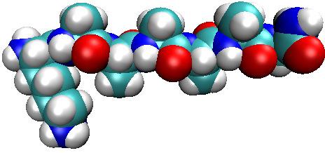
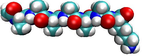
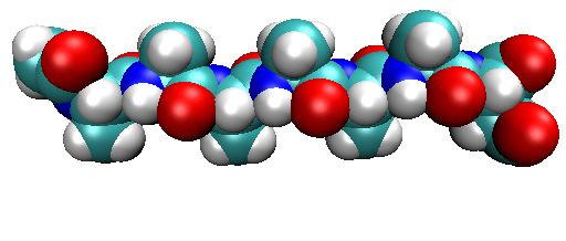
4 The importance of Control: 3. Liposomes PEG protective layer Antibody Associated with cell toxicity effects Immune response in some patients Research started in late 196s Water soluble drug Lipid bilayer Not many unique liposome-based drugs (most contain phosphatidylcholine) Doxil (1993, ovarian cancer, myeloma) Lipid soluble poor industrial reproducibility drug Self-assembling peptides 5-7 nm aa 2.5 nm 5-7 aa 1-cm 1-nm 4


5 Hydrogel consisting of self-assembling peptides (RADA-RADA-RADA-RADA) At physiological conditions.15 M NaCl & 3 < ph < 8 Liquid Gel Hydrogel consisting of self-assembling peptides Water solution Drugs Tissue specific injectable drug release system 5
.5 % w/v 1 cm Release of model drugs through peptide hydrogels Absorbance.14.12.1.8.6.4.2 1-15min 1-3min 1-45min 1-1.")
6 Release of model drugs through peptide hydrogels Phenol Red Bromophenol blue 8-hydroxypyrene- 1,3,6-trisulfonic acid trisodium salt (3-PSA) Phenol1,3,6,8- pyrenetetrasulfonic acid tetrasodium salt (4-PSA).5 % w/v 1 cm Release of model drugs through peptide hydrogels Absorbance min 1-3min 1-45min 1-1.5hrs 1-3hrs 1-6hrs 1-9hrs 1-12hrs 1-24hrs 1-48hrs 1-96hrs 1-168hrs Wavelength (nm).5 % w/v 5 mm Measure diffusion for 1 week 6
7 Release of model drugs through peptide hydrogels Slow release depending on charge and MW of the model drug Release of model drugs through peptide hydrogels r h =.9 nm r h = 1. nm Release depending on charge of the model drug 7


8 Release of model drugs through peptide hydrogels r h =.8 nm r h = 1.1 nm Release due to specific interactions of molecules with nanofibers Release of model drugs 8
9 Protein drug delivery FDA has approved >2 proteins for therapeutic uses In 214, protein drugs returned a revenue of ca. $7 billion Large and unstable molecules (structure held by weak forces) Easily damaged at mild storage conditions Rapidly eliminated by the body Some are difficult to obtain in large quantities Common administration routes are not suitable for proteins Oral Proteins/enzymes are degraded in the stomach or they are not absorbed through the intestines Skin Low permeability of large molecules Inhalation Protein delivery through the lungs is difficult (e.g., Exubera) Injection, subcutaneous or intravenous Proteins are cleared from circulation (frequent injections) 9
10 Controlled release of proteins Study protein delivery through peptide hydrogels using proteins that: are well characterized and widely used have a wide range of MW and pi have therapeutic interest Lysozyme MW 14.7 kda pi 11. Trypsin inhibitor MW 2.1 kda pi 4.6 BSA MW 66. kda pi 5.3 Human IgG MW 145. kda pi 7.2 Fluorescence Correlation Spectroscopy (FCS) High sensitivity and resolution (time/spatial) Detect molecules inside the hydrogel & in the supernatant Measure diffusion coefficients and concentration inside and outside the gel laser beam 1 µm.4 µm Detection volume (1 μm x 1 μm x 2 μm) 2 µm Volume of detection is 1.5 fl 1


 4 hydrogel.")


 Slow")

11 Protein release experiment PBS Water solution Protein PBS Gel + Protein Protein release through (RADA) 4 hydrogel M t /M.4 Lysozyme.2 Trypsin inhibitor BSA IgG Time (hours) Slow release depending primarily on the MW of the protein 11


 3.6 % (RADA) 4 1.")
 8 M t /M.6.4.2.")
12 Protein release through (RADA) 4 hydrogel % M t /M % Time (days) 1.5 % Protein release through peptide hydrogels 1..8 (RADA) 4 1. % (KLDL) 3.6 % (RADA) 4 1. % (core) + (KLDL) 3.6 % (shell) 8 M t /M Time (days) (RADA) 4 + (KLDL) 3 12


 -4-8")
 13")
13 Protein release through peptide hydrogels Question: What is the state of the proteins after being released from the hydrogels? Study the released proteins: Spectroscopy (CD, Fluorescence) Activity tests, bioassays Protein release through peptide hydrogels: CD spectroscopy 4 [θ ] x 1-4 (deg cm 2 mol -1 ) -4-8 Trypsin Inhibitor BSA Lysozyme IgG Wavelength (nm) 13

 Protein release")


14 Protein release through peptide hydrogels: Fluorescence Fluorescence Intensity (a.u.) Trypsin Inhibitor BSA Lysozyme IgG Wavelength (nm) Protein release through peptide hydrogels: Biological activity Normalized activity of Lysozyme Native Lsz Released Normalized acitivity of Trypsin 8 Native Trypsin Inhibitor Released Trypsin Inhibitor conc. of Trypsin Inhibitor (mg/ml) Measure the hydrolysis of the cell membrane of M. lysodeiktikus Measure the suppression of trypsin activity 14
15 Protein release through peptide hydrogels: Biological activity Measure binding efficiency of monoclonal IgG against antigen Protein release through peptide hydrogels: Biological activity Native or released antibodies Immobilized antigen -df (Hz) -df (Hz) IgG injection IgG injection Native IgG Hydrogel released IgG 4. µg/ml 2. µg/ml 1. µg/ml.5 µg/ml 4. µg/ml 2. µg/ml 1. µg/ml.5 µg/ml Time (s) 15


16 Smart hydrogel material for controlled release Lipid-like self assembling peptides 2.6 nm 2.4 nm ac-a 6 K-NH 2 (acetyl-aaaaaak-conh 2 ) KA 6 -NH 2 (KAAAAAA-CONH 2 ) 2.6 nm 2.4 nm ac-a 6 D-OH (acetyl-aaaaaad-cooh) DA 6 -COOH (DAAAAAA-COOH) 16

 Count number 2 15 1 5 1")

 17")
17 Lipid-like self assembling peptide nanovesicles: AFM ac-a 6 K-CONH 2 ac-a 6 K-CONH 2 KA 6 -CONH 2 Count number Nanovesicle size (nm) Count number Nanovesicle size (nm) Lipid-like self assembling peptide nanovesicles: AFM ac-a 6 D-COOH DA 6 -COOH Count number Nanovesicle size (nm) Count number Nanovesicle size (nm) 17
18 Lipid-like self assembling peptide nanovesicles: Carboxyfluorescein encapsulation and release % Cumulative CF Release ac-a 6 K-CONH 2 2 KA 6 -CONH 2 ac-a 6 D-COOH DA 6 -COOH Time (hours) Lipid-like self assembling peptide nanovesicles: Nile Red integration in the peptide bilayer % Cumulative Nile red release ac-a 6 K-CONH 2 KA 6 -CONH 2 2 ac-a 6 D-COOH DA 6 -COOH Time (hours) Fluorescence intensity Wavelength (nm) 18
19 Lipid-like self assembling peptides: Epithelial cell proliferation Cells/well Control.2 mg/ml ac-a 6 K-CONH 2 1. mg/ml ac-a 6 K-CONH 2.2 mg/ml KA 6 -CONH 2 1. mg/ml KA 6 -CONH 2.2 mg/ml ac-a 6 D-COOH 1. mg/ml ac-a 6 D-COOH.2 mg/ml DA 6 -COOH 1. mg/ml DA 6 -COOH 3 h 24 h 48 h Cumulative FITC-dextran transport (pmol/cm 2 ) Lipid-like self assembling peptides: Drug absorption Caco-2 cell monolayer model and rat intestine (everted sacs) are used to predict drug absorption (oral delivery) 14 Control ac-a 12 6 K-CONH 2 KA 6 -CONH 2 1 ac-a 6 D-COOH DA 8 6 -COOH Time (min) Caco-2 cell monolayer FD-4 permeability (nmol/cm 2 ) FD-4 FD-4 + A 6 D 5% increase FD-4 FD-4 + A 6 D 3 min 12 min Rat intestine 19
20 Conclusions Injectable peptide hydrogel Release small molecules & proteins of broad MW (e.g., Lsz, IgG) Hydrogel contains up to 99.5% water, active compound loading depends on the solubility of the drug Release kinetics depend on hydrogel density Functionality assays show that released proteins are active Lipid-like peptide nanovesicles Release of hydrophilic and hydrophobic drugs Release kinetics depend on amino acid sequence and drug properties Peptides are not cytotoxic Increase transport through the epithelial layer Self-assembling peptides are biocompatible, biodegradable, non-toxic, nonimmunogenic, transparent which are ideal for biomedical applications 2