biologics,llc The Practitioner s Solution for Autologous Biologics since 1999
|
|
|
- Dominic McDowell
- 5 years ago
- Views:
Transcription
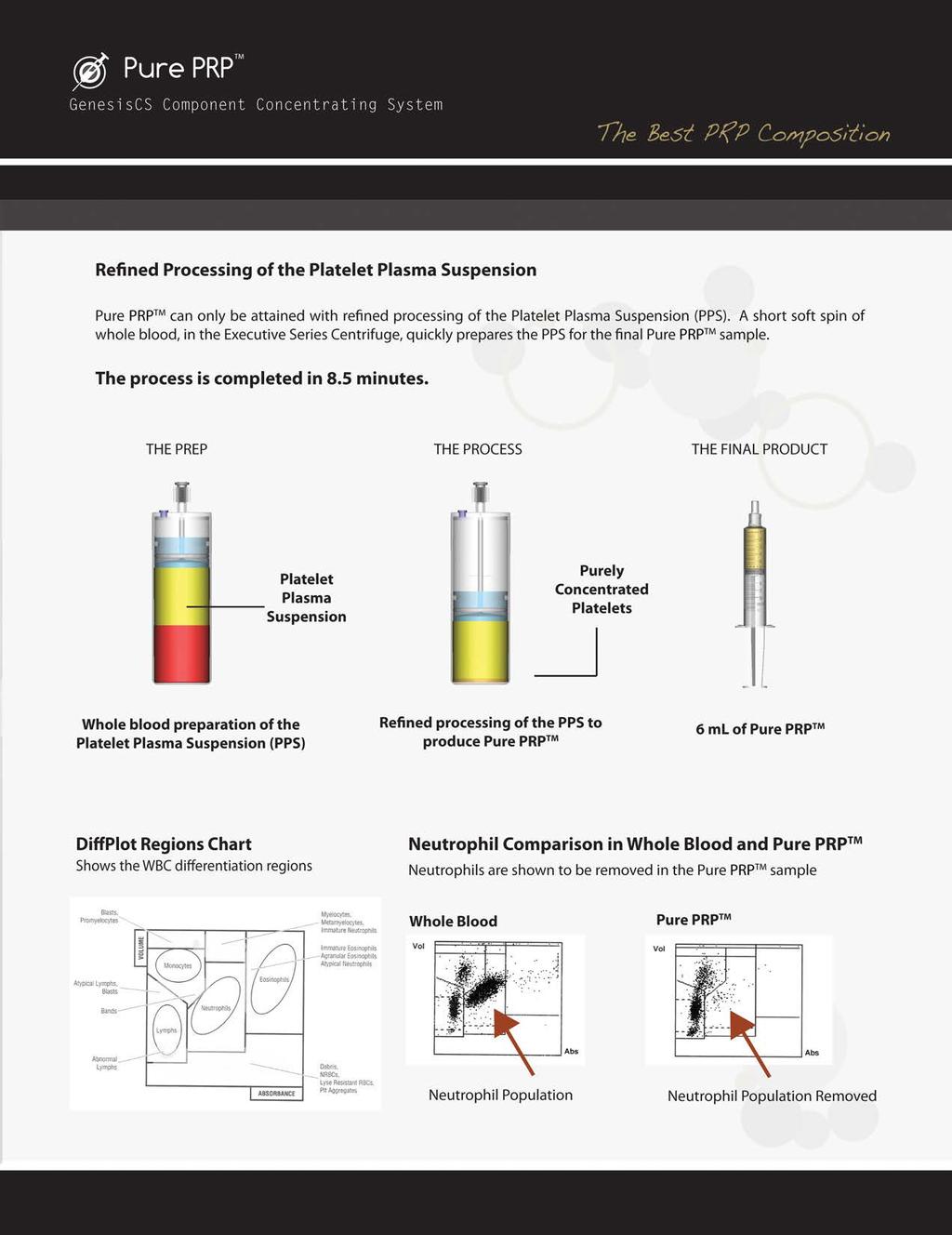
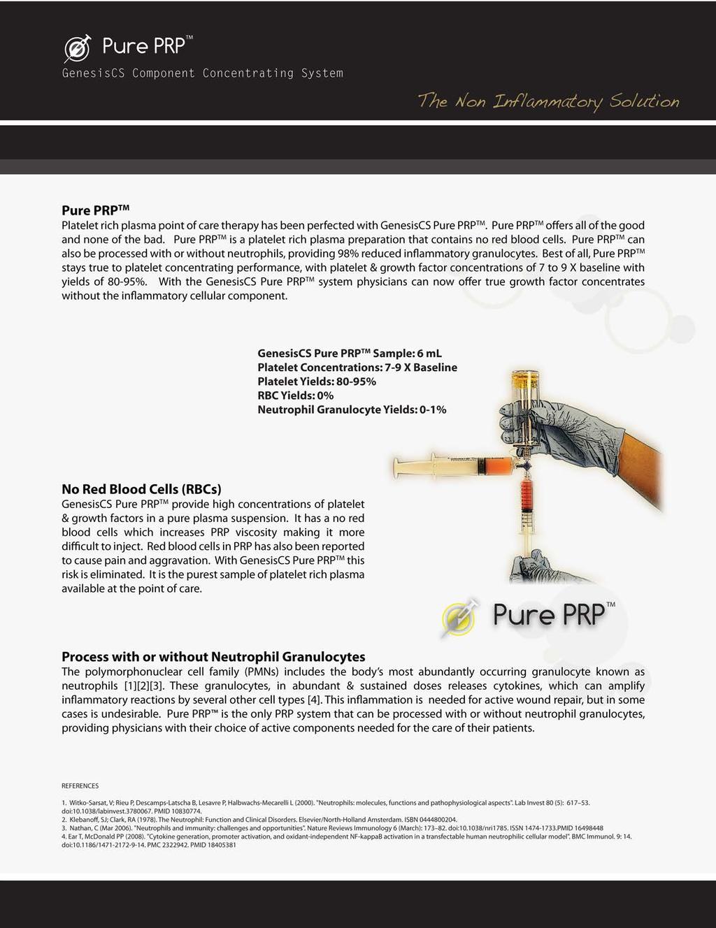
1 accellerated biologics,llc The Practitioner s Solution for Autologous Biologics since 1999 accellerated biologics, LLC is a nationwide medical distribution company focused on consulting and providing the physician, their staff and community with quality information and products related to Platelet Rich Plasma (PRP), Pure PRP, Bone Marrow Aspirate/Adipose Tissue (Stem Cells), Photo Modulation (PAPRP), Nutraceuticals and AmnioFix Sports Injectable.
2 TABLE OF CONTENTS PURE PRP Platelet Rich Plasma (PRP) Bone Marrow Concentrate (BMC) Analysis of EmCyte Corporation Concentrating Systems PhotoActivated Platelet Rich Plasma (PAPRP) accellerated biologics PRP & PAPRP Products References
3 2
4 3
5 4
6 5
7 6
8 7
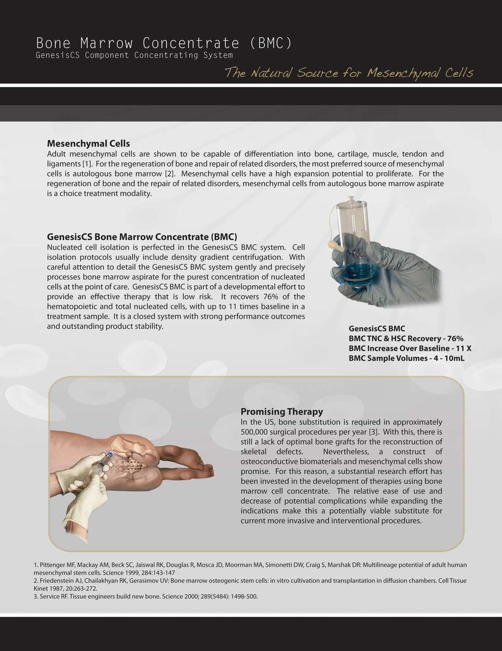
9 Analysis of EmCyte Corporation Concentrating Systems An independent review of pre-clinical performance data Principle Investigator(s): Dr. Robert Mandle Ph.D. 1. Children s Hospital, Harvard Medical School 2. The CBR Institute for Biomedical Research 3. Biosciences Research Associates, Boston, MA CONTENTS: Analysis of GenesisCS For Concentration of Human Bone Marrow Aspirate 60mL Point of Care Preparation of Autologous Platelet Products for Regenerative Medicine: Comparison of Four Market Leading Commercial Methods Analysis of GenesisCS Pure PRP Concentrating System for Platelet Concentration with RBC & Neutrophil Reduction (Data prepared internally at EmCyte Corporation) Nuclear Cell Count Analysis of Human Adipose Tissue Concentrate Processed with the Secquire 2 Concentrating Device In Vitro Characteristics of Platelets Collected with the GenesisCS Concentrating System
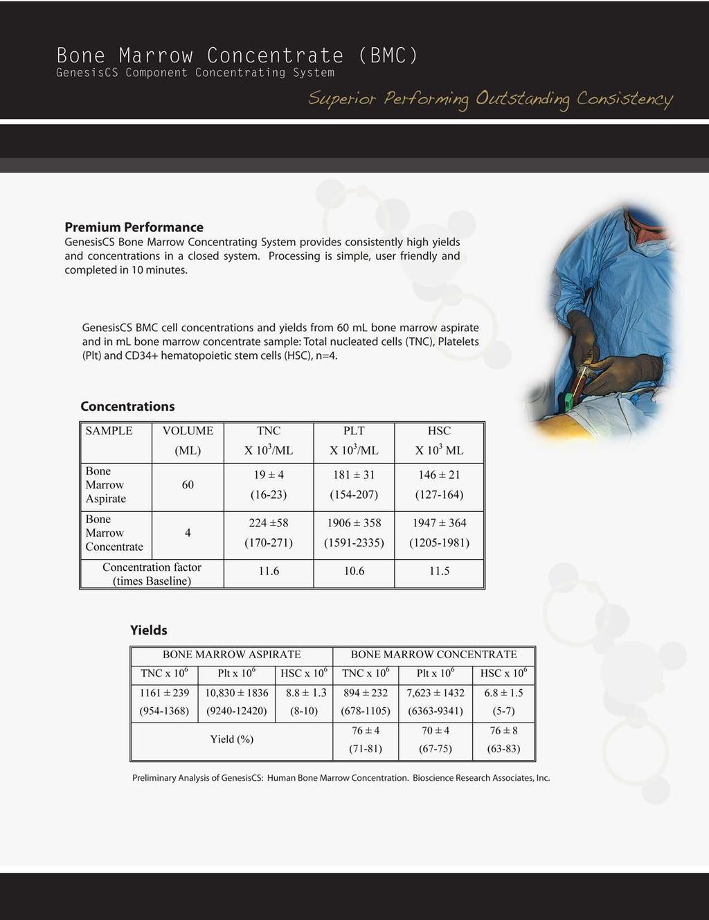
10 Analysis of GenesisCS For Concentration of Human Bone Marrow Aspirate 60mL IN VITRO TESTING RESEARCH STUDY PLAN: Title: Evaluation of GenesisCS with Bone Marrow Aspirate Revision: 2 Revision Date: January 12, 2012 TEST OBJECTIVE: Preliminary evaluation of GenesisCS for concentration of human bone marrow aspirate. Preclinical and clinical studies have suggested the benefit of using concentrated autologous bone marrow aspirate in bone repair, myocardial infarct and peripheral vascular disease. Bone marrow aspirate is often not sufficient for clinical efficacy in the absence of concentration1,2. This report represents results from an evaluation of GenesisCS device for the concentration of human bone marrow-derived stem cells. Sixty ml of human bone marrow aspirate were concentrated to approximately 6 ml with the GenesisCS. Samples of the bone marrow aspirate (BMA) and resulting bone marrow concentrate (BMC) were analyzed for Total Nucleated Cells (TNC), Platelets (PLT), and CD34 positive Hematopoietic Stem Cells (HSC). Yield calculation were done for TNC, PLT and HCS. EXPERIMENTAL DESIGN: Donor bone marrow samples, approximately 120mL, collected from two sites of the iliac crest, were obtained from Poetics (Cambrex). Bone marrow samples were collected in units/ml of heparin. Processing and all testing were initiated within 24 hr of collection. After obtaining a 1mL start sample from a well mixed transfer pack of BMA, two 60 ml syringes were filled with approximately 60 ml of marrow aspirate and the volumes recorded. GenesisCS disposables were filled from these syringes through the luer-lock fitting at a fill rate of approximately 1 ml/sec. Disposables were centrifuged at 2400 rpm (1020 x g) for 12 min. Two independent centrifuge runs were performed for each donor BMA from two separate donors collected on separate days for a total of four runs. Following centrifugation, the plasma layer was removed, by lowering the collection head to within 2-5mm above the buffy coat layer which contained the concentrated nucleated cells and platelets. Next, 2 ml of the remaining plasma and an additional 4 ml of the buffy coat was removed (4 ml following the first flash of RBC observed in the suction tubing above the collection device) for a total of 6mL of BMC. Analysis of BMA and BMC consisted of: Complete blood counts utilizing a Medtronic parameter hematology analyzer with extended platelet range. Cytometric analysis of CD34 positive hematopoietic stem/progenitor cells Manual differential counts on BMA and BMC samples. Yield of nucleated cells, platelets and CD34 positive HSCs were calculated for bone marrow concentrates RESULTS: Characterization of GenesisCS BMC: The TNC values from the hematology analyzer for pre-sample (BMA) and for product (BMC) and the calculated concentration over baseline values are shown in Table I. Table I: Total nucleated cells (2 donors with duplicate runs) Volume Total Nuclear Total Cells x 10 3 Concentration /µl Above Baseline Bone Marrow Aspirate 60mL x Bone Marrow Concentrate 4mL x Bone Marrow Concentrate 6mL x Table II lists the calculated total number of cells (volume x concentration) in BMA and BMC. TNC and PLT counts represent the values from the hematology analyzer times the volumes of BMA or BMC. HSC numbers are calculated from the percent of CD34+ cells gated with CD45+ events times the number of WBC (TNC minus nucleated red blood cells). Table II: The recovery of TNC, Plt and CD34+ HCS. Total cell numbers ± SD (yield percentages) BONE MARROW ASPIRATE BONE MARROW CONCENTRATE TNC x 10 6 PLT x 10 6 HSC x 10 6 TNC x 10 6 PLT x 10 6 HSC x ± 239 ( ) 10,830 ± 1836 ( ) Yield (%) 8.8± 1.3 (8-10) 894 ± 232 ( ) 76 ± 4 (71-81) 7,623 ± 1432 ( ) 70 ± 4 (67-75) 6.8 ± 1.5 (5-7) 76 ± 8 (63-83) 9

11 Figure 1. Recovery of TNC, Plt and CD34+ HCS DISCUSSION: The percent of TNC, Plt, and CD34+ HSC were calculated by dividing the total number of cells recovered in the BMC by the total number present in 60ml of BMA and are represented as mean plus standard deviation for 2 donors with duplicate runs. CONCLUSION: The product (BMC) yields were 76% for TNCs and CD34+ HSC. These yields are consistent with other point of care bone marrow concentrating devices. Platelet yields in the BMC averaged 70% and the product Hematocrit averaged 31.6% with a range of 31-40% (data not shown). Hematocrit can be adjusted by including more or less of the plasma layer during the collection of BMC. Variation within donor samples appears to less than between donors. Between donor variation will need to be determined in a larger study. However, the data from this preliminary evaluation with two donors run in duplicate, is very encouraging. 10 3
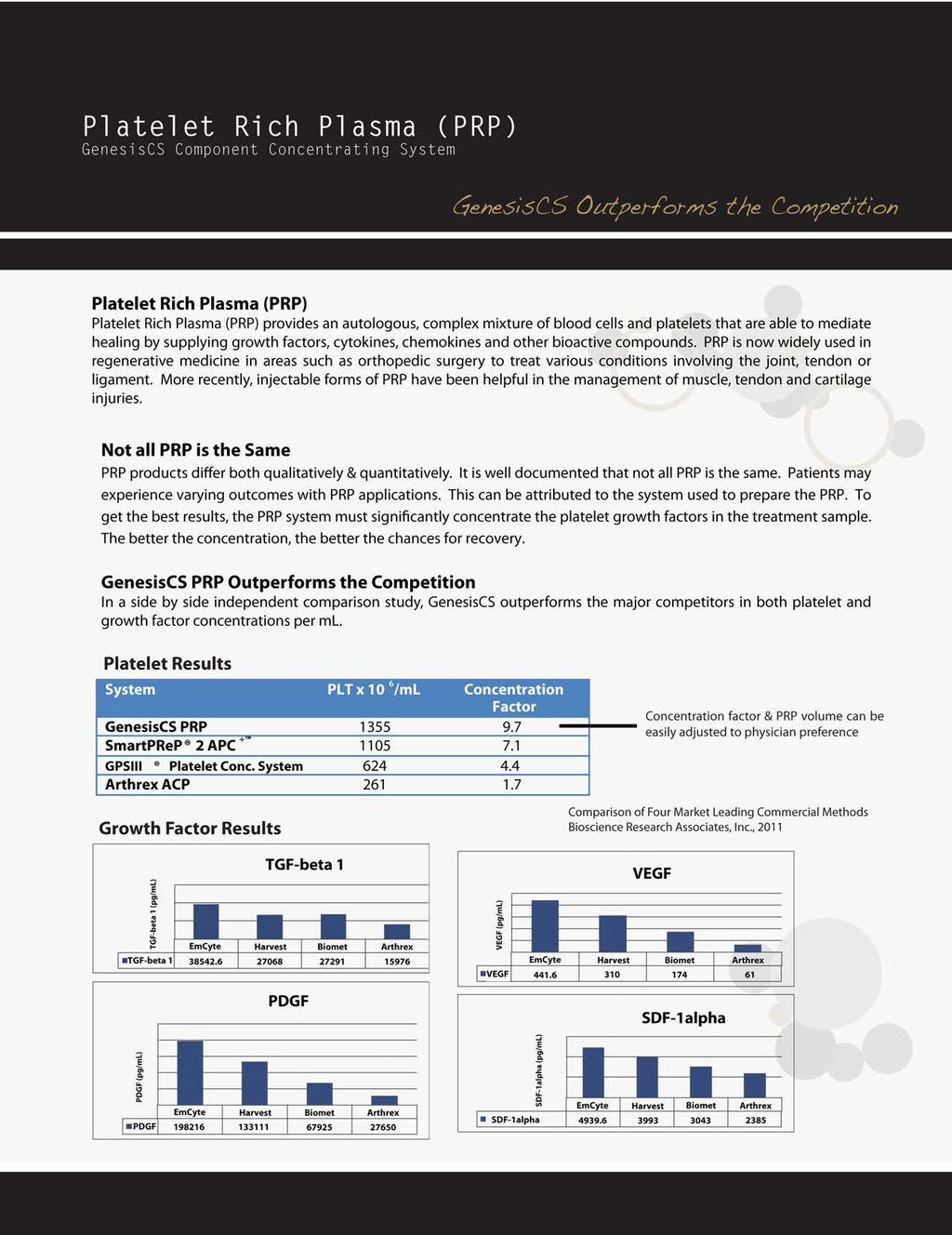
12 Point of Care Preparation of Autologous Platelet Products for Regenerative Medicine: Comparison of Four Market Leading Commercial Methods IN VITRO TESTING TEST OBJECTIVE: Platelet Rich Plasma (PRP) provides an autologous, complex mixture of blood cells and platelets that are able to mediate healing by supplying growth factors, cytokines, chemokines and other bioactive compounds. PRP technology that was initially used in dentistry and maxillofacial surgery to improve bone healing, is safe and capable of promoting and of accelerating the healing processes. PRP is now widely used in regenerative medicine including orthopedic surgery involving shoulder, hip and knee anterior cruciate ligament (ACL) reconstruction and meniscus repair. More recently, injectable forms of PRP have been helpful in the management of muscle, tendon and cartilage injuries. PRP products differ both qualitatively, e.g. the presence of absence of leukocytes, and quantitatively, including platelet concentration leukocyte differential and the concentration of bioactive compounds. The purpose of this study was to compare key parameters of the PRP product from four commercial point-of-care technologies using paired samples from 3 normal donors. EXPERIMENTAL DESIGN: Donor Selection: Blood was obtained from 3 normal donors following informed consent. All blood collection protocols and donors met requirements of the American Association of Blood Banks (AABB) and the United States Food and Drug Administration (FDA) Center for Biologics Evaluation and Research (CBER). The phlebotomy protocol, including informed consent was approved by the New England Institutional Review Board and was conducted in accordance with the Helsinki Declaration of 1975 as revised in Blood was drawn from the Median-cubital vein using a 16g apheresis needle and sliconized cannula (Reference Number 4R2441, Fenwal). Blood was drawn into transfer packs with the required ACD-A anticoagulant to blood ratio as suggested by each device manufacturer (See Table I). Point of Care PRP Systems: Four of the leading commercial point-of-care systems for autologous PRP production were tested with paired samples such that blood from each of three donors was tested in duplicate runs with each system. Table I lists the test device names, distributers, blood volume processed and the amount of anticoagulant used. Device Name Manufacturer Process Volume GenesisCS PRP SmartPReP 2 APC + GPSIII Platelet Concentrating System Arthrex ACP EmCyte Corporation Harvest Technologies, Corp. Biomet Biologics Arthrex Orthobiologics ACD-A: ml Blood Lot Number 60 ml 5: ml 6: ml 5: ml 1: Arthrex ACP was filled directly from the transfer pack; all others were loaded form a 60 ml syringe that was drawn from the transfer pack. Baseline samples were drawn from each transfer pack. Two device disposables were processed for each donor. Complete blood count (CBC) analysis was done on a Medonic CA 620 Hematology Analyzer. Platelet relative concentration and platelet yield were calculated by comparison to baseline unprocessed whole blood samples. Growth factors PDGF A/B, VEGF, SDF-α and TGF-β1 were measured by quantitative ELSAs (R&D Systems Quantikine kits) in platelet releasates prepared from PRP by addition of 1 part thrombin (1000U/ml in 10% CaCL2 ) per 10 parts PRP. RESULTS: The baseline WBC, Platelet and hematocrit values for three donors are shown in Tables II & III for samples collected in 8.3% (5:55 ratio) ACD-A anticoagulant and 10% (6:45 ratio) ACD-A. Table II. Baseline hematology data for ACD-A: Blood ratio 5:55 Donor WBC x 10 6 /ml PLT x 10 6 /ml HTC % Donor Donor Donor Table III. Baseline hematology data for ACD-A: Blood ratio 6:54 Donor WBC x 10 6 /ml PLT x 10 6 /ml HTC % Donor Donor Donor Duplicate PRP samples were produced, for each donor, on each of the four systems tested. The average WBC, platelet and hematocrit values are shown in Table IV for 6 runs on each system. 11 4
13 Table IV. Hematology data for PRP products System WBC x 10 6 /ml PLT x 10 6 /ml Concentration factor HTC % GenesisCS PRP SmartPReP APC + GPSIII Platelet Conc. System Arthrex ACP The average volume of PRP and the platelet yield were calculated for each PRP system. The platelet yields were measured by: YYYYYYYYYY =!"#!"!!!"!!"#$%&!"#$%&'%!!"#$%&&!"#$%& Where PLTPRP and PLTstart are the platelet counts in the PRP sample and baseline sample respectively. Platelet yields are the average of 6 PRP production runs with three donors. Four statistics of platelet yield are shown in Table V: mean value, plus the coefficient of variation about the mean, median value, and minimum and maximum values in the range of yield values. Table V. PRP product volumes and PLT yields System PRP vol. (ml) Platelet Yield Range GenesisCS Platelet 5 70%-96% Concentrating System SmartPReP 2 APC %-89% GPSIII Platelet %-82% Concentrating System Arthrex ACP %-85% Thrombin-generated releasates prepared from the PRP product of each system were analyzed with ELISA for PDGF-A/B, TGF-β1, VEGF, and SDF-1α. The relative concentrations of these growth factors and chemokine are shown in Figure1, 2, 3, and 4 respectively. Figure 1. PDGF-A/B Comparison in PRP releasate PDGF (pg/ml) PDGF EMCYTE Harvest Biomet Arthrex Figure 2. TGF-beta 1 Comparison in PRP releasate TGF- β1 (pg/ml) TGF- beta EMCYTE Harvest Biomet Arthrex Figure 3. VEGF Comparison in PRP releasate VEGF (pg/ml) VEGF EMCYTE Harvest Biomet Arthrex Figure 4. SDF-1α Comparison in PRP releasate SDF- 1alpha SDF - 1α (pg/ml) EMCYTE Harvest Biomet Arthrex 12 5
14 DISCUSSION Four of the most frequently used point-of-care autologous PRP systems were compared. All four systems are centrifuge based, and with the exception of loading the disposable with anticoagulated blood and harvesting the PRP product, the separations are automated. All systems concentrated platelets and WBC to varying degrees. Part of the variance was related to efficiency of platelet recovery and part was due to the volume of the PRP product produced. PRP volume collected can be adjusted during collection continuously on the Genesis system and in discrete increments of 10, 7 ml on the Harvest APC system. The Biomet GPSIII system is essentially fixed in PRP volume all though all PRP could be further diluted with the PRP fraction. The Arthrex ACP system contained the lowest concentration of WBC and platelets, with a mean platelet concentration of 70% greater than baseline levels. With respect to efficiency of platelet recovery, the GenesisCS and systems excelled with an average of 80% platelet yield across 3 donors. The highest yields were seen with the Genesis system; however the Smart PreP2 APC system was slightly more consistent between donors as reflected in the greater difference in sample median vs. sample mean in Table V. All systems recovered viable platelets, with an average process dependent platelet activation of approximately 10%. The Biomet GPSIII system demonstrated the least process dependent activation, but only recovered approximately half of the platelets. The measured concentration of growth factors, PDGF-A/B, TGF-β1, VEGF, and SDF-1α were all highest in the PRP produced with the GenesisCS system. The releasate concentrations of PDGF-A/B, TGF-β1 and to a lesser extent SDF-1α, correlate with the platelet count in the PRP. VEGF concentrations are influenced by both platelet and WBC concentrations. The efficiency of platelet and WBC recovery, the ability of the recovered platelets to retract the thrombin clot and ration of PRP volume to processed volume affect these results. The Arthrex ACP system despite only a 4mL PRP volume, only processed 9mL of blood vs. 54 or 56 ml for the other systems. In addition the PRP from the Arthrex ACP system did not have significant concentrations of platelets or WBC. There was a large variation in number of RBC in the PRP products across the platforms with GenesisCS> Smart PreP2 APC> GPSIII>ACP. There has been no clinical data concerning adverse events due to RBC contamination in PRP and as the RBC are autologous, there are no antigen cross match or agglutination issues. Furthermore, a typical pooled buffy coat platelet concentrate for transfusion has a hematocrit of approximately 50%. In testing done in our laboratory, we have shown that contaminating RBCs do not activate platelets in PRP. 13 6
15 Analysis of GenesisCS Pure PRP Concentrating System for Platelet Concentration with RBC & Neutrophil Reduction IN VITRO TESTING TEST OBJECTIVE: The use of platelet rich plasma has been documented in many fields. These fields include orthopedics, sports medicine, cosmetic, neurosurgery, maxillofacial surgery, and wound healing. Platelet rich plasma has emerged as a preferred treatment modality because it's a safe, effective, and natural alternative to surgery. Studies suggest that platelets contain growth factors and other mediators that play an important role in the repair of soft tissue and bone. When these bioactive proteins are concentrated they attract a host of other cellular components that work together to quickly and effectively repair damaged tissue. It is well known that not all platelet rich plasma preparations are the same. They vary in growth factor concentrations, white blood cell concentrations, red blood cell concentrations and volume. These components can greatly influence the efficacy of the final preparation. Preparations that contain platelet concentrations of fivefold or greater has been the preferred regimen for many practicing physicians. Physicians are also looking for platelet rich plasma preparations that contain reduced neutrophils and red blood cells. These components are reported to increase pain and inflammation at the injection site. To date, EmCyte Corporation is the only biologic company that has produced a device claiming to effectively concentrate platelets while eliminating up to 99% of the neutrophils and red blood cells at the point of care. In this study the performance of the GenesisCS Pure PRP TM system is carefully evaluated for the key parameters that are most important to physicians, to include platelet concentrations, white blood cell agranulocyte retention, neutrophil elimination, red blood cell elimination and adequate volume samples. EXPERIMENTAL DESIGN: Donor Selection: Blood was obtained from 6 normal donors following informed consent. All blood collection protocols and donors met requirements of the American Association of Blood Banks (AABB) and the United States Food and Drug Administration (FDA) Center for Biologics Evaluation and Research (CBER). The phlebotomy protocol, including informed consent was conducted in accordance with the Helsinki Declaration of 1975 as revised in Blood was drawn from the Median-cubital vein using an 18g apheresis needle and sliconized cannula (Reference Number 4R2441, Fenwal). Fifty (50) ml of blood was drawn from each patient into a 60mL syringe (Terumo Reference # SS-60L) filled with 10mL of sodium citrate anticoagulant (Fenwall Reference# 4B7867Q). Baseline samples were drawn from each syringe prior to processing. The blood samples were processed according to the manufacturer s instruction for processing Pure PRP TM. This required a 3.5 minute spin and a 5 minute spin. At the end of the processing steps approximately 6mL of Pure PRP TM was collected. Complete blood count (CBC) analysis was done on a Beckman Coulter AcT 5 Diff Hematology Analyzer. Platelet relative concentration and platelet yield were calculated by comparison to baseline unprocessed whole blood samples. Total white blood cell and red blood cell relative concentrations were calculated by comparison to the baseline of the unprocessed whole blood samples. The white blood cells were further broken down into its differentials and the relative percentages of the granulocytes and agranulocytes were calculated by comparison to the baseline unprocessed whole blood samples. RESULTS: The hematology comparison data for Pure PRP TM WBC, RBC and Platelet values for the six donors are shown in the following Tables I, II, and III respectively for samples collected in 16% (10:50 ratio) sodium citrate anticoagulant. Pure PRP TM volume is 6mL. Table I. Pure PRP TM 6mL hematology data for WBC from Baseline Donor Baseline WBC x 10 6 /ml Pure PRP TM WBC x 10 6 /ml Pure PRP TM Yields Donor % Donor % Donor % Donor % Donor % Donor % Average % Table II. Pure PRP TM 6mL hematology data for RBC from Baseline Donor Baseline RBC x 10 6 /ul Pure PRP TM RBC x 10 6 /ul Pure PRP TM Yields Donor % Donor % Donor % Donor % Donor % Donor % Average % 14 7

16 Table III. Pure PRP TM 6mL hematology data for Platelet concentrations by comparison to Baseline Donor Baseline Platelets x 10 6 /ml Pure PRP TM Platelets x 10 6 /ml Pure PRP TM Concentrations X Baseline Donor Donor Donor Donor Donor Donor Average The hematology comparison data for Pure PRP TM Neutrophil and Monocyte percentage values for the six donors are shown in the following Tables IV and V respectively for samples collected in 16% (10:50 ratio) sodium citrate anticoagulant. Pure PRP TM volume is 6mL. Figure 1. Figure 1. Pure PRP TM performance yields (6mL sample) Pure PRP 100% 90% 80% 60% 33% 40% 16% 20% Pure PRP 0% 1% 0% Table IV. Pure PRP TM 6mL hematology data for percentage of Neutrophil yields by comparison to Baseline Donor Baseline Neutrophil % Pure PRP TM Neutrophil % Pure PRP TM Yields Donor 1 67% 3.4% 0.51% Donor % 2.3% 0.55% Donor % 8.6% 2.00% Donor % 8% 1.47% Donor % 5% 1.03% Donor % 8.3% 2.06% Average 49.2% 5.9% 1.27% Table V. Pure PRP TM 6mL hematology data for percentage of Monocyte yields by comparison to Baseline Donor Baseline Monocyte % Pure PRP TM Monocyte % Pure PRP TM Yields Donor 1 7.1% 16.6% 23.38% Donor 2 7.7% 11.4% 14.81% Donor 3 8.1% 11.7% 14.44% Donor % 14.2% 13.92% Donor 5 9.8% 16.0% 16.33% Donor % 12.7% 11.65% Average 9.0% 13.8% 15.8% DISCUSSION The GenesisCS Pure PRP TM System presents a viable solution for PRP where the platelets can be retained to a degree that is equal to or better than comparable systems while removing better than 99% of RBCs and better than 98% of neutrophils on average. To date, there has been no other point of care system than have been shown to accomplish these three events in a single process. The system offers a two spin solution that has a total processing time that is still less than the standard average processing time. The system is equipped with a modified piston that traps platelets at the base of the concentrating device after final processing. This data have established that the GenesisCS Pure PRP TM is capable of preparing a platelet concentrate that is significantly stripped of neutrophils and red blood cells. Testing from in vitro studies, intended to evaluate the quality of the performance characteristics have demonstrated that the functional abilities are compatible to those using predicate devices or standard blood bank techniques. The GenesisCS Pure PRP TM system provided consistent concentrated platelet product with predictable yields and concentration factors of the investigated components. The yields were measured by: YYYYYYYYYY =!"#$"%&%'!"#$!%!!!"#$!%!!"#$%&!"#$"%&%'!"#$%&'$!!"#$%&&!"#$%& Where the ComponentPureRP and ComponentBaseline are the cell counts in the PRP sample and baseline sample respectively. The PurePRPvolume and Processvolume are the volumes of the Pure PRP TM sample and the baseline sample respectively. A summary of the performance outcomes for Pure PRP TM is shown in 15 8
17 Nuclear Cell Count Analysis of Human Adipose Tissue Concentrate Processed with the Secquire 2 Concentrating Device IN VITRO TESTING TEST OBJECTIVE: This study evaluated the product produced by the centrifuged-based Secquire-2 Cell Separator. Human adipose tissue was concentrated from lipoaspirate, and the nucleated cell concentration estimated by flow cytometry or fluorescent microscopy as a measure of product qualify. BACKGROUND: Adipose tissue provides a readily accessible source of autologous stem/progenitor cells and proangiogenic pericytes. Typically, the lipoaspirate contains 50% to 75% tumescent fluid. Centrifugation removes the fluid and condenses the buoyant adipose tissue. Concentrates of cellular and extracellular elements in the natural biological scaffolding of adipose tissue may promote wound healing and have applications in regenerative medicine. EXPERIMENTAL DESIGN: Lipoaspirate collection: Harvest of adipose tissue from lower abdomen via lipoaspiration was performed using standard of care, closed syringe method. A multiport infiltrator sterile cannula attached to a cc syringe was used to infiltrate tumescent solution (0.5gm Lidocaine with 1mg epinephrine per 1L of normal saline) into the subdermal fat plane. Adipose tissue suspended in the fluid media provided by the tumescent fluid was withdrawn by applying gentle suction with the syringe. Adipose Concentrate Production: The lipoaspirate was transferred immediately following harvest from the harvesting syringe into a Secquire-2 disposable and centrifuged for 3.5 min at approximately 140 x g according to manufacturer s instructions. Study Outcome Measures: An aliquot of the concentrated fat sample was shipped to BSR laboratories for analysis within 24hr of harvesting. A summary of the methods is listed below Nuclear cell counts and cell viability: The entire sample of adipose concentrate was digested with collagenase enzyme solution and the stromal vascular fraction (SVF) collected by centrifugation. The concentration of the nucleated cells was determined in the SVF by manual counting using a hemocytometer and fluorescent staining or by flow cytometer. Cell counts are reported as the concentration per ml of starting adipose concentrate. Cell phenotype and estimate of adipose derived stem cell concentration: The SVF cells were stained with fluorescent-labeled antibodies to CD45 a pan leukocyte marker) and CD31 a marker found on some white blood cells and on endothelial cells. Total nucleated cells were estimated by inclusion of the nuclear stain Syto-13. The fraction containing Adipose Derived Stem Cells (ASC) was determined by eliminating CD45 positive and CD31 positive cell populations from the nucleated cell population, as a maximum estimate of ASC. In separate experiments, 50% of cells in this fraction are positive for CD105, CD73 and CD90 ASC markers. Cell Viability: Viability was determined by dye exclusion (ethidium bromide homodimer) and with a viability stain (calcein AM ) using a fluorescent microscope. RESULTS: Concentrated adipose samples from nine donors were analyzed. The nucleated cell counts expressed as per ml of starting concentrated adipose sample are shown in Table I. Table I. Nucleated cell counts per ml of sample. Harvest Date Analysis Date Sample ID Cells x 10 5 /ml sample 13 Dec Dec 2011 # Sep Sep 2011 # Sep Sep 2011 # Aug Aug 2011 # Aug Aug 2011 # Aug Aug 2011 # Aug Aug 2011 # Aug Aug 2011 #BSR May May 2011 #BSR
18 The average cell count per ml of concentrated adipose sample was 5 x 10⁵ with a range of 2.4 to 9.8 x 10⁵. Sample # was also analyzed for cell viability. The percent viability of the nucleated cells was 81%. The volumes of lipoaspirate processed and concentrated adipose tissue produced are shown in Table II. Table II. Process volumes Harvest Date Analysis Date Sample ID LA Vol ml Prod Vol ml 13 Dec Dec 2011 # Sep Sep 2011 # Sep Sep 2011 # Aug Aug 2011 # Aug Aug 2011 # Aug Aug 2011 # Aug Aug 2011 # aspirate and concentrated product may not be valid. For these reasons, baseline aspirate samples are not included in the analysis. The nuclear cell number per ml of adipose tissue is variable and highly sensitive to the harvest method. In our experience manual syringe methods produce higher average cell numbers compared to wall vacuum assisted aspiration. The average for these samples, 5.0 x 10⁵ is consistent with our laboratory average of 4.8 x 10⁵ cells per ml of decanted adipose tissue (tumescent fluid removed by allowing fluid to settle below the buoyant fat) from manual method aspiration (N=10) and is higher than our laboratory average of 1.7 x 10⁵ cells per ml of decanted fat from vacuum assisted aspiration (N=20). The average aspirate volume was reduced by 2.1 fold (range ) for seven samples. Phenotypic analysis of SVF cells by flow cytometer is shown in Table III. The fraction containing the ASC is calculated by eliminating endothelial and white blood cells populations. Table III. Percentage of total nuclear cells in the ASC fraction. Harvest Date Analysis Date Sample ID ASC% 30 Aug Aug 2011 # % 17 Aug Aug 2011 # % 17 Aug Aug 2011 # % 10 Aug Aug 2011 # % 04 Aug Aug 2011 #BSR 02 40% 17 May May 2011 #BSR 01 20% The fraction containing the ASC constitutes on average 39% of the total nuclear cells in the SVF. The remainder is grouped as either CD45 positive, CD31 negative (lymphocytes); CD45 positive, CD31 positive (granulocytes); or CD 45 negative, CD31 positive (endothelial cells). CONCLUSIONS: Determining a concentration factor for the adipose product is difficult for the following reasons: a) Obtaining a representative sample is difficult because of the speed with which the sample separates into infranatant, fat and oil layers, and the great differences in viscosity between the various fractions. And b) The efficiency of digestion is difficult to assess and an assumption of consistent digestion between 17 10
19 In Vitro Characteristics of Platelets Collected with the GenesisCS Concentrating System IN VITRO TESTING TEST RESULTS: ph: There were no ph values less than 6.6 for any CPP at Time 0 or + 4 hr. These values are within acceptable range for platelet concentrates. ph 6.2 correlates well with platelet survival and function3. While there was a statistically significant difference between the means for Time 0 CPP (6.74) and Time +4 hr CPP (6.70) the difference is not clinically significant. P-selectin: The in vitro p-selectin test is used to evaluate the quality of platelet products. Detection of p-selectin on platelet membranes correlates with platelet activation. High percentage of p-selectin positive platelets measured direct (unactivated) is associated with loss of viability. For comparison, the values of p-selectin for day 1 apheresis platelet concentrates collected on centrifugal equipment is approximately 8-23 percent4. The direct p-selectin values (averaging 14 percent, Table IV) observed for the Time 0 and +4 hr CPP from the GenesisCS were consistent with these values. Functional reactivity of the platelets is demonstrated by adding an exogenous platelet agonist (ADP). The ADP-stimulated p-selectin values for Time 0 and +4 hr CPP were similar to ADP-stimulated values for paired whole blood samples. The low direct p-selectin values observed for the GenesisCS prepared CPP and the increase in p-selectin expression following exposure to ADP (averages greater than 60 percent) demonstrate the functional activity of the platelets is preserved. Collagen-dependent Platelet Aggregation: Platelet aggregation studies were performed using a collagen agonist. GenesisCS prepared CPP samples and their paired whole blood samples all had normal aggregation response (greater than 60 percent of maximum) with average values greater than or equal to 80 percent. to recover their resting volume after exposure to a hypotonic environment and demonstrates platelet membrane integrity5. The optical method used in this study is that of Valeri et al6 as modified by Farrugia et al7. The reported values are the percent of recovery of platelet volume (assessed by change in light transmission) in platelets diluted in water as compared to control platelets diluted in isotonic buffer. The observed hypotonic stress values for Accelerateprepared CPP were similar for paired, whole blood samples. Table IV: In Vitro Characteristics of Platelets Collected with the GenesisCS System. Mean ± 1 SD (Range) Parameter Whole Blood Time 0 hr Time 4 hr P-Value (0 hr vs. 4 hr) ph 6.78± ± ± ( ) ( ) ( ) p-selectin (%) Direct Measurement 1±4 (-2-10) 63±7 14±8 (1-24) 64±10 16±11 (4-33) 69±10 NS* NS* ADP (20 µm) Activation (51-76) (54-83) (50-82) Platelet 80±7 84±9 81±6 NS* Aggregation (%) Collagen agonist (190 µg/ml) (68-91) (66-97) (66-87) Hypotonic Stress Response 85±17 (43-107) 90±12 (64-110) 77±13 (55-95) NS* *NS=Not significant, p>0.05 Student s t-test (paired, 1 tail) CONCLUSION: These data have established that the GenesisCS system is capable of preparing a platelet concentrate suitable for the purpose intended. Testing from in vitro studies, intended to evaluate the quality of the platelets have demonstrated that the functional characteristics are compatible to those using predicate devices or standard blood bank techniques. The GenesisCS system provided consistent concentrated platelet product with predictable platelet yields and concentration factors. Hypotonic Stress Response: The hypotonic stress response assay measures the ability of platelets 18 11
20 REFERENCES 1. Marx R, Carlson ER, Eichstaedt RM. Platelet rich plasma: Growth factor enhancement for bone grafts. Oral Surg 85:638, Monteleone K, Marx R. Healing enhancement of skin graft donor sites with platelet rich plasma. 82nd Annual American Academy of Oral and Maxillofacial surgery meeting. Sept 22, 2000,San Francisco 3. Weibrich G, Kleis W, Hafner G. Growth factor levels in the platelet rich plasma produced by 2 different methods: Curasan-type PRP kit versus PCCS PRP system. Oral Maxillofac Implants 2002;17: Kallianinen L, Hirshberg J. Role of platelet-derived growth factor as an adjunct to surgery in the management of pressure ulcers. Plast Reconstr Surg 106: 1243, Rosenberg E, Dent H, Torosian J. Sinus grafting using platelet rich plasma- initial case presentation. Pract Periodont Aesthet Dent 2000; 12(9): Anitua E. The use of plama-rich growth factors (PRGF) in oral surgery. Pract Proced Aesthet Dent 2001;13(6): Hood A, Hill A, Reeder G. Perioperative autologous sequestration III: a new physiologic glue with wound healing properties. Proceedings of the American Academy of Cardiovascular Perfusion, volume 14 January Green D, Whitman D, Goldman C. Platelet gel as an intraoperatively procured platelet based alternative to fibrin glue: program implementation and uses in noncardiovascular procedures Presented at the proactive hemostasis management: The emerging role of platelets symposium. Jan 23-24, 1997 Aspen Co. 11. Robiony M, Polini F. Costa F. Osteogenesis distraction and platelet-rich plasma for bone restoration of the severely atrophic mandible: preliminary result. J Oral Maxillofac Surg 60: , Whitman D, Berry R, Green D. Platelet gel: an autologous alternative to fibrin glue with applications in oral and maxillofacial surgery. J Oral Maxillofac Surg 55: , 1997 PRP Protocols Page 7 of Slater M, Patava J, Kingham K, Mason R. Involvement of platelets in stimulating osteogenic activity. Journal of Orthopedic Research 13: , Krupski W, Reilly L, Perez S. Moss K, Crombleholme P, Rapp J. A prospective randomized trial of autologous platelet derived healing factors for treatment of chronic nonhealing wounds: A preliminary report. J Vasc Surg 1991; 14; Clark RAF. The Molecular and Cellular Biology of Wound Repair, 2nd ed. New York, London: Penum, Saigaw, T, et al, Clinical Application of Bone Marrow Implantation in Patients with Arteriosclerosis Obliterans, and the Association between Efficacy and the Number of Implanted Bone Marrow Cells, Circulation Journal, 68(12): , Hernigou, P.H., Percutaneous Autologous Bone Marrow Grafting for Nonunions. Influence of the Number and Concentration of Progenitor Cells, Journal of Bone & Joint Surgery, 97-A: , Powell D, Chang E. Recovery from deep-plane rhytidectomy following unilateral wound treatment with autologous platelet gel. Arch Facial Plast Surg/Vol3, Oct-Dec Man D, Plosker H, Winland-Brown J. The use of autologous platelet-rich plasma (Platelet gel) and autologous platelet poor plasma (fibrin glue) in cosmetic surgery. Plast. Reconstr Surg 107:229,
21 PAPRP - PhotoActivated Platelet Rich Plasma AdiLight-2 What is PhotoActivation? The wavelength, or bandwidth of wavelengths, is one of the critical factors in selective photomodulation. Pulsed or continuous exposure, duration and frequency of pulses (and dark off period) and energy are also factors as well as the presence, absence or deficiency of any or all cofactors, enzymes, catalysts, or other building blocks of the process being photomodulated. Different parameters with the same wavelength may have very diverse and even opposite effects. When different parameters of photomodulation are performed simultaneously, different effects may be produced. When different parameters are used serially or sequentially, the effects are also different. The selection of wavelength photomodulation is critical as is the bandwidth selected as there may be a very narrow bandwidth for some applications in essence these are biologically active spectral intervals. Generally the photomodulation will target flavins, cytochromes, iron-sulfur complexes, quinines, heme, enzymes, and other transition metal ligand bond structures but is not limited to these. Using the patient s own blood, the specially prepared PRP platelets are taken and re-injected into the same patient s affected area. The whole simple process is performed in the physician s clinic on the same day a point of service treatment. These platelets release growth factors that lead to accelerated tissue healing. By using the concentration of platelets, the activated growth factors promote temporary relief and stop inflammation creating a painless and faster healing treatment. Note: PRP also signals the body to call in stem cells to repair any area of injury. Stem cells also encourage damaged cells to repair themselves. How PhotoActivation Works As a concentrated source of platelets, PRP contains several different growth factors and other cytokines that accelerate and enhance the healing of bone and soft tissue. The PRP is then activated under AdiLight-2 for 10 minutes since this has been shown to significantly reduce pain and further accelerate healing. While PRP treatment (without photoactivation) is fast becoming a popular new treatment for muscular and skeletal injuries, it is also known to cause aggravated pain in the affected area for 2-10 days after injection. AdiStem Ltd. has researched the effect of different monochromatic light intensities and frequencies in the colored spectrum on various human and animal cell populations such as mesenchyme stem cells and white blood cells. The company has found that low-level light photoactivation or photomodulation can be utilized for significant benefit in stimulating the proliferation, differentiation, and inhibition/induction release of growth factors/cytokines of cells from any living organism. Healing is Accelerated and Post-Treatment Pain for PRP Patients Reduced: Once the PRP is prepared, it is activated briefly using AdiLight-2 before being injected back into the affected area. In most cases, photoactivation using AdiLight-2 increases Interleukin-1 Receptor Antagonist (IL-1RA) which decreases the pain and inflammation associated with PRP injections. In other cases, the duration of any pain is significantly reduced. Benefits to Doctors Using PhotoActivation: The PhotoActivation Process Takes Only 10 Minutes. AdiLight-2 is Simple to Use. No Monitoring Required. No Training Necessary. Can be Used with Any High Quality PRP Kit. Used for both Orthopedic or Cosmetic PRP Applications. Protocol: One Injection per Week for 3 Weeks. 20
22 How does AdiLight-2 work? AdiStem Ltd. has researched the effect of different monochromatic light intensities and frequencies in the colored spectrum on various human and animal cell populations such as mesenchyme stem cells and white blood cells. Low-level light photoactivation or photomodulation can be utilized for significant benefit in the stimulation of proliferation, differentiation, and inhibition/induction release of growth factors/cytokines of cells from any living organism. The wavelength or bandwidth of wavelengths is one of the critical factors in selective photomodulation. Pulsed or continuous exposure, duration and frequency of pulses (and dark off period) and energy are also factors as well as the presence, absence or deficiency of any or all cofactors, enzymes, catalysts, or other building blocks of the process being photomodulated. Different parameters with the same wavelength may have very diverse and even opposite effects. When different parameters of photomodulation are performed simultaneously, different effects may be produced. When different parameters are used serially or sequentially, the effects are also different. The selection of wavelength photomodulation is critical as is the bandwidth selected as there may be a very narrow bandwidth for some applications in essence these are biologically active spectral intervals. AdiStem has ongoing international research projects looking at the effects of different frequencies of monochromatic lights on various cells including mesenchyme stem cells and white blood cells. It has now found five frequencies (three are present in AdiLight-2) that can activate stem cells, in vitro, and two frequencies that inhibit them. AdiStem has also found similar frequencies to modulate pro-inflammatory and anti-inflammatory cytokine release from peripheral blood white blood cells. AdiStem is also exploring the direct effect of different low-level frequencies of light on endogenous cells (in vivo). AdiLight-2 AdiLight-2 is available from AcCELLerated Biologics for use in activating mesenchyme stem cells and modulating cytokine release by white blood cells. Mesenchyme Stem Cells When adipose-derived mesenchyme stem cells are taken out of a subject most of the cells are in a dormant state. In the body, stem cells and progenitor cells need to be activated by a physiological repair mechanism cascade, for example release of growth factor and chemokines by platelets. When the adipose-derived stem cells are photoactivated for 20 minutes with the AdiLight-2 device they show increased proliferation, increased production of integrins, vascular endothelial growth factor, thymosin beta 4 and interleukin 1 receptor antagonist. Hence, AdiLight-2 is of value in providing consistent clinical results, especially amongst age differences. Peripheral Blood White Blood Cells For many years internal medicine specialists in Eastern Europe and Korea have been using the photoactivation of blood, in vitro and in vivo, with various frequencies of light for immunomodulation in patients. When peripheral blood white blood cells (WBC) are photoactivated under AdiLight-2 for 10 minutes, an inhibition of pro-inflammatory cytokines (IL1, IL2, IL6 and TNFalpha) and induction of anti-inflammatory cytokines (IL1Ra and IL10) and beta endorphins are observed. Reduces Pain and Accelerates Healing Because of this property we have found AdiLight-2 to be a beneficial add-on to commonly used platelet rich plasma procedures in orthopedic and sports medicine procedures. One of the largest clinical drawbacks of the use of PRP in musculoskeletal healing is the aggravation of pain observed in the injected area post injection. Working with a group of Australian sports medicine specialists, we have deduced that a 10-minute exposure of WBC and platelets to AdiLight-2 prior to injection eliminates the aggravation of pain and potentiates the accelerated healing of PRP. It combines the benefit of autologous conditioned serum (ACS) with PRP in a simple 10-minute exercise. 21

 Concentrating Device 60mL, (1) Aspirating Accessory 60mL, (1) 60mL Syringe, (1) 170 Micron Filter, (1) VacLock Syringe, (1) Sterile Drape, (1) BMA Needle 11ga, 4 inches.")

23 accellerated Biologics PRP & PAPRP Products Premium Autologous Concentrating Systems GSBMA- 60 Bone Marrow Concentrating System 60mL A high performance concentrating system for bone marrow aspirate. This kit comes complete with a bone marrow filter, a bone marrow aspirating needle and a locking syringe to help maintain suction during the procedure. This all inclusive kit provides bone marrow concentrate that is up to 11X the baseline values. Produce 6-8mL of BMC from a 60mL sample of bone marrow aspirate. Processing time is 10 minutes. Each Kit contains: (1) Concentrating Device 60mL, (1) Aspirating Accessory 60mL, (1) 60mL Syringe, (1) 170 Micron Filter, (1) VacLock Syringe, (1) Sterile Drape, (1) BMA Needle 11ga, 4 inches. GSBMA- 120 Bone Marrow Concentrating System 120mL A high performance concentrating system for bone marrow aspirate. This kit comes complete with a bone marrow filter, two bone marrow aspirating needles and two locking syringes to help maintain suction during the procedure. This all inclusive kit provides bone marrow concentrate that is up to 11X the baseline values. Produce 12-16mL of BMC from a 60mL sample of bone marrow aspirate. Processing time is 10 minutes. Each Kit contains: (2) Concentrating Device 60mL, (1) Aspirating Accessory 60mL, (1) 60mL Syringe, (1) 170 Micron Filter, (2) VacLock Syringe, (1) Sterile Drape, (2) BMA Needle 11ga, 4 inches. GS- 30 Platelet Concentrating System 30mL The low volume, single spin solution for PRP applications. Produce 3-4 ml of PRP from a 30mL sample of anticoagulated whole blood. Processing time is 10 minutes. Each Kit contains: (1) Aspirating Accessory, (1) 30mL Syringe, (1) 12mL Syringe, (1) Concentrating Device 30mL, (1) Blood Draw Accessory, (1) Sterile Drape. 22

 Aspirating Accessory 60mL, (1) 60mL Syringe, (1) 12mL Syringe, (1) Concentrating Device 60mL, (1) Blood Draw Accessory, (1) Sterile Drape.")
24 accellerated Biologics PRP & PAPRP Products Premium Autologous Concentrating Systems GS-60 Platelet Concentrating System 60mL The optimal PRP system that outperforms the competition in growth factor concentrations. Offering versatility that allows you to select your concentrations in a closed, needle-less system. It's better for sterile environments and better for patients. Produce 6-8mL of PRP from a 60mL sample of anticoagulated whole blood. Processing time is 10 minutes. Each Kit contains: (1) Aspirating Accessory 60mL, (1) 60mL Syringe, (1) 12mL Syringe, (1) Concentrating Device 60mL, (1) Blood Draw Accessory, (1) Sterile Drape. GS-120 Platelet Concentrating System 120mL Providing a high volume point of care autologous PRP system that meets customer demands. It's better for sterile environments and better for patients. Produce 12-16mL of PRP from a 120mL sample of anticoagulated whole blood. Processing time is 10 minutes. Each Kit contains: (2) Aspirating Accessory 60mL, (2) 60mL Syringe, (1) 12mL Syringe, (2) Concentrating Device 60mL, (1) Blood Draw Accessory, (1) Sterile Drape. GS-60 PURE PRP 60mL The new premier PRP concentrating system. Pure PRP offers maximum flexibility in the least amount of processing time. Capturing up to 98% of the platelets in a bath of plasma that contains no red blood cells. This PRP preparation is a home-run for injection therapy, allowing physicians to get the highest concentrations of platelet growth factors better saturated into the injection site. This is accomplished using smaller needles because Pure PRP has almost zero viscosity when compared to conventional PRP preparations. Pure PRP is the only system that can be prepared with or without Neutrophils making it the most versatile system available to physicians, designed to meet every clinical need. Produce 6-10mL of PRP from a 60mL sample of anticoagulated whole blood. Processing time is 8.5 minutes. Each Kit contains: (1) Aspirating Accessory 60mL (1 60mL Syringe & 1 3 way Stopcock), (2) 60mL Syringe, (1) 12mL Syringe, (1) 12mL Syringe with (10mL Sterile Air), (2) PURE PRP Concentrating Devices 60mL, (1) Blood Draw Accessory, (1) Sterile Drape. 23


25 accellerated Biologics PRP & PAPRP Products Premium Autologous Concentrating Systems GS-120 PURE PRP 120mL The new premier PRP concentrating system. Pure PRP offers maximum flexibility in the least amount of processing time. Capturing up to 98% of the platelets in a bath of plasma that contains no red blood cells. This PRP preparation is a home- run for injection therapy, allowing physicians to get the highest concentrations of platelet growth factors and better saturated into the injection site. This is accomplished using smaller needles because Pure PRP has almost zero viscosity when compared to conventional PRP preparations. Pure PRP is the only system that can be prepared with or without Neutrophils making it the most versatile system available to physicians. It s designed to meet every clinical need. Produce up to 20mL of PRP from a 120mL sample of anticoagulated whole blood. Processing time is 6.5 minutes. Each Kit contains: (1) Aspirating Accessory 60mL (1 60mL Syringe & 1 3 way Stopcock), (3) 60mL Syringe, (1) 12mL Syringe, (1)12mL Syringe, (3) PURE PRP Concentrating Devices 60mL, (1) Blood Draw Accessory,(1) Sterile Drape. PL6050 PURE PRP PLUS Pure PRP Plus offers the benefits of the Pure PRP system along with the Secquire Concentrating Device. This system provides multifunctional uses aimed at maximizing patient outcome. Enjoy the benefits of this special kit and its remarkable uses. Each Kit contains: (1) Aspirating Accessory 60mL, (3) 60mL Syringe, (2) 12mL Syringe, (2) PURE PRP Concentrating Device 60mL, (1) Secquire Concentrating Device 50mL, (1) Blood Draw Accessory, (1) Sterile Drape. 24


26 accellerated Biologics PRP & PAPRP Products Centrifuge Machine Executive Series Centrifuge II Increased power and lighter buckets lead to improved separation in shorter time periods. Aerosol bucket caps add additional safety and improved braking technology maintains optimal separation in significantly shorter braking times when compared to the Elite Series Centrifuge. Optimal performance has now come to a machine that's elegant, lightweight, and digitally operated. The machine is totally mobile and has a smaller footprint that allows it to be used in the most confined spaces. Operating at just above a whisper, the new Executive Series Centrifuge II is a pleasure to use in any clinical situation. EmCyte Corporation, continues to move the bar of excellence in autologous regenerative therapies. PhotoActivation System (PAPRP) AdiLight- 2 PhotoActivation Technology is being used increasingly in Platelet Rich Plasma (PRP) preparations for Osteoarthritis, Degenerative Knees, Muscle Tears, Tendon Injuries, Joint Pain and Ligament Injuries. Ten minutes exposure of PRP to AdiLight-2 photoactivation, prior to injecting, has been found to significantly reduce inflammation and accelerate healing through enhancing growth factors and increasing Interleukin I Receptor Antagonist. Patients begin to feel relief from pain within a couple of days instead of weeks or months. 25
27 REFERENCES Robert E. Marx, DDS/David B. Harrell, PhD, OF, FRIPH Translational Research: The CD34+ Cell Is Crucial for Large-Volume Bone Regeneration from the Milieu of Bone Marrow Progenitor Cells in Craniomandibular Reconstruction Oral & Craniofacial Tissue Engineering Volume 2, Number 4, 2012 Foster, TE, Puskas, BL, Mandelbaum, BR, Gerhardt MB, Rodeo SA. Platelet-rich plasma: from basic science to clinical applications. Am. J. Sports Med. 37, (2009). Gosens T, Peerbooms JC, van Laar W, den Oudsten BL. Ongoing positive effect of platelet-rich plasma versus corticosteroid injection in lateral epicondylitis: a double-blind randomized controlled trial with 2-year follow-up. Am. J. Sports Med. 39(6), (2011). Mishra A, Woodall J Jr, Vieira A. Treatment of tendon and muscle using platelet-rich plasma. Clin. Sports Med. 28(1), (2009). de Vos RJ, Weir A, van Schie HTM et al. Platelet-rich plasma injection for chronic Achilles tendinopathy: a randomized controlled trial. JAMA 303(2), (2010). Nguyen RT, Borg-Stein J, McInnis K. Applications of platelet-rich plasma in musculoskeletal and sports medicine: an evidence-based approach. PM R. 3(3), (2011) Giusti I, Rughetti A, D Ascenzo S et al. Identification of an optimal concentration of platelet gel for promoting angiogenesis in human endothelial cells. Transfusion 49(4), (2009). Lisa A. Fortier, Hollis G. Potter, Ellen J. Rickey, Lauren V. Schnabel, Li Foong Foo, Leroy R. Chong, Tracy Stokol, Jon Cheetham and Alan J. Nixon Concentrated Bone Marrow Aspirate Improves Full-Thickness Cartilage Repair Compared with Microfracture in the Equine Model J Bone Joint Surg Am. 2010;92: doi: /jbjs.i Kenneth Mautner, Gerard Malanga & Ricardo Colberg Optimization of ingredients, procedures and rehabilitation for platelet-rich plasma injections for chronic tendinopathy /PMT Future Medicine Ltd Pain Management (2011) 1(6), ISSN Donna D. Alderman, DO, Robert W. Alexander, MD, DMD, FICS, Gerald R. Harris, DO, Patrick C. Astourian, MS, PA-C Stem Cell Prolotherapy in Regenerative Medicine Background, Theory and Protocols JOURNAL of PROLOTHERAPY VOLUME 3, ISSUE 3 AUGUST 2011 Kevin S. Sadati, DO; Anthony C. Corrado, DO; Robert W. Alexander, MD, DMD Platelet-Rich Plasma (PRP) Utilized To Promote Greater Graft Volume Retention in Autologous Fat Grafting The American Journal of Cosmetic Surgery Vol. 23, No. 4, 2006 Julien Ben Freitag, Adele Barnard CASE REPORT To evaluate the effect of combining photo-activation therapy with platelet-rich plasma injections for the novel treatment of osteoarthritis BMJ Case Report doi: /bcr Feng Lin, Steven F Josephs, Doru T Alexandrescu, Famela Ramos, Vladimir Bogin, Vincent Gammill, Constantin A Dasanu, Rosalia De Necochea-Campion, Amit N Patel, Ewa Carrier, David R Koos Lasers, stem cells, and COPD Journal of Translational Medicine 2010, 8:16 Hernigou, et al, Percutaneous Autologous Bone-Marrow Grafting for Nonunions: Influence of the Number and Concentration of Progenitor Cells, Journal of Bone and Joint Surgery, 87: , 2005 Yaokai Gan, Kerong Dai, Pu Zhang, Tingting Tang, Zhenan Zhu, Jianxi Lu The clinical use of enriched bone marrow stem cells combined with porous betatricalcium phosphate in posterior spinal fusion Biomaterials 29 (2008) Witko-Sarsat, V; Rieu P, Descamps-Latscha B, Lesavre P, Halbwachs-Mecarelli L (2000). "Neutrophils: molecules, functions and pathophysiological aspects". Lab Invest 80 (5): doi: /labinvest PMID Klebano, SJ; Clark, RA (1978). The Neutrophil: Function and Clinical Disorders. Elsevier/North-Holland Amsterdam. ISBN Nathan, C (Mar 2006). "Neutrophils and immunity: challenges and opportunities". Nature Reviews Immunology 6 (March): doi: /nri1785. ISSN PMID Ear T, McDonald PP (2008). "Cytokine generation, promoter activation, and oxidant-independent NF-kappaB activation in a transfectable human neutrophilic cellular model". BMC Immunol. 9: 14. doi: / PMC PMID NOTES: 26

28 Our position is that all PRP is not equal, and that biologics is the future and the future is now. Please contact accellerated biologics for additional information Fax: accellerated biologics,llc 801 Maplewood Drive Suite 15 Jupiter, FL 33458