Antimicrobial effectiveness evaluation of Isocide powder coating versus stainless steel plate
|
|
|
- Hugo Weaver
- 5 years ago
- Views:
Transcription
1 Antimicrobial effectiveness evaluation of Isocide powder coating versus stainless steel plate By: Dian Susanti and Alexander Atmadi Abstract Undesirable bioburdens can cause contamination on equipment and facilities used by laboratory professionals, where it is critical for them to keep everything clean and free from any health hazard. The bioburdens are present on many surfaces and it is hard to control their numbers effectively. Some laboratory and cleanroom products are equipped with organism growth control mechanisms. One such mechanism is Isocide powder coating. Two tests, based on JIS and ASTM standards,were performed to evaluate antimicrobial effectiveness of this powder coating. Stainless steel plate was used for this test as comparison. The test results showed that bacteria, yeast and mold that came into contact with Isocide coated surfaces were inhibited and effectively eliminated. Introduction Over the years, numerous companies have been manufacturing controlled environment, laboratory and cleanroom equipment including biological safety cabinets, laminar airflow clean benches, animal handling workstations, laboratory fume hoods, ovens, incubators, PCR and IVF workstations. These products are widely used in many applications and facilities which are exposed to germs and microorganisms. Thus, there is a valid concern that contact with such objects may inflict illness from microorganisms such as bacteria, viruses, fungi. It is very important to prevent biological contamination on the surfaces of laboratory equipment. Certain efforts have been undertaken to produce laboratory equipment with the ability to kill or inhibit the growth or reproduction of microorganisms including the introduction of antimicrobial paint or powder coating. There is no standard method published by either the EPA or BPD (EU Biocidal Products Directive)to determine the efficacy of antimicrobial paint. Many industry groups, such as ASTM, ISO, JIS, etc., publish their own standard methods that are primarily designed to determine the activity of antimicrobial agents on non-porous materials. Below, two testing procedures were developed based on basic principles of JIS and ASTM testing standards, to provide both qualitative and quantitative data on the antimicrobial activity ofisocide. Materials and methods Microorganism Preparation. The tests used three types of microorganisms shown in Table 1 Table 1 Bacteria Yeast Mold Bacillus subtilis var globigii Saccharomyces cerevisiae Aspergillus niger Streptococcus epidermidis Candida albicans Enteroccus faecalis Rhodotula rubra Escherichia coli Staphylococcus aureus Serratia marcescens
2 Bacteria and yeast cultures were prepared by passing 2 activation phases, and their vegetative cells were prepared in phosphate buffer solution at ph 7.3 with 24 hours of growth for bacteria, and 36 hours for yeast cells. Aspergillus niger spore suspension was also used in this experiment with concentration 5 x 10 8 spores/ml. Testing procedures A. Surface contact testing 1. Two type plates were used. Stainless steel and powder coating plates. Test plates were prepared from 50 x 50 mm electro galvanized steel plates, polished with zinc for rust protection, then sprayed with Isocide powder coating and baked in an oven at a temperature exceeding 180 C. These plates were swabbed with 70% Isopropyl alcohol to remove any visible dirt, left to dry and stored in the dark at 20 C prior to use. 2. All test microorganisms were pipetted with 0.1 ml pipettes into agar plates, and spread to the entire agar surface. Test plates were put in the center of inoculated agar. Bacteria cultures were spread into Trypticase soy agar, yeast culture inoculated to Saboraud dextrose agar, and mold was spread to Potato dextrose agar surface. 3. Growth of test microorganisms was observed hours for bacteria, hours for yeast, while mold was observed until 1 week of incubation period. Any growth between agar and test plate surface contact was documented. B. Determination of log reduction of bioburden 1. Test plates were prepared from 20 x 50 mm electro galvanized steel plates, polished with zinc rust protector, and coated with Isocide powder coating and baked at a temperature exceeding 180 C. Those plates were swabbed with 70% Isopropyl alcohol to remove any visible dirt, left to dry and stored in the dark at 20 C. 2. A stainless pan was prepared to hold the test plates during contact time, and this pan was equipped with organic decontaminator sheet, to remove any contaminant at the back surface of test plates. 3. Test plates were arranged accordingly on top of the decontaminator sheet in the pan, and pipetted with 0.1 ml of test microorganisms, and then the pan was covered with plastic wrap to keep the plates safe from unnecessary materials and contaminants. These plates were put inside incubator without humidity control at 25 C. 4. Tested plates were sampled at 24 and 48 hours of contact time, the remaining CFU (colony forming unit) of test microorganisms were quantified by serial dilution method, and log reduction was retrieved by comparing initial concentration with CFU remains in the test plates. Results The resulting different growth responses between the colonies with direct contact (under the test plate), minimum contact (on the plate s edge), and no contact (outside the plate) are shown in Figure 1 and described in Table 2.


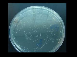
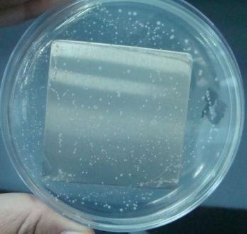
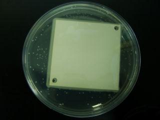
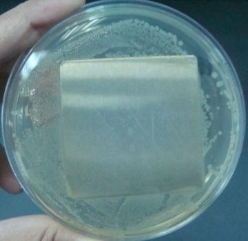
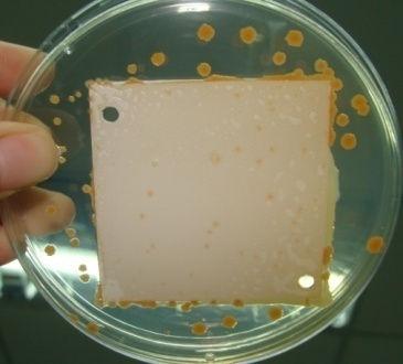
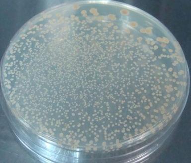
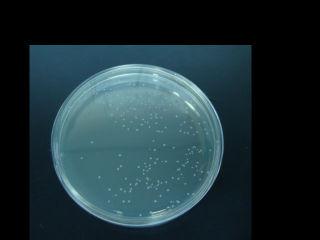
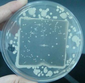
3 Agar control Agar with plate Agar with SS plate Bacillus subtilisvarglobigii Escherichia coli Enterococcus faecalis


 value remain reduction remain reduction Bacillus subtilisvarglobigii 3.0 x 10 5 5.48 57 3.70 36 3.")
4 Staphylococcus aureus Figure 1: Test plates from Test Procedure Aspergillus niger Table 2: Results from Test Procedure A Microorganisms Growth observation Isocide plate SS plate Outside of plate Bacillus subtilisvarglobigii Serratiamarcescens Staphylococcus aureus Streptococcus epidermidis Enterococcus faecalis Escherichia coli Saccharomyces cerevisiae Candida albicans Rhodotularubra Aspergillusniger (-) No growth; (+ to ++++) Representing more or less dense population of microorganisms Table 3: Results from Test Procedure B - Microbial log reduction after incubation with Isocide powder coating. Log reduction is calculated as the difference between log initial concentration and log final concentration.. Microorganisms Cells 24 hr 48hr Initial concentration logarithmic CFU Log CFU Log (CFU/ml) value remain reduction remain reduction Bacillus subtilisvarglobigii 3.0 x Serratiamarcescens 2.4 x Staphylococcus aureus 3.3 x Streptococcus epidermidis 1.2 x Enterococcus faecalis 1.6 x Escherichia coli 1.3 x Saccharomyces cerevisiae 9.0 x Candida albicans 1.0 x Rhodotularubra 4.0 x
5 Isocide performance 6 5 Bacillus subtilis globigii Cell logarithmic 4 3 Serratia marcescens Staphylococcus aureus Streptococcus epidermidis 2 Enterococcus faecalis Escherichia coli 1 Saccharomyces cerevisiae Candida albicans Rhodotula rubra Time (hour) Figure 2 Isocide performance between different microorganisms Observations 1.Table 2 show no microorganism growth, except for B. subtilis, in areas where Isocide was present. 2. During the time of observation, someb.subtilis colonies were growing under the test plate as described at figure Isocide performance varies, depending upon the type of microorganism. The best performance showed on Serratia marcescens and Enterococcus faecalis with no single colony growth after one day incubation 4. Table 2, show that all test microorganism capable growth under the SS plates. This indicating that SS cannot act as anti-microbial as powder coating plates. Conclusions This study supports the idea that the addition of antimicrobial coating can be effective in preventing or reducing microbial growth. The antimicrobial evaluations performed in this study were based on the zone of inhibition and JIS 2801 tests. We found that Isocide powder coating perfectly can serve as an antimicrobial agent compared with stainless steel plates since it does effect the microbial population growth. References Barret, L.,Emerentiana, S., Darrel, S Antimicrobial Coating. Tesh, E.M. The Regulation of Antimicrobials in Paints and Surface Treatments. Paint and Coatings Industry 2004, 7 Sadasivan, L Antimicrobial coating. Siva Microbiological Solutions LLC, Bristol, PA.
6