TR DISTRIBUTION STATEMENT A: Approved for public release; distribution is unlimited.
|
|
|
- Mae Thornton
- 5 years ago
- Views:
Transcription
1 Chapter 14. Intracellular detection of viral transcription and replication using RNA FISH i. Summary/Abstract Many hemorrhagic fever viruses require BSL-3 or 4 laboratory containment for study. The necessary safety precautions associated with this work often contribute to longer assay times and lengthy decontamination procedures. In this Chapter, we will discuss recent advances in RNA fluorescence in situ hybridization (FISH) that not only allow entirely new investigations into the replication of these viruses, but also how this method can be applied to any virus with a known sequence and how it can be rapidly performed to minimize time spent in high containment. We have adapted existing protocols for mrna detection with appropriate changes for examining viruses in a variety of containment laboratories [1, 2]. ii. Key Words Viral RNA detection, RNA FISH, RNA localization, TurboFISH, hemorrhagic fever virus replication 1. Introduction RNA FISH was developed as a method to visualize cellular RNA by binding a fluorescently-labeled oligonucleotide probe to a complementary target sequence. Unfortunately, early methods for RNA FISH required long staining times and the sensitivity was often not adequate to detect single molecules of RNA. A breakthrough in this technique was achieved when researchers showed that multiple probes could bind to a single RNA and the cumulative result of multiple binding events enabled the visualization of diffraction-limited, single molecule RNA spots [2, 3]. In this system, each oligonucleotide probe is tagged with a fluorophore and is complementary to a unique region of the target. Probe sets typically contain about 50 probes, enabling a much greater signal-to-noise ratio than previous methods. Probe sets are designed using freely available software to minimize cross-reactivity to other targets, to prevent probe overlap, and to minimize binding to predicted secondary structures. Multi-target probe sets enable single-molecule resolution of host mrnas using x oil objective lens to achieve such resolution. However, viral RNA tends to cluster in specific subcellular sites (e.g. viral replication factories). Thus while true single-molecule resolution may not be achievable due to the aggregation of multiple RNAs at one site, the combined signal intensity of these molecules is much greater than a single RNA and can be imaged using low-objective microscope lenses. Detection of viral RNA is therefore much more robust than host RNA and has resulted in the development of a variety of useful assays for virus detection. For hemorrhagic fever viruses with segmented genomes such as Crimean Congo hemorrhagic fever virus and Lassa fever virus, multiplexed RNA detection can simultaneously detect multiple genomic segments. In addition, probe sets can be designed to detect positive and negative sense RNA, which in some instances can delineate between genomic RNA, viral mrna, or viral crna. We and others have been expanded this technique to use in virus-infected tissue sections, high-throughput imaging, and for flow-cytometry based assays [4]. Detection of viral RNA allows for in depth interrogation of the subcellular sites of viral replication and such experiments will help further examine the mechanisms in which viruses replicate, assemble, and traffic through the cell. An additional benefit of this method is that the robust and quick staining methods described here enable rapid detection of virus infection which is useful for screening assays (Figure 1). 2. Materials
2 3. Methods Probes: Oligonucleotide probe sets are typically delivered as dried stocks. Reconstitute the probes in TE buffer to a final concentration of 25 µm. Probe stocks should be aliquoted and frozen at -20ºC. Buffers: Fixation buffers: 3.7% formalin solution (37% formalin diluted in PBS) or methanol. Make fresh for each experiment. Permeabilization buffer: 70% Ethanol Hybridization buffer: Add 1 ml deionized formamide 1 and 1 ml 20x saline-sodium citrate (SSC) to 8 ml of nuclease-free water. Add 1 gram of dextran sulfate and mix for 10 minutes at room temperature. Aliquot and store the hybridization buffer at 4ºC. Wash buffer: Add 20 ml of formamide and 20 ml of 20x SSC to 260 ml of nucleasefree water. Wash buffer can be kept at room temperature. Microcopy reagents: Cells can be grown on glass coverslips (#1.0 thickness recommended) placed in 24 well plates, chambered coverslips, or 96 well flat-bottom plates suitable for microscopy (Example: Greiner CELLSTAR 96 well plates). Microscope objectives: For screening purposes we recommend a 20x objective. For experiments designed to examine subcellular RNA localization we recommend a 63x oil objective. Immunostain blocking buffer: 5% bovine serum albumin (nuclease-free) diluted in PBS Antibodies for detecting protein targets DAPI staining solution: 4',6-diamidino-2-phenylindole (DAPI) diluted to 5 ng/ml in wash buffer or PBS I. Design probe sets appropriate for your target(s) of interest. Typically probe sets contain approximately 50 individual oligonucleotide probes. Probe sets with fewer probes may still yield adequate signal. Enter the target sequence in 5-3 orientation into a probe generator such as the Stellaris Probe Designer 2. For multiplexed probe detection, ensure that each probe set is tagged with a different fluorophore. II. Cell culture and infection conditions. Cells must be plated on an appropriate substrate for subsequent microscopy. For the best resolution, we recommend plating the cells on glass coverslips (#1.0). For testing or processing large numbers of samples, we recommend
3 microscopy-grade 96 well plates 3. Infect cells with chosen multiplicity of infection (MOI). We have found that depending on the virus, viral RNA is detectable within a few hours of infection. III. There are two methods for fixation and staining. The first method uses formaldehyde fixation followed by ethanol permeabilization. We find this method is preferable when the samples will also be immunostained. The second method, termed turbofish uses methanol as a fixative and permeabilizing reagent [1]. This method significantly reduces the time required to complete the assay but also requires larger concentration of probe. A. Formaldehyde fixation. 1. Rinse samples with PBS 1-2 times before fixing. 2. Remove the PBS and add enough 3.7% formalin fixation buffer to cover the cells. 3. Incubate at room temperature for 10 minutes. 4. Remove formalin and rinse the cells 1-2 times in PBS. 5. Remove PBS and replace with ethanol. Incubate cells in ethanol for at least 1 hour at 4⁰C Remove the probe sets from -20ºC and thaw at room temperature. Once thawed, vortex the probes for a few seconds and briefly centrifuge. 7. Remove hybridization buffer from 4ºC and equilibrate to room temperature 8. In a fume hood, aspirate ethanol from samples and replace with a generous amount of 10% formamide wash buffer (500 µl/well in 24 well plates) 9. Prepare the hybridization buffer by adding probe stock solution to the buffer. Each probe set must be empirically tested for the optimal concentration but we recommend starting with a 1:100 dilution of probe in hybridization buffer (0.25 µm). 10. Remove the wash buffer from the wells and add enough probe-hybridization buffer to cover the sample. For samples grown on coverslips, we recommend 100 µl of buffer to be added to the sample. Then carefully cover the sample with a second coverslip to evenly spread the hybridization buffer and to inhibit evaporation Wrap the sample in aluminum foil and place in a humidified 37ºC incubator for at least four hours or incubate overnight for convenience. 12. Remove the hybridization buffer from the samples and replace with 10% formamide wash buffer 6. If a secondary coverslip was used, carefully remove the top coverslip using tweezers. 13. Incubate the plate at 37ºC for minutes 7. Preferably rock the plates at a low speed (50 RPM). After 30 minutes, replace wash buffer with fresh wash buffer and incubate an additional minutes. 14. Remove the wash buffer and rinse the cells twice with 2x SSC buffer. 15. If co-immunostaining is desired, we recommend minimizing antibody staining times. We generally block and perform primary and secondary antibody incubations at 37ºC for 30 minutes each when possible. Use nuclease-free BSA or an alternative blocking buffer to minimize degradation of RNA targets. 16. Once the staining is completed, submerge the samples in 3.7% formalin for 24 hours in order to ensure complete virus inactivation 8.
4 4. Notes 17. Once the samples have been removed from containment, carefully decant the formalin. Rinse the samples 2-3 times with PBS. 18. Remove the coverslips from the wells using tweezers. Place the coverslips on glass slides using an appropriate mounting media. We prefer VectaShield Hardset mounting medium as it rapidly dries. 19. After minutes, ensure the mounting media is dry and begin to image the samples 9. B. Methanol fixation 1. Prior to fixation: place methanol at -20ºC for about 30 minutes (or until ice cold), place wash buffer at 37ºC, allow hybridization buffer to equilibrate to room temperature, and thaw, vortex, and centrifuge probes as before (A7). 2. In a fume hood, set a hot-plate to 37ºC. Alternatively, a 37ºC incubator can be used. 3. Rinse samples with PBS 1-2x. 4. Remove PBS and add ice-cold methanol to the samples (enough to cover each sample). 5. Place samples at -20ºC for 10 minutes While samples are fixing, prepare the hybridization buffer by adding 2-5 µl of probes to 100 µl of hybridization buffer ( µm) for each well. 7. When fixation is complete, aspirate the methanol from the samples and add 100 µl of hybridization buffer to each sample. 8. Wrap the sample in aluminum foil and place on the 37ºC hot-plate. Allow hybridization to proceed for at least 5 minutes. 9. Remove the hybridization buffer and replace with pre-warmed wash buffer. Incubate at 37ºC for one minute. 10. Remove the wash buffer and replace with fresh wash buffer. Repeat wash step twice. 11. Rinse cells 2-3 times with 2x SSC buffer 12. Process samples for complete inactivation, removal from containment, mounting, and imaging as discussed in A18-A20. 1 All work with formamide should be performed in a fume hood. Be sure to discard used formamide appropriately. 2 We frequently use sequences obtained from GenBank. However, we have found that the orientation and proper RNA species (crna versus genomic RNA) is not always properly labeled. For each sequence, we advise you to verify the sequence is correct. 3 When using 96 well plates, we recommend avoid using the edge wells as these wells tend to dry out more quickly than the interior wells. 4 Cells can be stored in ethanol for several days. Since ethanol tends to evaporate quickly, we recommend adding an excess of ethanol and wrapping the samples in paraffin to slow evaporation when permeabilizing the cells for longer times 5 For samples grown in 96 well plates, we recommend at least 50 µl of probe-hybridization buffer to prevent a surface tension meniscus from forming and causing the middle of the well to dry out.
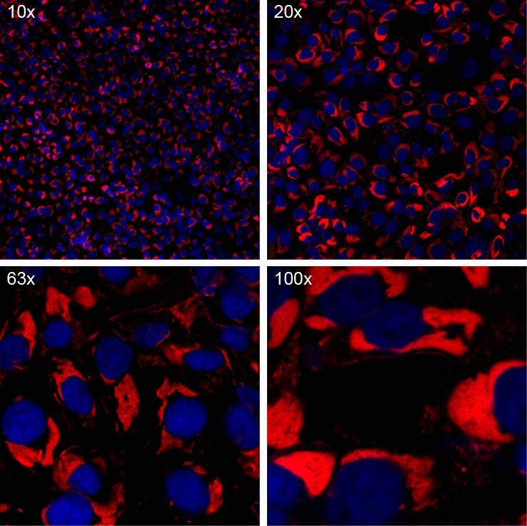
5 6 DAPI can be added to the formamide wash buffer or hybridization buffer when nuclear staining is desired. 7 Always ensure minimal light exposure to the samples once the RNA FISH staining has begun. 8 When using coverslips, be certain to keep the plates facing upward to prevent the coverslips from floating out of their appropriate well. 9 We recommend imaging the samples immediately after the mounting medium has hardened. Samples may be stored at 4ºC for prolonged use but RNA FISH signal tends to fade more rapidly than immunostaining. 10 If a -20C freezer is not available when performing the methanol fixation, we recommend placing the samples on ice. Alternatively, the fixation can be performed at room temperature if necessary. 5. Acknowledgments We thank Ron Cook and Marc Beal of Biosearch Technologies, Inc. for help in design and procurement of Stellaris FISH probes. We thank Dr. Arjun Raj (University of Pennsylvania) for previous protocols. Opinions, interpretations, conclusions, and recommendations are those of the author and are not necessarily endorsed by the U.S. Army 6. References 1. Shaffer, S.M., et al., Turbo FISH: a method for rapid single molecule RNA FISH. PLoS One, (9): p. e Raj, A., et al., Imaging individual mrna molecules using multiple singly labeled probes. Nat Methods, (10): p Femino, A.M., et al., Visualization of single RNA transcripts in situ. Science, (5363): p Jambo, K.C., et al., Small alveolar macrophages are infected preferentially by HIV and exhibit impaired phagocytic function. Mucosal Immunol, (5): p Figure caption Figure Error! Main Document Only.. Vero cells infected with Ebola virus were fixed in methanol. Viral RNA (red) was detected using a 5 minute RNA FISH incubation with hybridization buffer containing RNA FISH probes and DAPI. Cells were imaged on a confocal microscope with a 10x air objective (top left), a 20x air objective (top right), a 63x oil objective (bottom left), or a 100x oil objective.
6