Supplemental Information
|
|
|
- Avis Harrington
- 5 years ago
- Views:
Transcription
1 Supplemental Information DLA-matched bone marrow transplantation reverses the immunodeficiency of SCID dogs. Bone marrow transplantation studies were initiated with the goal of reversing the immunodeficiency in dogs affected with SCID. Supplemental Table 1 summarizes the results of DLA-identical marrow transplantation with or without initial marrow ablation in combination with post grafting immunosuppression. Transplants were performed on animals ranging from 5-7 weeks of age, when the animals still maintained significant levels of maternal antibody. All dogs received DLA-matched bone marrow (~0.5 x 10 9 nucleated cells/kg). Immediately after bone marrow transplantation, the animals were transported to the University Containment Facility, a barrier facility with BSL-3 capabilities. No other canines are maintained at this facility, and standard protocols are in place that minimize the chance of exposure to canine viral or bacterial pathogens or other opportunistic pathogens. Thus, SCID dogs are completed isolated from other animals during the critical period post-transplantation when they are most severely immunocompromised. Dogs in group 1 received no initial marrow ablation and post-grafting immunosuppression with the combination of cyclosporine (CSP) 5 mg/kg tid and mycophenolate mofetil (MMF). Four of eight recipients survived. Three of the four that did not survive succumbed from marrow failure even though significant donor hematopoietic chimerism developed after transplantation; these three animals developed acute pancytopenia 4-8 weeks after transplantation. Marrow failure in these dogs was attributed to either lack of sustained donor marrow engraftment or immune mediated graft rejection. The fourth animal succumbed to infectious complications 9 weeks after transplantation. The four surviving dogs had stable mixed chimerism for over 30 months.
2 Dogs in group 2 received initial marrow ablation with a single dose of cyclophosphamide (administered intravenously 24 hours prior to transplantation) and post-grafting immunosuppression with CSP and MMF. Three of four survived; the dog that succumbed showed sudden marrow failure after initial, transient donor chimerism. Graft failure occurred early in the experiment. We documented sub-therapeutic serum levels of cyclosporine, in the first dog with graft failure. Subsequently, we decided to increase the cyclosporine dose in the remaining three dogs. The three surviving dogs maintained stable mixed hematopoietic chimerism for over 18 months. Dogs in group 3 received no marrow ablation and the combination of CSP (15 mg/kg bid) and MMF. To date, all three recipients survive and show stable mixed chimerism for greater than six months. To assess the extent of chimerism in peripheral blood mononuclear cells (PBMC), chimerism was also assessed with a quantitative assay. DNA was prepared from PBMCs and PCR analyses of polymorphic micro-satellites (previously shown to be different in donor and recipient) were performed (Table 1). [Micro-satellites used to generate Table 1 were from the DNA-PKcs locus or the MEF2A locus.] By 32-P end labeling one of the amplification primers, donor chimerism could be quantified. The level of donor chimerism ranged from 10-34% in PBMC. To assess immune reconstitution, animals with sustained donor engraftment were immunized with a recombinant canine distemper vaccine (Meriel) at (median, 20) weeks after transplant; antibody titers were assessed two and four weeks later. All 10 animals mounted significant antibody titers, similar to those observed in a normal animal challenged with the same immunogen. Subsequently, all ten dogs received normal modified live canine vaccines with no ill effects; the transplanted recipients are currently housed in the general canine animal facility within the MSU s College of Veterinary Medicine. Although T and B cell functions were not assessed
3 separately, generation of protective immunity to distemper virus requires T/B collaboration, and we conclude that the surviving animals were effectively immune reconstituted. We conclude, that initial marrow ablation with cyclophosphamide does not improve marrow engraftment. Instead, the higher dose CSP regimen in combination with MMF provides adequate post-grafting immunosuppression to maintain donor hematopoietic engraftment. It is interesting that the residual immune system in these SCID dogs (perhaps NK cells, that cannot reject tumor xenografts) can reject DLAidentical bone marrow grafts if immunosuppression is inadequate. The higher CSP dose prevented graft rejection in 6 of 6 dogs (3 in group 2 and 3 in group 3); whereas only 4 of 9 grafts were successful using the lower CSP dose. Thus, there is good correlation between higher cyclosporine dose and graft survival. There is not a direct correlation between donor chimerism and immune reconstitution; this may be explained because chimerism was analyzed in PBMC that contain cells of non-lymphatic lineages. It is likely that cyclosporine is targeting either the extremely low numbers of residual T cells present in SCID dogs (unlike SCID mice, SCID dogs are minimally "leaky"). Alternatively, NK cells could theoretically be the target of cyclosporine's affect in these animals although NK cells are not known to mediate rejection of DLA matched bone marrow grafts. In any case, our data suggest that maintaining adequate cyclosporine levels are most critical to ensure successful engraftment. Development of this canine model of SCID should provide researchers in this field a new tool to address several outstanding questions in the field. First, the identification of a SCID dog with DNA-PKcs deficiency presents a unique opportunity to develop a clinically relevant large animal model for studying the role of NK-cells versus T-cells in allogeneic bone marrow transplantation. The SCID dog model which has intact NK-cell function but no mature T- or B-cell function will permit studies to identify
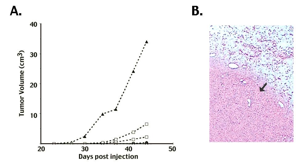
4 the role of NK cells in allogeneic transplantation in the major histocompatibility complex (MHC) mismatched setting. Second, the DNA-PKcs deficient SCID dog would serve as a valuable tool to study the in vivo function of ex vivo expanded T-cells for adoptive immunotherapy following allogeneic transplantation. The SCID dog model would permit studies to test if new approaches to ex vivo expanded cells can achieve in vivo function and a desired graft-versus-host effect without the issue of host immune response to genetically modified cells. Third, development of improved gene therapy strategies for hematopoietic stem cells has been limited by the need for myeloablative conditioning to suppress host immune responses. The SCID dog would permit the study of the in vivo function of genetically modified stem cells in a large animal without the confounding issue of a host immune response. Tumor Xenografts grow rapidly in SCID dogs. In addition to the ovarian carcinoma tumor xenograft, the fibrosarcoma HT1080 was introduced into SCID dogs. Six SCID puppies were injected subcutaneously with 10 7 tumor cells. Over the course of the experiment (6 weeks) five of six puppies developed readily palpable tumors (Supplemntal Fig. 1A) that were histologically consistent with a locally invasive fibrosarcoma (Supplemental Fig. 1B). As in nude mice, no evidence of metastatic disease was apparent on gross necropsy in dogs harboring HT1080 tumors. These experiments establish that SCID dogs do not reject human tumor xenografts; further experimentation is underway to determine whether these animals can also support normal skin xenografts as well as vascularized organ xenografts. Though the added expense and longer maturity time of dog models would preclude the use of SCID dogs in many research applications, the development of this alternative model of SCID may still have important advantages for certain applications. For example, SCID mice can be engrafted with normal or diseased human tissues for example with pieces
5 of diseased rheumatoid synovium, pieces of human kidney or ovary (usually in the peritoneal cavity or under the kidney capsule), pieces of diseased intestine, or human skin with a variety of dermatologic diseases. It may be similarly possible to graft diseased human tissues into SCID dogs, but in a more normal anatomical location. In recent years, there has been considerable effort on research focused at propagating fetal tissues with the goal of generating organs for transplantation. If fetal kidney cells are transplanted into mice (as xenografts) prior to fetal day ~35, organogenesis continues; however the blood supply in the developing organ is of host origin. Theoretically, it might be possible to transplant entire fetal organs after gestational day 35 into SCID dogs; these organs should develop human vasculature and theoretically might be useful as organs for transplantation. Finally, canine models are often utilized in pharmacological studies in drug development; it might be useful to assess drug efficacy and safety concurrently in dog models harboring xenogeneic grafts (for instance tumors, rheumatoid synovium, human pancreatic islets). In sum, these initial experiments provide support for the idea that SCID dogs may provide an important new tool for developing relevant models of human disease, and it is our goal to make these animals widely available to develop relevant models of human disease.
6 Supplemental Data Table and Figure Legends. Supplemental Table 1. DLA matched marrow grafts. Results of DLA matched bone marrow transplants described in this study. Transplants on two of the eight animals in group 1 were performed at the Fred Hutchinson Cancer Research Center; all other transplants were performed at the College of Veterinary Medicine, Michigan State University. Assessment of marrow engraftment in bone marrow transplanted animals was done by chimerism analysis using PCR amplifications of micro-satellites within the DNA-PKcs gene or MEF2A that are polymorphic between donor and recipient were done using one 32-P labeled primer with an unlabeled second primer. Amplified fragments were analyzed by denaturing acrylamide electrophoresis. Quantification of products deriving from donor and recipient was determined by phosphorimaging. Immune reconstitution was assessed by measuring antibody titers in response to a recombinant distemper vaccine. Supplemental Figure 1. Human fibrosarcoma tumor xenografts proliferate readily in SCID dogs. A. Growth of human fibrosarcoma cell line, HT1080 injected subcutaneously at one sites in six SCID dogs. Tumors were measured and tumor volume in each dog is shown. B. H+E staining of subcutaneous human fibrosarcoma (arrow).
7 Table 1. DLA Matched bone marrow grafts. Group (#) 1 Initial marrow ablation Allograft success rate 1 (8) none MMF 2 / CSP 3 2 (4) 3 (3) Duration of mixed chimerism 4, 7, 8, 9, 90 6,103 6, >139, >139 Post-grafting immunosuppression Cyclophosph MMF 2 / 3/4 4, >74, amide 4 CSP 5 >74, >78 none MMF 2 / CSP 5 3/3 >52, >52, >52 Graft assessment in Surviving Animals age at transplant (weeks) % donor chimerism Ab titer 7 Survival (weeks) 6 > > > [512] 7 >74 52 <4 [512] 8 > [512] 5 > [64] 5 > [128] 5 >52 32 <8 [32] 1 number of animals in group; 2 MMF 10mg/kg bid p.o. days 0-27; 3 CSP 5mg/kg tid p.o. on days 2 to 35; 4 10mg/kg sid i.v. 24 hours prior to transplantation; 5 CSP15mg/kg bid p.o. on days 2 to 35; 6 Euthanized with sustained allograft at the completion of the study; 7 Distemper antibody titers were measured 2 and 4 (in brackets) weeks after immunization. Group 1 was immunized 29 weeks post-transplantation. Group 2 was immunized 20 weeks post-transplantation. Group 3 was immunized 12 weeks post-transplantation.
8 Supplemental Figure 1.