Phage Antibody Selection With Reichert SPR System
|
|
|
- Caitlin Parrish
- 5 years ago
- Views:
Transcription

1 Phage Antibody Selection With Reichert SPR System Reichert Technologies Webinar April 4, 2016 Mark A. Sullivan, Ph.D. Department of Microbiology and Immunology University of Rochester
2 Presentation Outline Basics of SPR Technology Background on antibody generation and modification using phage display Objectives and rationale for investigating the Reichert instrument in isolation and analysis of phage displayed proteins and peptides Preliminary results obtained in fractionating phage in the SR7000DC instrument Binding and recovery of scfv-phage Effect of scfv valency on binding profile Separation of a positive-binding phage from a non-binder


3 Surface Plasmon Resonance Label-Free technique for analyzing biomolecular interactions in real time Extremely precise mass sensor for quantitative binding measurements Flow based measurement Key Components Sensor chip ligand immobilization Microfluidics analyte delivery Optical sensor measurement SPR is the Gold Standard for determining Kinetics Daghestani & Day. Sensors 2010, 10,
 Streptavidin/neutravidin -biotin")

4 Sensor Chip Sensor chip consists of glass coated with a thin gold layer Surface functionalized for immobilization of ligand Amine coupling (EDC/NHS) Streptavidin/neutravidin -biotin Gold-thiol Polymer matrix for balance between binding sites and signal strength N.J. de Mol, M.J.E. Fischer (eds.), Surface Plasmon Resonance, Methods in Molecular Biology, 2010.

5 Antibodies: Largest Class of New Therapeutic Agents Sources of Antibodies First commercialized monoclonal antibodies were humanized murine Mabs Human antibodies derived from high-throughput single B- cell cloning methods Limited to targets found in natural infections Display technologies Phage Yeast Ribosome / mrna Cell-based
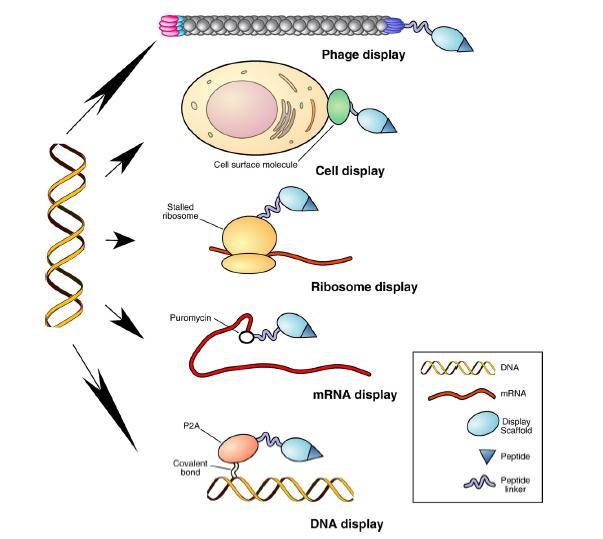
6 Display Technologies
7 Advantages of Display Technology for Antibody Isolation Fast - typically 1 week to isolate individual scfvs versus months for hybridomas Not subject to limitations of immunogenicity (self antigens) Avoids problems with immunodominance Alternative methods of antigen presentation Rapid production of proteins for analysis No animal use Yields DNA encoding antibody sequence Allows control of antigen presentation Use custom conditions during enrichment
8 Human Phage Antibody Library Naive Library Diversity ranges from ~1x10 9 to 1x10 11 clones Fab or scfv format Uses pooled cdna from many donors Binding is driven by shape complementarity Used to select antibodies against any target Affinities generally nm from 1x10 9 sized libraries Many applications for therapeutics and diagnostics require higher affinities (< 1nM Kd) Secondary libraries created by mutagenesis and re-selection can yield sub-nanomolar Kd Most common method uses biotinylated antigen and magnetic beads to enrich for clones with slower off rates Some methods employ very long incubations for off-rate selection (~1 week!)
9 Immunoglobulin Repertoire Cloning l1a l1b l1c l2a l2b l3 l4 l5a l5b VL5 PCR amplify pooled cdna with primers specific for immunoglobulin variable framework regions. The heavy and light chain variable regions are converted to a scfv format by SOE PCR. VL VH1,7 VH2 VH3 VH4 VH5 VH6 VL J VH5 VH VH J N-terminal Flag Epitope Assembled scfv cloned into AP-III 6 vector

10 Complementary Protein Surfaces
11 Phage Bio-Panning in an Antigen-Coated Well
12 Panning Profile of Phe scfv
13 Isolation 5 days Phage Display Cycle - Opportunity Bind Wash Elute Amplify Library Plate Repeat Enrichment 3x (2days/cycle) Secondary Mutegenesis Choose Binder Amplify Clone Binding ELISA 1) Binder Selection w/spr affinity in phage - skip protein expression 2) Eliminate rounds of enrichment - potential 6days->2days 3) Elution control to eliminate low affinity binders and improve reproducibility fine control of temp, time, flowrate & conc 4) Specificity w/selective subtraction 5) Run up to 4 enrichments simultaneously
 Outflow easily collected Flexibility to employ different elution buffers, flow rates and temperatures Capable of subtractive selection")
14 Phage Antibody Enrichment on a Reichert Instrument Potential Advantages Readily accessible fluidics system Inexpensive components (tubing and sensor slides) Outflow easily collected Flexibility to employ different elution buffers, flow rates and temperatures Capable of subtractive selection scheme
15 Key Questions to Answer Can antibody phage be applied, bind to a target and be recovered? Do bound phage generate a significant SPR signal that is useful for monitoring enrichment? How many phage can bind to immobilized target? Determines maximum diversity of a library that can be enriched How does valency of the displayed scfv affect the binding profile? What are the kinetics of separation of binders from non-binders?

16 Previous Use of SPR for Phage Binding Analysis Phage display and SPR both developed in the early 1990s. Very few publications describe phage selection with SPR analysis Early papers lacking in experimental details A recent paper employing peptide display offers a more thorough analysis of SPR and phage binding Implementing control of phage enrichment using instrument fluidics and the SPR siganl could offer improved discrimination of affinity variants
17 Binding of Phage to Immobilized BSA Preliminary experiments demonstrated that injection of standard monovalent preparations of a BSA scfv leads to high levels of phage in flowthough and wash fractions. Likely due to the 99% of phage particles with no scfv displayed. The presence of non-displaying phage obscures the detection by titering of bound phage after elution with acid. Very low SPR signal observed
18 Purification of scfv-displaying Phage The ~1% of displaying phage also have an N- terminal Flag sequence on the scfv Displaying phage can be selected by first binding on immobilized M2 anti-flag Mab Bound displaying phage can be recovered by incubation with 3X Flag peptide Use of the 3X peptide elution avoids possible damage of the scfv by low ph elution Injection of M2-3X phage allows recovery of bound phage after injection of elution solution (20 mm HCl).
19 Monovalent Phage Injection Inject (10 ml/min) Wash (50 ml/min) Elute (20 mm HCl) 100 ml Time (min) 250 ml fractions of outflow collected and phage titers determined by transduction into E. coli. Phage input: ~1x10 8 Phage output: ~3x10 6
20 Monovalent Phage Profile Phage binding Regenerate anti-bsa Mab Wash
21 Multivalent and Monovalent Display Infect with VCS M13 helper phage ~1% display the scfv 99% display no scfv Infect with Hyperphage helper (deleted for gene III) 100% display 3-5 copies of the scfv/ phage particle
22 Multivalent Phage Binding Phage binding Wash (Dissociation) A multivalent preparation of the BSA binding phage (~2x10 11 /ml ) was injected over a BSA surface at 10 microliters/min. The difference between left and right channels is shown. Bound phage were eluted ~25 minutes after binding by injection of 20 mm HCl for 1 minute and a 0.4 ml fraction was collected and titered by transduction. ~ 8x10 7 phage recovered or ~25 fold higher than monovalent recovery!
23 Kinetics of Separation Discriminating a Binder from a Non-binder Use a mixture of an anti-bsa scfv: V L -linker V H format and an anti-peptide scfv in the reverse format: V H -linker-v L. Colony PCR using an upstream vector primer and a primer targeting the VHJ yields fragments of ~800 bp or 400 bp that are easily resolved. VL linker VH ~800 bp VH linker VL ~400 bp
24 Separation of Monovalent anti-bsa Phage and Anti-peptide Phage Inject (10 ml/min) Wash (50 ml/min) Elute (20 mm HCl) 100 ml Time (min) 250 ml fractions of outflow collected and phage titers determined by transduction into E. coli. Colonies derived from each fraction are subjected to colony PCR and the products resolved by agarose gel electrophoresis.

25 Selection of BSA Binding scfv Flow-through Wash 1 Wash 2 BSA scfv Anti-peptide scfv Wash 3 Elute 1 Elute 2

26 Phage Titer of Fractions Bind Wash Elute Log (#phage) (Amp transducing) 1x10 8 1x ml 1x10 6 1x10 5 FT Outflow fraction number BSA scfv Peptide scfv
27 Key Results Part 1 Phage can be easily applied, fractionated and recovered from the outflow after elution. Recovery is approximately 1-5% when applying ~1x10 8 binding phage Yield of phage likely dependent on target levels immobilized and affinity Maximum binding is significantly higher as observed with a multivalent binder Increasing the number of displaying phage in the input is desirable
28 Key Results Part 2 Binding phage can be readily separated from non-binders Parameters such as flow rate and elution buffer composition will likely require customization depending on relative affinities of the population SPR signal is unlikely to be helpful when enriching for low abundance binders May be useful in characterization of panels of similar clones with varying affinities Will be essential to use only monovalent phage
29 Isolation 5 days Phage Display Cycle - Opportunity Bind Wash Elute Amplify Library Plate Repeat Enrichment 3x (2days/cycle) Secondary Mutegenesis Choose Binder Amplify Clone Binding ELISA 1) Binder Selection w/spr affinity in phage - skip protein expression 2) Eliminate rounds of enrichment - potential 6days->2days 3) Elution control to eliminate low affinity binders and improve reproducibility fine control of temp, time, flowrate & conc 4) Specificity w/selective subtraction 5) Run up to 4 enrichments simultaneously
30 Future Directions Optimization of regeneration/elution buffer conditions to maximize affinity discrimination Soluble target elution Flow rate control Temperature, ph, etc. Characterization of individual phage binding kinetics to rank order affinities Subtractive selection schemes Immobilize homologs of target in left channel, actual target in right channel Select phage that fail to bind the homolog but retain binding to the target Switch inlet tubing to elute only phage in the right channel

31 See Reichert SPR at CHI-PEGS! See ReichertSPR At PEGS Boston Booth #207
32 APPENDIX

33 Knez et al. Affinity Comparison of p3 and p8 Peptide Displaying Bacteriophages Using Surface Plasmon Resonance Anal. Chem :

34 Knez et al. Affinity Comparison of p3 and p8 Peptide Displaying Bacteriophages Using Surface Plasmon Resonance Anal. Chem :
35 Surface plasmon resonance characterizes the binding of the CEFESC-phage to the BIR2 domain of XIAP. Recombinant BIR2 of XIAP was immobilized on a sensor chip, and a range of concentrations of CEFESC phage was injected over the chip surface. Ingo Tamm et al. J. Biol. Chem. 2003;278: by American Society for Biochemistry and Molecular Biology