Molecular diagnosis of haemophilia and other bleeding disorders
|
|
|
- Joan Golden
- 5 years ago
- Views:
Transcription
1 Molecular diagnosis of haemophilia and other bleeding disorders Jayandharan G Rao Associate Professor Department of Haematology Christian Medical College Vellore, India.


2 Jayandharan et al, J Genet Synd Gene Ther, 2012
3 General characteristics of inherited bleeding disorders Deficiency Prevalence Gene on chromosome No of reported mutations in literature Fibrinogen 1:1 million Prothrombin 1:2 millions FV 1:1 million FVII 1: FV + FVIII 1:1 million 2, FVIII 1:5000 X ~2700 FIX 1:25000 X ~1150 FX 1:1 million FXI 1:1 million FXIII 1:1 million A subunit: Glycoprotein Ib/IIIa?1:1 million BSS 1:1 million 17, 22,3 50.


4 Hereditary bleeding disorders in India Population ~ 1.2 billion Incidence patients for each of the disorders 84% - Severe; 16% mild fold increased in populations practicing consanguineous marriage (South India) 2 1 CMC data unpublished 2 Mannucci et al, Blood, 2004
5 Molecular diagnosis of bleeding disorders
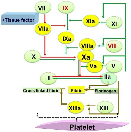
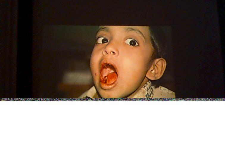
6 Haemophilia


7 Factor 8 gene defect haemophilia A 1 Factor 9 gene defect haemophilia B 2 Human chromosomes X chromosome Factor 9 Factor 8 35cM 1 Gitscheir et al, Nature, Yoshitake et al, Biochemistry, 1985.

8 FACTOR VIII AND FACTOR IX FACTOR 8 GENE -186 Kb FACTOR 9 GENE - 34 Kb mutation UTR- 150nt EXONS 8 EXONS 3 UTR nt 5 UTR- 29nt 3 UTR nt mrna 9010 nt mrna 2803 nt Pre A1 A2 B A3 C1 C2 R372 R740 R1689 Pre Pro GLA EGF1 EGF2 R145 Activation peptide Catalytic R180 N A1 A2 Ca2+ A3 C1 C2 C N GLA EGF1 EGF2 S S Catalytic C Heavy chain Light chain Light chain Heavy chain FACTOR VIII PROTEIN FACTOR IX PROTEIN
 Quantitative change")


9 Gene defect Qualitative (or) Quantitative change in protein Haemophilia F8 or F9 gene mutation FVIII:C or FIX:C-1-5% - moderate White et al, Thromb Haemost, 2011.
10 Control of haemophilia Carrier detection Prenatal diagnosis
11 Who seeks genetic testing? Eg: Pedigree with haemophilia??? Carrier status determination Prenatal diagnosis
12 Components of genetic testing Clinical history /phenotypic assays Pedigree analysis Genetic screening [Informed Consent] Counseling [Pre and post testing]

 \" Women are carriers")
13 Genetics of haemophilia X- linked inheritance 25% 25% " Both VIII and IX deficiency are X linked " Men are affected (25%) " Women are carriers (25%)
14 Genetics of Haemophilia Who is a carrier? XY X X X Y XX Eg: A Mother having a affected son
15 Genetics of Haemophilia If the father is a haemophiliac, the daughters will be obligate carriers and all the sons would be normal X Y X X X Y X X
16 Sporadic and Familial Haemophilia Only one known case in the family - Sporadic More than one haemophiliac in the family exists - Familial
17 Pedigree analysis Obligate and Possible Carriers
18 Approach for genetic diagnosis of haemophilia 1. Linkage analysis 2. Direct mutation detection


19 Linkage analysis -Track the defective chromosome F8 gene F9 gene -Technically simple, low cost -Multiple members and informativeness of polymorphism required -Informative in ~80% families Peake et al, WHO Bull, 1993.
 G A + XbaI - +/- + λbst")
 - CACACA CACACACA CA")
20 Linkage analysis Methods PCR Restriction fragment length polymorphism (RFLP) G A + XbaI - +/- + λbst EII UD Repeat sequence polymorphisms (microsatellites and VNTRs) - CACACA CACACACA CA 19/20 Intron13 CAn


21 Direct Mutation detection - Direct identification of disease causing mutation Extract DNA or RNA -Highly sensitive PCR -Gold standard F8-exon 3 Screening -Expensive Informative in >95% of families DNA sequencing

22 PCR based direct mutation detection in haemophilia A Intron 22 inversions account for ~45-50% of severe haemophilia A phenotypes 1 Intron 1 inversions account for ~2-5% of severe haemophilia A phenotypes 2 Int22h2/h3 int22h1 Intron 22 inversion MW +/ Kb 11Kb 10Kb Intron 1 inversion 1 Kb 1.5 Kb Exon Intron Homologous sequence 1 Kb 1.5 Kb 1 Lakich et al, Blood, Bagnall et al, Blood, 2003
23 DNA samples required for genetic diagnosis Affected patient Indirect Direct DNA samples Proband requesting genetic diagnosis

24 Chorionic villus sampling at weeks for prenatal diagnosis Placenta DNA Genetic diagnosis
25 Haemophilia Approach to molecular diagnosis at CMC, Vellore Factor 8 gene Factor 9 gene Intron 1 & 22 inversions (FVIII:C<1%) Direct mutation detection Direct mutation detection Linkage analysis- not preferred Indirect Linkage analysis


26 Multiplex PCR and Conformation sensitive gel electrophoresis CSGE is a rapid, heteroduplex based detection method for mutation screening. The method relies on the differential migration of DNA heteroduplexes in comparison with homoduplexes during polyacrylamide gel electrophoresis under mildly denaturing conditions. PATIENT DNA + NORMAL CONTROL DNA CON ABI 310 GENETIC ANALYSER 14EE2 F EXON R + 14B1B PATIENT AMPLICON H D NORMAL CONTROL AMPLICON 26AA2 A A C A A G T T G T T C A A C T A G T T G T T C MPCR group 3 CSGE 12AB F8:38->13 PCRs F9: 9->4PCRs Jayandharan et al, J Thromb Haemost, Jayandharan et al, Haemophilia, Jayandharan et al, Thromb Haemost, 2005.
![Algorithm for genetic diagnosis of bleeding disorders [CMC, Vellore] Uniplex PCR Fibrinogen](/docs-images/86/93489127/images/27-0.jpg "deficiency Prothrombin deficiency Combined F5F8 deficiency Factor VII deficiency Factor X")
 (-)")
 Multiplex PCR Haemophilia A Haemophilia B Factor XIII deficiency Glanzmann Thrombasthenia")
27 Algorithm for genetic diagnosis of bleeding disorders [CMC, Vellore] Uniplex PCR Fibrinogen deficiency Prothrombin deficiency Combined F5F8 deficiency Factor VII deficiency Factor X deficiency Factor XI deficiency Protein C deficiency Protein S deficiency Wiskott-Aldrich syndrome Bernard Soulier syndrome Common mutation Haemophilia A* (Intron 1 & 22 Inversion) (-) Direct mutation detection PCR-CSGE-Seq. (-) Multiplex PCR Haemophilia A Haemophilia B Factor XIII deficiency Glanzmann Thrombasthenia Indirect Linkage analysis Haemophilia A Glanzmann Thrombasthenia
28 Validation of molecular algorithms for genetic diagnoses Bleeding Disorder Gene/ Chromosome Reference Haemophilia A F8 / Ch.X Jayandharan et al, Haemophilia, 2004 Haemophilia B F9 / Ch.X Jayandharan et al, Haemophilia, 2005 Jayandharan et al, J Thromb Haemost, 2003; Thromb Haemost, 2005 Fibrinogen disorders FGA/FGB/FGG-Ch.4 Sumitha et al, Haemophilia, Prothrombin F2 / Ch.11 Jayandharan et al, J Thromb Haemost, 2005 Factor V deficiency F5/Ch.1 Chapla et al, Thromb Haemost, Factor V+VIII deficiency LMAN1-Ch.18 MCFD2-Ch.2 Jayandharan et al, Haemophilia, 2007 Factor VII deficiency F7/ Ch.13 Jayandharan et al, Haematologica, 2007 Factor X deficiency F10 / Ch.13 Jayandharan et al, J Thromb Haemost, 2005 Factor XI deficiency F11/ Ch.4 Jayandharan et al, J Thromb Haemost, 2005 Factor XIII deficiency F13A/ Ch.6 Jayandharan et al, Thromb Haemost, 2006 Glanzmann Thrombasthenia Bernard Soulier syndrome Jayandharan et al, J Thromb Haemost, 2007; Srivastava ITGA2B-Ch.17 et al, Natl Med J India, 2003; Nelson e t al, J Thromb ITGB3- Ch.17 Haemost, GP1BA-Ch.17 GP1BB-Ch-22 GP9-Ch.3 Sumitha et al, J Thromb Haemost, 2011 Wiskott Aldrich syndrome WASP-Ch.X David et al, Eur J Haematol, 2012
29 Setting up a genetic diagnosis facility Laboratory space: ~ sq.ft Personnel: Equipments: 2 [Technician, research officer] PCR machine, electrophoresis, 4 C/-20C storage, centrifuge, ph meter, waterbath,?centralized DNA sequencer HR Training/ collaboration with established centers
30 EQAS scheme for genetic diagnosis of bleeding disorders CMC-EQAS
31 Conclusions Molecular studies in bleeding disorders useful for: prevention and control Need to generate diagnostic algorithms to each population Can be done at significantly lower cost by process optimization