A Method for Staining Nematode Secretions and Structures 1
|
|
|
- Ronald Pierce
- 5 years ago
- Views:
Transcription
1 Journal of Nematology 20(1): The Society of Nematologists A Method for Staining Nematode Secretions and Structures 1 D. PREMACHANDRAN, 2 N. VON MENDE, ~ R. S. HUSSEY, 2 AND M. A. MCCLURE ~ Abstract: Secretions from amphids, phasmids, and excretory system were stained by incubating nematodes in 0.1% coomassie brilliant blue G-250 in 40% aqueous methanol containing 10% acetic acid on slides with coverslips sealed with nail polish or Zut. Nematodes incubated in this staining solution usually produced copious amounts of secretions from their amphids and excretory pore. Phasmids also stained dark blue, enabling them to be easily observed. Other biological dyes stained these secretions or were useful for differentiating specific morphological features of nematodes. Key words: secretion, amphid, phasmid, excretory system, stain, coomassie brilliant blue, exudate. Few biological dyes have been identified that stain specific morphological features of nematodes (3,5). Spicules of Ostertagia bisonis stain with acid fuchsin (7). Gold chloride stains phasmids, nerve rings, and gonads (4,10); picric acid-iodine differentiates glandular structures (4). Methods for inducing and staining secretions from nematode esophageal glands, amphids, or phasmids have not been developed previously. Preliminary observations showed that second-stage juveniles (J2) of Meloidogyne incognita (Kofoid & White) Chitwood incubated in coomassie brilliant blue R in an acetic acid-methanol solution on a slide sealed with nail polish produced amphidial, phasmidial, and excretory secretions that stained dark blue (Premachandran and Hussey, unpubl.). This study was expanded to identify other biological dyes that might differentiate secretions or morphological features of nematodes. Received for publication 1 June ' Research at Georgia was supported by a grant from Agrigenetics Research Corporation, Boulder, Colorado, and by state and Hatch funds allocated to the Georgia Agricultural Experiment Stations. Research at Arizona was supported by USDA grant 84- CRCR-I-1401 and Hatch funds allocated to the Arizona Agricultural Experiment Station. Deparnnent of Plant Pathology, University of Georgia, Athens, GA Present address of first author: Abbott Laboratories. Agricultural Research Center, Oakwood Road, Long Grove, IE Department of Plant Pathology, University of Arizona, Tucson, AZ Present address of second author: Department of Biological Sciences, University of Missouri, Columbla. MO MATERIALS AND METHODS Nematodes: The following nematode species were used in these studies: M. /ncognita (eggs and J2), Scutellonema brachyurum (Steiner) Andrassy, Tylenchulus semipenetrans Cobb, Belonolaimus longicaudatus Rau, Ditylenchus dipsaci (Kuhn) Filipjev, Hoplolaimus galeatus (Cobb) Thorne, Xiphinema spp. and an unidentified microbivore. Staining procedure: Although 68 biological dyes were tested, only those that stained specific nematode structures or their secretions are reported. Before use, staining solutions were filtered through Whatman ~1 filter paper to remove insoluble material. A small drop (ca. 10 #1) of nematode suspension was placed on a clean microscope slide. An approximately equal volume of stain in methanol, acetic acid, methanolic acetic acid, or distilled water was added to the slide. An alternative procedure involved mixing equal volumes of a nematode suspension and staining solution in a microcentrifuge tube before adding a drop to a slide. Pieces of glass wool were placed on the slide to support the coverslip which was slowly lowered onto the nematode sample. The coverslip was sealed with either clear nail polish, Zut, or white petroleum jelly. Nematodes were observed with a compound microscope immediately or were incubated at room temperature and observed at B0-minute intervals for 2 hours or after overnight incubation. 7O
 Amphidial secretions of Belonolaimus longicaudatus stained after incubation in 0.")
 Amphidial secretions ofmeloidogyne incognita second-stage juvenile stained following incubation in 0.")
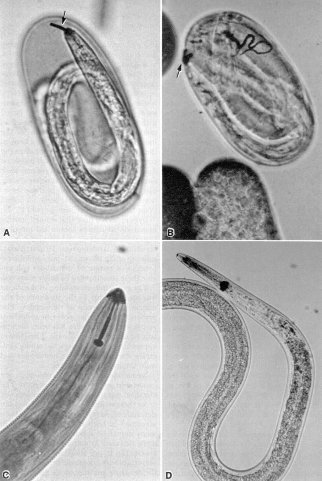
2 FIG. 1. Staining of amphidial and excretory secretions of plant-parasitic nematodes. A) Amphidial secretions of Belonolaimus longicaudatus stained after incubation in 0.2% coomassie brilliant blue G-250 in 40% methanol and 10% acetic acid for 10 minutes. B) Amphidial secretions ofmeloidogyne incognita second-stage juvenile stained following incubation in 0.2% coomassie brilliant blue G-250 in 20% methanol for 72 hours. C) Excretory secretion of Scutellonema brachyurum stained after incubation in 1% aqueous china blue for 72 hours. D) Amphidial and excretory secretions of M. incognita juvenile stained after incubation in 1% aqueous china blue for 14 hours.
 10% acetic acid induced production of blue-staining secretions from the amphids, phasmids, and excretory pore of tylenchid nematodes when")
3 72 Journal of Nematology, Volume 20, No. 1, January 1988 RESULTS Secretions: A solution of 0.1% coomassie brilliant blue G-250 in 40% methanol and (or) 10% acetic acid induced production of blue-staining secretions from the amphids, phasmids, and excretory pore of tylenchid nematodes when the coverslips were sealed with nail polish or Zut (Figs. 1A, B, 2A). The quantity of the secretion produced and time required for optimal production varied with nematode species. Some nematode species frequently produced exudates immediately following placement into this solution, and in some instances after continued incubation the secretions formed long strands. S. brachyurum produced amphidial and excretory secretions immediately after incubation in this staining solution, whereas these secretions from H. galealus formed very slowly. In general, secretions from nematodes incubated in coomassie brilliant blue R-methanol developed into long strands, whereas the secretions usually precipitated at the pore of the structures when stained with the dye dissolved in 10% acetic acid. Second-stage juveniles of M. incognita inside eggs also produced secretions that stained with coomassie brilliant blue G-250 (Fig. 4A, B). Aqueous solutions of 29 dyes were effective in staining secretions of four nematode species (Table 1, Figs. 1-3); however, secretions were induced and stained with 28 of the dyes only when coverslips were sealed with nail polish or Zut. Only carminic acid induced and stained secretions when coverslips were sealed with petroleum jelly. Staining of exudates from amphids, phasmids, or the excretory system varied with nematode species and the dye used. The excretory system usually produced the largest quantity of secretion which stained very intensely with acid fuchsin, carminic acid, china blue (Fig. 1C, D), coomassie brilliant blue R, cyanine, naphthol blue black, and nigrosin depending on the species (Table 1). The amphidial secretion ofm. incognita was most effectively stained with china blue, whereas naphthol blue black was more effective in staining this secretion from T. semipenetrans (Table 1). In general, the phasmidial secretion did not stain or only stained lightly with most dyes (Table 1). Six dyes effectively stained the secretion produced by phasmids of S. brachyurum. The large phasmids (scutellae) of S. brachyurum produced a dense secretion when incubated in methyl orange or naphthol blue black (Fig. 2C). In addition to differences among the nematode genera in secretion production and staining, differences occurred between developmental stages. For example, china blue stained secretions of the excretory system of males but not juveniles of T. semipenetrans (.Table 1). F1o. 2. Staining of phasmids and phasmidial secretions ofmeloidogyne incognita and Scutellonema brachyurum. A) Phasmids of M. incognita second-stage juvenile stained after incubation in 0.2% coomassie brilliant blue G-250 in 40% methanol and 10% acetic acid for 4 hours. B) Phasmid of S. brachyurum stained following incubation in 1% aqueous hematoxylin for 15 days. C) Phasmidial secretions of S. brachyurum stained after incubating 28 hours in 1% aqueous napthol blue black. D) Phasmids ofs. brachyurum stained after incubation in 1% aqueous trypan blue for 30 hours. FIG. 3. Staining of nematode reproductive structures or exudate. A) Testis of Ditylenchus dipsaci male stained after incubation in 1% aqueous toluidine blue O for 7 hours. B) Genital primordium of D. dipsaci fourth-stage juvenile stained after 24 hours in 1% aqueous azure blue. C) Testis of microbivorous nematode stained after incubation in Diene's stain. D) Exudate produced at vulva of Belonolaimus longicaudatus stained after incubation in 0.2% coomassie brilliant blue G-250 in 40% methanol and 10% acetic acid for 10 minutes. FIG. 4. Staining of secretions and structures of plant-parasitic nematodes. A, B) Staining of amphidial and excretory secretions of Meloidogyne incognita second-stage juvenile in egg after incubation in 1% aqueous coomassie brilliant blue G-250 for 72 hours. C) Styler ofscutellonema brachyurum stained after incubation in 1% aqueous iodine green for 48 hours. D) Staining of anterior part of metacorporus of Ditylenchus dipsaci after incubation in 1% aqueous toluidine blue O for 48 hours. ---9

4 Staining Nematodes: Premachandran et al. 73


5 74 Journal of Nematology, Volume 20, No. 1, January 1988

6 i Staining Nematodes: Premachandran et al. 75
.")
7 76 Journal of Nematology, Volume 20, No. 1, January 1988 An exudate was produced at the vulva of B. longicaudatus when specimens were incubated in coomassie brilliant blue G-250 in the methanol-acetic acid solution (Fig. 3D). Staining structures: Many of the dyes were capable of differentiating specific nematode morphological features. Almost half of the dyes provided visual definition of phasmids of S. brachyurum, and many stained the phasmidial pores within seconds. Rapid and intense staining was observed with azure blue, carminic acid, coomassie brilliant blue R and G-250, evans blue, hematoxylin (Fig. 2B), rosaniline hydrochloride, and toluidine blue. The phasmidial sheath cell also stained when specimens were incubated in solutions of azure blue, chlorazol fast pink, eosin B, erythrosin, evans blue, giemsa, malachite green, and trypan blue (Fig. 2D). The small phasmids of M. incognita J2 were easily observed when specimens were incubated 4 hours in coomassie brilliant blue G-250 to allow sufficient secretion production (Fig. 2A). Several dyes were very effective in staining nematode reproductive systems. The testis of D. dipsaci stained intensely with toluidine blue O (Fig. 3A), and the genital primordium of D. dipsaci J4 stained with azure blue (Fig. 3B). The reproductive system of a microbivorous nematode stained when specimens were incubated in Diene's stain and heated briefly (Fig. 3C). The stylet of S. brachyurum stained following incubation of adult female nematodes in a 1% solution of iodine green (Fig. 4C). Staining of unidentified bodies in the procorpus and a dense deposit of dye in the anterior part of the metacorpus was observed when D. dipsaci was incubated, uncovered, in B.P.I. dishes overnight in a toluidine blue O solution (Fig. 4D). The staining method was not suitable for dorylaimid nematodes. Coomassie brilliant blue R in 10% acetic acid rapidly penetrated the cuticle of Xiphinema species, intensely stained the entire body, and obscured most morphological features. DISCUSSION This new staining method is a simple, rapid, and reliable procedure for inducing and staining secretions from amphids, phasmids, and the excretory system of many nematode species. Several biological dyes in addition to coomassie brilliant blue were identified that stained these secretions equally well. This method rapidly induced secretions from amphids and the excretory system for most nematode species examined; however, the reaction time varied with nematode genera and the type of secretion. Although the mechanism for the induction of these secretions is not known, the greatest production occurred when the coverslips were sealed with nail polish or Zut, which indicates the secretions were chemically induced. Several other chemicals that induce formation of these secretions have subsequently been identified (Von Mende and McClure, unpubl.). Dyes reported here that stained specific nematode structures should aid morphological investigations and be useful in instructional laboratories. In particular, staining of the phasmidial pore and the reproductive system of nematodes will enable these structures to be easily observed. The rapid intense staining of dorylaims was probably due to the structure of the cuticle in this group of nematodes. The presence of cuticular pores in Longidorus macrosoma and X. index has been demonstrated using methyl blue (6). These pores apparently allow the dyes to rapidly penetrate the body of these nematodes and stain the internal structures. Nematode amphids and phasmids are considered secretory structures (6,8). The chemical nature of the material induced from these structures in this study was not determined. Acetylcholinesterase has been identified in both of these structures in many nematode species (6), and esterases were detected in amphids of Meloidogyne species (2). In our study, staining of these secretions with coomassie brilliant blue indicates their proteinaceous nature, since this dye specifically stains proteins.
8 TABLE 1. Staining of materials secreted by the excretory system, amphids, and phasmids of nematodes treated with various biological dyes. CA. Excretory system Amphid Dye numbert Mi Sb Dd Ts Mi Sb Dd Ts Mi Phasmid Sb Dd Ts Acid fuchsin J Alizarin fluorine blue Azure blue J+M M.... Bromocresol purple N.A Bromophenol blue N.A Bromothymol blue N.A J++M J Carminic acid China blue M Chlorazol fast pink J Coomassie brilliant blue R J+++M Coomassie brilliant blue G J++M J+M Cyanine N.A J Erythrosin bluish J.... Evans blue o3 Fast green FCF J Hematoxylin J Indigocarmine ~ Lacmoid Light green J t~ Methyl orange J go Naphthol blue black J&M ~- Nigrosin J+M k, Orange G M Orseillin BB Phloroglucin N.A Phloxine Poneeau S Solantine pink 4BL Trypan blue J.... ~, Concentration of staining solution of each dye was 1% except for bromothymol blue which was 0.04%. Nematode species: Mi = Meloidogyne incognita (second-stage juveniles). Sb = ScuteUonema brachyurum (females). Dd = Ditylenchus dipsaci (juveniles). Ts = Tylenchulus semipenetrans (juveniles U] or males [M]). Secretion production: - = none, + = little, + + = good, and = excellent. ~" Colour index, 3rd ed The Society of Dyers and Colourists. Bradford, Yorkshire, U.K.N.A. = not applicable. ** g~,,q
9 78 Journal of Nematology, Volume 20, No. 1, January 1988 The copious production of a proteinaceous secretion by the excretory system was unexpected; however, secretions from the excretory pore were previously shown to be antigenic (1,9). This method is a simple and useful procedure for staining secretions and specific morphological features of nematodes. The secretions are probably artificially induced, however, since large quantities of secretions are induced only when nematodes are incubated in staining solutions on slides sealed with nail polish or Zut. LITERATURE CITED 1. Bird, A. F Serological studies on the plant parasitic nematode, Meloidogyne javanica. Experimental Parasitology 15: Bird, A. F Esterases In the genus Meloidog)'ne. Nematologica 12: Bird, A. F The structure of nematodes. New York: Academic Press. 4. Hasbrouck, E Gold chloride and picric acid-iodlne as in toto stains for free living and plant parasitic nematodes. Phytopathology 49: Hooper, D.J Handling, fixing, staining and mounting nematodes. Pp inj. F. Southey, ed, Laboratory methods for work with plant and soil nematodes, 6th ed. Reference Book 402, Ministry of Agriculture, Fisheries and Food. London: Her Majesty's Stationery Office. 6. McLaren, D.J Nematode sense organs. Advances in Parasitology 14: Stringfellow, F Technique for staining spicules and gubernaculum in whole mounts of nematodes. Proceedings of the Helmintholog[cal Society of Washington 38: Wang, K. C., and T. A. Chert Ultrastructure of the phasmid of Scutellonema brachyurum. Journal of Nematology 17: Webster, J. M., and D. J. Hooper Serological and morphological studies on the inter- and intraspecific differences of the plant parasitic nematodes Heterodera and Dit)'le~zchus. Parasitology 58: Yadav, S. M., and M, L. Chawla Use of gold chloride for staining scutellae/phasmids. Indian Journal of Nematology 10:260.