Ch 6. Microbial Growth
|
|
|
- Stanley Hall
- 5 years ago
- Views:
Transcription

1 Ch 6 Microbial Growth
2 Student Learning Outcomes: Classify microbes into five groups on the basis of preferred temperature range. Explain the importance of osmotic pressure to microbial growth. Provide a use for each of the four elements (C, N, S, P) needed in large amounts for microbial growth. Explain how microbes are classified on the basis of O 2 needs. Identify 2 ways in which aerobes avoid damage by toxic forms of O 2. Explain biofilms, describe their formation and their potential for causing infection. Distinguish between chemically defined and complex media. Justify the use of each of the following: anaerobic techniques, living host cells, candle jars, selective and differential media. Define colony and CFUs and describe how pure cultures can be isolated with streak plates. Explain how microbes are preserved by deep-freezing and lyophilization. Distinguish between binary fission and budding. Define generation time and explain the bacterial growth curve. Review some direct and indirect methods of measuring bacterial cell growth.
3 SLOs cont.: Check Your Understanding How would one determine whether a microbe is a strict anaerobe? If bacterial cells were given a sulfur source containing radioactive sulfur ( 35 S) in their culture media, in what molecules would the 35 S be found in the cells?

4 Microbial Growth Microbial growth: Increase in cell number, not cell size! Physical Requirements for Growth: Temperature Minimum growth temperature Optimum growth temperature Maximum growth temperature Five groups based on optimum growth temperature 1. Psychrophiles 2. Psychrotrophs 3. Mesophiles 4. Thermophiles 5. Hyperthermophiles Fig 6.1


5 Figs 6.2/3: Food preservation temperatures and Effect of amount of food on its cooling rate
6 Physical Requirements for Growth: ph and Osmotic Pressure Most bacteria, the so-called, grow best between ph 6.5 and 7.5. Some bacteria prefer a ph range 1 to 5. These are the What bacterium lives in the stomach? Some bacteria do well in hypertonic (?) environments: Obligate vs. facultative


7 What happens if bacterium is not salt tolerant? Fig 6.4

 Vit B 7 Vit B")
8 Chemical Requirements for Growth: C, N, S, P, etc. Carbon Half of dry weight Chemoheterotrophs use organic carbon sources Nitrogen and Sulfur Found in? Needed for? S also in thiamine and biotin Phosphorus: Phosphate ions (PO 3 4 ) Vit B 7 Vit B 1 Also needed K, Mg, Ca, trace elements (as cofactors), and organic growth factors

9 Chemical Requirements for Growth: Oxygen O 2 requirements vary greatly Table 6.1: The Effects of Oxygen on the Growth of Various Types of Bacteria
10 Toxic Forms of Oxygen Superoxide free radicals (also known as superoxide anions) 2 Peroxide anion: O 2 2
 Cause of most nosocomial infections, i.e.: : Indwelling catheters Bacteria communicate by chemicals via quorum sensing The secret social lives of bacteria TED talk by Bonnie Bassler https://www.")
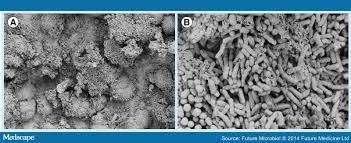
11 Biofilms Fig 6.5 Microbial communities form slime or hydrogels and share nutrients 1 st step: Attachment of planctonic bacteria to surface structures. Bacteria in biofilm are sheltered from harmful factors (disinfectants etc.) Cause of most nosocomial infections, i.e.: : Indwelling catheters Bacteria communicate by chemicals via quorum sensing The secret social lives of bacteria TED talk by Bonnie Bassler
12 Culture Media Culture medium: Nutrients prepared for microbial growth Have to be sterile (?) Inoculum: Introduction of microbes into medium Culture: Microbes growing in/on culture medium Chemically defined media: Exact chemical composition is known (for research purposes only) Complex media: Extracts and digests of yeasts, meat, or plants, e.g.: Nutrient broth Nutrient agar Blood agar

13 Agar Complex polysaccharide Used as solidifying agent for culture media in Petri plates, slants, and deeps Generally not metabolized by microbes Liquefies at 100 C Solidifies ~40 C
 that combine with O 2.")

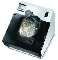
14 Anaerobic Culture Methods Reducing media contain chemicals (e.g.: thioglycollate) that combine with O 2. Keep in tightly capped test tubes. Novel method in clinical labs for Petri plates: Add oxyrase to growth media OxyPlate Anaerobic jars Anaerobic chambers Figs 6.6 & 6.7


15 Capnophiles Aerobic bacteria grow better in high and low In human body, these conditions found in E.g: Campylobacter jejuni Use candle jar, CO 2 - generator packets, or CO 2 incubators Candle jar


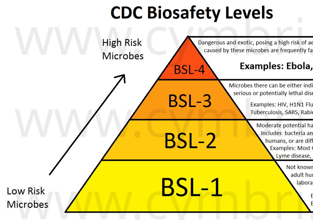
16 Biosafety Levels Biosafety Level 3 Biosafety Level 4

17 Selective and Differential Media Selective media: Additives suppress unwanted and encourage desired microbes e.g. EMB and mannitol salt agar etc. Differential media: changed in recognizable manner by some bacteria Make it easy to distinguish colonies of different microbes. MacConkey agar EMB Mannitol salt agar and hemolysis on blood agar Compare to Fig 6.9

18 MacConkey EMB

19 Compare to Fig 6.10 MSA
.")
20 Pure Cultures Contain only one species or strain. Most patient specimens and environmental samples contain several different kinds of bacteria Streak-plate method is commonly used. More details in lab. Colony formation: A population of cells arising from a single cell or spore or from a group of attached cells (also referred to as CFU). Only ~1% of all bacteria can be successfully cultured Aseptic technique critical! Compare to Fig. 6.11
: Frozen ( 54 to 72 C) and dehydrated in a vacuum.")
21 Preserving Bacterial Cultures Deep-freezing: Rapid cooling of pure culture in suspension liquid to 50 to 95 C. Good for several years. Lyophilization (freeze-drying): Frozen ( 54 to 72 C) and dehydrated in a vacuum. Good for many years.

 Ranges from")
22 The Growth of Microbial Cultures Binary fission exponential growth Budding Fig 6.12a Generation time time required for cell to divide (also known as doubling time) Ranges from 20 min ( ) to > 24h ( )


23 Consider reproductive potential of E. coli! Fig 6.13 Type of curve? Fig 6.14
 phase phase phase Compare growth in liquid and on solid media Foundation Fig 6.")
24 Bacterial Growth Curve Illustrates the dynamics of growth Phases of growth phase Exponential or logarithmic ( ) phase phase phase Compare growth in liquid and on solid media Foundation Fig 6.15

25 Direct Measurements of Microbial Growth Viable cell counts: Plate counts: Serial dilutions put on plates CFUs form colonies Fig 6.16


26 Fig 6.17 Figure 6.15, step 1


27 Additional Direct Counts 1. Direct microscopic count: Counting chambers (slides) for microscope 2. Filtration method of choice for low counts Fig 6.18
 is function of cell")
28 Indirect Count: Spectrophotometry Measures turbidity OD (Absorbance) is function of cell number Fig 6.21


29 H 2 O 2 and wound care Clinical Case: Glowing in the Dark