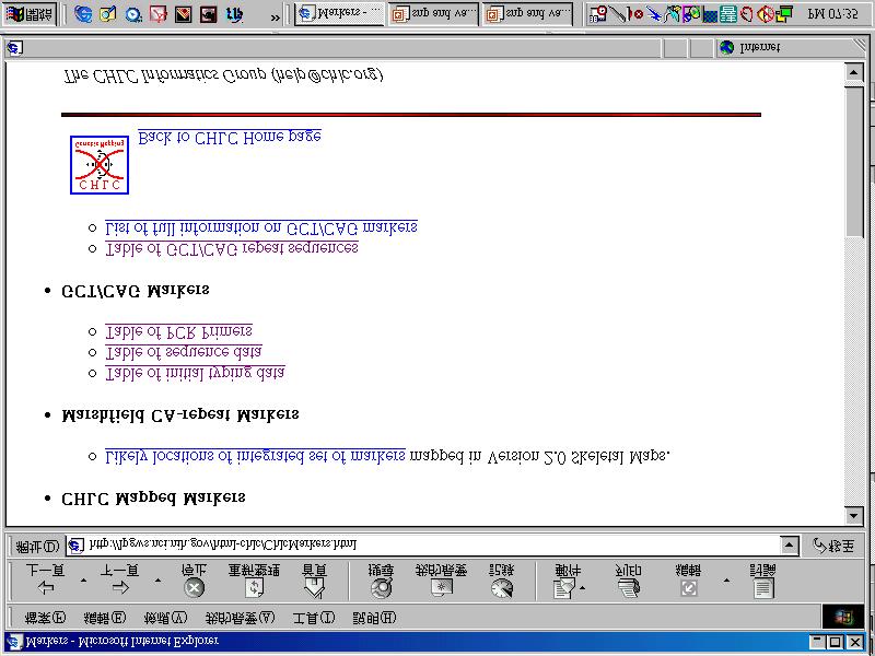
Microsatellite markers
|
|
|
- Claude Hicks
- 5 years ago
- Views:
Transcription
1 Microsatellite markers
2 Review of repetitive sequences

3 25% 45% 8% 21% 13% 3%
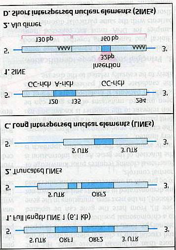
4 Mobile genetic elements: = dispersed repeat included: transposition: moving in the form of DNA by element coding for transposases. retrotransposition: moving in the form of RNA by element coding for reverse transcriptase. including: LINEs SINEs retrovirus-like elements (e.g,ltr; long terminal repeat)
5

6 LINE = A type of large repetitive DNA segment found throughout the genome. SINE = A type of small repetitive DNA segment found throughout the genome. Repeats such as Alu sequences are collectively called SINE.

7 Figure 9.25 Non-autonomous This refers to the fact that many of the transposable elements are missing some of the genes required for transposition; however, these elements can still move because other copies of the element in the genome encode the necessary gene products
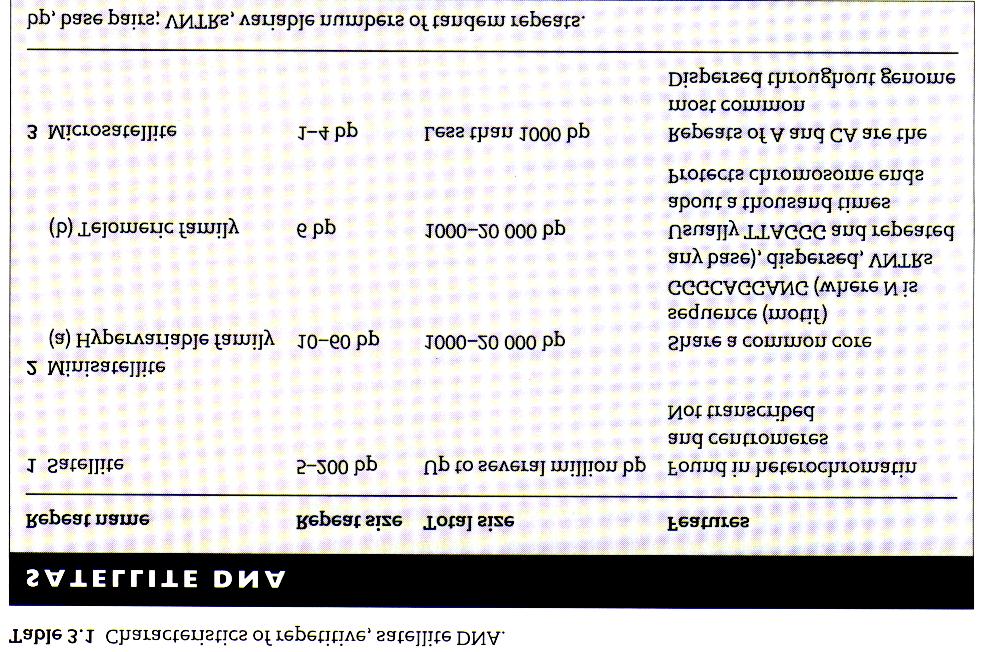
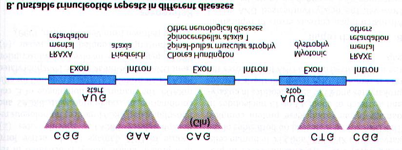
8 Tandem repeats (SSR; Simple sequence repeat): Sequences that are present in multiple copies adjacent to one another. SSLP (Simple sequence length polymorphism) e.g., satellite, minisatellite (VNTR; variable number TR), microsatellite (STR; short tandem repeat) All belong to SSR but with different repeat length.
, that are dispersed throughout the")
9 = VNTR = variable numbers of tandem repeats Consists of runs of dinucleotide repeats, most commonly CA(or TG on complementary strand), that are dispersed throughout the genome.
10
11 Microsatellite is distributed along the chromosome arm, in contrast to minisatellites, which are more abundant close to the telomeres in human.

12 Figure 9.27
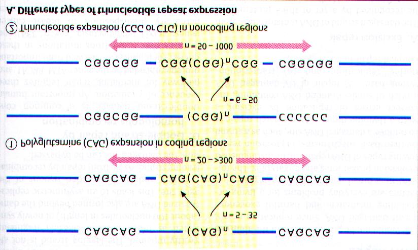
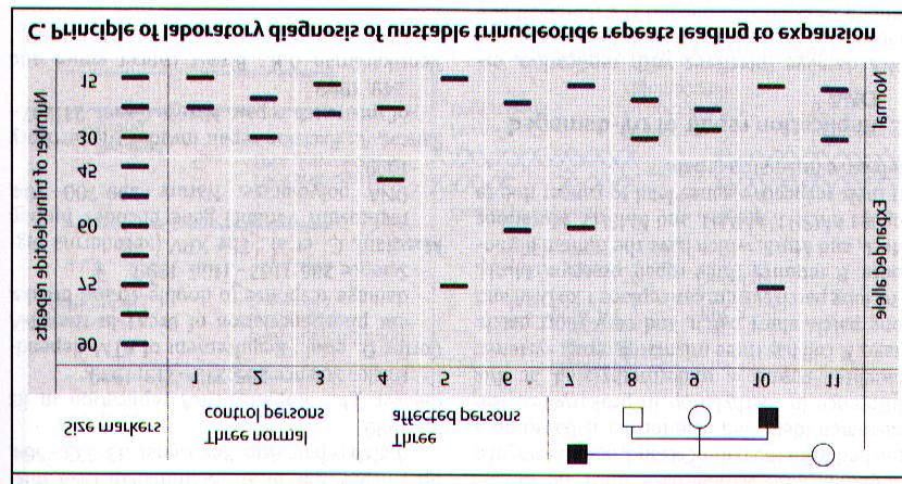
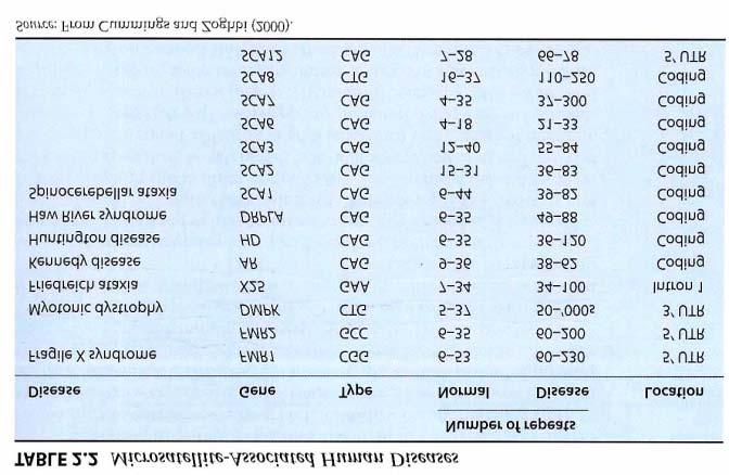
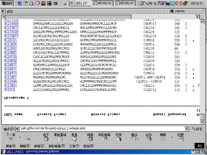
13 Microsatellite markers 1. Consist of STR, one to six nucleotide long. 2. In human, the five most abundant microsatellites contain repeat seq of A, AC, AAAN, AAN, AG. (decreasing order) (N= C, G, T) 3. The most common classes are : di-, tri- and tetra-nucleotide Freq rate 1/10kb 4. CA is the most common dinucleotide repeats. but tri- & tetra-nucleotide are often high polymorphic. 5. Microsatellites have a much higher mutation rate than standard sequences (up to gamates/generation)
14 Microsatellite markers (Cont ) 6. Microsatellites have a high probability of back mutation 7. Polymorphism is generated by loss or gain of repeats (MIN; microsatellite instability). Tool for cancer genetics. 8. Human microsatellites average at least 10 alleles with heterozygosity per locus over 80% A crucial tool in forensic analysis and paternity test. 9. As markers for pedigree analysis and family disease tracing.
15 Genetic mapping & resolution RFLP (coverage 1/ 10 cm) map single gene disorder. Microsatellite markers (1/10 kb) map more complex diseases SNP (1/1~1.5 kb) the third generation map & map functional variation in gene
16
17
18
19
20
21 Characterization of new genetic markers Submit the sequences to search for tandem repeat by program.
22

23 Protocol to detect multi-allelic markers
24 Database review
25
26
27
28
29
30
31
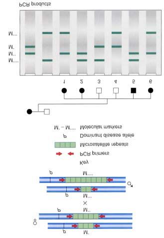
32
33
34
35
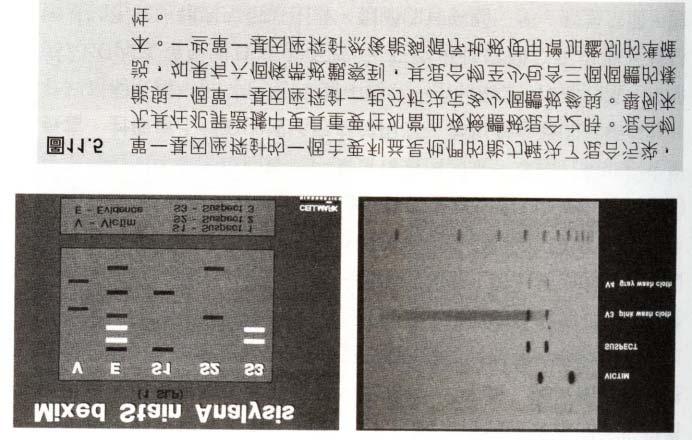
36
37
38 Genetic Instability of Cancer Genetic instability is a defining molecular signature of most human cancers. At the molecular level, it is characterized by microsatellite instability (MIN) or allelic imbalance (AI), representing losses or gains of defined chromosomal regions. Analysis of MIN/AI is useful in elucidating the molecular basis of cancer and also provides a molecular basis for cancer detection.
39 Two problems for Traditional Methods 1.Sample DNA is a mixture of neoplastic and non-neoplastic DNA 2.Sample DNA is often degraded to a variable extent
40 Parents Microsatellite marker Primers heterozygous
41 Parents delete Primer Microsatellite marker Normal Delete
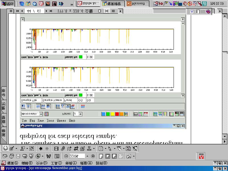
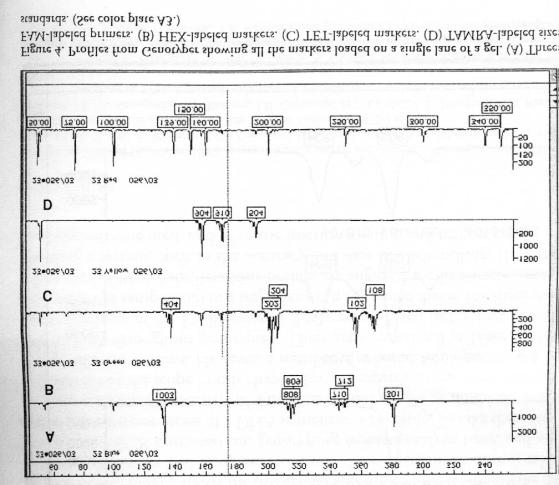
42 Parents delete destroy Primers Microsatellite marker Normal Delete Lose of heterozygosity (LOH)
43 Intact DNA Degraded DNA [degrade partly] No PCR false-positive for LOH
44 Normal Cancer Mixed
45 Cancer Normal Cancer Normal
46 Method for genotyping by microsatellite marker

47 SSLP (Simple sequence length polymorphism)

48 Microsatellite marker needs the long template (~200 to 350 bp)
")
49 Oral Oncology 36 (2000)

50 Fluorescence-based microsatellite genotyping Short tendem repeat
51
52

53 Sequencing for determining microsatellites
54
55
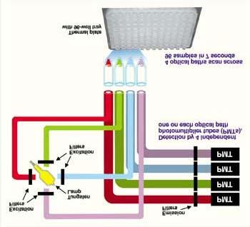
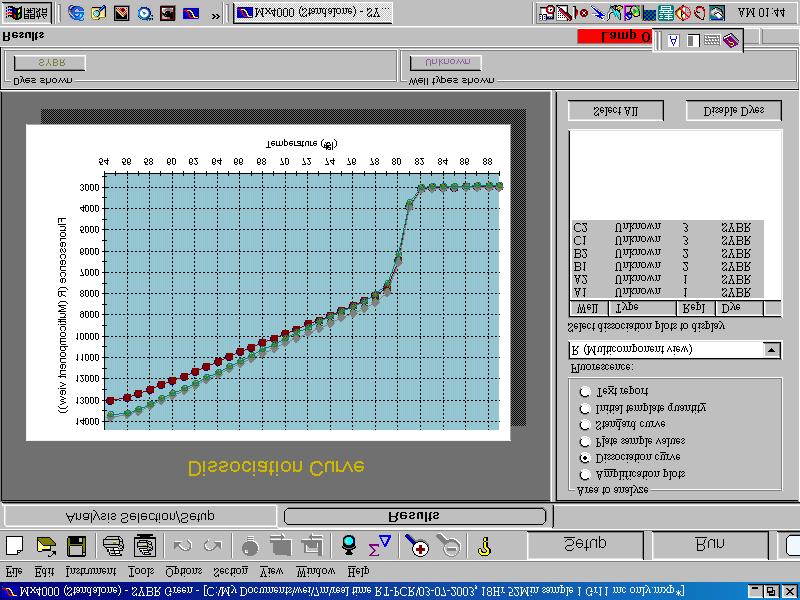
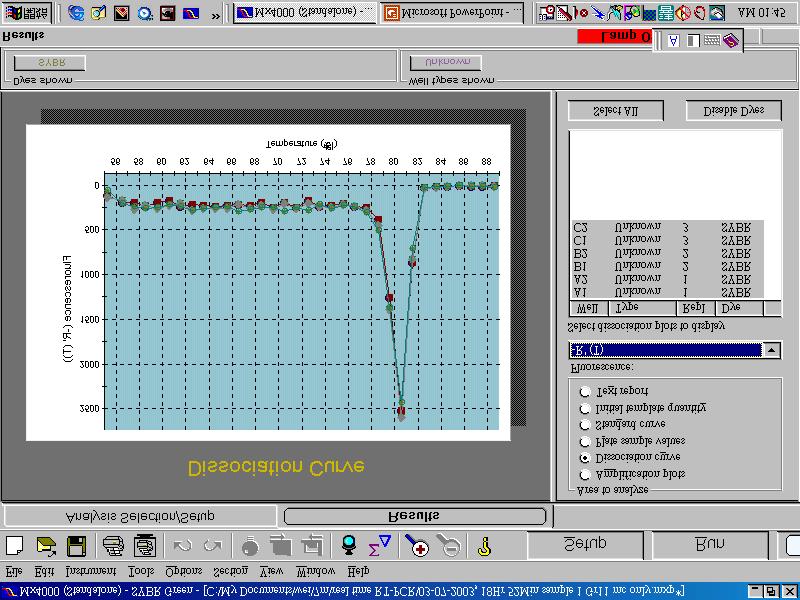
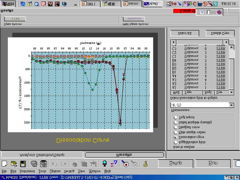
56 microsatellite marker detection By melting curve
57
58
59
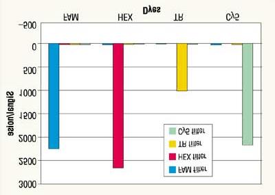
60 DNA released from non-neoplastic cells, can mask AI because it is difficult to quantify the allelic ratio using microsatellite markers.
61 Melting curve & DNA length
62
63

64
65 Real Time PCR
66
67
68

69 SYBR Green I stain is a highly sensitive fluorescent stain for detecting nucleic acids in agarose and polyacrylamide gels. The detection limit using SYBR Green I stain is as low as 60 pg per band of ds DNA using 300 nm transillumination. With 254 nm epi-illumination, as little as 20 pg of ds DNA can be detected. This is approximately 25 to 100 times more sensitive than ethidium bromide staining. The stain can also detect single stranded DNA and RNA, although the sensitivity is lower. The detection limit for oligonucleotides stained with SYBR Green I stain is 1-2 ng with 300 nm transillumination.


70 Gel Visualization New generation fluorescent nucleic acid stains SYBR Gold SYBR Green I for DNA SYBR Green II for RNA The best high-sensitivity reagents for staining DNA and RNA in electrophoretic gels post-electrophoresis stains
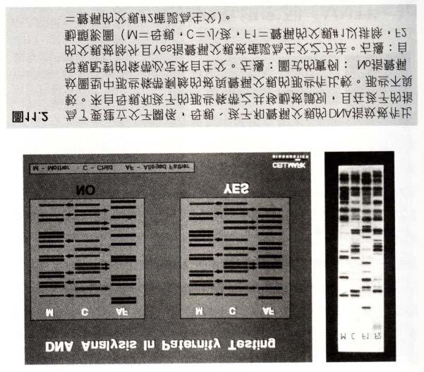
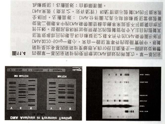
71 254nm

 Sensitivity")
72 Comparison of SYBR Green I and EtBr Mutagenicity: Ames test Mutat Res 439, (1999) Sensitivity

73 Real-Time PCR with SYBR Green

74 Real-Time PCR with SYBR Green

75 Real-Time PCR with SYBR Green
76
77
78
79
80 Paternity & microsatellite marker Microsatellite marker = Short tandem repeat (STR)
81
82
83

84 mother son daughter
85
86
87
88
89
90