[KCl] Heated. No SS III. No NE. unspliced spliced. b kda
|
|
|
- Thomasine Bridges
- 5 years ago
- Views:
Transcription
1 rrna level a 1 MDQGYGGYGA WSAGPANTQG AYGTGVASWQ GYENYNYYGA QNTSVTTGAT YSYGPASWEA 61 AKANDGGLAA GAPAMHMASY GPEPCTDNSD SLIAKINQRL DMMSKEGGRG GSGGGGEGIQ 121 DRESSFRFQP FESYDSRPCL PEHNPYRPSY SYDYEFDLGS DRNGSFGGQY SECRDPARER 181 GSLDGFMRGR GQGRFQDRSN PGTFMRSDPF VPPAASSEPL STPWNELNYV GGRGLGGPSP 241 SRPPPSLFSQ SMAPDYGVMG MQGAGGYDST MPYGCGRSQP RMRDRDRPKR RGFDRFGPDG 31 TGRKRKQFQL YEEPDTKLAR VDSEGDFSEN DDAAGDFRSG DEEFKGEDEL CDSGRQRGEK 361 EDEDEDVKKR REKQRRRDRT RDRAADRIQF ACSVCKFRSF DDEEIQKHLQ SKFHKETLRF 421 ISTKLPDKTV EFLQEYIVNR NKKIEKRRQE LMEKETAKPK PDPFKGIGQE HFFKKIEAAH 481 CLACDMLIPA QPQLLQRHLH SVDHNHNRRL AAEQFKKTSL HVAKSVLNNR HIVKMLEKYL 541 KGEDPFTSET VDPEMEGDDN LGGEDKKETP EEVAADVLAE VITAAVRAVD GEGAPAPESS 61 GEPAEDEGPT DTAEAGSDPQ AEQLLEEQVP CGTAHEKGVP KARSEAAEAG NGAETMAAEA 661 ESAQTRVAPA PAAADAEVEQ TDAESKDAVP TE b c Pol II p mock RNAse A hnrnp M mock RNAse A 6.2 5S 18S Supplementary Figure 1. associates with proteins involved in transcription and RNA processing through its YG-rich N-terminal region and in an RNA-independent manner. (a) Human protein sequence. YGG/YG sequences are highlighted in cyan, and RG sequences are in yellow. The cysteines in the two zinc finger domains are in red and underlined. (b) Immunoblot analysis of endogenous -associated proteins following immunoprecipitation by control (normal rabbit IgG) or anti- antibody from mock-treated or RNAse A-treated HeLa cell nuclear extract. (c) The 5S and 18S rrna levels in the mock-treated or RNAse A-treated HeLa cell nuclear extract were determined by RT-qPCR.
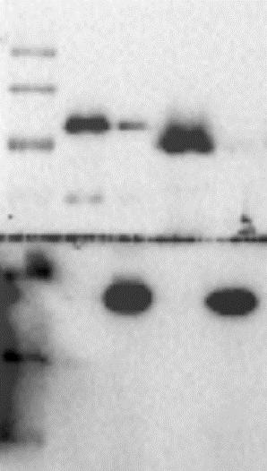
![a [KCl] 3 6 1 15 Heated No NE No SS III unspliced spliced 3 1 b kda 2 115 95 54 37 29 2 Supplementary Figure 2. is directly required for efficient minigene splicing.](/docs-images/86/93796483/images/2-0.jpg "(a) In vitro splicing assay under different conditions, including indicated final KCl concentrations in millimolar, using heat-inactivated nuclear extract, or no nuclear extract (no NE).")
2 a [KCl] Heated No NE No SS III unspliced spliced 3 1 b kda Supplementary Figure 2. is directly required for efficient minigene splicing. (a) In vitro splicing assay under different conditions, including indicated final KCl concentrations in millimolar, using heat-inactivated nuclear extract, or no nuclear extract (no NE). The last lane was from a normal splice reaction but no reverse transcriptase SS III was used in the reverse transcription reaction. All reactions here used HeLa nuclear extract batch 1, which was much more concentrated and thus more active than batch 2 used in Fig. 2e. (b) Coomassie staining of FLAG-tagged wild type or indicated mutants purified from Sf9 cells.
3 Relative mrna level a 293 H1299 mouse ESC hnrnp F hnrnp F Akap95 hnrnp F b hnrnp F GAPDH H1299 Mouse ESC hnrnp A1 hnrnp M hnrnp F hnrnp H GAPDH Supplementary Figure 3
4 % Input HA GAPDH FH- FH-(11-692) FH-ZF(C-S) no SSIII UV Lowly-expressed exogenous FH- FH-(11-692) FH-ZF(C-S) % Input % Input % Input % Input c Formaldehyde Highly-expressed exogenous FH- FH-(11-692) FH-ZF(C-S) d e UV Endogenous.25 No CL, anti-.2 UV, control Ab UV, anti Ab anti- f UV Lowly-expressed exogenous hnrnp F.2.1 FH-hnRNP F FH-hnRNP F kb 9 1 FAM126A FAM126A g FH-hnRNP F plasmid (ng) h.1, control Ab, anti-akap95 KD, control Ab KD, anti-akap95 FH-hnRNP F Endo. hnrnp F Anti-HA Supplementary Figure 3 (continued) 9 Mouse Fam126a 1 11 ENSMUST11519
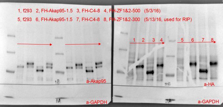
5 Supplementary Figure 3. directly regulates alternative splicing of FAM126A. (a) Expression of and hnrnp F was determined by RT-qPCR from the shrna- or sirna-treated cells as indicated, and normalized to GAPDH. Average ± SD from 3 biological repeats for each are plotted. (b) Immunoblotting of and hnrnp proteins following KD by shrna or microrna (mir#8 and mir#12). (c) Overexpression of FH- or its indicated mutants was induced by doxycycline in stable Flp-In T-REx 293 cell lines. The arrow points to the endogenous in the western blot on the left. The control is the parental Flp-In T-REx 293 cells. Anti-FLAG RIP assays were performed in these cell lines after formaldehyde-crosslinking, and followed by qpcr by a series of primers at the FAM126A locus. Reverse transcriptase was omitted for the no SSIII sample. Two independent repeats of RIP assays are shown. (d) This is another biological repeat of RIP assays shown in Fig. 3b, after UV-crosslinking of cells expressing exogenous wild type or mutant at levels comparable to the endogenous level. (e) These are two more independent repeats of RIP assays for endogenous shown in Fig. 3c, after UV-crosslinking of normally growing 293 cells. (f) These are two more independent repeats of RIP assays shown in Fig. 3d, after UVcrosslinking of 293T cells transfected with hnrnp F plasmid dose equivalent to that circled in Supplementary Fig. 3g. (g) Immunoblot analysis of 293T cell lysates following transfection of indicated amount of hnrnp F plasmid. Anti- and anti-ha antibodies were used for the top and bottom images, respectively. The circled dose represents the plasmid dose used in transfections for RIP assays in Fig. 3d and Supplemental Fig. 3f. (h) RIP assays using control (normal rabbit IgG) or anti-akap95 antibody were performed in control or Akap95-depleted (KD) mouse ES cells after UV cross-linking, and followed by qpcr at the mouse Fam126a locus.
6 % Input % Input % Input Binding sites per 1 6 nt a Binding sites b c Proximal intron Distal intron 5' UTR 3' UTR Exon Not annotated Binding coverge d Formaldehyde RIP Cross-link, RIP Ab, cells: no CL, anti-flag, FH- Formaldehyde, anti-flag, control Formaldehyde, anti-flag, FH- Formaldehyde, anti-flag, FH-(11-692) Formaldehyde, anti-flag, FH-ZF(c-s) UV RIP Cross-link, RIP Ab, cells: No CL, anti-flag, FH- UV, anti-flag, control UV, anti-flag, FH- UV, anti-flag, FH-(11-692) UV, anti-flag, FH-ZF(c-s) UV RIP Cross-link, RIP Ab: No CL, anti- UV, rigg UV, anti-.1 NPDC1 LENG8 ACTN4 CPSF1 TTLL3 PI4KAP1 SMPD4 Supplementary Figure 4
.74.74 FH-(ZF C-S ).74 KD.74.74 Input KD.74.74 PLCXD1 Supplementary Figure 4 (continued)")
7 RPM RPM RPM e RIP: cell: 5kb.1 1kb.46 FLAG FH- FH-(11-692) FH-(ZF C-S ).1.46 Input KD KD FAM126A FOXO3 RIP: cell: 1kb.29 FLAG FH- FH-(11-692) FH-(ZF C-S ).29 Input KD KD KDM2A RIP: cell: 5kb.74 FLAG FH- FH-(11-692) FH-(ZF C-S ).74 KD Input KD PLCXD1 Supplementary Figure 4 (continued)
8 RPM RPM RPM f RIP: cell: 5kb 1.6 FLAG Input FH- FH-(11-692) FH-(ZF C-S ) KD KD ZNF451 RIP: FLAG Input cell: FH- FH-(11-692) FH-(ZF C-S ) KD KD 5kb kb ANKLE2 NPDC1 RIP: cell: 5kb.73 FH-.73 FLAG FH-(11-692).73 FH-(ZF C-S ).73 Input KD KD AP3D1 Supplementary Figure 4 (continued)
9 Supplementary Figure 4. preferentially binds to introns of cellular pre-mrnas. (a) Distribution of the numbers of FH- binding sites within different categories of genic regions, as calculated from MACS peak counts of the anti-flag RIP results from 293 cells induced to express FH-. The color legend applies to a-c. (b) FH- binding sites in each 1 million nucleotides of different genic regions, using anti-flag RIP results from 293 cells induced to express FH-. (c) Distribution of the genomic region coverage of binding sites within different categories of genic regions, as calculated from MACS peak coverage of anti- RIP results from 293 cells (left) or anti-flag RIP results from 293 cells induced to express FH- (right). (d) qpcr results of UV RIP assays for binding at introns of indicated genes, where strong binding peaks were found in the formaldehyde-based RIP-seq. (e and f) Representative RIP profiles. In blue are the Anti-FLAG RIP-seq profiles from parental 293 cells (control) or stable 293 cells overexpressing the FLAG-tagged wild type or indicated mutants. In red are the anti- RIP-seq profiles from parental 293 cells (control) or 293 cells stably expressing micrornas ( KD). In black are the sequencing profiles of total input RNAs from the corresponding cells for the anti- RIP-seq. All profiles have the same y-axis scale for each gene so that they are directly comparable. Note that, compared to the FH- binding, FH-ZFc-s binding was drastically reduced at all shown transcripts, but FH-(11-692) binding was either unaffected (e) or modestly reduced (f) at different transcripts or different regions within the same transcript. Arrows at the FAM126A panel point to the two intronic binding peaks shown by qpcr in Figure 3. The broken vertical lines in the KDM2A and PLCXD1 panels were positioned at an exon to clearly show that the nearby binding peaks were not at the exon, but rather, proximal to the exon. In the ZNF451 panel, the left sub-panel shows a zoomed-in image of the first intron. Arrows on the gene diagrams indicate the direction of the gene transcription.


 Volcano plot for exons whose normalized usage was significantly affected (Padj<.5 for red and yellow, red for fold change over 1.")
10 Gene function Percent included -Log 1 Padj -Log 1 Padj a 1 b c down >1.5 fold up >1.5 fold Log 2 fold change Log 2 fold change d Tmem258 Aa Wsb1 Etv4 VPS9D1 CRBN e ATP-binding chromatin regulator chromatin modification histone methyltransferase complex transcription PHD Zinc finger chromosome organization RNA-binding mrna metabolism RNA processing RNA splicing cell cycle protein localization microtubule cytoskeleton down up f Chromatin/ transcription regulation RNA processing Gene expression 1E+ 1E-1 1E-2 P value Supplementary Figure 5. preferentially promotes exon inclusion. (a) Volcano plot for exons whose normalized usage was significantly affected (Padj<.5 for red and yellow, red for fold change over 1.5) or not (Padj>.5, blue) by Akap95 shrna #1 compared to scramble shrna in mouse ES cells. Fold change is the ratio of the normalized exon level in the knockdown over that in the control cells. See Supplementary Table 2 for the gene list.
11 (b) Pie chart showing the number of exons whose normalized usage was significantly (P<.5) reduced (blue) or enhanced (red) by Akap95 shrna #1 compared to scramble shrna in mouse ES cells. (c) Volcano plot for genes whose expression was significantly affected (Padj<.5 for red and yellow, red for fold change over 1.5, and only very few significantly affected genes were changed less than 1.5 fold) or not (Padj>.5, blue) by Akap95 shrna #1 compared to scramble shrna in mouse ES cells. Fold change is the ratio of the normalized exon level in the knockdown over that in the control cells. (d) Validation of the DEXseq analyses on the effects of knockdown on inclusion of exons in mouse ES cells (for Tmem258, Chpt1, As465934, Wsb1, and Etv4) or human 293 cells (for VPS9D1 and CRBN). Following quantification of band intensity by the ImageJ program, the percentage of the PCR product representing exon 11-included transcript in the total transcripts was calculated and shown as percent included on the Y axis. Because the exons shown in this panel appear to be mostly skipped, plotting percent skipped as in Fig. 3a shows only modest (yet statistically significant) changes (data not shown). Average ± SD from biological duplicates are plotted. P<.5 between control and KD for all these genes except Tmem258. (e) Gene ontology analyses of genes that contain significantly (P<.1) affected exons upon knockdown by shrna #1 in mouse ES cells, based on 632 DAVID IDs with downregulated exons and 322 DAVID IDs with up-regulated exons. Gene functions in light blue shade are for chromatin/transcription regulation, and gene functions in pink shade are for RNA binding and processing. (f) A diagram showing that may regulate gene expression through regulating alternative splicing of RNAs encoding factors involved in chromatin and transcription regulation and factors involved in RNA processing, based on results in Fig. 5f and Supplementary Fig. 5e.

 RNA binding profiles of exogenous and endogenous (blue and red, respectively) at RNA")

 The model shows how might regulate the splicing of FAM126A pre-mrna by binding")
12 RPM RPM a USPL1 ASPH FH- 1kb.1 1kb hnrnp H hnrnp F hnrnp A1 PICALM 5kb.1 PPP1R7 1kb.1 FH-.1.1 hnrnp H hnrnp A2B1 b Supplementary Figure 6. A possible model for the role of in splice regulation. (a) RNA binding profiles of exogenous and endogenous (blue and red, respectively) at RNA regions flanking the exons (marked by red circle) regulated by and hnrnps, as shown in Fig. 6a. Shown at the bottom are binding regions of hnrnp proteins taken from (b) The model shows how might regulate the splicing of FAM126A pre-mrna by binding to intronic regions near the splice sites and also facilitating splice site communication via its interaction with hnrnps and/or itself.


")


 1e")

13 For Fig. 1d KDa Pol II H5 (S2-P) 2 DDX5 DDX17 1 Pol II H14 (S5-P) Pol II 8WG16 (un-p) 2 KDa 2 A: control IP B: IP C: DDX5 IP D: hnrnp U IP E: hnrnp K IP KDa 2 N-2 (total) For Fig. 1e Input pull-down a-hnrnp H1 Supplementary Figure 7


14 For Fig. 1f Pull down M2-pull down Input For Fig. 1g For Fig. 1h a-flag a-his Supplementary Figure 7 (continued)


15 For Fig. 2c For Fig. 2d For Fig. 2e For Fig. 3a For Fig. 3b , con-sh 2, Akap95-sh#2 3, Akap95-sh#1 Supplementary Figure 7 (continued)


16 For Fig. 6a USPL1 PPP1R7 KRIT1 1, f293-scr 2, f293-shakap95#1 3, f293-shakap95#3 WDR85 PICALM G2AD ASPH , f293-scr 2, f293-shakap95 For Fig. 7a Supplementary Figure 7. Uncropped blots. Note that some of these blots are merged with marker to show size, and blots in the main figures are unmerged and some may be from different exposure times.
17 Supplementary Table 1. Mass spectrometry results for -associated proteins. Major proteins co-immunoprecipitated with FH- or FH- (11-692) were identified by MALDI mass spectrometry, categorized and listed in the left column, and followed by their molecular weights (MW), mass spectrometry protein scores and numbers of matched peptides. Mass spectrometry protein scores are defined as the combined ion scores of all peptides matched from mass spectrometry peak lists. The ion score is calculated by the Mascot software as -1Log P, where P is the probability that the observed match of each peptide sequence is a random event. As the same protein was often detected in multiple segments excised from the gel (Figure 1b) with different scores and numbers of matched peptides, only the highest score and number of matched peptides for that protein are listed. Proteins in red are those with protein scores over 2. NF, not found. FH- FH-(11-692) MW (kd) score # peptides score # peptides hnrnps hnrnp M hnrnp H hnrnp H hnrnp H hnrnp F hnrnp D NF hnrnp U NF hnrnp K NF DEAD-box helicases DDX5 (p68) NF DDX17 (p72) NF DDX17 (p82) NF DDX3X NF DDX3Y NF RNA helicase A (DHX9) NF Other RNA-associated proteins RBM14 (CoAA) NF EWS NF NHN1 (ZC3H18) NF HA NF ZNF NF DBC1 (CCAR2) NF FAM12A (C9orf1) NF KHDRBS1 (SAM68) NF ZCCHC NF
18 NOP NF NOL NF NOP NF SFRS NF SFRS14 (SUGP2) NF Others KLHL NF PELP NF DNA-PKc NF CAD NF NFYC NF Supplementary Table 2. shrna, sirna, and mirna sequences RNA format Species target Sequence 5-3 Akap95 #1 GCCCGACAAGACAGTAGAATT Mouse shrna Akap95 #2 GCCTGTTCTGTATGCAAGTTT Human ATTAGTTACTACCACTCAAAT 3 UTR GAAAGGAGGCUGUAGAAUA coding GAAUUGAUGGAGAAAGAAA sirna Human GUCCAUGGCUCCCGACUAC ON-TARGET plus GGAACGAGCUGAACUACGU SMARTpool GAACAACAGACAUAUAGUG GAUCGAGGCUGCUCACUGC MiRNA Human mir 1 TAAAGTTGGAACTGCTTCCGT mir 2 TTTCGGAGAAATCTCCTTCAC Supplementary Table 3. PCR or qpcr Primers App. Species gene Forward primer (5-3 ) Backward primer (5-3 ) expression Human GAPDH CCTTCATTGACCTCAACTACATGG TCGCTCCTGGAAGATGGTGATGGG expression human ACTB CCTTCAACACCCCAGCCATGTACG GGCACAGTGTGGGTGACCCCGTC expression human 5S rrna GATCTCGTCTGATCTCGGAAG GGTATTCCCAGGCGGTCT expression human 18S rrna ACAGGATTGACAGATTGA TATCGGAATTAACCAGACA expression human GCCAAGGCCAATGATGGCGGCCTG GCCCCTGCCTCCTTCCTTGGAC expression human hnrnp F GGGAAACACAAGGAGAGGATAG GCACGGACATGAACTTCAGA expression mouse Gapdh CATCTTCTTGTGCAGTGCCAG GGCAACAATCTCCACTTTGCC expression mouse Akap95 TGTACCGACAACTCAGACTCG GTAGGACTCGTATGGCTGGAA Splicing- ptn23/2 PCR 4 SplicingqPCR minigene Spliced ptn24 TTCACCAGCTGTTGGGTAA TGAGGTTGTTGGTGACATTCT splicing mouse Fam126a CACACCAACCTCCTCTAGAATATC CGTCTCTGGTTCTTTCCTACAC splicing human FAM126A TGCAGTAACCAGCATGTCAA CTAGACGCAGCCCTGGAATA splicing human KRIT1 ATCTCGGTGGTCCAACTCAG TGGCAGTATTCTTTGGACGA splicing human PPP1R7 AGTCGCAGGAGATGATGGAG TCCCTATGCGATAGTGATTCAA splicing human ASPH TCGAAGATGAAGCAAAAGAACA CTTCCACGTGGTAACTATGCTC splicing human WDR85 CTGGTCGAGGTCCAAAGAAA TCCTCCCCAACCCTCTCTAT minigene CGCGGATCCGTACTCCCTCTCAAAAGC CCGGAATTCCTTCTCCGCCTGAGCCTC
19 splicing human USPL1 ACGTTGCAACTAGGGTGGAG CAAGCAGGGCAATACTCATCT splicing human G2AD AGCTGGCCAAACTGCTCTAC CAGGTCTCGGCACATCTCA splicing human PICALM GCCCAATGATCTGCTTGATT ATTAAGGCCAGCTGAAGGGT splicing mouse Tmem258 GAGCTCGAAGCCATGAGTAG GACTCCAAAGCCCATGAAGA splicing mouse Aa TTTGAGAACCTGTGTGTAAGTCC ATTGATTCCAATTCAGGGCAGA splicing mouse Wsb1 CATTCTCTCCCGACTGTTCTATG TACTCAGGAGTCTGGACATTACT splicing mouse Etv4 GCAGAACTTCAAGCAGGAGTA GGGAATGGTCGAAGGGATTT splicing human VPS9D1 GCCATGAAGCTTGCCAAC GGCCCTCTCCAGACACT splicing human CRBN GGAAGCACAGTTTGGAACAAC CTGGCACTTATTGAGGGATTCT App. Species gene Forward Backward Forward primer (5-3 ) start* start* Backward primer (5-3 ) RIP human FAM126A GGCAGAAGTGACCTAGGATTG GAGGACGAAGCAGGAGTATTAAG RIP human FAM126A CCTATACCATCAAGGGCAGTTT CATACAAGTGGGAGCCTGTATC RIP human FAM126A GGAGAATGGGATCTAGCTCAA CAGCAATGGCTCTGGATATAAT RIP human FAM126A CTCCCGAGTAGCTGGGATTA GATCAGCCTGACCAACATGAA RIP human FAM126A TCCCTCTTAGCCTCCTATTCTT CAACATGAGCAGATAGAGCAGATA RIP human FAM126A ACACCAACTTCCTCTCGAATATC ACAATGGTTTGACTTGAAGAGTTAC RIP human FAM126A TCAGTGAACTCTTAAGCAAGTGA CGTCAACACTATGGCATTCTTTAC RIP human FAM126A TCCCTTCTTGTCTGCTTCATT AAGGGAGGGAAATGGGAAAG RIP human FAM126A GTACTGGTTGTGTTGTGGTTTC CAATGGCAACTGTTCTCATCAC RIP human FAM126A -118 ACTGCTACAGGAAGCACAAG CTGCAGGACCTACATTTCTCTC RIP human FAM126A TCCCTTTATCCAGTGGACTCTA GGAAAGCCAAGACTACCCTAAA RIP human FAM126A TTGGAGAGATCTGTTTGACAACT CACATTAAGTTAAGCCATGTCCATAC RIP human FAM126A ACTCCTCCTTTCTCCACCTT ACAGGTCAGCAACACCTATTAC RIP human FAM126A -518 TCCCAGACCACCCATCTAA -544 GGAACAGAGCTCACACTGTAATA RIP human FAM126A AAGGAAGAGGGTATGTGAAACG -348 CTTCTCCTGGTGGCTGAATG RIP human FAM126A GTTGTGTAGCTCTTCCTGAACT -28 TTGCCCTAAGTCGCTTTCTT RIP human FAM126A -196 TGGACTGGAGATCCCTTTAGTA GAGGTCACTGCAACTGAGATAA RIP human FAM126A GGCTCCCAGAAGTGCATATT CTGGGAACCACTGCTCTATTC RIP human FAM126A -135 CAGCTAAGTTAGGGAACCACAG -127 CAGACTCCCACTAATGATGCTAAA RIP human FAM126A -119 GAATTGGGCATCCTTGTCATTC -15 CTGAGCATCAATCTCTCCTAGTG RIP human FAM126A 81 TGGAGGATGGAATAAACTCACC 198 CTGTGCAACTGCTGCTTAAC RIP human FAM126A 936 GGAAATACAGAATTAACAGGTCAAGAA G 115 GACGCAGCCCTGGAATAAA RIP human FAM126A 1177 CAGAGAGCCCAAAGTGAGAAT 1278 CTTAGCCAAACCATGGCTACTA RIP human FAM126A 2132 TGGTAACCCAGTGCCTAGAA 2189 ACAACACACCTACCAGCAATC RIP human FAM126A 2889 GACCGTTGAGGAAGACCTAAG 2938 GTGACTACAGCAAGGGACTATG RIP human FAM126A 4666 AAATGTCTTCCCTTCCCTTCC 4713 AGTCTTGAGAGCTTGCCTTAAA RIP mouse Fam126a AGTAAGGAAGGTACAAGGTGTATTC CCTCGTATTGACATGCTGGTAA RIP mouse Fam126a GACTGTCTCAGGCTACAACTTC ATCATAAACAGGAGGCACTCAA RIP mouse Fam126a CCACAGTGGCCATTCTGTATT -185 CTTACCCAGGATCCCATCAAAC RIP mouse Fam126a -981 GCTCGCTCACCTTCTTCTTT CCTCTGATGGATGCTTGTCTTC RIP mouse Fam126a -895 GAGTGTCTGTGTCTGCTCTTAAA AACGACTGGCTTGGAAAGTTA RIP mouse Fam126a CTGGCAAGTGGTTGCAAATAA -751 GGACAGACACACAGAGGAAAG RIP mouse Fam126a TTTCAGAGGTGGGAGGGATTA GTTGGTACCTAAGAGGCTGAAG RIP mouse Fam126a -622 GGAAGGTCTGGAGTCTGTTATG ATGGATGTACTGAGCACCTATG RIP mouse Fam126a AGGATGTGTATGGCTCTTTGG AGGCTCCTGAAACTTCTTTGT RIP mouse Fam126a GTGGAGGTCAGAGGACAATTT -469 TAGGGAAAGGCACTTGCTATG RIP mouse Fam126a GCATATGAAATGTAGGGATGTTGAG -282 AGCTGGAAGGCACATCTTAC RIP mouse Fam126a TTCTTGACGTGGTTTCTACAGTAT -163 GGTGTGTGGCAGAGGTTATAG RIP mouse Fam126a -745 GTCTCAATAGGAACCTGAAGAAATG -624 CAATCATGCCTTGGTGCTTAG RIP mouse Fam126a -26 GACATTGACGCCCAGCATA 99 GGACATTCCATCCTCCATCATT RIP mouse Fam126a 515 TGGAGAACACAACCTTCCTAAA 651 TGCAAAGAGCAGAAGCAATTAG RIP mouse Fam126a 113 GAGCGAAGGAGAGAGAAACTG 126 CTTCAGAGAGAGAAGCTCAAGG RIP mouse Fam126a 4219 CCAAGTGAAGAGAGAAGCAGAG 438 GTGCTACAGAGATGGGCTTAAA RIP human NPDC CCCTAGTTCCTAACCACACTTTC TCTCCAGCACCAGGATAAGA RIP human LENG CAGTGTCCTTCGTTCACTAGAC CACCTCAATCACCAGACAGAA RIP human ACTN GAGTATGGAGCCCGACAAAG AGTGCATGGCTAGGAAATGG RIP human CPSF GGTGGTGTGGTGGCTTAAATA CTTGGCTGCTCTGTGACTTT RIP human TTLL GCTGAAGCTCTGGCTCATATT GGGCACATTAGGGAAGGATAAG RIP human PI4KAP GGGAAGGTGGTTGGAATTAGAT CTGACCTACACCTGTCCTTTG RIP human SMPD CCATACAAACAGCCCACTCT CTGCCATCTCTTGCATCTCATA * The position is relative to the center of exon 11 of human FAM126A or exon 1 of mouse Fam126a, which is set as. For the rest genes in RIP assays, the position is its chromosomal coordinate on hg19 (GRC37).