Supplementary Figure 1. Additional RNAi screen data
|
|
|
- Bruce Sparks
- 5 years ago
- Views:
Transcription
1
2 Supplementary Figure 1. Additional RNAi screen data A. Cisplatin induced ATR autophosphorylation. Western blot illustrating ATR and phospho-atr (T1989) in cells exposed to 1 µm cisplatin for 24 hours prior to cell lysis. B. Cisplatin induced ATR autophosphorylation. Quantification of the level of cisplatin induced ATR autophosphorylation determined in (A). C. Sensitivity of MCF12A and HCC1143 to VE-821 in RNAi screen format assay. Dose response curves illustrating MCF12A and HCC1143 sensitivity to VE-821 following five days of drug exposure in a 384 well plate format. D. Bar chart illustrating VE-821 DE Z-scores from the highthroughput screen for genes implicated in ATR activation. ATR was represented two times in the RNAi screening library and both replicates are shown. E. Bar chart illustrating VE-821 DE Z-scores from the high-throughput screen for ERCC1, ERCC4 and ATM in HCC1143 and MCF12A. ERCC4 was represented two times in the RNAi screening library and both replicates are shown.
3
4 Supplementary Figure 2. Validation of ATR/ARID1A synthetic lethality A. Dose response curves illustrating 384 well plate cell survival data from HCC1143 cells transfected with sirna SMARTpool targeting ARID1A (red) or sicon (blue). Twenty-four hours after transfection, cells were exposed to VX- 970 for five continuous days. Error bars represent SD from triplicate experiments. Survival curve siarid1a vs. sicon p-value <0.0001, ANOVA. B. Bar chart illustrating Log2 surviving fractions at 1 µm VX-970 in HCC1143 cells. Cells were transfected with individual ARID1A sirna (#01, #02, #03 and #04). 24 hours after transfection cells were exposed to VX-970 (0.1 µm) or DMSO for five days. Viability was then estimated using CellTitre-Glo. Asterisk indicates statistically significant difference (Student s t-test, p<0.05). C. Dose response survival curves of HCT116 ARID1A +/+ and ARID1A -/- cells exposed to methotrexate for five days prior to estimating cellular viability using CellTitre-Glo. D. Dose response survival curves of HCT116 ARID1A +/+ and ARID1A -/- cells exposed to taxol for five days prior to estimating cellular viability using CellTitre-Glo. E. Dose response clonogenic survival curves of HCT116 ARID1A +/+, ARID1A +/- and ARID1A -/- exposed to increasing concentrations of VX-970. Dose response curves from clonogenic assay are shown. Error bars represent SD (n=3), ANOVA p-value of <0.001 comparing ARID1A +/+ vs. ARID1A +/- and ARID1A +/+ vs ARID1A -/-. F. Western blot of ARID1A protein expression in HCT116 ARID1A isogenic cells. G. Arid1a -/- mouse ES isogenic cells are sensitive to VE-821. Dose response curves from clonogenic assay are shown. Arid1a mouse ES isogenic cell lines were exposed to increasing concentrations of VE-821 for 10 days in a colony formation assay. Cells were then fixed, stained with SRB and colonies of 30 or more cells counted to estimate cellular viability compared to DMSO treated controls. Error bars represent SD from triplicate experiments. ANOVA p-value of < H. ARID1A-deficiency induces comparable ATRi sensitivity to loss of ATM. Dose response curves for a panel of human tumor cell lines annotated by ATM or ARID1A mutation status. The indicated cell lines were exposed to VX-970 for five days prior to estimating cellular viability using CellTitre-Glo.
5
6 Supplementary Figure 3. Olaparib, BMN673, VX-970 and cisplatin ARID1A -/- selectivity. HCT116 ARID1A isogenic (+/+ and -/-) cell lines were exposed to increasing concentrations of (A) olaparib, (B) BMN673, (C) VX- 970 and (D) cisplatin. Following five days of drug exposure cellular viability was estimated by CellTitre-Glo. E. Bar chart represents the therapeutic window as represented by the fraction of AUC ARID1A+/+ / AUC ARID1A-/- for the indicated drug. F. Box and whisker plots illustrating AUC data from ARID1A wildtype (WT, n= 3) or mutant (Mut, n=6) OCCC cell lines exposed to VE-821. Cells were exposed to VE-821 for five days prior to estimating cellular viability by CellTitre-Glo. Asterisk indicates statistically significant difference (p<0.05). G. As (F) for ARID1A wildtype or mutant (mut) OCCC cell lines exposed to BMN 673. H. As (F) for ARID1A wildtype or mutant (mut) OCCC cell lines exposed to olaparib.
7
8 Supplementary Figure 4. Additional validation of ATR/ARID1A synthetic lethality A. Dose response curves illustrating 384 well plate cell survival data from HCT116 p53-/- cells transfected with sirna targeting ARID1A (red) or sicon (blue). Twenty-four hours after transfection, cells were exposed to VX-970 for five continuous days. Error bars represent SD. Survival curve siarid1a vs. sicon p-value <0.001, ANOVA. B. Dose response curves illustrating 384 well plate cell survival data from HCT116 and HCT116 ARID1A -/- cells transfected with sirna targeting TP53 (red dotted) or sicon (blue, red solid). Twenty-four hours after transfection, cells were exposed to VX-970 for five continuous days. Error bars represent SD. C. Z score box whisker comparison plot for 86 human tumor cell lines transfected with ATR sirna. Cells lines were transfected with sirna and cell viability was estimated five days later by CellTitre-Glo reagent. ATR sirna Z-scores for ARID1A wildtype (n=65) and ARID1A mutant (n=21) were found to be statistically different (p=0.0084, median permutation test). D. Synthetic lethality with ATR sirna HCT116 ARID1A +/+ and -/- cells were transfected with control sirna or sirna targeting ATR. Western blot demonstrating ATR protein level 96 hours following sirna transfection. E. Bar chart illustrating cell viability 96 hours following sirna transfection. Surviving fractions are normalized to control sirna for each cell line. Asterisk indicates statistically significant difference (Student s t-test, p<0.05) between HCT116 ARID1A -/- and HCT116 ARID1A +/+. F. Dose response curves for TOV21G (ARID1A mutant) and RMG1 (ARID1A wildtype) cells exposed to VE-821 for five days prior. G. Western blot illustrating γh2ax in TOV21G cells exposed to VX-970 (0.5 µm). Unt = untreated. H. Western blot showing Cyclin B1 levels in cytoplasmic (Cyt) and nuclear (Nuc) fractions of HCT116 ARID1A isogenic cells following exposure to VX-970 (0.5 µm) for 24 hours. Quantified levels of Cyclin B1 are shown. I. Silencing TOP2A sensitises HCT116 cells to VX-970. Surviving fraction curves from 384 well plate cell of HCT116 cells transfected with sirna targeting TOP2A (red) or sicon (blue). Forty-eight hours after transfection, cells were exposed to VX-970 for five continuous days. Error bars represent SD. Survival curves sitop2a vs. sicon p-value <0.001, ANOVA. J. Western blot illustrating TOP2A protein silencing.

9
10 Supplementary Figure 5. ATRi induced γh2ax in ARID1A -/- cells prior to mitotic entry A. Representative immunofluorescence images of HCT116 ARID1A +/+ and ARID1A -/- cells stained with DAPI, Cyclin A, γh2ax and a merged image, following treatment with DMSO, IR (10 Gy, 2 hours) and VX-970 (0.5 µm, 2 hours). Scale bar equals 75µM. B. Fraction of Cyclin A negative staining positive for γh2ax (>10 foci and/or pan nuclear staining) following DMSO or VX-970 (0.5 µm, 2 hours) treatment in HCT116 ARID1A +/+ and ARID1A -/-. Error bars represent SD for 3 biological replicates. No less than 200 cells were counted on four large field images for each replicate. P-values calculated using Student s t-test and * indicates statistical significance (p<0.05). C. Fraction of Cyclin A positve staining positive for γh2ax (>10 foci and/or pan nuclear staining) following DMSO or VX-970 (0.5 µm, 2 hours) treatment in HCT116 ARID1A +/+ and ARID1A -/-. Error bars represent SD for 3 biological replicates. No less than 200 cells were counted on four large field images for each replicate. P-values calculated using Student s t-test and * indicates statistical significance (p<0.05). NS indicates the comparison is not statistically significant (p>0.05 by Student s t-test). D. Representative images of the various classifications used to score cells as H2AX positive in panels B and C. Scale bar equals 6 µm. E. Western blot of cell lysates taken from the indicated time points from HCT116 ARID1A +/+ and ARID1A -/- cells following release from synchronisation in G 1 /early S phase by double thymidine block. Cells were released from thymidine block into media containing DMSO or VX- 970 (0.5 µm).

11
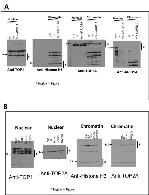
12 Supplementary Figure 6. A. Uncropped scans of western blots presented in Figure 1F. B. Uncropped scans of western blots presented in Figure 1H.

13
14 Supplementary Figure 7. A. Uncropped scans of western blots presented in Figure 2B and 2L.

15
16 Supplementary Figure 8. A. Uncropped scans of western blots presented in Figure 3H.
17
18 Supplementary Figure 9. A. Uncropped scans of western blots presented in Figure 4D. B. Uncropped scans of western blots presented in Figure 4F.

19
20 Supplementary Figure 10. A. Uncropped scans of western blots presented in Figure 5H. B. Uncropped scans of western blots presented in Figure 5F.