Plasma cell neoplasms
|
|
|
- Angel Douglas
- 5 years ago
- Views:
Transcription


1
2 Plasma cell neoplasms
3 WHO CLASSIFICATION OF PLASMA CELL NEOPLASMS Plasma Cell Myeloma Variants - Non-secretory myeloma Indolent myeloma Smoldering myeloma Plasma cell leukemia Plasmacytoma Solitary plasmacytoma of bone Extramedullary plasmacytoma
4 Immunoglobulin Deposition Diseases Primary amyloidosis Systemic light and heavy chain deposition diseases Osteosclerotic Myeloma (POEMS Syndrome) Lymphoplasmacytic Lymphoma (Waldenstrom s s Macroglobulinemia) Heavy Chain Diseases Gamma HCD Mu HCD Alpha HCD
5 Multiple Myeloma is Malignant proliferation of plasma cells Accompanied by production of onoclonalimmunoglobulin Clinical forms: multiple myeloma solitary plasmacytoma plasma cell leukemia
6
7
8
9 Incidence Increases with age Median age 68 years <2% of patients are < 40 years >40% of patients are >70 years Male > Females Black > White (2:1) Incidence is 1% of all malignancies in white 2% of all malignancies in black
10 Etiology - Unknown Various Predisposing Factors - Ionizing radiation - Chromosomal translocation & (11; 14) commonest Other 13q 14 deletion 17p 13 deletion - Over expression of MYC or Ras gene - Mutation in P53 &Rb-1 gene - Exposure to metals (Nickel) Benzene & Petroleum product Silicon, wood and Leather Industries - Infection of marrow macrophages with human herpes virus 8
11 Clinical features of MM Bony lesions :- Increased osteoclastic function Lytic lesion Bone pain, pathological fractures Hypercalcemia Cord compression Osteoporosis Anemia :- Bone marrow replacement by tumor cell Inhibition of Hematopoiesis Mild Hemolysis Folate, Vit B12, Iron deficiency
12 Renal Failure in MM Interstitial nephritis due to light chain tubular cast Hypercalcemia Amyloidosis Direct involvement by Myeloma cells. Light chain deposition in glomerulus Nephrotoxic chemotherapy Hyperuricemia NSAIDS for pain Recurrent infection Bleeding :- Platelet dysfunction Clotting disturbance
13 Neurological Symptoms Hypercalcemia :- Lethargy, depression, confusion, weakness. Hyperviscosity :- Headache, Fatigue, Retinopathy, Visual changes Bone disease :- Cord compression, Radicular pain Peripheral neuropathy :- Amyloid Infections :- Hypogammaglobulinemia Decreased neutrophil migration T cell Function Normal Common are Pneumonia & Pyelonephritis by Capsulated bacteria.
14
15 Pathogenesis Multiple myeloma is a malignant condition caused by clonal proliferation of plasma cells.these plasma cells are usually confined to BM but may be seen on PB in end stage myeloma However myeloma cells is a long lived plasma cell,which has been exposed to antigen having undergone the B-cell maturation processes Maturation of normal B cell precursors to mature plasma cell involves rearrangement og the Ig genes with subsequent somatic mutation of the (V ) region
16 1-The myeloma cell is is a post germinal center plasma cell which has undergone Ig gene recombination,class switching and somatic mutation, and homes to the BM.
17 2-A chromosomal translocation between the immunoglobulin heavy chain gene (on the fourteenth chromosome, locus 14q32 32) and a oncogene (often 11q13 13, 4p , 6p21 21, 16q23 and 20q11 11[1]) is frequently observed in patients with multiple myeloma. This mutation results in dysregulation of the oncogene which is thought to be important initiating event in the pathogenesis of myeloma. The result is proliferation of a plasma cell clone and genomic instability that leads to further mutations and translocations. The chromosome 14 abnormality is observed in about 50% of all cases of myeloma. Deletion of (parts of) the thirteenth chromosome is also observed in about 50% of cases.
18 3-IL-6 is an important cytokine originally identified as B cell differentiation Factor that causes proliferation of Plasma blastic cells and induces terminal differentiation of B cells into antibody producing cells. Close cell to cell contact between Myeloma cell and bone marrow stromal cell trigger a large amount of IL-6 production by stromal cell.
19
20 WHO DIAGNOSTIC CRITERIA FOR PLASMA CELL MYELOMA Major criteria I. Plasmacytoma on tissue biopsy II. Bone marrow plasma cell > 30% III. Monoclonal M spike on electrophoresis IgG > 3,5g/dl, IgA > 2g/dl, light chain > 1g/dl in 24h hurinesample Minor criteria a. Bone marrow plasma cells % b. M spike but less than above c. Lytic bone lesions d. Normal IgM < 50mg, IgA < 100mg, IgG < 600mg/dl
21 Major criteria Diagnosis: *I I+b b, I+c c, I+d *II + b, II + c, II + d *III + a, III + c, I II + d *a +b+ + c, a +b+ + d I. Plasmacytoma on tissue biopsy II. Bone marrow plasma cell > 30% III. Monoclonal M spike on electrophoresis IgG > 3,5g/dl, IgA > 2g/dl, light chain > 1g/dl in 24h urine sample Minor criteria a. Bone marrow plasma cells % b. M spike but less than above c. Lytic bone lesions d. Normal IgM < 50mg, IgA < 100mg, IgG < 600mg/dl
22 Diagnosis: I + b, I + c, I + d II + b, II + c, II + d III + a, III + c, I II + d a + b + c, a +b + d
23 Various immunoglobulin in MM IgG 53% %ofcases Slow growth rate Less Hypercalcemia & Amyloidosis IgA 25% %ofcaess Hypercalcemia Hyperviscosity Amyloidosis not common IgD 1% of cases Hepatosplenomegaly LAP, Extraosseous lesions Renal failure, bad prognosis Light chain disease 20% of cases More aggressive course Renal failure, lytic lesion Hypercalcemia Short survival
24 Myeloma staging system Clinical staging (Salmon-Durie) is based on level of haemoglobin, o serum calcium, immunoglobulins and presence or not of lytic bone lesions correlates with myeloma burden and prognosis I. Low tumor mass II. Intermediate tumor mass III. High tumor mass subclassification A - creatinine < 2mg/dl
25 Stage Criteria I All of the following : II Estimated Tumor Burden, x cells/m² 1. Hemoglobin >100 g/l (<10 10g/dL) 2. Serum calcium <3 mmol/l (<12 mg/dl) 3. Normal bone x-ray or solitary lesion 4. Low M-component production a. IgG level <50 g/l (<5 g/dl) b. IgA level <30 g/l (<3 g/dl) c. Urine light chain <4 g/24h <0.6 (low) Fitting neither I nor III (intermediate) III One or more of the following : 1. Hemoglobin >85 g/l (<8.5g/dL) >1.20 (high) 2. Serum calcium >3 mmol/l (<12 mg/dl) 3. Advanced lytic bone lesion 4. High M-component production a. IgG level >70 g/l (>7 g/dl) b. IgA level >50 g/l (>5 g/dl) c. Urine light chain >12 g/24 24h
26 Diagnosis of MM The diagnosis of multiple myeloma is often made incidentally during routine blood tests for other conditions. For example, the existence of anemia and a high h serum protein may suggest further testing. diagnosis of multiple myeloma is difficult to make on the basis of any single laboratory test result. Accurate diagnosis generally results from consideration of a number of factors, including physical evaluation, patient history, symptoms, and lab results.
27 A number of laboratory tests and medical procedures are used to help confirm a diagnosis of myeloma. These tests should be conducted on all patients as part of an initial evaluation. It is very important for patients to undergo all appropriate tests, as these tests help physicians determine treatment options. Many of these tests are also used to assess the extent of disease and to plan and monitor treatment. Standards d for diagnosis i currently require confirmation i of 1 major and one minor criteria or 3 minor criteria in a patient displaying symptoms of myeloma.
28 Blood and Urine Tests A Complete Blood Count (CBC) measures the number of red blood cells, white blood cells, and platelets in the blood, as well as the number or relative proportion of the different types of white blood cells present, to detect t if any of these are outside the normal range. anaemia, thrombocytopenia rouleaux in peripheral blood smears
29
30 ESR > 100 Chemistry profile checks levels of various blood components such as albumin, blood urea nitrogen (BUN), calcium, creatinine, and lactate dehydrogenase (LDH). Increased BUN and creatinine indicate decreased kidney function, while LDH levels help assess tumor cell burden. Serum levels of beta-2 microglobulin (β2-m) reflect the tumor mass and are now considered a standard measure of tumor burden. C-reactive protein is a surrogate marker (meaning it follows the same pattern of increased or decreased levels) for IL-6 6, a growth factor for myeloma cells. Quantitive immunoglobulins (QIGs) measure the levels of different types of antibodies (IgG, IgA, IgM).
31 Electrophoresis (EP) measures the levels of various proteins in the blood or urine. When performed on blood, it is called serum protein electrophoresis (SPEP). When performed on urine, it is called urine protein electrophoresis (UPEP). An additional test, t called an immunoelectrophoresis (IEP) or immunofixation, may also be performed to provide more specific information about the type of abnormal antibody proteins present. Assessing changes and proportions of various proteins, particularly M protein, helps track the progression of myeloma disease and response to treatment. Myeloma is characterized by a large increase in M protein, which appears as a "spike" on electrophoresis
32 24 hour urine protein and UPEP measure the amount of the urine for patients (about 50%) who show the presence of myeloma protein in the urine. These assessments help stage the patient and assess progression of the disease and response to treatment.
33
34 Tests Conducted on the Bone X-rays and other imaging g tests can assess changes in the bone structure and determine the number and size of bone lesions. All patients should have a bone (skeletal) survey, which is a series of x-rays of the skull, spine, arms, ribs, pelvis, and dl legs (see x-ray). Some patients may also require: Magnetic Resonance Imaging (MRI), a technique that t uses magnetic energy to provide detailed images of bone and soft tissue. Computerized axial tomography (CAT), also known as computed tomography (CT), which uses a computer to generate 3-dimensional x-ray pictures.
35
36 Bone marrow aspiration or bone marrow biopsy
37
38


39
40


41
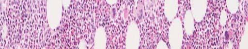
42 PATTERNS OF BONE MARROW INVOLVEMENT IN MYELOMA Interstitial Focal Mixed Diffuse
43


44
45 IMMUNOHISTOCHEMISTRY IN PLASMA CELL DYSCRASIAS Assessment of quantity of plasma cells on bone marrow sections Identification of a monoclonal plasma cell proliferation Distinction of myeloma from other neoplasms
46 IMMUNOPHENOTYPE OF PLASMA CELL MYELOMA CD138+ C D10-/+ CD5--/+ Clonal CIg+ CD45-/+ CD19--/+ CD38++ HLA-Dr-/+ CD20--/+ CD56+ (most) EMA-/+ CD22--/+ CD79a+ SIg--/+ CD34
47 CYTOGENETICS OF PLASMA CELL MYELOMA 50% have abnormalities by conventional cytogenetic studies 70% to 100% by FISH Hyperdiploidy and complex structural rearrangements most common Most common involved chromosomes: 13 [monosomy or del(13q14)] 14 [t(11;14)(p13;q32)] 1 [duplications of q and deletions of p] 11,8,6q,5q,7q,12 also common
48 PROGNOSTIC ASSOCIATIONS OF CYTOGENETICS INMYELOMA Unfavorable del 13q hypodiploidy 11q More favorable no growth of metaphases
49 DIAGNOSTIC CRITERIA FOR MGUS More common <10% Plasma cell M protein present, stable levels l of M protein: IgG < 3,0g IgA < 2g LC<1g/day Normal immunoglobulins - normal levels No Bunce-Jones protein No anemia No Renal failure No lytic lesion & hypercalcemia Normal β2 microglobulin level No treatment required
50
51 COURSE OF PATIENTS WITH MGUS (FOLLOW-UP YEARS) Group Description I No substantial ti increase in MCP 12% II MCP >3g/dL, but no myeloma or related 10% diseases III Died of unrelated causes 52% IV Development of myeloma (68%), macroglobulinemia (11%), amyloidosis (13%) 26% or related diseases (8%)
52 Smoldering multiple myeloma M protein present, stable levels of M protein: IgG 3,0g IgA 2g LC 1g/day normal immunoglobulins - normal levels marrow plasmacytosis 10% complete blood count - normal no lytic bone lesions no signs of disease
53 INDOLENT MYELOMA M component: IgG <7g/dl, IgA <5g/dl Rare bone lesions (< 3 lytic lesions), without compression fractures Normal hemoglobin, serum calcium and creatinine No infections
54 COMPARISON OF MGUS, INDOLENT AND SMOLDERING MYELOMA MGUS SMM IMM Plasma cells (BM) <10% 10-30% >30% M component IgG<3.5, IgA<2 IgG>3.5, IgA.>2 IgG 3.5-7, IgA2-5 Lytic bone lesions None None < 3 Symptoms/infection None None None
55 PLASMA CELL LEUKEMIA >2 X 10⁹/L plasma cells in blood Younger age Higher incidence of organomegaly and lymphadenopathy More extensive bone marrow infiltration Renal failure more common Less bone pain, fewer lytic lesions Poor response to therapy


56
57 NON-SECRETORY MYELOMA <1% of fmyelomas No serum or urine monoclonal protein Renal failure and hypercalcemia are generally lacking Immunostaining for a monoclonal protein on bone marrow sections may establish the diagnosis Rarely there is no monoclonal protein synthesized Must rule out IgD and IgE myeloma
58 PLASMACYTOMAS Solitary Plasmacytoma of Bone Extramedullary Plasmacytomas
59 SOLITARY PLASMACYTOMA OF BONE Localized plasma cell tumor Absence of a plasma cell infiltrate in random marrow biopsies No evidence of other lesions by radiographic examination Absence of renal failure, hypercalcemia, anemia
60 EXTRAMEDULLARY PLASMACYTOMAS Most Common Primary Sites Upper air passages and oropharynx (May involve draining lymph nodes) Less Common Sites Lymph nodes (primary), salivary glands, spleen, liver, etc. 25% have small monoclonal spike Rare dissemination, rarer evolution to myeloma
61
62
63
64
65 WALDENSTROM SMACROGLOBULINEMIA (LYMPHOPLASMACYTOID LYMPHOMA) Lymphadenopathy or splenomegaly 20%-40% Bone marrow involvement 90% Lytic bone lesions 2% Hypercalcemia 4% Hyperviscosity syndrome 15%
66 Hyperviscosity syndrome Causes sludging of RBCs in capillaries and increased thrombus formation leading to: Headache Dizziness Vertigo Deafness Seizures Bleeding
67 TUMOR INFILTRATION BONE MARROW LYMPH NODES SPLEEN MONOCLONAL MACROGLOBULINEMIA CIRCULATING IgM HYPERVISCOSITY CRYOGLOBULINEMIA COLD AGGLUTININ ANEMIA TISSUE IgM NEUROPATHY GLOMERULAR DISEASE AMYLOIDOSI
68 Epidemiology and Demographics 1500 people diagnosed d each year in the US Occurrence is 2x higher in men than in women Usually occurs in people > 65 More common among caucasians Mean life span after diagnosis is 5-7 years (longer than MM) 10% of patients ts attain complete remission
69 Diagnosis of WM Good physical exam including : fundoscopy to look for sausage links in the retina abdominal palpation to look for hepatosplenomegaly Several e different ent lab tests t GOLD STANDARD is serum electrophoresis
70 Labs CBC with differential Hgb is low (mild anemia) WBC count is normal to low Platelet count may be low Blood smear can show malignant lymphoid cells Elevated ESR, with rouleaux
71 Serum electrophoresis This will show the overproduction of Ig in the serum as an M spike in the gamma region Immunoelectrophoresis If a large M spike is seen, serum immunoelectrophoresis will determine the isotype of the offending macroglobulin
72 Determining Severity Test IgM levels in the serum The higher the levels, the more severe Test serum viscosity Uses a viscometer to measure viscosity it of serum compared to water Results are more sensitive than ESR Symptoms will appear when serum viscosity is 4x normal
73 Why do we think someone has WM? Anemia signs (fatigue, weakness, dyspnea, etc.) Symptoms of hyperviscosity y syndrome Unexplained weight loss Unexplained easy bleeding (due to interference with clotting factors - including epistaxis and purpura) Engorgement and narrowing of retinal veins Lymphadenopathy Peripheral neuropathy (due to acculumations of paraproteins in perivascular and perineural tissues) Hepatosplenomegaly l Increased bacterial infections
74 OSTEOSCLEROTIC MYELOMA (POEMS SYNDROME) Polyneuropathy (progressive, demyelinating) Organomegaly (hepatosplenomegaly, lymphadenopathy) Endocrinopathy (hypogonadism, hypothyroidism) Monoclonal gammopathy (usually IgA lambda or IgG lambda) Skin changes (hyperpigmentation) pg
75 The level of paraprotein in the serum and urine is usually low Anemia,hypercalcemia,renal dysfunction and pathological fracture are rare BM trephine shows a characteristic osteosclerotic plasmacytoma which h may occur singly or multiply l.the lesion shows thickened trabecular bone with closely associated peritrabecular fibrosis i with entrapped plasma cells,the rest of the bone marrow away from the lesion is relatively l normal with les than 5% plasma cells. Lymph node biopsy shows a follicular proliferation with regrssed and reactive follicles and interfollicular plasma cells accumulation
76 IMMUNOGLOBULIN DEPOSITION DISEASES Primary amyloidosis Systemic light and heavy chain deposition disease
77 Amyloidosis Definition It is a disorder of protein folding in which normally soluble protein are deposited in the extracellular space as insoluble fibrils that progressively disrupt tissue structure and function About 20 different unrelated proteins can form amyloid in vivo The term amyloid is derived from the Greek for starch like
78 Amyloid deposition may be : Systemic or localized Hereditary or acquired Life threatening or incidental finding
79 Pathgenesis of amyloid Amyloidosis breaks the dogma that tertiary structures t of proteins is dertermined d solely l by their primary amino acid sequences Amyloid forming proteins can exist in two completely different stable structures
80 The transformation evidently involving massive refolding of the native form into one that can autoaggregate in a highly ordered manner to produce the characteristic predominantly B-sheet.rigid,non-branching fibrils of nm in diameter and of indeterminate length Acquired biophysical properties common to all amyloid fibrils include insolubility in physiological solution,relative resistance to proteolysis,and ability to bind Congo red in an ordered manner that give the the diagnostic green birefringence under cross-polarized light Amyloid depoition can occur in three circumstances.
81 I-When there is a sustained abnormally high concentration of certain normal protein,such as amyloid A protein (SAA) in chronic inflammation and B2microglobulin in renal failure (AB2M) II-When there is normal concentration of a normal,but inherently amyloidogenic,protein over a very prolonged period,such as transthyretin in senile amyloidosis (ATTR) and B-protein in Alzheimer s disease
82 III-When there is a production of an acquired or inherited variant protein with and abnormal structure such as,amyloidogenic monoclonal Ig light chain The genetic and environmental factors that influence individual susceptibility to and timing of amyloid deposition are unclear
83 Systemic AL amyloidosis Formerly known as primary Occur in a small proportion p of monoclonal B-cell dyscrasias AL fibrils are derived from monoclonal Ig light chain,which are unique in each patient AL fibrils are more commonly derived from lambda than kappa light chain Virtually any organ or combination of organs other than the brain may be directly affected commonly including the heart
84 Diagnosis and investigation of amyloidois Amyloid should be considered in the DD of : Renal failure NS Restrictive cardiomyopathy Peripheral or autonomic neuropathy Hepatomegaly
85 Early symptoms are often very non-specific and insidious The index of suspicion should be high in patients known to have clonal B-cell dyscrasias But in practice the diagnosis of amyliodosis is usually an unexpected finding following biopsy of an organ with disturbed function
86 DIAGNOSTIC CRITERIA FOR PRIMARY AMYLOIDOSIS Tissue biopsy showing typical morphology Apple green birefringence i under polarized light after Congo Red staining Typical fibrillar ultrastructure
87 BIOPSY DIAGNOSIS OF AMYLOIDOSIS Diagnostic % Bone marrow examination 56% Abdominal fat aspiration 80% Combined BM & fat aspirate 89%
88
89 HEAVY CHAIN DISEASE Monoclonal l protein composed of incomplete heavy chains devoid of light chains Lymphoproliferative p disorders - often CLL or macroglobulinemia-like picture Alpha chain disease Gamma chain disease MU chain disease
90 ALPHA CHAIN DISEASE Involves GI track Lamina propria is heavily infiltrated with plasma cells Uniform clinical picture Bone marrow is normal
91 GAMMA CHAIN DISEASE Hypogammaglobulinemia li i and incomplete monoclonal gamma chain Variable clinical picture 50% have a lymphoplasmacellular proliferation similar to macroglobulinemia 15% are predominantly plasmacytic Occasional cases are associated with CLL or large cell lymphoma 30% benign-appearing or no lymphoproliferative disease
92 MU CHAIN DISEASE CLL-like picture More often hepatosplenomegaly Less lymphadenopathy Characteristic vacuolated plasma cells Large amounts of Bence-Jones protein
93
94 PROBLEMATIC MORPHOLOGIC FEATURES IN MYELOMA Mature cytology Pleomorphic cytology Convoluted plasma cell variant Phagocytosis Cytoplasmic inclusions Plasmablastic myeloma
95